TABLE OF CONTENTS
RIASSUNTO 3
ABSTRACT
8
INTRODUCTION
12
1. Neuroglobin 13
Globins family 18
Heme group 24
2. Flotillin 30
Lipid rafts 36
3. Hematopoiesis 45
4. Cellular stress condition: 50
· Oxidative stress 50
· DNA damage 56
· Phagocytosis 63
AIMS
70
MATERIAL AND METHODS
72
RESULTS AND DISCUSSION 79
2. Ngb expression levels increase during myeloid differentiation 80
3. Ngb expression increases during phagocytosis 83
4. Ngb expression does not change during serum deprivation and DNA
damage 85
5. Ngb expression increases during oxidative stress conditions in
hematopoietic cells. 87
6. Ngb expression increases during Inh treatment 92
7. Oxidative stress effect on Ngb expression is increased by vitamin
D₃-induced differentiation in U937 cells 95
8. Flotillin-1 knockdown decreases Ngb expression 97
9. Ngb physically interacts with Flotillin-1 in an oxidative stress dependent
manner in myeloid cells 100
CONCLUDING
REMARKS
AND
FUTURE
PERSPECTIVES 102
IL RUOLO DELLA NEUROGLOBINA NEL SISTEMA
EMATOPOIETICO
Emoglobina e Mioglobina sono due proteine appartenenti alla famiglia delle globine in grado di trasportare e conservare l’ossigeno. Nel 2000 è stato identificato un terzo membro di questa famiglia: la Neuroglobina (Ngb), particolarmente espressa nel sistema nervoso, (da cui il nome) ma non solo, infatti la si ritrova anche nel tratto gastrointestinale, nel tessuto endocrino e nella retina. Si tratta di una proteina monometrica costituita da 151 amminoacidi (17 KDa) con un’alta affinità per l’ossigeno. Il gene della Ngb umana è situato sul cromosoma 14q24. Nel corso dell’evoluzione il gene della Ngb non ha subito molti cambiamenti e ciò ne sottolinea un ruolo rilevante. La Ngb, come tutti i membri della famiglia delle globine, è una emo proteina, con un gruppo prostetico eme, grazie al quale è in grado di legare reversibilmente l’ossigeno ed altri ligandi. L’atomo di ferro del gruppo prostetico può trovarsi nello stato ferroso (Fe2+) o ferrico (Fe3+). Sia nella forma ferrica che ferrosa la Ngb è esacoordinata con ligandi endogeni (istidina prossimale e distale) e O2 o
CO spiazzano l’istidina distale per legarsi essi stessi.
Nonostante i numerosi studi effettuati, non si conoscono ancora perfettamente quali siano tutte le funzioni della Ngb. Sicuramente è chiaro che la Ngb svolge una funzione protettiva nei neuroni in condizioni di ipossia ed ischemia. Inoltre la Ngb svolge un’azione protettiva in condizioni di stress ossidativo e perossidazione lipidica. Però il meccanismo completo con cui riesce a svolgere questa sua funzione neuroprotettiva non è stato ancora bene delucidato. Diversi studi hanno sottolineato come questa funzione possa essere collegata: 1-alla capacità della Ngb di eliminare tutte le specie reattive dell’ossigeno e dell’azoto; 2-alla sua elevata affnità per l’ossigeno che garantisce una riserva d’ossigeno utile in condizioni di ipossia;
3-alla sua capacità di intervenire nel meccanismo di trasduzione del segnale. Infatti la letteratura suggerisce che la Ngb nella forma ferrosa (legante l’O2) viene subito convertita
nella forma ferrica e solo quest’ultima è in grado di legare la subunità Gα della proteina G
legata a sua volta al GDP e ne impedisce il rilascio di quest’ultimo, inibendone quindi l’attivazione. In questo modo la Ngb impedisce l’attivazione della trasduzione del segnale che porta poi alla morte cellulare, garantendo di conseguenza la sopravvivenza cellulare. E’ noto che solo nella forma ferrica la Ngb può legare (oltre a Gα) la Flotillina-1, una
proteina particolarmente espressa nei rafts, piccoli domini di membrana, ricchi in sfingolipidi e colesterolo che funzionano da piattaforme per concentrare e segregare
molecole segnale in modo da compartimentalizzare i processi cellulari. Nei rafts sono quindi presenti diverse proteine coinvolte nella trasduzione del segnale, tra cui le proteine G. La Flotillina è una proteina integrale di membrana che svolge un ruolo chiave nell’assemblaggio dei rafts e nella trasduzione del segnale. Se ne conoscono due isoforme: Flotillina-1 e -2. Il gene della Flotillina-1 è localizzato sul cromosoma 6 e codifica per una proteina di 47 KDa, particolarmente espressa nei neuroni, ma anche nella membrana dei fagosomi, degli endosomi e nel nucleo. La funzione della Flotillina è associata alla sua localizzazione nei rafts ed è coinvolta in molti processi cellulari: partecipa all’ingresso del glucosio nelle cellule stimolate con insulina, induce la rigenerazione dei neuroni, partecipa alla maturazione dei fagosomi, stimola la proliferazione di cellule cancerose di prostata e definisce un pathway endocitico Clatrina-indipendente.
In base a tutte queste informazioni lo scopo di questo lavoro è quello di andare a valutare innanzitutto se la Ngb sia espressa nelle cellule mieloidi e poi eventualmente capire quali siano le sue funzioni in questo sistema e se esista anche qui una relazione funzionale tra Ngb e Flotillina-1.
E’ stata individuata un’ampia espressione della Ngb nelle cellule mieloidi utilizzate (THP1, U937 e HL60) ed anche in linee cellulari derivanti dal cancro ai polmoni (H1299, H1650 e H1975). Sulla base di questo rsultato siamo andati poi a valutare l’espressione della Ngb durante il differenziamento mieloide. Abbiamo riscontrato che l’espressione della Ngb, così come quella della Flotillina, aumenta durante questo processo ed in particolare la Ngb è espressa a più alti livelli in cellule macrofagiche. Visto che nei neuroni la Ngb ha una funzione protettiva nei confronti dello stress ossidativo, siamo andati a studiare l’espressione della Ngb durante una condizione di stress ossidativo che si verifica nelle cellule mieloidi, vale a dire durante la fagocitosi. Abbiamo osservato che l’espressione della Ngb aumenta nel corso di questo processo, così come la Flotillina-1, ed aumenta ancora di più durante la fagocitosi a carico di macrofagi maturi, che hanno un ruolo fondamentale nel processo stesso. Questi risultati suggeriscono un possibile coinvolgimento di queste due proteine in questo processo. Siamo andati poi a studiare l’espressione della Ngb in cellule mieloidi in condizioni che potrebbero indurre stress cellulare o apoptosi: durante la deprivazione di siero (testando diverse concentrazioni di siero al di sotto di quella utilizzata in condizioni standard) ed in seguito a trattamento con idrossiurea (un inibitore della sintesi di DNA). In entrambe le situazioni non abbiamo riscontrato nessuna varazione nei livelli di espressione della Ngb, suggerendo probabilmente un mancato coinvolgimento della Ngb stessa. Mentre abbiamo registrato un
considerevole aumento di espressione della Ngb in seguito a trattamento delle cellule mieloidi con H2O2 per indurre stress ossidativo. Questo risultato ci consente di ipotizzare
che durante condizioni di stress ossidativo la Ngb, oltre ad una funzione neuroprotettiva, svolge la stessa funzione di protezione anche nelle cellule mieloidi. Diversi farmaci sono in grado di indurre stress ossidativo, uno di questi è l’isoniazide (Inh), farmaco anti tubercolare di prima linea. Inh inoltre è in grado di legare reversibilmente il Fe in forma ferrica e ferrosa del gruppo eme dell’emoglobina troncata del Mycobacterium tuberculosis, legame che perturba le proprietà del gruppo eme, renendo l’emoglobina incapace di svolgere le sue normali funzioni. Dato che anche la Ngb ha un gruppo eme, che viene convertito nella forma ferrica in condizioni di stress ossidativo per poter espletare la sua funzione neuroprotettiva, siamo andati a valutare l’espressione della Ngb in seguito a trattamento delle cellule mieloidi con varie concentrazioni di Inh. Abbiamo riscontrato un aumento di espressione della Ngb all’aumentare delle dosi di Inh somministrata. Avendo osservato che l’espressione della Ngb aumenta durante il differenziamento mieloide ed in condizioni di stress ossidativo, abbiamo pensato di combinare i due processi e vedere cosa accadesse alla Ngb. Abbiamo così osservato che l’espressione della Ngb aumenta ancora di più nel caso di stress ossidativo indotto in cellule mieloidi dfferenziate. In fine siamo andati a studiare la relazione tra Ngb e Flotillina-1 nel sistema ematopoietico. Prima di tutto siamo anadati a valutare se l’espressione della Flotillina-1 influenza quella della Ngb. Pertanto abbiamo prodotto cellule mieloidi sovra-esprimenti la Flotillina-1 e cellule in cui la Flotillina-1 è stata sileziata. Nel primo caso non abbiamo riscontrato nessuna variazione di espressione della Ngb, mentre abbiamo osservato una notevole diminuizione di espressione della Ngb nelle cellule in cui la Flotillina-1 era stata silenziata. L’assenza della Flotillina-1 impedisce l’aumento di espressione della Ngb anche nel caso di trattamento con H2O2 per indurre stress ossidativo. Questi risultati suggeriscono una possibile funzione
protettiva e di stabilizzazione da parte della Flotillina-1 sulla Ngb. Per confermare questa ipotesi siamo andati a studiare se esiste un’interazione tra queste due proteine. Pertanto abbiamo messo a punto un esperimento di co-immunoprecipitazione utilizzando un anticorpo anti-Flotillina-1, seguito da Western Blotting con un anticorpo anti-Ngb. Questo è stato fatto in cellule mieloidi sia in condizioni basali che in seguito a trattamento con H2O2 per indurre stress ossidativo. Abbiamo ottenuto un risultato positivo, ossia un
interazione, solo dopo trattamento con H2O2. Quindi in cellule mieloidi la Ngb interagisce
In conclusione possiamo affermare che la Ngb è espressa in cellule mieloidi e di cancro al polmone; la sua espressione aumenta durante: il differenziamento mieloide, la fagocitosi e lo stress ossidativo; la sua espressione diminuisce in cellule in cui la Flotillina è stata silenziata; interagisce con la Flotillina-1 durante lo stress ossidativo.
In base ai risultati ottenuti sarebbe interessante andare a studiare il ruolo della Ngb nei progenitori ematopoietici sottoposti a fattori di crescita e di differenziamento verso la linea granulocitaria o monocitaria. Inoltre per rafforzare i dati che sostengono un potenziale ruolo protettivo della Ngb in condizioni di stress ossidativo in cellule mieloidi, andrebbe valutato l’effetto conseguente alla sua sovra-espressione e al suo silenziamento.
THE
ROLE
OF
NEUROGLOBIN
IN
THE
HEMATOPOIETIC SYSTEM
Hemoglobin and Myoglobin are proteins that belong to the globins family and are able to deliver and conserve oxygen. In the 2000 a new component of this family has been identified and named Neuroglobin (Ngb). Ngb is mainly expressed in the nervous system, from which it takes its name, but it is located also in the gastrointestinal tract, in the endocrine tissue and in the retina. Ngb is a monometric protein formed by 151 aminoacids (17 KDa) and has a high affinity for oxygen. The human Ngb gene is located in the chromosome 14q24. Over the evolution, the Ngb gene has been conserved and this suggests its relevant role in biological systems. Ngb, as all the globins family’s members, is an heme protein that shows the heme prostetic group, by which can reversibly bind oxygen and other ligands. The iron atom in the prostetic group can have the oxidative state +2 (ferrous) or +3 (ferric). Both in the ferrous and in the ferric state the Ngb is hexacoordinated with other endogenous ligands (proximal and distal hystidine) and O2 or
CO crowd-out the distal hystidine to link themselves to the protein. Even though many studies have been conducted, it is not completely clear which functions the Ngb has. It is known that the Ngb plays a protective role in neurons under conditions of hypoxia and hyschemia. Moreover it plays, too, a protective function during the oxidative stress and lipidic peroxidation. However the mechanism by which Ngb can protect neurons has not been completely elucidated yet. Several studies have shown which mechanisms this function can be linked to: 1) the Ngb ability to scavenge reactive oxygen and nitrogen species; 2) its high affinity for oxygen that allows the organism to have a bulk of oxygen during hypoxia conditions; 3) its role in the signal transduction. In fact, the body of literature suggests that Ngb, in its ferrous state and linking oxygen, is quickly converted in the ferric state and only the latter is able to bind the Gα subunit of the G protein, that in turn
is bound to the GDP. The link between the Ngb in the ferric state and the G protein does not allow the release of the GDP and consequently the activation of the G protein itself. This way Ngb halts the signal transduction that leads to cell death and guarantees the survival of the cells. Additionally, in the ferric state Ngb can bind Flotillin-1, a protein mainly express in rafts, small membrane domains, rich in sfingolipids e cholesterol, that function as platforms to concentrate and segregate signal molecules during cellular processes. Thus in rafts there are several proteins involved in the signal transduction, among which G proteins. Flotillin is an integral membrane protein that plays a key role in
assembling rafts and in the signal transduction. Two different isiform of Flotillin are known: Flotillin-1 and Flotillin-2. The Flotillin-1 gene is localized in the chromosome 6 and code for a 46 KDa protein, particularly expressed in neurons, but also present in the fagosomes membrane, in endosomes and in the nucleus. The function of Flotillin is associated with its localization in rafts and it is involved in many cellular processes: it participates in the glucose income in insulin-stimulated cells; it induces the neuronal regeneration; it participates in the fagosome maturation; it drives the proliferation of prostatic cancer cells and finally it defines an endocytic Clatrin-dependent pathway. According to this information, the aim of the project is to evaluate either the possible expression of Ngb in myeloid cells, and to analyze its function in this specific cell system, and to find a functional relationship between Ngb and Flotillin-1. In this project, a significant expression of Ngb has been individuated in myeloid cells (THP1, U937 and HL60) and in lung cancer cells (H1299, H1650 and H1975). According to these findings, the expression of Ngb during the myeloid differentiation has been analyzed. The expression of Ngb, as well as of Flotillin, increases during the differentiation. Additionally, Ngb is mainly expressed in macrophagic cells. Because of the neuroprotective role of Ngb during the oxidative stress, its expression has been evaluated during a specific condition of stress that occurs in myeloid cells, that is the fagocytosis. The Ngb level improves during the fagocytic process, as well as Flotillin-1, and increases even more during the fagocytosis realized by mature macrophages, which have a basic role in this process. These results suggest a possible involvement of these two proteins in this mechanism. Moreover, the Ngb expression has been analyzed in specific conditions that could have induced cellular stress and apoptosis, particularly during serum deprivation (by testing concentrations of serum lower than the concentration used in standard conditions) and after treatment with hydroxyurea (a DNA synthesis inhibitor). In both these situations there was not a modification in the Ngb expression, suggesting that probably the protein is not involved. Conversely, a remarkable increase of the Ngb expression in myeloid cells has been observed after H2O2 treatment, inducing oxidative stress. This result could suggest that,
during oxidative stress conditions, Ngb may play a protective role in myeloid cells as well as shown in neurons. Different drugs can induce oxidative stress, such as Isoniazid (Inh), a first line anti-tubercolar drug. Inh is able to reversibly bind both the ferric and ferrous iron of the heme group in the truncated hemoglobin of the Mycobacterium Tuberculosis. This link modify the heme group properties, making the hemoglobin unable to perform its normal functions. The Ngb, too, has an heme group whose ferrous iron is converted in
ferric iron under oxidative stress conditions to protect neurons. Thus, the Ngb expression has been evaluated after treatment with different concentrations of Inh in this cellular system and consequently a dose-dependent increase of Ngb expression has been observed. Afterwards, combining the myeloid differentiation with oxidative stress conditions (H2O2
-induced), a higher Ngb level has been measured in mature myeloid cells compared to the Ngb expression in undifferentiated cells. Finally the functional relationship between Ngb and Flotillin-1 in the hematopoietic system has been studied. First of all the possible influence of Flotilin-1 on Ngb has been evaluated. According to this purpose, over-expressing Flotillin-1 cells and Flotillin-1 knockdown cells have been generated. In the first condition no variations in the expression of Ngb have been observed, in the latter a remarkable decrease in the protein have been noticed. The lack of Flotillin-1 prevent Ngb from increasing also in H2O2-induced oxidative stress conditions. This data suggests a
possible protective and stabilizing function of Flotillin-1 on Ngb. To confirm this hypothesis, the physical interaction between these two proteins has been investigated by setting up a co-immunoprecipitation assay with an antibody anti-Flotillin-1, and then a Western Blotting with an antibody anti-Ngb, in myeloid cells both in basal conditions and after an H2O2-induced oxidative stress treatment. The results obtained showed that, in
myeloid cells, the physical interaction occurs only after H2O2 treatment, thus in conditions
of oxidative stress. In conclusions, to sum the data collected during this project: Ngb is expressed in myeloid cells and in lung cancer cells, too; its expression increases during the myeloid differentiation, the fagocytosis and the oxidative stress; additionally, its level tends to diminish in Flotillin-1 knockdown cells; and finally, Ngb interacts with Flotillin-1 during the oxidative stress. According to these results, further studies could be conducted to elucidate and better understand the functions of Ngb in different cellular systems. As examples, the role of Ngb could be analyzed in myeloid progenitors treated with growth factors and induced to differentiate in granulocytes or monocytes. Moreover, as a support to the hypothesis that Ngb has a potential protective effect in myeloid cells under oxidative stress conditions, consequences of the silencing or over-expression of Ngb could be evaluated.
1.NEUROGLOBIN
Hemoglobins and Myoglobins are related protein families that function in oxygen transport and storage. In 2000 a third globin type has been identificated, predominantly expressed in the brain and consequently named Neuroglobin (Ngb). Mouse Ngb is a monomer with a high oxygen affinity [half saturation pressure P50=0,9 to 2.2 Torr (0,12-0,29 kPa)]. The
human Ngb gene (NGB) is located on the chromosome 14q24 and it is constituted by a unique exon-intron. The promoter region contains several putative Sp1-binding sites and at least three transcription starting points but there is not a TATA domain. NGB gene has three introns at positions B12-2, E11-0 and G7-0, of which B12-2 and G7-0 are conserved in Hemoglobins and Myoglobins.
Both mouse and human genes code for proteins of 151 amino acids (17 KDa) that are 94% identical, that is a higher conservation than that between the orthologous Hemoglobins or Myoglobins of these species (77-85 % identity). Even if this proteins belong to the globin superfamily, they have a low amino acid sequence similarity with vertebrate Myoglobins (<21% identity) and Hemoglobins (<25% identity) insinuating a distinct evolution and function (Burmester T. et al; 2000).
Figure 1: Comparison between Human Ngb, mouse Ngb, Myoglobins and Hemoglobins α and β. (Burmester T. et al. Nature, 2000)
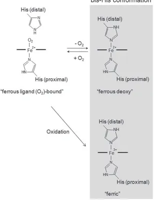
Ngb as well as all members of globins superfamily is a heme protein, with a heme prosthetic group (Fe2+-protoporphyrin IX), by which reversibly binds oxygen or other ligands. The iron atom in the heme prosthetic group exists in either the ferrous (Fe2+) or ferric (Fe3+) redox state. Both the ferrous and ferric Ngbs forms are hexacoordinated to
endogenous ligands (namely proximal and distal histidine residues), and O2 or CO displace
the distal His residue to produce ferrous O2- or CO-bound Ngb (Dewilde et al., 2001).
Figure 2: Illustration of the ferrous and ferric form of Ngb (Watanabe et al.,Journal of Biological Chemistry, 2012).
It is highly expressed in brain neurons but also in the peripheral nervous system, endocrine tissues and retina (Reuss et al., 2002). Ngb has a neuroprotective role against hypoxic/ischemic insults that occur in neurons. In fact antisense-mediated Ngb knockdown rendered cortical neurons more vulnerable to hypoxia, whereas Ngb over-expression gives protection of cultured neurons against hypoxia (Sun et al., 2001).
In animal stroke models, Ngb over-expression reduced infarct size (30% compared to wild type) in rats after a middle cerebral artery occlusion (MCAO), and the outcome was reversed by Ngb knockdown thanks to anti-sense oligonucleotide (Sun et al., 2003). The reduction of brain infarction in Ngb-over-expressing transgenic mice can be sustained up to 14 days after ischemia (respect to wild type), underlined that Ngb over-expression offers neuroprotection against transient focal cerebral ischemia (Zhanyang et al., 2012), but the involved mechanisms it is not yet completely clear.
Ngb has also a protective role against other neurological disorders, like β-amyloid and NMDA toxicity (Khan et al., 2007; Li et al., 2008) and then protects retinal ganglion cells (RGC) against ocular Hypertension and glaucomatous damages (Wei et al., 2011).
Therefore it has been widely accepted that Ngb has a protective role, but the underlying mechanism remains poorly defined. An initial evidence suggests that the neuroprotective role may be largely linked to its ability to bind O2 and NO. In addition, putative signal
transductions and a mitochondrial function preservation may also be involved.
Several studies have indicated that Ngb neuroprotective function is related to its ability in scavenging reactive species, in fact Ngb can bind nitric oxide (NO) with a high intrinsic affinity and a low dissociation rate (Van Doorslaer et al., 2003). In addition, Ngb-O2 reacts
with NO quickly to produce NO3- and ferric Ngb (HNgb). This pathway would dispose of
NO and may protect cellular respiration by the inhibitory effect of NO on cytochrome c oxidase activity (Moncada et al., 2002; Brunori et al., 2008).
Furthermore, Ngb over-expression rendered neuroblastoma cells more resistant to NO-induced cell death, underlined Ngb ability in neutralizing neurotoxic effects of reactive nitrogen species (Jin et al., 2008).
Ngb also has a protective function against other oxidative challenges in neurons, such as H2O2-induced oxidative stress or lipid peroxidation (Fordel et al., 2006; Sun et al., 2005).
HNgb binds the GDP-bound form of the α subunit of G protein (Gα), competing with Gβγ
subunit (to binds Gα)(Kitatsuji et al., 2007; Wakasugi et al., 2003).
G proteins are heterotrimeric proteins constituted by a Gα subunit with GTPase activity and a Gβγ dimer, and belongs to a protein family whose signal transduction functions
depend on the binding of guanine nucleotides (Gilman et al., 1992; Simon et al., 1991). Ligands- or signal-activated G protein-coupled receptors (GPCRs) cause GDP release from Gα to bind GTP. The GTP bind to Gα causes conformational changes that comport dissociation of GTP-Gα from Gβγ, that can separately regulate the activity of different
effector molecules (adenylyl cyclase, ion channels,..). Thanks to the intrinsic GTPase activity of Gα (which hydrolyzes bound GTP to GDP), the signal transduction is turned off and GDP-Gα reassociated with Gβγ. Gα proteins are divided in four families: Gαi/o, Gαs,
Gαq/11 and Gα12/13(Hepler et al., 1992; Simon et al., 1991).
Ferric HNgb binds exclusively the GDP-bound form of Gαi/o and functions as a guanine
nucleotide dissociation inhibitor (GDI) for Gαi/o (Wakasugi et al., 2003). On the contrary
ferrous-ligand-bound HNgb under normoxia does not interact with Gαi/o nor has GDI
stress-responsive sensor for signal transduction in the brain (Wakasugi et al., 2003 and 2005).
Moreover, Ngb exists in lipid rafts only during oxidative stress. In fact, the addition of a lipid rafts distruptor (MβCD) reduces neuroprotective activity of HNgb, that is restored by reconstruction of lipid rafts. This suggests that lipid rafts are fundamental for Ngbs neuroprotection (Watanabe et al., 2012).
Wakasugi et al in 2004 have demonstrated that Ngb interacts with Flotillin-1 by using a yeast two-hybrid screening, evidence that was confirmed by GST pull-down assays. Flotillin-1 is a lipid raft microdomain-associated protein, and also Gαi/o exists in lipid rafts
(Moffet et al., 2000; Yuyama et al., 2007). Therefore, Flotillin-1 recruits HNgb to lipid rafts, where HNgb binds Gαi/o and acts as a GDI for Gαi/o preventing neuronal death
(Watanabe et al., 2012).
Figure 3: Neuroprotective mechanism of HNgb (Watanabe et al., Journal of Biological chemistry, 2012).
Ngb expression is confined to metabolically active and oxygen-consuming cell types (Burmester et al., 2004). At the subcellular level, Ngb is associated with mitochondria, linked it to the oxidative metabolism (Burmester et al., 2007). Mitochondria have a key roles in energy production, ROS homeostasis and cell death signaling. They respond to various insults to cells and its dysfunction is related with a large variety of clinical phenotypes. Mitochondria comprise a central locus for energetic perturbations and oxidative stress in hypoxia/ischemia (Nicholls et al., 2000; Sims et al., 2002). Ngb over-expression improves mitochondrial function and reduces oxidative stress after hypoxic insults that occur in neurons (Liu et al., 2009). Ngb might affect both mitochondrial functions and free radical generations as its potential neuroprotective mechanisms. Hypoxia and OGD (Oxygen-Glucose Deprivation) develop mitochondrial depolarization (Larsen et al., 2006). The mitochondrial permeability transition pore (mPTP) is a protein pore created across the inner and outer membrane of the mitochondria in pathological conditions (such as stroke). During hypoxia/ischemia, mPTP opening determined release of Cyt c from mitochondria to cytosol (Zhang et al., 2008) and next activation of caspase-dependent or -incaspase-dependent apoptosis pathways (Petit et al., 1998; Zhu et al., 2002). Ngb over-expression is correlated with reduced mPTP opening, and decreased cytochrome c release, highlighting an inhibitory role of Ngb in OGD-induced mPTP opening, that is one of the major causes of cell death in a variety of tissue ischemic damages, like heart attack and stroke. Therefore, Ngb inhibitory effect in mPTP opening may be an important mechanism of Ngb neuroprotection. Several Ngb-binding proteins have been identified by yeast two-hybrid assay: Na/K ATPase β1, cytochrome c1, ubiquitin C, voltage-dependant anion channel (VDAC) and a few more (Yu et al., 2012), that some are biologically relevant for neuronal function and survival (Yu et al., 2012).
Ngb gene expression is tissue-specific and Ngb mRNA is widely distributed throughout the adult rat brain, including cerebral cortex, hippocampus, and subcortical structures (thalamus, hypothalamus, olfactory bulb, and cerebellum) (Wystub et al., 2003; Zhang et al., 2002; Geuens et al., 2003). Ngb protein distribution is compatible with its mRNA localization and the subcellular immunoreactivity is restricted to the cytoplasm. The highest Ngb expression is in the retina with a concentration 100-fold higher than in the brain (Schmidt et al., 2003). Ngb mRNA was observed in the perikarya of the nuclear and ganglion layers of the neuronal retina, whereas the protein was present mainly in the plexiform layers and in the ellipsoid region of the photoreceptor inner segment (Hundahl et al., 2005). The distribution of Ngb correlates with the subcellular localization of
mitochondria and with the relative oxygen requests. So Ngb supplies oxygen to the retina, as well as Myoglobin in the myocardium and skeletal muscle.
The expression of Ngb gene is up-regulated in:
1) hypoxic/ischemic conditions in cultured cells (Schmidt-Kastner et al., 2006; Shao et al., 2009) and stroke animals brain (Sun et al., 2001 and 2003, Shang et al., 2006; Fordel et al., 2007)
2) in the cerebellum of mouse pups in response to hypoxic-ischemic insults caused by maternal seizures during intrauterine life (Lima et al., 2011)
3) in the cortical peri-infarct region in stroke patients (suggesting its clinical relevance for endogenous neuroprotection ) (Jin et al., 2010)
While expression level decreased to about a half in aged rats (24 months) compared to young ones (3, 12 months) in various regions of brain, implying the pathophysiological importance of Ngb in age-related neurodegenerative diseases (Sun et al., 2005).
Transcription factors Sp1 and Sp3 can bind to the human Ngb promoter region causing transactivation of Ngb promoter (Zhang et al., 2011). Transcription factors, members of the NFkB family (p65, p50, cRel), Egr1, and Sp1, bind the Ngb promoter, causing basal Ngb expression. Moreover, NFkB (p65) and Sp1 as well as Hif1, were also responsible for hypoxia-induced up-regulation of Ngb expression (Yu et al., 2012).
Globins family
The term globin regroups a variety of proteins, structurally and phylogenetically related but present in organisms of developmental levels also very different. All the members of this family share the typical globin fold and the presence of the prosthetic group heme, that allows them to bind oxygen and other small diatomic molecules. The most famous representatives of this family are undoubtedly Hemoglobin (Hb), the more studied protein in the world, present in red blood cells and responsible for transporting oxygen (necessary for the oxidative respiration of the cells) through the blood in vertebrates, and Myoglobin (Mb), the first protein whose structure has been solved by X-ray (by John Kendrew and Max Perutz in 1958) and whose function is to store oxygen in the muscles tissue. But recent studies have revealed the existence of a high number of members of this family, classified on the basis of structural, functional and evolutionary parameters and present not only in animals but also in bacteria, protozoa, plants and mushroom.
Globins are small metalloproteins that generally comprise around 150 amino acids, but may also have N- and/or C-terminal extensions. Most globins cover eight α-helical segments (named A through H) with a characteristic 3-over-3 α-helical sandwich structure
(referred to the globin fold) and with a heme prosthetic group (Fe2+-protoporphyrin IX), by which they reversibly bind oxygen and other ligands (Perutz 1979; Bolognesi et al., 1997).
Figure 4: Example of the globin fold in tertiary structure. (Pesce et al., Biochemistry and Molecular Biology Education, 2001).
While their overall structures are conserved, the primary sequences of globins often change. In fact, only the proximal histidine adjacent to the Fe2+, is present in all globins, and most of them also show a phenylalanine in the inter-helical region CD1, which stabilizes the heme.
Although the globin polypeptide chain consists of about 150 residues, lately it was discovered a "truncated" hemoglobin, shorter than about 20-40 amino acid residues and having a three-dimensional structure partially different. These so-called truncated hemoglobins are expressed in eubacteria, cyanobacteria, protozoa and plants, and were divided into three phylogenetic groups (I, II, III) according to an amino acid sequence analysis. Their structure is different compared to the classic folding globin, although it retains some features. The pairs of antiparallel α-helical B-E and G-H form a sandwich "two-over-two", which can be considered as a reduction to the minimum of the "three-over-three” globin fold (Pesce et al., 2001). The helix A and the region CD-D are almost completely absent and the helix F is partly replaced by a long pre-helix loop. A characteristic common to all "truncated" hemoglobins is the presence of an apolar tunnel that connects the distal heme pocket with the solvent; this channel might have a role in facilitating the binding of gaseous ligands.
A further subgroup is called "mini-hemoglobins" and is characterized by a sequence even shorter. One of which is the hemoglobin of marine worm Cerebratulus lacteus, the shortest hemoglobin known so far, that consists of only 109 amino acids. Deletions respect to the
classic folding globin affecting different areas. Even the mini-hemoglobin have a hydrophobic channel within the protein matrix (Pesce et al., 2002).
Figure 5: Two -over -two fold globin in hemoglobin truncated Paramecium caudatum.
(Pesce et al., Biochemistry and Molecular Biology Education, 2001).
The quaternary structures of globins is very various, from simple to complex monomers consisting of hundreds of subunits. The most famous example of monomeric globin is Myoglobin. Hemoglobin is instead a tetramer, more precisely a "dimer of dimers", composed by two α subunit and 2 β subunits. It is interesting to note that this structural difference is reflected on the function. Hemoglobin binds oxygen in the lungs, transports it in the organism, transfers it to tissues, and binds CO2 thanks to amino-terminal groups of
the four globin chains. Myoglobin, present in muscle tissue, stores oxygen and releases it during intense aerobics activity. At the oxygen partial pressures of the pulmonary alveolus (PpO2~100 mmHg ), the Hemoglobin in the blood is almost completely saturated with
oxygen. While in tissues (PpO2 ~25-30 mmHg) Hemoglobin largely releases oxygen that
binds Myoglobin for which has a great affinity at those partial pressures of oxygen. The binding process and the release of O2 by Hemoglobin is closely related and controlled by
its quaternary structure. Hemoglobin indeed can exist in two quaternary forms, a high affinity for oxygen (R form) and a low affinity (T form). The deoxy protein is in a low affinity state, the binding of the first oxygen induces a conformational change (to the R form) which increases the affinity for the binding sites. This conformational change involves the heme group and, by the binding to the proximal histidine of the F helix, the change involves the tertiary and quaternary structure of the hemoglobin, and facilitates the binding of other molecules of oxygen in the free binding sites of the protein (cooperative binding).
Myoglobin, in addition to the main function of supplying oxygen, has a role as scavenger of intracellular NO (strong inhibitor of cytochrome c oxidase, terminal enzyme of the respiratory chain) in cardiac muscles and skeleton (Brunori et al., 2001).
Even dimeric globin are quite widespread in nature. Some examples are: Cytoglobin, rice hemoglobin HB1, hemoglobins of Scapharca and Vitreoscilla.
While the majority level of complexity in vertebrates is constituted by tetrameric structures, in invertebrates there is a wider variety of aggregation states. They were in fact identified both multisubunit hemoglobins, in which the number of monomers can be up to 144 units, and multidomain hemoglobins. These are proteins originated by gene duplication and containing more domains having globin folding. The multidomain proteins can in turn aggregate to form multidomain multisubunit hemoglobin. So globin fold, in these cases, represents a sort of "brick" for the construction of complex aggregates, which show interesting symmetries in the structure. There are also the so-called "globin fusion" in which the globin domain is combined with a non-globin domain. From a phylogenetic analysis, it seems likely that these proteins have evolved following the merger of a primitive globin gene with the gene coding for another protein having transduction or enzymatic function. A subset of these are Flavohemoglobins, expressed in bacteria and pathogenic mushrooms, and consist of a "classic" globin domain fused to a domain containing a group FAD and NADH with oxydoreductase activities. In Escherichia coli, the flavohemoglobin Hmp is expressed following an increase in the concentration of O2
and NO and catalyzes the oxidation of oxygen-dependent NO. The nitro oxidation reaction occurs at the level of heme globin, thanks to electrons provided by one molecule of NADH. The intramolecular electron transfer occurs through the group FAD.
The large diffusion of Hemoglobin in the living realm allows to assume that the origin of these proteins is very old. The study of functions carried out in various organisms also shows that hemoglobin may have other functions different from the transportation of oxygen, such as catalytic functions, detoxification or sensors gaseous ligands. The role of iron-porphyrin group in the electron transfer has established soon at the evolution, as shown by the ubiquity of cytochromes, involved in many oxide-reductive reactions. The atom of porphyrin iron, during these reactions, cycles from 2+ oxidation state to 3+. At some point in the evolution, estimated around about 1800 million years ago, oxygen began to accumulate in the atmosphere and probably hemo proteins developed some of the protective capabilities against this potential toxic agent (Kiger et al., 2002). Some Hemoglobins have assumed the role of scavenger also of other substances, such as NO and
CO, through redox catalytic activity. Flavohemoglobins, truncated hemoglobin of Mycobacterium tuberculosis, hemoglobin of Ascaris suum and even Myoglobin of vertebrates, are examples of hemoglobins still existent that retain this catalytic role. Available oxygen, as a final electron acceptor, has probably boosted the evolution of heme proteins that can bind reversibly. This type of function, unlike the cytochromes, requires the iron atom in oxidation state 2+. These primitive carrying oxygen hemoglobins were supposedly expressed in small amounts within cells and facilitated the supply of oxygen for the oxidative respiration. With the evolution of multicellular organisms, we have developed specialized structures, like erythrocytes, in which they expressed high levels of hemoglobin responsible for the transport of oxygen (Hardison et al., 1996). In today's organisms, the transport of oxygen is probably the main role of globin, but not the only one: some globins act as oxygen sensors, other as final acceptors of electrons in redox cycles. In invertebrates, in particular, there are hemoglobins involved in the acquisition of sulfur, which is used as electron acceptor in the respiratory chain, or have a role in phototropism (Kiger et al., 2002). The hypothesis of the existence of an ancestral hemoglobin, from which all hemoglobin present in prokaryotes, mushrooms, plants and animals derived is evaluated. The analysis of genes coding for different hemoglobins widespread in nature seems to reinforce this hypothesis. From these studies the existence of a gene coding for an ancestral hemoglobin dating back to more than 1500 million years ago has been hypothesized, before the divergence between plants and animals, as shown by the figure below.
Figure 6: Scheme for the origin of the family of hemoglobin from a common ancestor
The function of many hemoglobins is unknown. Studies on the spectroscopic property, investigations to measure the affinity for gaseous ligands, determination of three-dimensional structures through X-ray analysis are some of the methods used to derive structural and functional informations of these proteins.
Heme group
A common feature of the super family of globins is the presence of heme, a prosthetic group (i.e. non-protein) that can be bound to the protein both with a covalent and with a non-covalent bound, although in most cases this bond is rather weak. The Fe-protoporphyrin IX is the prosthetic group of globin; it is derived from an organic macromolecule, the porphyrin (a macromolecule heterocyclic, highly conjugated, it’s composed of four pyrrole subunits interconnected through their coals at by means of methine bridges=CH), which coordinates an iron atom. In the Fe-protoporphyrin IX tetrapirrolic ring is replaced with two vinyl groups in position 2 and 4, two propionyl groups in position 6 and 7 and four methyl groups in the remaining positions.
Figure 7: A diagram of the heme group B and the direct link between the heme group and
the side chain of proximal histidine.
The iron atom is coordinated by four nitrogen atoms of the pyrrole rings, all of which are planar; in all globins, too, Fe atom of the tetrapirrolic ring ties a histidine residue, named proximal (HisF8, using the numbering used for the classical HbA), which instead acts as a Lewis base donating an electron pair to "coordinate" the iron, which acts as a Lewis acid. The proximal histidine is the only residue conserved in all known globins, along with a phenylalanine (CD1) implicated in the stabilization process of heme within the protein
matrix. For this stabilization process hydrophobic interactions of the prosthetic group shape with the side chains of leucine, isoleucine, valine and fenilalanine residues are also important. The iron atom is directly bound to the pyrrole nitrogen in a coordination compound (or complex); in fact all the cations of any metal in the periodic table are capable of accept electronic density and can therefore coordinate around itself electron donor groups, exceeding in number the own number of oxidation (charge electricity). The ability to form directed and enough strong bonds, by accepting electronic couples from surrounding molecules or ions, it is a characteristic of transition metals. The coordinative bond is usually of medium strength; it ranks among intermolecular interactions energetically weak present in solids, ionic links and covalent bonds, the strongest type of chemical known bond. The geometry of the compound is the simplest and as more symmetric as possible: for example, if the metal has six identical coordinates molecules, it will form a regular octahedron.
The theory on the energetic structure of coordination compounds is called theory of crystal field and is based on the description of the ionic metal-ligand bond. It describes the complex as a central metal positively charged and negatively charged ligands, which approach the central metal disrupting it from the energy point of view. The entity of perturbation will be different from the various d orbital, depending on their spatial orientation compared to that of perturbing species. This breaks the symmetry energy of electrons in the d orbitals: in the octahedral field two groups of orbitals, whose energetic difference is called “separation energy” of the crystal field and is denoted by Δ0, are
formed. Depending on the extent of this magnitude, that is compared with the electron-electron repulsion that takes place when two electron-electrons are in the same orbit, configurations for different electronic ground state are possible. If the separation, that occurs between two orbitals, is not very high, a high spin elettronic disposition is favourite. But, if the disturbance is very strong and there is therefore a great separation between orbitals, it can be energetically cheaper to pair the binding electrons in a lower energetic orbital (low spin). The iron coordination number, that is the maximum number of coordination bonds, is six: there may be six atoms (called ligands) around the iron that bring into sharing the binding electrons. In fact iron has six electrons of the state 3d in 5 orbitals and four out of five electrons are unpaired. When iron (ferrous state) binds with the four pyrrole nitrogen atoms has no more unpaired electrons and the bonds that it forms become covalent links. The iron atom within the heme group can exist in oxidation state Fe2+ (also called Fe II, reduced or ferrous state), which has six valence electrons in the d orbitals, or Fe3+, (Fe III,
oxidized or ferric state), with 5 valence electrons. In both oxidation states the coordination number of the iron is six. As mentioned before, iron ligands must have an octagonal coordination geometry; porphyrin provides four coordination bonds and another is created with the proximal histidine: so remains a free coordination bond. In most of the heme proteins, in the absence of exogenous ligands this coordination site is empty (or occupied by a water molecule bound very weakly) and can be then used to reversibly bind O2, but
also other molecules such as CO and NO, or ions such as OH-, CN- or N3. When one of
these molecules binds it, they complete the octagonal coordination of the iron atom. There is a close connection among the coordination number, molecular structures and magnetic properties of heme. When the iron is in the form of free ion, all its d orbitals have the same energy; within the heme group, the ion iron is bound to protoporphyrin and histidine: these species magnetically disturb d orbitals, by creating the separation of levels as discussed above. There are five types of d orbitals, called dxy, dxz, dyz, dx2-y2, dz2. Under the influence
of the "crystal field", the energetic level of these orbitals tend to split; about orbitals dx2-y2
and dz2, that point in the direction of ligands, there is an increase in the energy due to the
repulsion between the electrons of ligands and those present in these orbitals. The other three orbitals dxy, dxz, dyz not point directly towards ligands therefore repulsion and its
variation is less energetic.
Figure 8: Spatial orientation of d orbitals.
In configurations with low spin, 6 (Fe2+) or 5 (Fe3+) valence electrons are all in orbital dxy,
dxz, dyz, and antibonding orbitals dx2-y2, dz2 are empty. When the complexes are in high-spin
orbitals dx2-y2, dz2, each orbital contains an electron. Between high and low spin there is
also a difference in the length of iron-ligand bonds, which in the first case are longer; this applies both to axial bonds and to the porphyrin macrocycle, which then widens.
Furthermore in the forms with 6c the iron atom is nearly located in the heme’s plane, instead in those with 5c it is out of the heme’s plane but in the fifth ligand, the distal histidine, causing a contraction of the cavity of the porphyrin. This happens because in the complex with 5c the perturbation of the field is so high that the state with minimum energy is at high-spin and the emic iron is too large (its radius is 92 pm in human hemoglobin A) to insert the porphyrin in the ring. When the O2 binds, the field perturbation grows and
increases the separation of levels enough to make the shape more prone to a low spin: the radius of the iron is contracted to 75 pm, and the iron is in the tetrapirrolic ring. Thus the distal histidine approaches to the heme and in the human HbA always generates the sequence of structural changes that favor the following links with O2. For other axial
ligands, the field strength can be minor and high-spin forms can be observed even in a hexacoordinated state.
Figure 9: Heme in deoxygenated and oxygenated state.
Hemoglobin and Myoglobin are the best characterized examples of pentacoordinated globins, as discussed before. By forming five coordination bonds, Fe2+ is in a high-spin state and is able to reversibly bind one molecule of O2 through the free valence of
coordination.
Up until recently, the unbound form of the heme group was thought to be pentacoordinated in all globins.
Therefore, a large curiosity arose about the discovery of so-called "hexacoordinated hemoglobin" in which, even in the absence of ligands, the atom of iron porphyrin, both ferrous and ferric, is involved in six bounds with four pyrrole rings: one with the proximal
histidine and one with another compound (generally another histidine) on the distal surface in the traditional site of oxygen or other exogenous ligands link. This characteristic ensures that the iron is in a state of low spin even when the heme is not bound to exogenous ligands. Hemoglobins, functionally active in pentacoordinate form, may undergo irreversible. Changes that lead to the formation of derivatives with intramolecular hexacoordination but inactive against their physiological ligands. Also in different hexacoordinated hemoproteins not belonging to the globin family, but having often a redox or catalytic function, the endogenous ligand generally can not be displaced by exogenous ligands. On the contrary, hexacoordinated hemoglobins show their biological functions in this state of coordination, by reversibly binding small diatomic ligands, such as O2, CO and
NO, that compete with the endogenous ligand. In the presence of exogenous ligands, hexacoordinated hemoglobins pass from a reduced form to a state of extramolecular hexacoordination, always with a low spin. In such conditions, even reduced Myoglobin and Hemoglobin pass to hexacoordinated low spin state, for which the spectral differences between penta- and hexacoordinated hemoglobins become less obvious and are attributable to different surroundings in which the chromophores are. Usually the residue which completes the coordination bonds of the iron in the absence of ligands it is the exogenous histidine E7 (called "distal"), which in most cases is also present in the pentacoordinated hemoglobin, but which does not directly bind the heme: as mentioned above, the E7 has a function of stabilize the complex when it binds a ligand, by forming with it a hydrogen bond. Despite the coordination bonds of heme iron are complete even in the absence of exogenous ligands, hexacoordinated hemoglobins are able to bind O2 and other diatomic
molecules with high affinity, compared to that of other hemoglobins and myoglobin (Kundu et al., 2003). To explain this feature has been speculated that the distal histidine easily dissociates from the iron but remains close enough to stabilize the ligand forming a strong hydrogen bond with it (Hargrove et al., 2000; Arredondo-Peter et al., 1997). However, the distal histidine causes a slowdown on the binding process, and its dissociation may require a structural rearrangement that is also extended to the distal pocket (Milani et al., 2005). The deoxy proteins may be presented in the purely hexacoordinated form or can be created an equilibrium with the pentacoordinated state of heme iron particularly in cases in which there are extensive conformational changes between the two species. The constant equilibrium can depend on several factors; apart from the temperature (Uzan et al., 2004). Recently it has been observed that the fraction of the hexacoordinated form in the Ngb and in other proteins depends on the pressure and
viscosity of the medium in which it’s located (Hamdane et al., 2005). Regarding the oxidized form of this type of hemoglobins, by measures of spectra in the UV-visible absorption is evidence of maintaining a shape in hexacoordinated low spin, in all similar to that of the oxidized form of the cytochromes of the type His-Fe-His. Contrary to the metHb, metMb and any of the hexacoordinated hemoglobins studied so far, a coordination with a molecule of water was observed in the oxidized form. For water is deprotonable, this kind of link may show pH-dependent spectral properties. It would appear that almost all members of hemoglobins, without exogenous ligands, remain in the hexacoordinated state either in the reduced and in the oxidized form. Today, the note and solved structures indicate that the axial ligands are the same regardless of the state of oxidation.
The group of hemoglobins with intramolecular hexacoordination is a subject of considerable and recent interest in the context of the globin superfamily. Both truncated and mini hemoglobin, and protein with "classic" globin folding belong to this group. Ngb is an example of hexacoorinated globin.
Isoniazid (that represents a first line anti-tuberculosis medication in prevention and treatment) binds reversibly the ferric and ferrous M. tuberculosis trHb type N with a simple bimolecular process, which perturbs the heme-based spectroscopic properties. Isoniazid (Inh) binds to the heme-Fe atom, suggesting a direct role of Inh in impairing crucial functions of Mycobacterium tuberculosis, such as the scavenging of reactive nitrogen and oxygen species and metabolism (Ascenzi et al., 2013). But to date there is not data about an interaction between Inh and Ngb.
2.FLOTILLIN
Flotillins, also called Reggies, are integral membrane proteins that plays a key role in assembling rafts and signal transduction. There exist two isoforms of Flotillin: Flotillin-1 and -2, with an homology of 47% (Slaughter et al., 2003). Both isoforms are evolutionarily well conserved and ubiquitously expressed from fly to man (Malaga-Trillo E. et al, 2009). Initially it was thought that Flotillin was an integral membrane protein present in caveolae, but following studies on the myeloid cell line THP1 have revealed that in absence of Caveolins, Flotillins become structural proteins which assist the assembly of the rafts. They are proteins that interact with membrane domains rich in cholesterol and sphingolipids and are used as markers of lipid rafts (Morrow et al., 2005). Thus, if co-expressed in the caveolae, Flotillins can interact with Cav-1 and additionally serve as functional substitutes in Cav-1 deficient cells (such as breast cancer cells MCF-7) (Staubach et al., 2011).
The gene of Flotillin-1 is located on chromosome 6 and encodes a protein of 47 kDa, mainly expressed in neurons and hematopoietic cells lacking in Caveolin.
Its location is highly dynamic: it is also expressed in the membrane of phagosomes, endosomes and in the nucleus.
Flotillin-2 instead is a protein of 42 kDa, present in the plasma membrane of epithelial cells and is concentrated mostly in regions of cell-cell contact. It plays an important role in the regulation of signal mediated by raft, including growth factor receptors and adhesion to the extracellular matrix (Slaughter et al., 2003). The Flotillin-2 associates with the membrane after miristoylation in Gly2 and multiple palmitoylation of cysteine residues present at the N-terminus region, which constitute the insertion of the trans-membrane domain in membrane regions with saturated fatty acids modified at N-terminus.
Mutations of Gly2 block miristoylation and palmitoylation of Flotillin-2 so that it becomes soluble and unable to bind to the membrane (Morrow et al., 2005).
Flotillins are proteins that consist of a single polypeptide of 428 amino acids; the N-terminal portion has a region homologous to the Stomatin and Prohibitin (PHB) domain which includes amino acids 83-226. On the contrary the C-terminus region has a small repeating pattern (Morrow et al., 2005). Many studies show that the protein containing the domain PHB have a high affinity for lipid rafts and, since it is present in many proteins, it could represent an important sequence that allows the entry of proteins in rafts.
Flotillin-1 is palmitoylated at Cys34, an essential modification for its localization in the plasma membrane: in fact, mutations of this conserved cysteine residues at the N-terminus reduce palmitoylation and the subsequent incorporation of Flotillin-1 in the plasma membrane. Moreover, the N-terminal region has two hydrophobic regions (aa 10-36 and 134-151): the first region interacts with membrane rafts, the latter is required for the localization in the plasma membrane. The C-terminal region of Flotillin-1 which extends from aa 328 to aa 355 is an α-helix that may form a triple helix wrapped with other monomers of Flotillin.
Flotillin-1 is associated with the membrane compartments of the endocytic pathway (lysosomes and phagolysosome). Traffic Flotillin presents an unusual feature: it reaches the plasma membrane following the N-terminal palmitoylation but not with classical secretory pathway.
Proteins containing the PHB domain have two hydrophobic domains and a trans-membrane domain at the N-terminal that is missing in Flotillin (Morrow et al., 2005). The difference at the N-terminus contributes to differentiate the strategy to target those proteins. In the passage to the plasma membrane Flotillin does not pass trough secretory organelles, unlike the other proteins containing the N-terminus transmembrane domain,
such as Stomatin. This sequence at N-terminus functions as signal for the insertion of the protein that is synthesizing in the endoplasmic reticulum and then will move to the Golgi (Morrow et al., 2005). The absence of this signal sequence induces Flotillin to an independent Golgi traffic (Morrow et al., 2002). The association between Flotillin and the plasma membrane is mediated by two hydrophobic regions in the PHB domain. Thus, the PHB domain plays an important role in the distribution of different groups of membrane proteins having specialized domains with specific lipid composition (Liu et al., 2005). Flotillin function is associated to its localization in rafts and is involved in many cellular processes (Liu et al., 2005):
1. It takes part in the entrance of glucose into the cells stimulated with insulin. It is involved in the activation of the insulin receptor and recruitment of the glucose transporter: Cbl protein is linked to the insulin receptor through one of three SH3 domains of CAP. Following the activation of the insulin receptor and phosphorylation of Cbl, the complex CAP-fosfoCBL moves in rafts forming a ternary complex with Flotillin. This association is required for the signal transduction induced by insulin that regulates the translocation of the glucose transporter (GLUT4) from the cytoplasm to the membrane to require glucose (Chiang et al., 2001).
2. It induces the regeneration of neurons: Pyk2 is a protein associated with the growth factor receptors tyrosine kinase and regulates cytoskeletal remodeling, a key event in the growth of neurites. Pyk2 is associated with Cbl and Argbp2. The expression of Pyk2, Cbl and Argbp2 facilitates the growth of neurites induced by growth factors. Flotillin, binding Argbp2 that binds Cbl and Pyk2 with SH3 domain, drags this complex in lipid rafts (Haglund et al.,2004).
3. It participates in the maturation of phagosomes. Studies show that Flotillin is located in phagosomes after that phagosomes have acquired the lysosome-associated membrane glycoprotein (LAMP 1) following the fusion with endocytic structures. The absence of Flotillin in primary phagosomes indicates that it is not in their plasma membrane; indeed, immunofluorescence studies demonstrate that Flotillin is located in the endocytic structures and is associated with phagosomes only after the fusion of these with late endosomes. The mechanisms are still unclear, but the presence of Flotillin in mature phagosomes and its absence in primary phagosomes suggests a possible role of Flotillin in the maturation of phagosomes (Dermine et al., 2001).
4. It induces cell proliferation of PC3 cells (cell cultures of cancerous prostate). Flotillin, after stimulation with mitogenic agents, translocates to the nucleus and stimulates the proliferation. This process seems to be mediated by a protein PTOV-1, over-expressed in prostatic tumor cells, that translocates to the nucleus of cancer cells. The involvement of Flotillin in cell proliferation is shown by using RNA interference (i.e., gene silencing by small RNAs): silencing both Flotillin-1 and PTOV-1 genes leads to a reduction of cell proliferation. In addition to this, over-expression of Flotillin stimulates proliferation, but the proliferative effect is evident only in the presence of PTOV-1 (Santamaria et a., 2005).
5. It is involved in a Clathrin-independent endocytic pathway. The mechanisms of this process are still not very clear. Some studies show that Flotillin, Caveolin and Clathrin define different regions of the membrane that can be internalized. These three regions can be differentiated according to the mechanism of internalization: Flotillin-dependent endocytosis requires an increase in expression of Flotillin itself. Flotillin localization in endocytic structures is demonstrated by immunofluorescence studies with monoclonal antibodies anti Flotillin and by studies of RNA interference (RNAi). Confocal microscopy analysis demonstrates that Flotillin is located on the plasma membrane and on endocytic structures, and Flotillin’s RNAi reduces the signal in these regions. The endocytosis of dextran also shows the co-localization of dextran and Flotillin in endosomes and the co-location of dextran and LAMP1 in lysosomes, suggesting that Flotillin is present in the endocytic pathway. Analysis by fluorescence microscopy, also reveal that regions of the membrane containing Flotillin-1 labeled with the fluorescent protein EGFP internalize in a different way compared to clathrin coated vesicles and caveolae. This further endocytic mechanism is demonstrated by the absence of Flotillin in transferrin positive endocytic structures because transferrin internalizes through the classic endocytic clathrin-dependent mechanism (Oleg et al., 2006).
Figure 11: Flotillin’s functions in Receptor TK and endocytosis (Kurrle et al Intech, 2012).
These data suggest that Flotillin interacts with a small family of adapter proteins that carry a homology domain to Sorbina (SoHO). In mammals three proteins with this domain have been identified: the CAP protein (protein associated with c-Cbl), Vinexina and binding proteins Argbp2. All these proteins have the N-terminus domain SoHO which is a module that links both the Flotillin-1 and the C-terminus domain of the SRC homology (SH3), and allows them to act as adapter proteins. The SH3 domain is rich in Proline and recognizes proteins that have the amminoacidic sequence PxxP (P = proline; x = amino acid). Additionally, proteins with the SoHO domain form a bound between lipid rafts and signaling proteins or cytoskeleton.
The presence of the PHB domain and the ubiquitous expression of Flotillin suggest its importance. One hypothesis could be that Flotillin participates in the generation of the rafts and the stabilization of proteins contained in the rafts themselves through transient ties or with high affinity (Kimura et al., 2001).
In addition Flotillins interact with several proteins.
Flotillin-1 is a crucial molecule in IgE receptor-mediated mast cell activation, and involved in the activation of Lyn (Kato et al. 2006).
The Flotillin-1 interaction with CAP, Vinexin α and ArgBP2 (member of the SoHo family) indicates that Flotillin-1 related to the organization of cytoskeleton (Kimura et al. 2001). The Flotillin-1 interaction with Ngb suggests that Flotillin-1 might recruit Ngb to rafts to preventing neuronal death (Wakasugi et al., 2004).
Flotillin-2 interacts with F-actin thanks to its SPFh domain regulating its lateral mobility at plasma membrane (Langhorst et al., 2007).
Flotillin-2 interacts with kinesis KIF9 and Flotillins knockdown reduced matrix degradation by macrophage podosomes, so Flotillin and KIF9 can regulate matrix degradation by podosomes (Comfine et al, 2011).
Flotillins are associated also with several cytoskeletal proteins (particularly myosin IIa and spectrin), therefore they play an important roles during neutrophil migration in uropod formation and in the regulation of myosin IIa (Ludwig et al, 2010).
Flotillins are involved also in tumor progression and in neurodegenerative diseases.
Both Flotillin-2 protein and mRNA were increased in tumorogenic and metastatic melanoma cell line in vitro. SB2 melanoma cells altered to highly tumorigenic and metastatic in nude mice after transfection of Flotillin-2. These cells proliferated fast in absence of serum and thrombin enhanced their migration. Additionally, the expression of protease activated receptor 1 (PAR-1) mRNA increased in this cells (Hazarika et al., 2004). In this way Flotillin.-2 may play an important role in affecting tumor progression through interacting with PAR-1.
Several studies revealed that Flotillins play a role in the pathogenesis of neurodegenerative diseases (BSE, scrapie and CJD), Parkinson’s and Alzheimer’s diseases (AD).
Prion diseases are caused by misfolding of cellular prion protein (PrPc). It was showed that PrPc was closely associated with Flotillins at plasma membrane in lymphocytes. Furthermore, cross-linking of PrPc appeared in its clustering in the region of the preformed Flotillin cap (Stuermer et al., 2004). Flotillins were found in lipid-rich vescicles from jurkat T cells together with PrPc (Reuter et al., 2004). Additionally, scrapie prion protein PrPSc is localized in Flotillin-1 positive late endosomes in the central nervous system cells (Pimpinelli et al., 2005). So, clustering of PrP may contribute to the spreading of prion diseases.
Flotillin-1 was overexpressed in the substantia nigra of Parkinson’s patients (Jacobowitz et al., 2004). Cellular amyloid β-protein (Aβ, a phatological hallmark of AD) is accumulated in Flotillin-1 positive endocytic vescicles. Moreover, Flotillin-1 associated with extracellular Aβ plaques in AD patient brain sections (Rajendran et al., 2007). Sratins, that
reduced the Aβ load by modulating the processing of the amyloid beta precursor protein and reduced the prevalence of AD, also reduced the expression of Flotillin-1 (Kirsch et al., 2003). So, these may indicate an association of Flotillin-1 with AD.
Lipid rafts
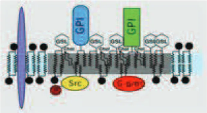
Biological membranes are complex two-dimensional structures arising from the assembly of various species of lipid and membrane proteins.
Figure 12: Plasma membrane structure.
The fluid mosaic model proposed by Singer and Nicolson (Singer et al., 1972), according to which the cell membranes are fluid and characterized by a random distribution of the molecular components (lipids and proteins) to form a homogeneous structure, has been revisited in years. In fact, several studies suggest that , instead of being fluid and uniform, plasma membranes are characterized by the presence of highly specialized regions, particularly enriched in sphingolipids and cholesterol, named Rafts. These microdomains play an important role in various cellular processes such as membrane trafficking, signal transduction and regulation of the activity of membrane proteins.
Originally called detergent-resistant membranes (DRM), the lipid rafts are defined small (10-200 nm), heterogeneous, highly dynamic, enriched in cholesterol and sphingolipids domains which act as platforms both to concentrate and segregate signaling molecules (Simons et al., 1997) and to compartmentalize cellular processes. In fact, the cell membranes are complex in the composition but highly precise in the purpose.