UNIVERSITÀ DEGLI STUDI DI ROMA
"TOR VERGATA"
FACOLTA' DI MEDICINA
DOTTORATO DI RICERCA IN
BIOTECNOLOGIE DELLA RIPRODUZIONE E DELLO SVILLUPPO
CICLO DEL CORSO DI DOTTORATO
XX
Titolo della tesi
REGOLAZIONE DELL'ESPRESSIONE GENICA NELLECELLULE GERMINALI MASCHILI
Nome e Cognome del dottorando
ANNARITA DI SAURO
Docente Guida/Tutor: Prof. Pellegrino Rossi - Prof. Claudio Sette
Coordinatore: Prof. Raffaele Geremia
INDEX
INTRODUCTION ... 3
I) Spermatogenesis... 4
I.1) Generals... 4
I.2) Transcription Regulation... 7
I.3) Translation Regulation ... 10
II) Regulation of Cap Dependent Translation... 18
RESULTS ... 24
I) [35S]methionine incorporation in isolated testis germ cells ... 25
II) Immunohistochemistry analisys of adult mice testis sections ... 28
III) Analysis of the expression of factors involved in the control of translational activity .. 29
IV) Analysis of [35S]methionine incorporation in cells treated with inhibitors of protein synthesis... 31
V) Analysis of cap-dependent initiation complex formation in the presence of rapamycin and Mnk inhibitor ... 32
VI) Analisys of translational activity of spermatocytes after ocadaic acid (OA) treatment 34 VII) Analysis of formation of the translational initiation complex in different germ cell types ... 37
DISCUSSION ... 39
MATERIALS AND METHODS ... 44
Cell isolation, culture and treatment ... 45
Preparation of cell extracts and Western blot analysis. ... 46
7-methyl-GTP-Sepharose chromatography. ... 46
REFERENCES... 48
ONGOING PROJECT ... 55
Ongoing Project: RNA Interference in Vivo to study the role of Germ cell specific proteins ... 56
Transcriptome analysis of differentiating spermatogonia ... 69 stimulated with kit ligand ... 69 Dynamic Regulation of Histone H3 Methylation at Lysine 4 in Mammalian
I) Spermatogenesis
I.1) Generals
In male mammals, development and differentiation of the germline is a dynamic process. Primordial germ cells (PGCs) migrate to the developing testis where they form prospermatogonia that subsequently enter mitotic arrest and remain in this state for the duration of fetal development. Shortly after birth in the mouse, these cells resume mitotic activity and a subset of these cells seed basal compartments of the developing seminiferous tubules to form stem spermatogonia. The spermatogonial stem cell population replicates mitotically to both maintain itself and ultimately give rise to differentiating spermatogonia. These cells then enter meiosis as primary spermatocytes that proceed through the first and second meiotic divisions to yield postmeiotic spermatids that differentiate via the process of spermiogenesis to form spermatozoa. (Jeremy Wang et al, 2005).
Spermatogenesis is the differentiation process of male germ cells to produce mature spermatozoa, and is divided into three distinct stages: the mitotic proliferation of spermatogonial stem cells, meiotic division of spermatocytes, and spermiogenesis of haploid spermatids (Zirkin, 1993). In mice, the proliferation of spermatogonia occurs soon after birth, at around 5 days post-partum (dpp), and this is followed by the first wave of spermatogenic differentiation, which gives rise to primary spermatocytes at around 10 dpp, haploid round spermatids at 20 dpp, and mature spermatozoa at 35 dpp (Bellve et al., 1977; Zhao and Garbers, 2002). This differentiation takes place in the seminiferous tubules, in which spermatogonia and Sertoli cells sit on the basement membrane, with spermatocytes interior to spermatogonia, and spermatids and mature spermatozoa facing the lumen (Figs. 1A and B). Through the continuous proliferation of spermatogonia and subsequent differentiation, functional spermatozoa are produced throughout male life. (Shoji et al, 2005)
Figura 1 (A) Schematic representation of spermatogenesis in mouse seminiferous tubule. (B)
Differentiation time course of the first wave of spermatogenesis.
Spermiogenesis is divided into 16 steps in mice and 19 steps in rats based partially on morphological features of the nucleus and acrosome, an spermatogenic cell-specific (SCS) secretory organelle derived from the Golgi apparatus (Russell et al., 1990). Cells in steps 1±8, round spermatids, have round transcriptionally active nuclei, whereas cells in steps 9±11, elongating spermatids, undergo nuclear elongation and progressive transcriptional inactivation. Transcriptional activity is not detectable by electron microscopic visualization of transcription in elongated spermatids in mice (steps 12±16), a very sensitive procedure (Kierszenbaum and Tres, 1975). Spermiogenesis ends with the elimination of the cytoplasm as a residual body and the release of spermatozoa into the lumen of the seminiferous tubule. The testis also has several somatic cell types. The development of male germ cells is intimately associated with Sertoli cells, which form the basal membrane of the seminiferous tubules. Several interstitial cell types, including Leydig cells (which produce testosterone) lie between the tubules (Russell et al., 1990).
The differentiation of spermatozoa involves profound changes in the structure of organelles and the synthesis of many SCS proteins. Some of the genes encoding SCS proteins are not homologous to any genes that are expressed in somatic cells. For example, the transition proteins and protamines, a family of highly basic chromosomal structural proteins that sequentially replace the histones during elongating and elongated spermatids, convert
chromatin from a nucleosomal organization to smooth fibrils (Meistrich,1989). (Kleene C. K, 2001)
I.2) Transcription Regulation
Transcriptional and translational control plays a crucial role during the spermatogenesis. The pattern of ribonucleic acid synthesis during germ cell development from the stem cell to the mature spermatid was studied in the mouse testis, by using uridine-H3 labelling. Autoradiographic studies in the mouse (Monesi, 1964, 1965, 1971) have shown that the incorporation of tritiated precursors into the RNA, occurs in spermatogonia. RNA synthesis is a continuous process throughout the cell division cycle in spermatogonia and stops only for a very short interval (1 hour) during metaphase and anaphase. (Monesi 1964). The rate of RNA synthesis in the autosomes is very low in early meiotic prophase: leptotene, zygotene and in the first half of pachytene, then rises rapidly to a peak in middle-late pachytene and decreses again in diplotene until a complete arrest in diakinesis and metaphase-anaphase1. After meiosis, RNA synthesis is resumed in early spermiogenesis but stops completely in mid-spermiogenesis after the beginning of nuclear elongation ( Monesi et al 1978) Fig 2
Figure 2. Pattern of >3H@uridine and >3H@arginine incorporation into RNA and nuclear protein, respectively, 1h after intratesticular administration of radioactive precursors.
Pachytene spermatocytes
Round spermatids Elongating spermatids Mitotic
spermatogonia
(arginine (uridine
Expression profiling analysis has been conducted with RNA from testes of animals of different ages (Almstrup et al., 2004; Shima et al., 2004) or with RNA from highly enriched cell populations (Schlecht et al., 2004). A recent study of RNA from purified cell types reported 405 transcripts expressed in mitotic spermatogonial cells (Schlecht et al., 2004). Over 200 of the transcripts in this study were uncharacterized mRNAs that were upregulated in spermatogonia. Among about 60 loci associated with spermatogonia in the literature were messages of genes required for cell cycle regulation (Ccnd2), components of extracellular matrix (Col3a1 and Mgp), hormone signal transduction (Cfgf, Egr1, Fgfr1, Igfbp2, Igfpb3, Pdgfa, and Vegf), and serum response and transcriptional regulation (Jun, JunB, Id2, Klf9, Stat3, and Zfp36) Expression levels of about one-half of the genes increase in spermatogonia and decrease at later meiotic stages including some histones (H1f0 and H3f3b), ribosomal proteins (Rpl35, Rps3, and Rps4), and motor proteins (Mrlcb, Myh9, Mylc2a, Tpm1, and Tpm4) (Schlecht et al., 2004).
Similar progress has been made in the study of control of gene expression in spermatocytes. DNA replication does not occur in spermatocytes, but DNA repair is critical during this time period. A more recent study reveals that approximately 442 transcripts are highly induced in spermatocytes (Schlecht et al., 2004). Transcripts identified in that study include genes involved in synaptonemal complex (SC) formation (Sycp2 and Sycp3), DNA repair (Polh), and chromatin condensation (topoisomerase 2a, Top2a). The metalloproteases (Adam2, Adam3, and Adam5), factors necessary for ubiquitin-mediated protein degradation (Ube2d2 and Psmc3), transcriptional regulators (Crem and Miz1), and enzymes involved in energy metabolism (Cyct and Ldhc) are expressed. (Grimes 2004).
Six novel genes specifically expressed in pachytene spermatocytes has been observed: a chromatin remodeling factor (chrac1/YCL1), a homeobox gene (hmx1), a novel G-coupled receptor for an unknown ligand (Gpr19), a glycoprotein of the intestinal epithelium (mucin 3), a novel RAS activator (Ranbp9), and the A630056B21Rik gene (predicted to encode a novel zinc finger protein). (Rossi et al., 2004)
Another factor that has been found to have a differentially expression in the postnatal testis of mouse is the c-kit transmembrane tyrosine-kinase receptor . It is expressed in type A spermatogonia and its transcription ceases at the meiotic phase of spermatogenesis. On the other hand alternative, shorter c-kit transcript is expressed in post-meiotic germ cell. This
transcript encodes a truncated version of the c-kit protein (tr-kit), lacking the extracellular, the transmembrane and part of the intracellular tyrosine kinase domain. It contains only the phosphotransferase domain and the carboxyterminal tail of c-kit and it is catalytically inactive because it lacks the ATP binding site (Rossi et al., 1992)
The alternative mRNA is transcribed from an haploid-specific alternative promoter localized within the 16th intron of the c-kit gene, is active in postmeiotic haploid cells and the protein is expressed in spermatids and mature spermatozoa (Rossi et al., 1992; Albanesi et al., 1996) Many experimental works allow to candidate tr-kit as a sperm factor at fertilization. Infact the microinjection of recombinant tr-kit protein or of synthetic tr-kit RNA in MII oocytes induces: the parthenogenetic activation of the oocytes and the extrusion of the 2nd polar body, the formation of pronuclei and the reaching of two blastomere stage in the activated eggs (Sette et al., 2004). The microinjection of tr-kit into metaphase-arrested oocytes triggers cell cycle resumption via the sequential activation of Fyn and PLCȖ1 (Sette et al., 2002). Several data suggest that tr-kit promotes the formation of a multimolecular complex composed of Fyn, PLCȖ1, and Sam68, which allows phosphorylation of PLCȖ1 by Fyn, and may modulate RNA metabolism (Paronetto et al., 2003)
I.3) Translation Regulation
Protein synthesis occurs at nearly all stages of spermatogenesis in the mouse, with elevated incorporation of labeled amino acids during the pachytene stage of meiosis (Monesi, 1965, 1967). Other proteins are synthesized in a stage specific manner during meiosis and spermiogenesis are retained in spermatozoa (O’Brien and Bellvè. 1980 a,b). These include a number of germ cell specific proteins such as lactate dehydrogenase C4, synthesized at maximal levels in late pachytene spermatocytes and both protamines and phosphoglycerate kinase B which are sinthesized during the haploid stages.
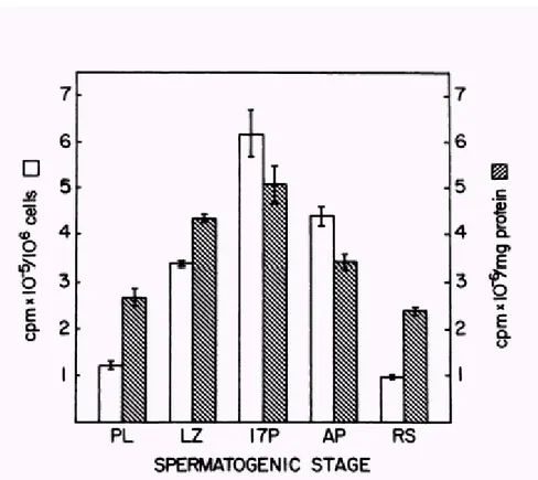
An accurate analysis of the stage specific protein synthesis by isolated spermatogenic cells throughout meiosis and early spermiogenesis in the mouse has been made by O’Brien (1987). Spermatogenic cells from prepubertal and adult mice were separated by unit gravity sedimentation. Pre-leptotene, leptotene/zygotene, and pachytene spermatocytes were isolated from 17-day-old mice. Adult pachytene spermatocytes and round spermatids were isolated from mature animals. These germ cells were then cultured in defined medium with [35S] methionine for 4-5 h. For each cell type, relative [35S] met incorporation was determined and labelled proteins were compared by two dimensional (2D) polyacrilammide gel electrophoresis and autoradiography. Levels of [35S] met incorporation by isolated germ cells correlate closely with previous autoradiographic estimates of protein synthesis during spermatogenesis (Monesi, 1967). Pachytene spermatocytes from prepuberal mice incorporate the highest levels of [35S] met. Similar patterns of incorporation were observed when the data were expressed as cpm/106 cells or cpm/mg protein. However, greater differences between cell types were apparent when incorporation was expressed per 106 cells, reflecting the marked changes in cell size and protein content that occur during germ cell differentiation. Preleptotene, leptotene/zygotene, and 17d pachytene spermatocytes exhibited progressive increase in incorporation that correlated with advancing stages of meiosis. Maximal levels of incorporation were detected for 17d pachytene spermatocytes. Adult pachytene spermatocytes incorporated §30% less [35S] met than the 17 pachytene population. On a per cell basis, pachytene spermatocytes incorporated 4-6 times more [35S] met than round spermatids. (Fig 3)
Figure 3. Incorporation of >35S@methionine >35S@met into trichloracetic acid (TCA )-precipitabie macromolecules for purified spermatogenic cells. preleptotene (PL), leptotene/zygotene (LZ), and
pachytene (1 7P) spermatocytes were isolated from 17-day-old mice. Adult pachytene spermatocytes (AP) and round spermatids (RS) were isolated from mature animals. Cells were incubated with >35S@met (50PCi/ml) for 4 h at 320C prior to TCA precipitation. For each cell type, data are expressed both as incorporation/106 cells and as incorporation/mg protein. Bars represent mean values for triplicate determinations and vertical lines represent SE.
Comparisons of 2D autoradiograms indicated that many proteins, including actin and tubulins, are synthesized at approximately equal levels in all stages examined. Other proteins, including heat-shock proteins and multiple plasma membrane constituents, are synthesized in a stage specific manner in leptotene/zygotene spermatocytes, pachytene spermatocytes and round spermatids (O’Brien 1987)
At the onset of spermiogenesis, transcription and translation become temporally uncoupled because elongating spermatids are transcriptionally incompetent for remodelling and condensation of the chromatin due to replacement by protamines of DNA-binding histones (reviewed in Steger 1999)
During spermiogenesis infact, transcription ceases prior to the differentiation of the mature cells. To complete germ cell differentiation and initiate early embryogenesis, proteins are synthesized from pre-existing mRNAs that are stored for several days. Some mRNAs are stored for more than a week before they are recruited for translation at specific times during sperm morphogenesis (Braun 2000).
De novo protein synthesis is ensured by the mRNAs that elongating spermatids inherit from primary spermatocytes and round spermatids and that are translated several days after their synthesis (Braun, 2000).
The pattern of translational activity during spermatogenesis varies according to the cellular differentiation of germ cells (Kleene, 1996). Similarly to proliferating somatic cells, mitotic spermatogonia readily utilize the transcribed mRNAs (Cataldo et al., 1999). By contrast, although the transcriptional activity of primary spermatocytes and round spermatids is copious, only a very small percentage (5–10%) of these mRNAs are actively translated and most of them are accumulated as ribonucleoproteins (RNPs; Biggiogera et al., 1990).
Most of the testicular mRNAs known to be translationally regulated are initially transcribed in postmeiotic cells. Because protein synthesis occurs on polysomes and translationally inactive mRNAs are sequestered as ribonucleoproteins (RNPs), movement of mRNAs between these fractions is indicative of translational up- and down-reguation (Iguchi et al., 2006)
Microarrays analysis of mRNAs in RNPs and polysomes from testis extracts of prepuberal and adult mice (17d and 22d) has been made to characterized the translational state of individual mRNAs as spermatogenesis proceeds. It was observed that many of the translationally delayed postmeiotic mRNAs shift from the RNPs into the polysomes: 742
mouse testicular transcript show a dramatic shift between RNPs and polysomes. One subgroup of 35 genes containing the known translationally delayed phosphoglicerate kinase 2 (Pgk2) is initially transcribed during meiosis and is translated in later stage cells. Another subgroup of 82 meiotically expressed genes is translationally down regulated late in spermatogenesis (Iguchi et al., 2006)
The fundamental role played by RNPs during gametogenesis is suggested by their high levels of expression in germ cells (Venables and Eperon, 1999).
Accordingly, knockout mouse models for RNA-binding proteins expressed in different
phases of gametogenesis, like MSY2, Tenr, Miwi, Mili, and Dazl (Deng and Lin, 2002; Saunders et al., 2003; Kuramochi-Miyagawa et al., 2004; Connolly et al., 2005; Yang et al., 2005), display a sterile phenotype. Remarkably, gametogenesis is under a tight translational control of gene expression by RNA-binding proteins also in lower organisms like Caenorhabditis elegans and Drosophila melanogaster (Kuersten and Goodwin, 2003).
About two-thirds of all mRNAs in adult mammalian testes is mRNP particle-associated (Kleene 1993, 1996; Cataldo et al. 1999; Schmidt et al. 1999). While some mRNP particle-associated mRNAs share conserved sequences others exhibit no obvious sequence similarities (Kleene 1996).
In mouse and rat, both mRNP particles and chromatoid bodies, electron dense material in the vicinity of the nucleus, can be observed in pachytene spermatocytes and round spermatids (Biggiogera et al. 1990; Moussa et al. 1994). Within chromatoid bodies, mRNAs have been demonstrated by 3H-uridine incorporation (Söderström 1981), ethidium bromide phosphotungstic acid staining (Biggiogera et al. 1990), and immunocytochemistry. Therefore, it is assumed that chromatoid bodies may serve as storage organelle for translationally repressed mRNAs (Review Steger 2001)
Among the mRNAs that are under translation control in spermatids are protamine 1 (Prm1) and 2 (Prm2). The protamines mediate nuclear condensation during the terminal stages of spermatid differentiation. They substitute the nucleosomal histones operating a profound remodelling of the chromatin and rendering the nucleus more compact and unable to transcribe new messengers (Sassone- Corsi., 2002) (Fig 4). Translational delay of the Prm1 message is essential for normal spermatogenesis in mammals.
Figure 4 Histone-to-protamine exchange during spermatogenesis requires three
developmental steps: first, part of somatic histones are replaced by testis-specific histones; second, both somatic histones and testis-specific histones are exchanged by transition proteins; third, transition proteins are replaced by protamines. The DNA protamine-interactions result in increased chromatin condensation causing cessation of transcription. Therefore, in haploid spermatids, gene expression requires temporal uncoupling of the processes of transcription and translation (SCN Sertoli cell nucleus, Sg spermatogonia, Scy I primary spermatocyte, Scy II secondary spermatocyte, rSpd round spermatid eSpd elongated spermatid)
Mutations that result in premature translation of the Prm1 mRNA cause precocious nuclear condensation and sterility (Lee et al., 1995)
Like many localized or stored mRNAs, sequences responsible for the translational silencing of Prm1 lie within the 3’ UTR of its mRNA (Braun et al., 1989). The 17-nucleotide translational control element (TCE) mediates translational repression of the Prm1 mRNA. Mutation of the TCE causes premature synthesis of protamine protein and sterility. The Prm1 mRNA is stored as a cytoplasmatic ribonucleoprotein (mRNP) particle in spermatids. Contained within the particle are several members of the Y box family of nucleic acid binding proteins. The murine Y box proteins MSY1, MSY2 and MSY4 bind in a sequence-dependent manner to a conserved region in the proximal portion of the Prm1 3’ UTR. Sequence-specific binding by MYS4 to the Y box recognition sequence (YRS) is dependent on the highly conserved cold shock domain, possibly through the RNP1 and RNP2 motifs present within it. The Y box proteins may function as translational repressor in vivo. Alternatively their primary function may be to protect mRNAs from degradation during their extended period of storage. Translational activation of stored mRNAs is essential for the completion of gametogenesis. Proper translational activation of the Prm1 in elongated spermatids requires the cytoplasmatic double-stranded RNA binding protein TARBP2. Tarbp2 is expressed at low levels in many cells but is expressed at robust levels in late stage meiotic cells and in postmeiotic spermatids. Mice mutant for Tarbp2 are defective in proper translational activation of the Prm1 and Prm2 mRNAs and are sterile. (Braun 2000)
Another protein that may be implicated in translational control of spermatogenesis is Sam68, a protein belonging to the STAR family (signal transduction and activation of RNA metabolism), involved in RNA homeostasis (Vernet and Artzt 1997) STAR proteins are conserved across eukaryotes and may represent a direct link between signal transduction pathways and RNA metabolism. They bind RNA through a GSG (Grp33/Sam68/GLD-1) domain, which contains a single KH domain (hnRNP K homology domain) flanked by conserved N- and C-terminal sequences named QUA1 and QUA2 domains required for homodimerization and specificity in RNA binding (Vernet and Artzt, 1997; Lukong and Richard, 2003). It has been observed that Sam68 is down-regulated at the onset of meiosis and it accumulates again during the pachytene stage and until spermiogenesis begins (Paronetto et al., 2006). Interestingly, Sam68 is localized in the cytoplasm during the meiotic divisions and
it associates with the polysomes engaged in active translation Translocation of Sam68 from the nucleus to the polysomes correlates with its phosphorylation by ERK1/2 and MPF. Moreover, direct cloning experiments reveals that Sam68 binds mRNAs encoded by genes required for the spermatogenetic program. Thus, these studies suggest a novel role for Sam68 as mRNA carrier that shuttles from the nucleus to the cytoplasm and may facilitate translation during the meiotic divisions. (Paronetto et al., 2006)
Figure 5. Sam68 cosediments with the polysomes in secondary spermatocytes. Germ cells
isolated from mouse testis by elutriation were analyzed for DNA content by FACS. Cells from fraction 5 (A), 4 (B), or 3 (C) were fixed in 1% paraformaldehyde and stained with propidium iodide for the analysis. Nuclei of cells obtained from the same fractions were processed for cytological analysis and stained with Giemsa (D–F). Fractionation on sucrose gradients of cell extracts obtained from fraction 5 (G), or 4 (H), or 3 (I): absorbance profiles at 254 nm show the distribution of soluble RNP, single ribosome and polysomes (top panels); Western blot analyses of each fraction from the gradients indicate the distribution of Sam68 (second row panels), of the ribosomal S6 protein (third row panels), used as standard of distribution of the ribosome (80S) and the polysomes, and of initiation factor eIF4E (bottom panels), used as standard of distribution of smaller RNPs.
II) Regulation of Cap Dependent Translation
Translational control plays a key role in temporal regulation of development events that must be executed largely in the absence of transcription. Emerging evidence suggests that distinct mechanisms of translational repression and activation act on specific mRNAs at different steps in the cell cycle progression.
A key regulatory point for translational control in eukaryotes is initiation, instigated by binding of the translational initiation complex to the 5’ cap of the mRNA, leading to recruitement of the ribosomal subunits (Backer and Fuller., 2007)
Ribosome binding is facilitated by a number of translation initiation factors that guide the ribosome to an mRNA’5 end, except for mRNAs which initiate by binding to an internal ribosome-binding site (IRES). The 5’ end of all nuclear-transcribed mRNAs possess a cap structure (m7GpppN, in which “m” represents a methyl group and “N”, any nucleotide) that is specifically recognized by eukaryotic translation initiation factor 4E (eIF4E). eIF4E binds the 5’ cap as a subunit of a complex (termed eIF4F) containing two other proteins, one of two large scaffolding proteins, termed eIF4GI and eIF4GII
(encoded by two different genes), and the RNA helicase eIF4A (Review Sonenberg 2006)). Following its binding to the 5’ cap, eIF4F is thought to unwind the mRNA 5’-proximal secondary structure to facilitate the binding of the 40S ribosomal subunit in association with several other initiation factors (Gingras et al. 1999b). Unwinding requires another initiation factor, eIF4B (Hershey and Merrick., 2000)
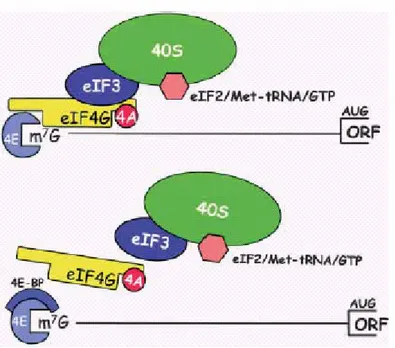
Figure 6. (A) Assembly of the mammalian ribosome initiation complex at the 5’end of an
mRNA. eIF4E, as part of the eIF4F complex, binds them7G-cap structure. eIF4G binds eIF3, which, in turn, recruits the 40S ribosomal subunit along with its associated ternary complex (eIF2/Met-tRNA/GTP). Not shown are other initiation factors that participate in ribosome recruitment. 4E-BPs binds the dorsal convex surface of eIF4E to prevent its interaction with eIF4G, thereby abrogating ribosome binding.
Strikingly, several components of the ribosome recruitment machinery as well as ribosomal components are either direct or indirect targets of mTOR. These include eIF4B, eIF4G, and eIF4E, the latter of which is activated by the phosphorylation of its repressors, the 4EBP proteins. In addition, S6K and its targets, the ribosomal protein S6 and elongation factor 2 (eIF2Į), are also targets of this pathway.
The interaction between eIF4E and eIF4G is regulated by members of the eIF4E-binding proteins (4E-BPs), a family of translational repressor proteins. The mammalian family consists of three low molecular weight proteins, 4E-BP1, 4E-BP2, and 4E-BP3, encoded by three separate genes.
The 4E-BPs compete with eIF4G proteins for an overlapping binding site on eIF4E, such that the binding of a 4E-BP or an eIF4G protein is mutually exclusive (Haghighat et al. 1995; Mader et al. 1995; Marcotrigiano et al. 1999).
Whereas hypophosphorylated 4E-BPs bind with high affinity to eIF4E, the hyperphosphorylation of 4E-BPs prevents this interaction (Sonenberg., 2006).
mTOR regulates protein synthesis through the phosphorylation and inactivation of the repressor of mRNA translation, eukaryotic initiation factor 4E-binding protein (4E-BP1), and through the phosphorylation and activation of S6 kinase (S6K1). These two downstream effectors of mTOR whose phosphorylation is inhibited by rapamycin in vivo, can be phosphorylated by recombinant mTOR in vitro (Brunn et al. 1997; Burnett et al. 1998). Moreover, substitution of Asp 2338 with alanine in the catalytic domain of mTOR is sufficient to inhibit mTOR kinase activity toward S6K1 and 4E-BP1 in vivo and in vitro.
Thus, S6K1 or 4E-BP1 phosphorylation is often used as an in vivo readout of mTOR activity (Sonenberg., 2006).
The most widely used inhibitor of mTOR activity is rapamycin, a natural compound that acts as non competitive inhibitor. It acts by forming an inhibitory complex with its intracellular receptor, the FK506-binding protein, FKBP12, which binds a region in the C terminus of TOR proteins termed FRB (FKB12–rapamycin binding), thereby inhibiting TOR activity (Chen et al. 1995; Choi et al. 1996).
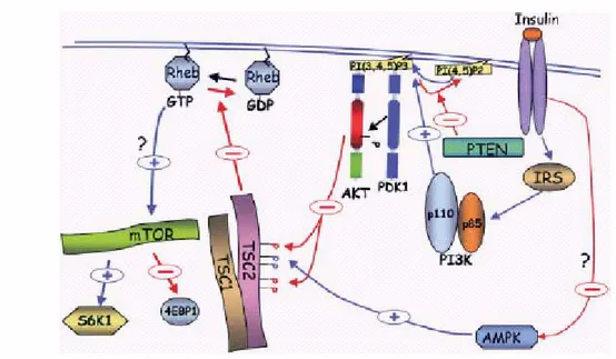
The mammalian ortholog of the yeast TOR proteins was independently cloned and identified by using an FKBP12–rapamycin affinity purification by four groups and named FRAP (FKBP–rapamycin-associated protein) RAFT1 (rapamycin and FKBP target), or RAPT1 rapamycin target; (Brown et al. 1994;). mTOR is the master regulator of cap-dependent mRNA translation and ribosomal biogenesis. Its activity is regulated by growth factors by the PI3K/Akt signalling pathway. This pathway lead to phosphorylation and inhibition of TSC2 by Akt and to the subsequent activation of Rheb, which activates mTOR by an as yet unknown mechanism Fig 7 (Sonenberg., 2006)
Figure 7. The regulation mTOR activity by growth factors is mediated by the PI3K/Akt
signaling pathway leading to phosphorylation and inhibition of TSC2 by Akt and to the subsequent activation of Rheb, which activates mTOR by an as yet unknown mechanism. In addition, TSC2 is activated by AMPK (+) Activation; (-) inhibition.
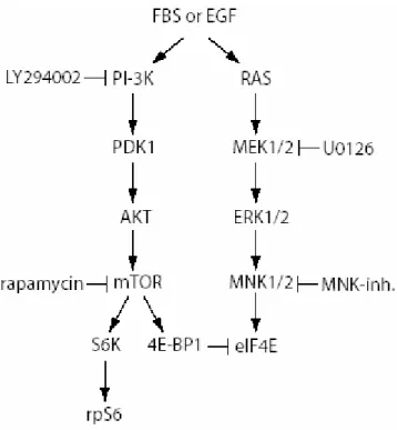
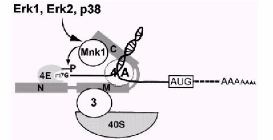
The RAS/MAPK pathway integrates mTOR function in the activation of the eIF4F complex. Its activation cause the activation of MAP Kinase interacting Kinase 1 (MNK1), the kinase that phosphorylates eIF4E on serine 209 (Sonenberg., 2006) (Fig 8)
Figure 8. Schematic representation of the signal transduction pathways involved in the
regulation of mRNA translation mediated by growth factors. Only some mediators of PI3K/Akt/mTOR and MAPK pathways are indicated. The inhibitors used in this study and their targets are indicated.
Mnk1 is associated with the eIF4F complex via its interaction with the C-terminal region of eIF4G. According with the model proposed by Pyronnet and coauthours (Embo.,1999), the phosphorylation of eIF4E occurs in the eIF4F complex, suggesting that eIF4F assembles prior to eIF4E phosphorylation. eIF4E can interact in a mutually exclusive manner with either eIF4G or 4EBPs. The association of eIF4E with eIF4Gs enhances binding of eIF4E to the 5’
eIF4E might enhance its affinity for eIF4Gs and stabilize its interaction with the mRNA 5’ end (Fig 9).
Figure 9. Mnk1 phosphorylates eIF4E as a component of the eIF4F complex. The model
shows that phosphorylation of eIF4E occurs in the eIF4F complex, suggesting that eIF4F assembles prior to eIF4E
phosphorylation. eIF4E can interact in a mutually exclusive manner with either eIF4Gs or 4E-BPs. The association of eIF4E with eIF4Gs enhances binding of eIF4E to the 59 cap structure (Haghighat and
Sonenberg, 1997; Ptushkina et al., 1998) and brings Mnk1 into the vicinity of eIF4E. The resulting phosphorylation of eIF4E might enhance its affinity for eIF4Gs (Bu et al., 1993) and stabilize its
Results
Study of the translational activity during the various differentiation steps of spermatogenesis.
An important question, regarding how sequential developmental events can be ordered by translational control, is how the translational initiation machinery can become targeted to and activated at specific subsets of mRNAs, and how this machinery might change in different cell types and stages during spermatogenesis.
Indeed, an important mechanism that controls differentiative events during spermatogenesis is exerted at the translational regulation level.
Translational control plays a key role in temporal regulation of developmental events that must be executed largely in the absence of transcription. As stated in the introduction section, it has been observed that pachytene spermatocytes incorporate the highest levels of [35S]methionine indicating a major protein syntesis in these cells compared to round spermatids (O’Brien 1987)
We have studied whether and how these data correlate with biochemical parameters that characterize the control operated at the level of the translational initiation machinery.
I) [35S]methionine incorporation in isolated testis germ cells
Before analysing the translational activity at the level of the formation of the initiation machinery, we re-evaluated the rate of [35S]methionine incorporation in germ cells at different developmental stages.
Germ cells were isolated from adult mice testis by elutriation technique to obtain cell populations enriched in specific cellular types. Isolated spermatocytes, round spermatids and elongating spermatids, were used to examine alterations in [35S]methionine incorporation
during differentiation. In these experiments cells were cultured for 1h in MEM medium in the presence of [35S]met.
Fig 1, shows that the [35S]met incorporation into TCA precipitable proteins detectable in different cellular types is maximal for spermatocytes when expressed either as cpm/106cells or as cpm/mg protein. Round spermatids and elongating spermatids incorporate less [35S]met than spermatocytes. However, greater differences between cell types were apparent when incorporation was expressed on a per cell base, reflecting the marked changes in cell size and protein content that occur during germ cell differentiation, as already published by O’Brien (1987)
Similar variations in [35S]met incorporation were observed in two separate triplicate experiments.
[35S]met incorporation in elongating spermatids is slightly more elevated compared with round spermatids. 0 50 100 150 200 250 300 350 400 450 cpm/ncellx(10x10³) cpm/prote cpm/ncellx(10x10³) 392 33,33 19,8 cpm/prote 169,1091954 64,41223833 79,33333333
sp.cytes sp.tides elong.sp.tides
Figure 1 shows that the [35S]met incorporation into TCA precipitable proteins detectable in different cellular types is maximal for spermatocytes when expressed either as cpm/106cells or as cpm/mg protein.
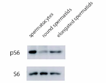
In perfect agreement with this observation, we found, by western blot, a strongly reduced level of S6 phosphorylation in round spermatids compared to spermatocytes, and an increase in elongating spermatids when compared to round spermatids, as showed in Figure 2. Indeed, phosphorylation of S6 is a well known marker of active translation. The western blot analysis was confirmed by immunocytochemistry studies on testis sections from adult mice.
Figure 2. Western Blot analysis reveals a reduced level of S6 phosphorylation in round
spermatids compared to spermatocytes, and an increase in elongating spermatids when compared to round spermatids
II) Immunohistochemistry analisys of adult mice testis sections
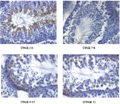
Figure 3 shows that strong immunoreactivity with an antibody directed against pS6240/244 was evident in middle-late pachytene spermatocytes (stage 9-10 of the cycle of theseminiferous epithelium), in spermatocytes undergoing the meiotic divisions (stage 12), in the cytoplasm of elongating spermatids (stage 2-6), whereas no or, at least, little staning was evident in lepto-zygotene (stages 9-10 and 12), early pachytene (stages 2-6), round spermatids (stage 2-6, 7-8, 9-10) and spermatozoa near to the spermiation (stage 7-8)
Figure 3 Immunohistochimestry analysis direct against pS6240/244 reveals a strong
immunoreactivity of antibody in middle-late pachytene spermatocytes (stage 9-10 of the cycle of the seminiferous epithelium), in spermatocytes undergoing the meiotic divisions (stage 12), in the cytoplasm of elongating spermatids (stage 2-6), wherease no or, at least, little staning was evident in lepto-zygotene (stages 9-10 and 12), early pachytene (stages 2-6), round spermatids (stage 2-6, 7-8, 9-10) and spermatozoa near to the spermiation (stage 7-8)
III) Analysis of the expression of factors involved in the control of
translational activity
To study at the biochemical level how translational control can operate in different cellular types during sequential developmental events of spermatogenesis, we analyzed the expression of some proteins involved in the pathway that modulate translational activity.
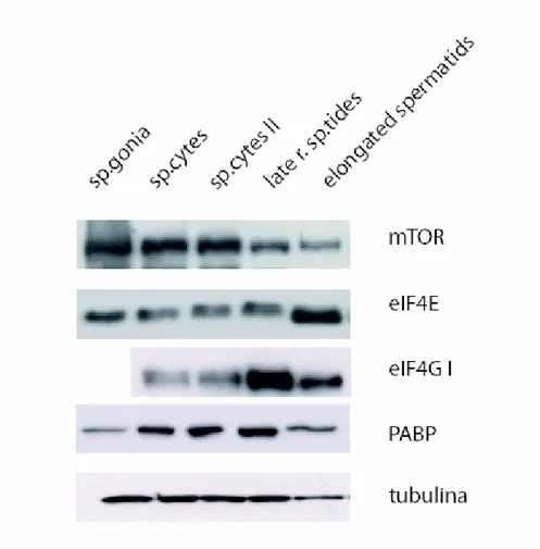
Figure 4 shows that all factors analysed are present in different cellular types. mTor expression decreases progressively in round spermatids and in elongating spermatids compared to spermatocytes, wherease eIF4E levels do not change significantly, eIF4G seems more abundant in round spermatids with respect to meiotic cells; PABP levels substantially do not change.
Figure 4 Analysis by western blot of expression of some proteins involved in the pathway that modulate translational activity.
IV) Analysis of [35S]methionine incorporation in cells treated with
inhibitors of protein synthesis
To evaluate whether the traslational ativity seen in spermatocytes, round spermatids is affected by the inhibition of mTor or Erk-dependent pathways, we performed [35S]methionine incorporation experiments after pre-incubation with specific inhibitors of these pathways, i.e. Rapamycin for the mTor pathway and Mnk inhibitor for Erk-dependent pathways.
Spermatocytes and round spermatids were isolated by elutriation technique and cultured in MEM medium in the precence of rapamycin, or Mnk inhibitor. Cells were treated with these drugs for 3 h, and [35S]met was added in the last hour of incubation. The analysis of [35S]met incorporation into TCA-precipitable proteins of spermatocytes revealed that both rapamycin and Mnk inhibitor reduce the [35S]met incorporation of § 20% compared to untreated control, whereas no apparent effect was evident in spermatids( Fig 5)
0 100 200 300 400 500 600
citi c citi rapa citi Mnk Tidi c Tidi rapa Tidi Mnk
cpm/n cell cpm/prot
Figure.5 Spermatocytes, round spermatids isolated by elutriation technique and cultured in MEM medium in the precence of rapamycin, or Mnk inhibitor for 3 h were analysed for [35S]met incorporation. [35S]met was added in the last hour of incubation.
V) Analysis of cap-dependent initiation complex formation in the presence
of rapamycin and Mnk inhibitor
To better understand the role of cap dependent translational activity in different cellular types during spermatogenesis, we analysed the assembly of the translation initiation complex by assaying binding of eIF4F components to m7-GTP-sepharose beads (mimicking the cap mRNA structure) in the presence or absence of rapamycin or Mnk inhibitor.
Spermatocytes, round spermatids and elongating spermatids were isolated from adult mouse testis and cultured for 3 h in medium supplemented with rapamycin or Mnk inhibitors. Cells were harvested and the extracts processed for binding to m7-GTP-sepharose beads.
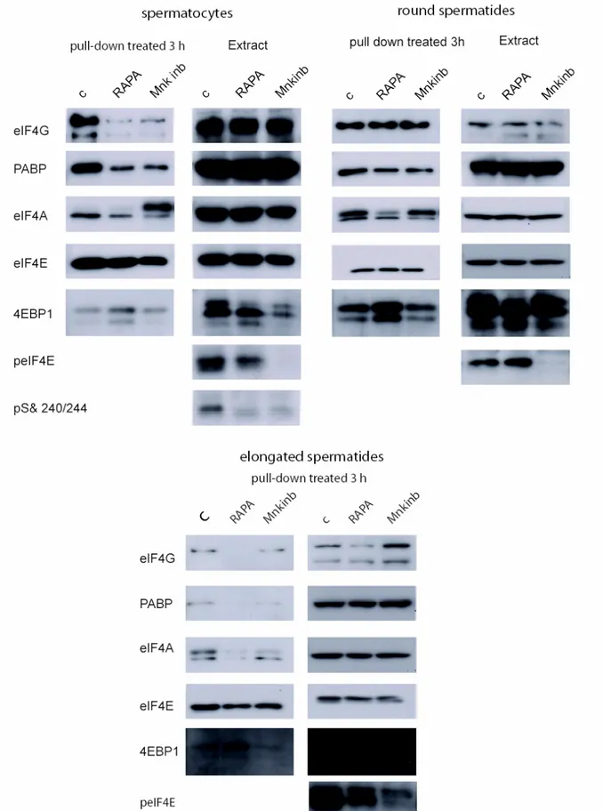
Figure 6 shows that treatment with both drugs decreases the binding of several components that take part to the translation initiation complex formation in spermatocytes, such as eIF4G, PABP, eIF4A and eIF4E. 4EBP1 (which competes with eIF4G for the binding site on eIF4E, thus inhibiting cap-dependent translational initiation) appears to increase its binding to m7 -GTP-sepharose beads after rapamycin, but not after Mnk inhibitor treatment, in agreement with the notion that it is specific target of phosphorylation by mTor. Indeed we also noticed in rapamycin treated samples an increase of a faster migrating 4EBP1 band, indicative of dephosphorylation. On the contrary eIF4E phosphorylation was completely blocked, as expected, by inhibition of Mnk. Finally both drugs inhibited S6 phosphorylation
In round spermatids no effect was observed by treatment with these drugs (with the exception of inhibition of eIF4E phosphorylation by the Mnk inhibitor), whereas sensitivity to both drugs was evident again in elongating spermatids.
Figure 6 Binding of eIF4F components to m7-GTP-sepharose beads in the presence or absence of rapamycin or Mnk inhibitor. Spermatocytes, round spermatids and elongating spermatids isolated from adult mouse testis and cultured for 3 h in medium supplemented with rapamycin or Mnk inhibitors were harvested and the extracts processed for binding to m7-GTP-sepharose beads
VI) Analisys of translational activity of spermatocytes after ocadaic acid
(OA) treatment
Treatment of spermatocytes with the phosphatase inhibitor ocadaic acid OA induces their meiotic progression, in particular it induces their entry into methaphase I (MI), characterized by normal condensation of bivalent chromosomes and synaptonemal complex breakdown (Wiltshire et al., 1995). It has been described that OA-induced meiotic progression requires activation of the maturation promoting factor (MPF; Wiltshire et al., 1995), a complex of cyclin-dependent kinase cdc2 and the regulatory subunit cyclin B1, and of the extracellular signal-regulated protein kinases (ERKs; Sette et al., 1999), also known as the mitogen-activated protein kinases (MAPKs). To determine whether OA treatment increase the translational activity concomitant with the entry of spermatocytes into the meiotic divisions induced by OA, we first evaluated the rate of phosphorylation of some factors which normally correlates with an increase in translational activity.
Figure 7 Phosphorylation rate analysis of some factors which normally correlates with an increase in translational activity. by western blot in spermatocytes treated with ocadaic acid (OA).
Another approach was to analyse the assembly of the translation initiation complex by assaying binding of eIF4F components to m7-GTP-sepharose beads after OA treatment.
Figure 8 shows a clear increase in the binding to m7-GTP-sepharose beads in spermatocytes of all the factors that take active part to formation of the translational initiation complex (eIF4G, PABP and eIF4A), whereas the binding of the initiation inhibitor 4EBP1 was reduced. We also noticed an increase of the phosphorylation state of 4EBP1.
Figure 8 Assay of binding of eIF4F components to m7-GTP-sepharose beads after OA treatment.
VII) Analysis of formation of the translational initiation complex in
different germ cell types
To investigate whether the difference in the rate of translational activity observed in spermatocytes, round spermatids and elongating spermatids by [35S]methionine incorporation correlates with the formation of the translational initiation complex, we performed an experiment to assay the assembly of the eIF4F complex to the cap mimicking mRNA structure. Spermatocytes, round spermatids and elongating spermatids isolated from adult mouse testis were harvested after 1h of culture in MEM medium and the extracts processed for binding to m7-GTP-sepharose beads.
Figure 9 shows that there is no substantial difference in the binding of eIF4G, PABP, eIF4A and eIF4E to m7-GTP-sepharose beads between spermatocytes and round spermatids, whereas an increase of the binding of these factors occurs in elongating spermatids.
On the contrary, 4EBP1 binding appears to be strongly reduced in elongating spermatids, but this appears to be due mainly to a strong decrease in the total amount of this protein in these cells. Also in this experiment the pattern of S6 phosphorylation state during spermatogenetic differentiation was similar to that described in fig 2 and 3.
Figure 9 Assay of the assembly of the eIF4F complex to the m7-GTP-sepharose beads.
Spermatocytes, round spermatids and elongating spermatids isolated from adult mouse testis were harvested after 1h of culture in MEM medium and the extracts processed for binding to -GTP-sepharose beads.
Discussion
Translational control is crucial for proper timing of developmental events that take place in the absence of transcription during spermatogenesis.
As stated in the introduction section, it has been observed that pachytene spermatocytes incorporate the highest levels of [35S]methionine indicating a major protein syntesis in these cells. The aim of this project was to understand whether biochemical data of translational activity correlate with those of [35S]methionine incorporations and to investigate how the translation activity is regulated at the molecular level, in particular at the level of the initiation complex formation, in different cell types during spermatogenesis.
First of all we re-evaluated the rate of [35S]methionine incorporation in germ cells at different developmental stages. We observed that the highest [35S]methionine incorporation is present in spermatocytes indicating a stronger translational activity in meiotic cells compared to haploid cells.
Elongating spermatids show a higher rate of [35S]met incorporation compared to that of round spermatids. In agreement with these data are the finding of high levels of S6 phosphorylation ( a well known marker of active translation ) in spermatocytes, which is reduced in round spermatids, and which increase again in elongating spermatids, as evaluated by immunoblot analisys. Immunocytochemical analisys indicate a strong level of S6 phosphorylation in middle-late pachytene spermatocytes and in spermatocytes undergoing the meiotic divisions and in elongated spermatids. Immunoreactivity is low in early meiotic cells and in the cytoplasm of round spermatids and spermatozoa near to the spermiation step.
All these data indicate that during spermatogenesis there are two moments of high translational activity, one in late pachytene spermatocytes and one during spermatid elongation.
Thus, after a period of low protein syntesis at the beginning of meiosis, two periods of high translational activity are separated by a period of reduced protein synthesis corresponding with the beginning of the haploid stage of spermatogenesis.
Active translation in late pachytene spermatocytes, is supported by the increase of phosphorylation state of Akt, S6 and eIF4E after OA treatment, which is known to push meiotic progression of spermatocytes and their entry into methaphase I (MI).
Moreover, after OA treatment, we observed an increase in the binding to m7-GTP-sepharose beads of all the factors that take active part to the formation of the translational initiation complex (eIF4G, PABP and eIF4A) and a concomitant increase of the phosphorylation state 4EBP1, an inhibitor of the eIF4F complex formation which correlates with its reduced binding to capped mRNA.
After having evaluated the rate of translational activity in different cell types, we investigated the presence of some factors involved in the pathway that modulate translational activity, such as mTor, eIF4E, eIF4G and PABP, and found that they are expressed during all differentiative stages of spermatogenesis. The most evident difference was a decrease in the expression of mTor in the haploid stages.
In the [35S]methionine incorporation experiments made on spermatocytes and round spermatids after pre-incubation with rapamycin or Mnk inhibitor, we observed a reduction of protein synthesis of § 20% in spermatocytes, whereas no apparent effect was evident in round spermatids.
These data suggest that, while translational activity of spermatocytes is partially regulated by mTor and Erk pathways, this regulation is not occurring in round spermatids. These results were confirmed by binding assay of eIF4F components to m7-GTP-sepharose beads (mimicking the cap mRNA structure) in the presence or absence of rapamycin or Mnk inhibitor. Treatment with both drugs decreases the binding of eIF4G, PABP, eIF4A and eIF4E that take part to the translation initiation complex formation in spermatocytes, and increases the binding of 4EBP1 ( competitive inhibitor of eIF4G).
In round spermatids no effect was observed by treatment with these drugs confirming their indipendence from mTor and Erk pathways
However these pathways appeared to be active again in elongating spermatids.
These data would suggest that the lower level of translational activity in round spermatids with respect to spermatocytes and elongating spermatids is mainly due to a decrease in cap dependent translation.
However this conclusion is not supported by the analysis of the binding of initiation factors to m7-GTP-sepharose beads (mimicking the cap mRNA structure), which shows that there are no apparent differences between spermatocytes and round spermatids. Thus, a delay in the elongation step of translation, rather than reduced formation of initiation complexes, might account for the decrease in the rate of protein synthesis at the beginning of the haploid phase of spermatogenesis. Indeed, one would expect that, in the absence of active elongation, preformed initiation complexes are not disassembled, and this could explain the lack of effect of rapamycin and Mnk inhibitor in round spermatids. Infect mTor and Erk dependent phosphorylations are dynamically required to reform continuously initiation complexes at active translation sites (Figure 10).
On the other hand, we found a clear increase in the amount of initiation complex in elongating spermatids, accompanied by a strong reduction in the levels of the 4EBP1 inhibitor. This increase in the formation of initiation complexes correlates positively to the new increase in S6 phosphorylation and protein synthesis in the final steps of spermiogenesis, and might compensate the reduced efficiency of elongation in the haploid stages of spermatogenesis.
Figure 10 Hypothetical mechanism of translation activity of spermatocytes (A) and round spermatids (B): The CAP-dependent translation is active in spermatocytes (A) and inhibited in round spermatids (B) probably due to the absence of active elongation in these cells.
Cell isolation, culture and treatment
Testes of adult CD1 mice (Charles River Italia) were used to prepare pachytene spermatocytes, round spermatids and residual elongated spermatids. After dissection of the albuginea membrane, testes were digested for 15 min in 0.25% (w/v) collagenase (type IX, Sigma) at room temperature under constant shaking. Digestion was followed by two washes in minimum essential medium, hence seminiferous tubules further digested in minimum essential medium containing 1 mg/ml trypsin for 30 min at 30 °C. Digestion was stopped by adding 10% fetal calf serum and the released germ cells were collected after sedimentation (10 min at room temperature) of tissue debris. Germ cells were centrifuged for 10 min at 1,500 rpm at 4 °C and the pellet resuspended in 20 ml of elutriation medium (120.1 mM NaCl, 4.8 mM KCl, 25.2 mM NaHCO3, 1.2 mM KH2PO4, 1.2 mM MgSO4(7H2O), 1.3 mM CaCl2, 11 mM glucose, 1 3 essential amino acid (Life Technologies, Inc.), penicillin, streptomycin, 0.5% bovine serum albumin). Germ cells at pachytene spermatocyte, round spermatid, and elongated spermatid steps were obtained by elutriation of the unfractionated single cell suspension as described previously (25). Homogeneity of cell populations ranged between 80 and 85% (pachytene spermatocytes) and 95% (round spermatids), and was routinely monitored morphologically (Sette et al., 1999). Spermatogonia were obtained from mice 8-d-old as described previously (Rossi et al., 1993). After elutriation, pachytene spermatocytes, round spermatids and residual body were cultured in minimal essential medium, supplemented with 0.5% bovine serum albumin (BSA), 1 mM sodium pyruvate, 2 mM sodium lactate, at a density of 106 cells/ml at 32°C in a humidified atmosphere containing 95% air and 5% CO2. After 30 mitutes, cells were treated with Rapamycin 50nM, or Mnk inhibitor 10PM for 3h. For the in vivo labelling experiments, 1 x 10⁶ spermatocytes or 4 x 10⁶ per 35 mm well were incubated in the presence of inhibitors as indicated. In the last 1h [35S] cell labeling mix (PRO-MIX, Amersham >1000 Ci/mmol) was added to a final concentration of 20 Ci/ml. Cells were lysed in PBS-SDS buffer (150 mM NaCl, 2.7 mM KCl, 8 mM Na2HPO4, 1.4 mm KH2PO4, 0.1% sodium dodecy lsulfate) and proteins were precipitated in 10% trichloroacetic acid (TCA). After two washes with 5% cold TCA the insoluble material was collected on GFC filters (Whatman) and the incorporated radioactivity was measured in scintillation fluid.
For okadaic acid (OA) treatment, spermatocytes were treated for 4h with or 0.5 PM okadaic acid (OA; Calbiochem, San Diego, CA) to induce metaphase I entry (Wiltshire et al., 1995) or equal volumes of the solvent dimethyl sulfoxide (DMSO). At the end of the incubation, cells were harvested and washed twice with ice-cold phosphate-buffered saline (PBS) and protein extracted as described below.
Preparation of cell extracts and Western blot analysis.
For proteins extraction, spermatocytes, round spermatids, residual body were lysed by the addition of lysis buffer containing: 100 mM NaCl, 10 mM MgCl2, 30 mM Tris-HCl, pH 7.5, 1 mM dithiothreitol, 10 mM -glycerophosphate, 0.5 mM Na3VO4, 1 % Triton-X-100, protease inhibitor cocktail (Sigma-Aldrich). Extracts were homogenised with 15 strokes with a Tight glass pestle and immediately centrifuged for 10 min at 12,000xg at 4°C. The resulting supernatants were used as the cell extracts. Protein concentration was determined by using Bradford reagent (BioRad). Cell extracts were used for Western blot analysis as previously described with the following primary antibodies (1:1000 dilutions): rabbit anti- eIF4E, rabbit anti-pSer473-AKT, mouse, rabbit anti-4E-BP1, rabbit, rabbit anti-eIF4G and rabbit anti-rpS6 (Cell Signaling Technology); rabbit anti-pSer240/244 rpS6 and rabbit anti-pSer209 eIF4E (BioSource International,Inc.USA); mouse anti-tubulin (Sigma-Aldrich). After incubation with secondary anti-mouse or anti-rabbit IgGs conjugated to horseradish peroxidase (Amersham), immunostained bands were detected by chemiluminescent method (Santa Cruz Biotechnology).
7-methyl-GTP-Sepharose chromatography
.For the isolation of eIF4E and associated proteins, spermatocytes, round spermatids and residual body were lysed in buffer containing 50 mM HEPES, pH 7.4, 75 mM NaCl, 10 mM MgCl2, 1 mM DTT, 8 mM EGTA, 10 mM -glycerophosphate, 0.5 mM Na3VO4, 0.5 % Triton-X-100, protease inhibitor cocktail and Rnasi out. Cell extracts were incubated for 10 min on ice and centrifuged at 12,000xg for 10 min at 4°C. The supernatants were pre-cleared
for 1h on Sepharose beads (Sigma-Aldrich). After centrifugation for 1 min at 1000xg, supernatants were recovered and incubated for 1h and 30 minutes at 4°C with 7-methyl-GTP-Sepharose (Amersham) under constant shaking. Beads were washed three times with lysis buffer and absorbed proteins were eluted in SDS-PAGE sample buffer.
REFERENCES
Albanesi C, Geremia R, Giorgio M, Dolci S, Sette C, Rossi P. (1996). A cell- and
developmental stage-specific promoter drives the expression of a truncated c-kit protein during mouse spermatid elongation.
Development. 1996 Apr;122(4):1291-302.
Almstrup K, Nielsen JE, Hansen MA, Tanaka M, Skakkebaek NE, Leffers H. (2004). Analysis
of cell-type-specific gene expression during mouse spermatogenesis.
Biol. Reprod 70, 1751– 1761.
Bellvé AR, Cavicchia JC, Millette CF, O'Brien DA, Bhatnagar YM, Dym M.(1977).
Spermatogenic cells of the prepuberal mouse.Isolation and morphological characterization.
J. Cell Biol. 74, 68– 85.
Backer C C. and Fuller T M. (2007) Translational control of meiotic cell cycle progression
and spermatid differentiation in male germ cells by a novel eIF4G homolog
Development 134, 2863-2869
Bellvè A R and O’Brien DA, (1983). The mammalian spermatozoon: structure
and temporal assembly. In: Hartmann JF (ed.), Mechanism and Control of Animal Fertilization. New York: Academic Press, pp.55-137
Biggiogera M, Fakan S, Leser G, Martin TE, Gordon J. (1990). Immunoelectron microscopic
visualization of ribonucleoprotein in the chromatoid body of mouse spermatids.
Reprod. Dev. 26, 150–158.
Braun RE, Peschon JJ, Behringer RR, Brinster RL, Palmiter RD. (1989). Protamine
3’-untraslated sequences regulate temporal translational control and subcellular localization of growth hormone in spermatids of transgenic mouse.
Genes and development, 3, 793-802
Brown, E.J., Albers, M.W., Shin, T.B., Ichikawa, K., Keith, C.T., Lane, W.S., and Schreiber, S.L.1994. A mammalian protein targeted by G1-arresting rapamycin–receptor complex. Nature 369: 756–758.
Braun R.E. (2000) .Temporal Control of Protein Synthesis during spermatogenesis International Journal of Andrology , 23, Suppl.2 : 92-94 (2000).
Cataldo L, Mastrangelo MA, Kleene KC. (1999). A quantitative sucrose gradient analysis of
the translational activity of 18 mRNA species in testes from adult mice.
Chen, J., Zheng, X.F., Brown, E.J., and Schreiber, S.L. 1995. Identificationof an 11-k Da
FKBP12–rapamycin-binding domain within the 289-kDa FKBP12–rapamycin-associated protein
and characterization of a critical serine residue.
Proc. Natl. Acad. Sci. 92: 4947–4951.
Choi, J., Chen, J., Schreiber, S.L., and Clardy, J. 1996. Structure of the FKBP12–rapamycin
complex interacting with the binding domain of human FRAP.
Science 273: 239–242.
Connolly CM, Dearth AT, Braun RE (2005). Disruption of murine Tenr results in
teratospermia and male infertility.
Dev. Biol. 278, 13–21.
Deng, W. and Lin H. (2002). . miwi, a murine homolog of piwi, encodes a cytoplasmic protein
essential for spermatogenesis
Dev. Cell 2, 819–830.
Dolci, S., Pellegrini, M., Di Agostino, S., Geremia, R., Rossi, P., (2001).Signaling through
extracellular signal regulated kinase is required for spermatogonial proliferative response to stem cell factor.
J. Biol. Chem. 276, 40225–40233.
Wang PJ, Page DC, McCarrey JR. (2005). Differential expression of sex-linked and autosomal
germ-cell-specific genes during spermatogenesisin the mouse
Human Molecular Genetics, 2005, Vol. 14, No. 19 2911–2918
Gingras, A.-C., Raught, B., and Sonenberg, N. 1999b. eIF4 initiation factors: Effectors of
mRNA recruitment to ribosomes and regulators of translation.
Annu. Rev. Biochem. 68: 913–963.
Grimes S.R. (2004). Testis-specific transcriptional control Gene 343 11 –22 Review.
Haghighat, A., Mader, S., Pause, A., and Sonenberg, N.1995.Repression of cap-dependent
translation by 4E-binding protein1: Competition with p220 for binding to eukaryotic initiationfactor-4E.
EMBO J. 14: 5701–5709.
Hershey, J.W.B. and Merrick, W.C. (2000). Pathway and mechanism of initiation of protein
synthesis. In Translational control of gene expression (ed. M.B. Mathews),
Cold Spring Harbor Laboratory Press, Cold Spring Harbor, N.Y pp. 33–88.
Iguchi N, Tobias JW, Hecht NB. (2006). Expression profiling reveals meiotic male germ cell
mRNAs that are translationally up- and down-regulated.
Kierszenbaum, A.L., Tres, L.L. (1975). Structural and transcriptional features of the mouse
spermatid genome.
J. Cell Biol. 65, 258±270.
Kleene, K.C., (1993). Multiple controls over the efÆciency of translation of the mRNAs
encoding transition proteins, protamines and the mitochondrial capsule selenoprotein in late spermatids in mice.
Dev. Biol. 159, 720±731.
Kleene K.C (1996). Patterns of translational regulation in the mammalian testis. Mol Reprod Dev. 1996 Feb;43(2):268-81. Review.
Kleene K.C (2001). A possible meiotic function of the peculiar patterns of gene expression in
mammalian spermatogenic cells
Mechanisms of Development 106 (2001) 3±23 Review
Kuersten, S., and Goodwin, E. B. (2003). The power of the 3_ UTR: translational control and
development. Nat. Rev. Genet. 4, 626–637.
Kuramochi-Miyagawa S, Kimura T, Ijiri TW, Isobe T, Asada N, Fujita Y, Ikawa M, Iwai N, Okabe M, Deng W, Lin H, Matsuda Y, Nakano T. (2004). Mili, a mammalian member of piwi
family gene, is essential for spermatogenesis.
Development 131, 839–849.
Lee K, Haugen HS, Clegg CH, Braun RE. (1995). Premature Traslation of protamine 1 mRNA
causes precocious nuclear condensation and arrest spermatide differenciation in mice.
Proceedings of the National Academy of Science of the USA 2, 12451–12455.
Lee, M. H., and Schedl, T. (2001). Identification of in vivo mRNA targets of GLD-1, a
maxi-KH motif containing protein required for C. elegans germ cell development.
Genes Dev. 15, 2408–2420.
Lukong, K. E., and Richard, S. (2003). Sam68, the KH domain-containing
superSTAR.
Biochim. Biophys. Acta 1653, 73–86.
Mader, S., Lee, H., Pause, A., and Sonenberg, N. 1995. The translation
initiation factor eIF-4E binds to a common motifshared by the translation factor eIF-4 _ and the translationalrepressors 4E-binding proteins.
Mol. Cell. Biol. 15: 4990–4997.
Marcotrigiano, J., Gingras, A.-C., Sonenberg, N., and Burley,S.K. 1999. Cap-dependent
translation initiation in eukaryotesis regulated by a molecular mimic of eIF4G.
Meistrich, M.L. (1989). Histone and basic nuclear transitions in mammalianspermatogenesis. In: Hnilica L.S., Stein, G.S. (Eds.), Histones and Other Basic Nuclear Proteins. CRC Press, Boca Raton, FL, pp. 165±182.
Monesi , V. (1964). RIBONUCLEIC ACID SYNTHESIS DURING MITOSIS AND MEIOSIS
IN THE MOUSE TESTIS.
J. Cell Biol. 22, 521-532.
Monesi , V. (1965). Synthetic activities during spermatogenesis in the mouse RNA and
protein.
Exp. Cell. Res. 33, 197-224.
Monesi , V. (1967). Ribonucleic acid and protein synthesis during differentiation of male
germ cells in the mouse.
Arch Anat Microsc Morphol Exp. 1967;56(3):61-74.
Monesi , V. (1971). Chromosome activities during meiosis and spermiogenesis. J. Reprod. Fert. Suppl. 13, 1-14.
Monesi V, Geremia R, D'Agostino A, Boitani C. (1978).Biochemistry of male germ cell
differentiation in Mammals: RNA synthesis in Meiotic and Postmeiotic cells.
In: Current Topics in Developmental Biology (A.A.Moscona and A.Monroy eds ). Academic Press, New York and London.
Moussa F, Oko R, Hermo L. (1994) The immunolocalization of small nuclear
ribonucleoprotein particles in testicular cells during the cycle of the seminiferous epithelium of the adult rat. Cell Tissue Res 278:363–378
O'Brien DA (1987).Stage-specific protein synthesis by isolated spermatogenic cells throughout meiosis and early spermiogenesis in the mouse.
Biol Reprod. 1987 Aug;37(1):147-57.
O'Brien DA, Bellvé AR.(1980) a. Protein constituents of the mouse spermatozoon. II.
Temporal synthesis during spermatogenesis.
Dev Biol. 1980 Mar 15;75(2):405-18.
O'Brien DA, Bellvé AR.(1980) b. Protein constituents of the mouse spermatozoon. I. An
electrophoretic characterization.
Dev Biol. 1980 Mar 15;75(2):386-404.
Pellegrini, M., Grimaldi, P., Rossi, P., Geremia, R., Dolci, S., 2003Developmental expression of BMP4/ALK3/SMAD5 signaling pathway in the mouse testis: a potential role of BMP4 in spermatogonia differentiation.
Pyronnet S., Imataka H.,Gingras AC, Rikiro Fukunaga R.1,2,Hunter T.1 and Nahum Sonenberg N.3 (1999)Human eukaryotic translation initiation factor 4G(eIF4G) recruits
Mnk1 to phosphorylate eIF4E
The EMBO Journal Vol.18 No.1 pp.270–279, 1999
Paronetto M P, Zalfa F, Botti F, Geremia R,
Bagni C, and Sette C(2003). Tr-kit promotes the formation of a multimolecular complex
composed by Fyn, PLCgamma1 and Sam68.
Oncogene. 2003 Nov 27;22(54):8707-15.
Rossi P, Dolci S, Sette C, Capolunghi F, Pellegrini M, Loiarro M, Di Agostino S, Paronetto MP, Grimaldi P, Merico D, Martegani E, Geremia R. (2004). Analysis of the gene expression
profile of mouse male meiotic germ cells
Gene Expression Patterns 4 (2004) 267–281
Roeder, G.S., (1997). Meiotic chromosomes: it takes two to tango. Genes Dev. 11, 2600–2621.
Rossi P, Marziali G, Albanesi C, Charlesworth A, Geremia R, Sorrentino V. (1992). A novel
c-kit transcript, potentially encoding a truncated receptor, originates within a c-kit gene intron in mouse spermatids.
Dev Biol. 1992 Jul;152(1):203-7.
Rossi, P., Dolci, S., Albanesi, C., Grimaldi, P., Ricca, R., Geremia, R., (1993). FSH induction
of steel factor (SLF) mRNA in mouse Sertoli cells and stimulation of DNA synthesis in spermatogoný´a by soluble SLF.
Dev. Biol. 155, 68–74.
Russell, L.D., Ettlin, R.A., Hikim, A.P.S., Clegg, E.D.(1990) . Histological and
Histopathological Evaluation of the Testis. Cache River Press, Clearwater, FL. Sassone-Corsi, P. (2002). Unique chromatin remodeling and transcriptional
regulation in spermatogenesis.
Science 296, 2176–2178.
Saunders PT, Turner JM, Ruggiu M, Taggart M, Burgoyne PS, Elliott D, Cooke HJ. (2003).
Absence of mDazl produces a final block on germcell development at meiosis. Reproduction 126, 589–597.
Schlecht, U., Demougin, P., Koch, R., Hermida, L., Wiederkehr, C.,Descombes, P., Pineau, C., Jegou, B., Primig, M.,.( 2004). Expression profiling of mammalian male meiosis and
gametogenesis identifies novel candidate genes for roles in the regulation of fertility.
Mol. Biol. Cell 15, 1031–1043.
Schmidt EE, Hanson ES, Capecchi MR. (1999). Sequence-independent assembly of spermatid
Schrans-Stassen, B.H., van de Kant, H.J., de Rooij, D.G., van Pelt, A.M., (1999). Differential
expression of c-kit in mouse undifferentiated and differentiating type A spermatogonia.
Endocrinology 140, 5894–5900.
Sette, C., Bevilacqua, A., Geremia, R., and Rossi, P. (1998).Involvement of phospholipase
Cgamma1 in mouse egg activation induced by a truncated form of the C-kit tyrosine kinase present in spermatozoa..
J Cell Biol. 1998 Aug 24;142(4):1063-74.
Sette, C., Barchi, M., Bianchini, A., Conti, M., Rossi, P., and Geremia, R. (1999).
Activation of the mitogen-activated protein kinase ERK1 during meiotic progression of mouse pachytene spermatocytes.
J. Biol. Chem. 274, 33571–33579.
Sette C, Paronetto MP, Barchi M, Bevilacqua A, Geremia R, Rossi P. (2002). Tr-kit-induced
resumption of the cell cycle in mouse eggs requires activation of a Src-like kinase.
EMBO J. 2002 Oct 15;21(20):5386-95.
Shima, J.E.,McLean,D.J.,McCarrey, J.R., Griswold, M.D.,(2004) .Themurine testicular
transcriptome: characterizing gene expression in the testisduring the progression of spermatogenesis.
Biol. Reprod 71, 319–330.
Shojia M., Chumaa S., Yoshidab K.,Moritab T.,Nakatsujia N. (2005). RNA interference
during spermatogenesis in mice
.
Developmental Biology 282 524 – 534
Söderström KO (1981) Labeling of the chromatoid body by [3H]uridine in the rat pachytene
spermatocytes.
Exp Cell Res 131: 488–491
Sonenberg N and Hay N (2004) Upstream and downstream of mTOR Genes & Dev Review18: 1926-1945
Sorrentino, V., Giorgi, M., Geremia, R., Besmer, P., Rossi, P., 1991. Expression of the c-kit protooncogene in the murine male germ cells.
Oncogene 6, 149–151.
Steger K. (1999).Transcriptional and translational regulation of gene expression in haploid
spermatids.
Anat Embryol (Berl). 1999 Jun;199(6):471-87. Review.
Steger K.(2001) Haploid spermatids exhibit translationally repressed mRNAs Anat Embryol (2001) 203:323–334 Review
Venables JP, Eperon IC (1999) The roles of RNA-binding proteins in spermatogenesis and
male infertility.
Curr Opin Genet Dev9:346–354
Vernet, C., and Artzt, K. (1997). STAR, a gene family involved in signal
transduction and activation of RNA.
Trends Genet. 13, 479–484.
Wiltshire, T., Park, C., Caldwell, K. A., and Handel, M. A. (1995). Induced
premature G2/M transition in pachytene spermatocytes includes events unique to meiosis.
Dev. Biol. 169, 557–567.
Yang, J., Medvedev, S., Yu, J., Tang, L. C., Agno, J. E., Matzuk, M. M., Schultz,
R. M., and Hecht, N. B. (2005). Absence of the DNA-/RNA-binding protein MSY2 results in
male and female infertility.
Proc. Natl. Acad. Sci. USA 102, 5755–5760.
Yoshinaga, K., Nishikawa, S., Ogawa, M., Hayashi, S., Kunisada, T., Fujimoto, T., Nishikawa, S.-I., (1991). Role of c-kit in mouse
spermatogenesis: identification of spermatogonia as a specific site of c-kit expression and function.
Development 113, 689–699.
Zhao, G.Q., Garbers, D.L. (2002). Male germ cell specification and differentiation. Dev. Cell 2, 537– 547.
Zirkin, B.R. (1993). Regulation of spermatogenesis in the adult mammal: gonadotrophin and
androgens. In: Desjardins, C., Ewing, L.L. (Eds.),Cell and Molecular Biology of the Testis.
![Figure 1 shows that the [ 35 S]met incorporation into TCA precipitable proteins detectable in different cellular types is maximal for spermatocytes when expressed either as cpm/10 6 cells or as cpm/mg protein](https://thumb-eu.123doks.com/thumbv2/123dokorg/8025767.122144/27.892.224.790.582.905/incorporation-precipitable-proteins-detectable-different-cellular-spermatocytes-expressed.webp)