C
lass II nonextraction treat- ment can be particularly chal-lenging in adult patients. Although many appliances have been pro-posed for maxillary molar dis-talization in such cases,1,2 they generally require some degree of patient compliance, and their de -sign causes anchorage loss due to the incorporation of occlusal rests or bands on the premolars.The introduction of skeletal anchorage has radically changed orthodontists’ approach to Class II treatment.3,4 Still, there are a few cases in which buccal
mini-screws cannot be inserted because of a lack of interradicular space or an extended maxillary sinus.5 At first, only available edentulous spaces could be used for implant insertion,6 but the midpalate was subsequently confirmed as a po -tential site for skeletal orthodontic anchorage in the maxilla.7-16
This article shows the use of a modified Hilgers Pendulum17 appliance, supported by an osseo-integrated palatal mini-implant, to gain space in the maxillary arch of an adult patient.
Diagnosis and Treatment Plan
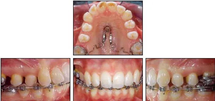
A 25-year-old female pre-sented with moderate crowding in both arches (Fig. 1, Table 1). Clinical examination revealed Class II skeletal and dental rela-tionships, a balanced profile, and a pleasant facial appearance.
Three treatment options were discussed with the patient: extraction of the upper first and lower second premolars to correct the Class II malocclusion; extrac-tion of the upper first premolars
CASE REPORT
Palatal-Implant Anchorage
in an Adult Class II Patient
ALDO GIANCOTTI, DDS, MS MARIO GRECO, DDS
GIANLUCA MAMPIERI, DDS CLAUDIO ARCURI, DDS, MS
Drs. Giancotti and Mampieri are Assistant Professors and Dr. Greco is a postgraduate student, Department of Orthodontics, and Dr. Arcuri is Professor and Chief, Department of Periodontology, University of Rome “Tor Vergata”, Rome, Italy. Contact Dr. Giancotti at Viale Gorizia 24/c, 00198 Rome, Italy; e-mail: [email protected].
Dr. Arcuri Dr. Mampieri
Dr. Greco Dr. Giancotti
Fig. 1 25-year-old female patient with upper and lower crowding and Class II skeletal and dental relation-ships before treatment.
to compensate for the malocclu-sion; and nonextraction treatment involving distalization of the illary molars. When the patient de clined extractions, the third op -tion was chosen. The treatment plan involved anchorage from a temporary midpalatal implant.
Treatment Progress
After some interproximal reduction to reduce crowding, the
lower arch was bonded. A self-threading Orthosystem mini-implant* (6mm × 3mm) was in serted at an angle of about 60° to the occlusal plane, and a heal-ing cap was placed.9 After 12 weeks, a polyvinyl siloxane im -pression of the maxillary arch was taken, using a transfer analog connected to the implant to repro-duce the exact position of the implant on the cast.
The distalizing device was
a modified version of the Hilgers Pendulum,8,17 which has the ad -vantages of flexibility and ease of activation due to its removable springs. Two arms ending in lin-gual sheaths were soldered to a stainless steel octagonal cap de -signed to fit over the mini-implant. Distalizing springs were fabricated from .032" TMA** wire, and the mesially oriented loops were inserted into the lin-gual sheaths (Fig. 2).
Following Hilgers’s recom-mended procedure for bodily dis-tal movement of the maxillary molars, the two distalizing springs were initially activated at 60° to the horizontal plane, pro-ducing 200g of distalizing force. A second activation was per-formed 12 weeks later to correct the root inclination by bending the two spring ends at 30° to the sagittal plane17 (Fig. 3).
The distalization phase was
Giancotti, Greco, Mampieri, and Arcuri
Fig. 2 A. Modified Hilgers Pendulum arms prepared for insertion into palatal-implant sheaths. B. Lingual sheaths soldered to palatal implant, and Pendulum arms activated. C. Completed appliance ready for placement.
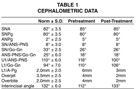
TABLE 1
CEPHALOMETRIC DATA
Norm ± S.D. Pretreatment Post-Treatment
SNA 82° ± 3.5 85° 85° SNPg 80° ± 3.5 80° 80° ANPg 2° ± 2.5 5° 5° SN/ANS-PNS 8° ± 3.0 8° 8° SN/Go-Gn 33° ± 2.5 26° 26° ANS-PNS/Go-Gn 25° ± 6.0 18° 18° U1/ANS-PNS 110° ± 6.0 118° 100° L1/Go-Gn 94° ± 7.0 110° 106° L1/A-Pg 2.0mm ± 2.0 6mm 3mm Overjet 3.5mm ± 2.5 4mm 2mm Overbite 2.0mm ± 2.5 4mm 2mm Interincisal angle 132° ± 6.0 112° 133°
*Registered trademark of Institut Straumann, Waldenburg, Switzerland; www.straumann. com.
**Registered trademark of Ormco, Orange, CA; www.ormco.com.
Fig. 3 Second activation of Pendulum springs, after 12 weeks of molar distalization.
Fig. 4 After six months of maxillary molar distalization, lower brackets removed and lingual retainer bonded. (Radiographs taken before removal of lower brackets.)
completed in six months, with the right side reaching a Class I posi-tion first (Fig. 4). A rigid .051" transpalatal bar was then con-nected to the palatal mini-implant
to maintain the molar positions (Fig. 5). The lower brackets were debonded, and a lingual 44 re -tainer was bonded.
As expected, the upper pre-molars drifted distally over the next five months of this molar-retention phase (Fig. 6). Addi-tional space closure and alignment were carried out in the upper arch using the Bidimensional tech-nique18 with an .018" × .022" stainless steel archwire. Nickel titanium closed-coil springs (300g) were attached to crimped hooks distal to the lateral incisor brackets for anterior retraction (Fig. 7).
The lower canines through first molars were rebonded for the
finishing phase. An .018" Austra-lian wire*** was placed in the upper arch from second premolar to second premolar, and an .018" Australian overlay archwire with an asymmetrical intrusion loop was inserted in the first-molar tubes to align the gingival mar-gins (Fig. 8). The palatal mini-implant was removed after 22 months of treatment.
Treatment Results
Post-treatment evaluation showed Class I molar and canine
Giancotti, Greco, Mampieri, and Arcuri
***Registered trademark of A.J. Wilcock Pty. Ltd., Whittlesea, Victoria, Australia; distributed in North America by G&H Wire Company, Franklin, IN; www.ghwire.com.
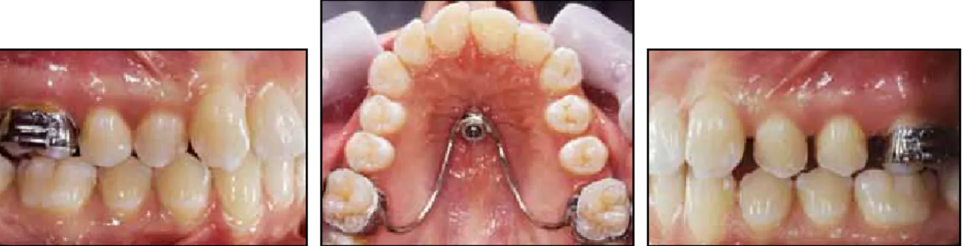
Fig. 5 Palatal bar connected to palatal implant for maintenance of molar positions.
Fig. 6 Distal drifting of premolars after molar distalization.
Fig. 7 Bidimensional technique used in upper arch for remaining space closure; hooks crimped on archwire distal to lateral incisors for anterior retraction with 300g nickel titanium closed-coil springs.
relationships with proper overbite and overjet (Fig. 9, Table 1). alometric data and superimposi-tions indicated no change in the skeletal pattern, except for some important variations in incisor inclination. The upper incisors were retruded 18°, compatible with the amount of space opening during molar distalization. The 4° improvement in lower incisor inclination was attributable to the interproximal stripping for relief of crowding and to the avoidance of Class II elastics, made possible by the use of implant anchorage.
Discussion
In this case, once the patient refused extraction treatment, skel-etal anchorage was needed to support the molar-distalization mechanics. Radiographs of the up -per arch showed inadequate
spac-es between the second-premolar and first-molar roots for miniscrew placement. Moving the in -sertion site more gingivally would have placed the screws in the unattached gingiva, increasing the risks of screw failure and sinus penetration.4,5 Furthermore, when buccal miniscrews are used as anchorage for maxillary space opening, they must generally be removed and replaced by screws in different locations for the retraction phase.12 Such a series of insertions, removals, and reinser-tions would be no less invasive for an adult patient than the insertion and removal of a single palatal implant would be.
Conclusion
The implant-supported modified Pendulum appliance shown here is an effective option
for Class II nonextraction treat-ment of adult patients, offering the following advantages:
• A constant force is applied to the molars by the removable dis-talizing springs, while the reac-tion force is dissipated through the osseointegrated implant to the palatal bone.
• All phases of treatment can be managed without any risk of anch-orage loss or the need for mini-screw repositioning.
• Patient cooperation with remov-able appliances is not required. • The system is well accepted by the patient because it is intraoral, fairly inconspicuous, and rela-tively comfortable to wear.
ACKNOWLEDGMENTS: The authors wish to thank Mr. Stefano Della Vecchia and his staff at Orthosystem Roma for their technical support in fabrication of the appliance.
(continued on p. 634)
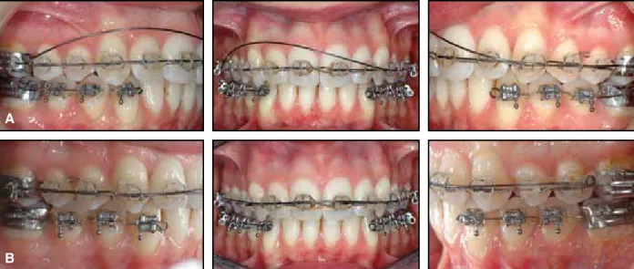
Fig. 8 A. Lower brackets bonded from canines to first molars for finishing; upper overlay wire placed for asymmetrical intrusion to align anterior gingival margin. B. After activation of overlay intrusion wire.
A
Giancotti, Greco, Mampieri, and Arcuri
Fig. 9 A. Patient after 22 months of treatment. B. Superimposition of pre- and post-treatment cephalometric tracings.
1. Antonarakis, G.S. and Kiliaridis, S.: Maxillary molar distalization with non-compliance intramaxillary appliances in Class II malocclusion: A systematic re -view, Angle Orthod. 78:1133-1140, 2008. 2. Kinzinger, G.S.M.; Eren, M.; and
Diedrich, P.R.: Treatment effects of intraoral appliances with conventional anchorage designs for non-compliance maxillary molar distalization: A litera-ture review, Eur. J. Orthod. 30:558-571, 2008.
3. Block, M.S. and Hoffman, D.R.: A new device for absolute anchorage for ortho-dontics, Am. J. Orthod. 107:251-258, 1995.
4. Carano, A.; Velo, S.; Incorvati, C.; and Poggio, P.: Clinical applications of the Mini-Screw-Anchorage-System in the maxillary alveolar bone, Prog. Orthod. 5:212-235, 2004.
5. Kravitz, N.D. and Kusnoto, B.: Risks and complications of orthodontic mini-screws, Am. J. Orthod. 131(4 suppl.): S43-51, 2007.
6. Roberts, W.E.; Helm, F.R.; Marshall, K.J.; and Gongloff, R.K.: Rigid endo-sseous implants for orthodontic and
orthopedic anchorage, Angle Orthod. 59:247-256, 1989.
7. Bondemark, L.; Feldmann, I.; and Feldmann, H.: Distal molar movement with an intra-arch device provided with the Onplant System for absolute anchor-age, World J. Orthod. 3:117-124, 2002. 8. Giancotti, A.; Muzzi, F.; Greco, M.; and
Arcuri, C.: Palatal implant-supported distalizing devices: Clinical application of the Straumann Orthosystem, World J. Orthod. 3:135-139, 2002.
9. Giancotti, A.; Muzzi, F.; Santini, F.; and Arcuri, C.: Straumann Orthosystem method for orthodontic anchorage: Step by step procedure, World J. Orthod. 3:140-146, 2002.
10. Giancotti, A.; Greco, M.; Docimo, R.; and Arcuri, C.: Extraction treatment using a palatal implant for anchorage, Austral. Orthod. J. 19:87-90, 2003. 11. Giancotti, A.; Greco, M.; Mampieri, G.;
and Arcuri, C.: Clinical management in ex traction cases using palatal implant for anchorage, J. Orthod. 31:288-294, 2004.
12. Giancotti, A.; Greco, M.; Mampieri, G.; and Arcuri, C.: Treatment of ectopic
maxillary canines using a palatal implant for anchorage, J. Clin. Orthod. 39:607-611, 2005.
13. Maino, B.G.; Mura, P.; and Gianelly, A.A.: A retrievable palatal implant for absolute anchorage in orthodontics, World J. Orthod. 3:125-134, 2002. 14. Maino, G.B.; Gianelly, A.A.; Bednar, J.;
Mura, P.; and Maino, G.: MGBM system: New protocol for Class II non ex -traction treatment without cooperation, Prog. Orthod. 8:130-143, 2007. 15. Triaca, A.; Antonini, M.; and
Winter-mantel, E.: Ein neues Titan-Flach-schrauben-Implantat zur orthodontisch-en Verankerung am anteriororthodontisch-en Gaumorthodontisch-en, Inf. Orthod. Kieferorthop. 24:251-257, 1992.
16. Wehrbein, H.; Feifel, H.; and Diedrich, P.: Palatal implant anchorage reinforce-ment of posterior teeth: A prospective study, Am. J. Orthod. 116:678-686, 1999. 17. Hilgers, J.J.: The Pendulum appliance
for Class II non-compliance therapy, J. Clin. Orthod. 26:706-714, 1992. 18. Gianelly, A.A.: Bidimensional Tech
nique: Theory and Practice, GAC national, Central Islip, NY, 2000.