ALMA MATER STUDIORUM – UNIVERSITÀ DI BOLOGNA
DOTTORATO DI RICERCA IN SCIENZE MEDICHE SPECIALISTICHE
Ciclo XXV°
Settore Concorsuale di afferenza: 06/D2
Settore Scientifico disciplinare: MED 14
Titolo tesi
A step forwards in immunomodulation and immunetolerance knowledge.
HLA-G expression in co-cultures of peripheral blood mononuclear cells and stem cells after in vitro NGAL stimulation.
Presentata da: Dr. Irene Capelli
Coordinatore Dottorato
Relatore
Prof. Sandro Mattioli
Prof. Gaetano La Manna
INDEX
ABSTARCT………...……….3
1. INTRODUCTION...5
1.1. Neutrophil Gelatinase Associated Lipocalin (NGAL)...5
1.1.2 NGAL, the iron binding and the bacteriostatic action ...6
1.1.3. NGAL and inflammation and oxidative stress ...8
1.1.4 NGAL in kidney formation...10
1.1.5. NGAL as a growth factor ...11
1.1.6 NGAL and pathological kidney conditions ...13
1.2 Immune tolerance ...15
1.2.1 Tolerance induction ...17
1.2.2 HLA-G ...19
1.2.3 HLA-G and Regulatory T cells...22
2. OBJECTIVES...26
3. MATERIALS AND METHODS...27
3.1 PBMCs isolation and cultures ...27
3.2 NGAL in vitro effect on HLA-G expression in PBMCs...27
3.3 NGAL in vitro effect on HLA-G expression in PBMCs after treatment with scalar doses of NGAL:Enterobactin:Iron………....28
3.4 NGAL:Enterobactin Iron Free effect on HLA-G expression………...28
3.5 NGAL:Enterobactin:Iron effect on HLA-G expression following anti-NGAL antibody in vitro administration……….28
3.6 Indirect evaluation of NGAL role on HLA-G expression after treatment with Ent:Iron……... 28
3.7 Soluble HLA-G evaluation trough ELISA after NGAL:Enterobactin:Iron treatment………….29
3.8 NGAL effect on T regulatory cells………...29
4. RESULTS... .29
4.1 NGAL in vitro effect on HLA-G expression in PBMC ... ..29
4.2 NGAL in vitro effect on HLA-G expression in PBMCs after treatment with scalar doses of NGAL:Enterobactin:Iron………30
4.3 NGAL:Enterobactin Iron Free effect on HLA-G expression………...32
4.4NGAL:Enterobactin:Iron effect on HLA-G expression following anti-NGAL antibody in vitro administration………...32
4.5 Indirect evaluation of NGAL role on HLA-G expression after treatment with Enterobactin:Iron ………33
4.6 Soluble HLA-G evaluation trough ELISA after NGAL:Enterobactin:Iron treatment………….33
4.7 NGAL effect on T regulatory cells………33
5. DISCUSSION and CONCLUSIONS ………..36
ABSTRACT
Ngal (Neutrophil Gelatinase-associated Lipocalin ) è una proteina appartenente alla famiglia delle
lipocaline verso cui la recente letteratura ha mostrato una notevole attenzione, soprattutto in
quanto biomarcatore in alcune condizioni patologiche (danno renale acuto e cronico, patologie
autoimmuni, neoplasie). Il ruolo biologico di NGAL non è però ancora del tutto compreso.
Numerose sono le dimostrazioni della sua azione batteriostatica. Recenti lavori hanno inoltre
evidenziato un ruolo di NGAL nella modulazione di NFkB. Nessun lavoro ha valutato il ruolo di
NGAL nell’immunità umorale.
Lo scopo dello studio è quello di capire se NGAL possa esercitare un ruolo di attivazione
(modulazione) della risposta T cellulare attraverso la regolazione del complesso HLA-G, un
mediatore di tolleranza.
Cellule mononucleate da sangue periferico (PBMCs) sono state ottenute da 8 donatori sani dopo
consenso informato e isolate tramite centrifugazione (Ficoll). PBMC sono poi state trattate con 4
concentrazioni crescenti di NGAL (da 40 a 320 ng/mL), associate o meno a ferro e analizzate con
tecnica fluorimetrica ed elisa.Alle analisi eseguite NGAL stimola l’espressione di HLA-G sulle
cellule T CD4+ con un andamento dose dipendente. L’effetto del ferro sull’espressione di HLA-G non è di univoca interpretazione.Inoltre L’aggiunta di NGAL in vitro modifica il pattern di espressione delle cellule T, aumentando la popolazione delle cellule CD4+ CD25+ FoxP3. L’utilizzo
di anticorpi anti NGAL limita l’espressione di HLA-G e diminuisce significativamente la percentuale di CD4+ CD25+ FoxP3+ . In conclusione abbiamo mostrato un coinvolgimento di
NGAL nell’immunità cellulare. Valutando il ruolo di NGAL come molecola immunomodulatoria, abbiamo mostrato che NGAL gioca un ruolo chiave neell’immunotolleranza aumentando l’espressione di HLA-G e cellule T regolatorie nei donatori sani. Un possibilAs potential future scenario applicativo di tale studio riguarda l’utilizzo in vivo di NGAL nell’immunomodulazione dei pazienti sottoposti a trapianto o affetti da patologie autoimmuni.
ABSTRACT
NGAL (Neutrophil Gelatinase-associated Lipocalin ) is a protein of lipocalin superfamily. Recent
literature focused on its biomarkers function in several pathological condition (acute and chronic
kidney damage, autoimmune disease, malignancy). NGAL biological role is not well elucidated.
Several are the demonstration of its bacteriostatic role. Recent papers have indeed highlight NGAL
role in NFkB modulation. The aim of this study is to understand whether NGAL may exert a role in
the activation (modulation) of T cell response through the regulation of HLA-G complex, a
mediator of tolerance.
From 8 healthy donors we obtained peripheral blood mononuclear cells (PBMCs) and we isolated
by centrifugation on a Ficoll gradient. Cells were then treated with four concentrations of NGAL
(40-320 ng/ml) with or without iron. We performed flow cytometry analysis and ELISA test.
NGAL increased the HLA-G expression on CD4+ T cells, with an increasing corresponding to the
dose. Iron effect is not of unique interpretation. NGAL adiction affects regulatory T cells increasing
in vitro expansion of CD4+ CD25+ FoxP3+ cells. Neutralizing antibody against NGAL decreased
HLA-G expression and reduced significantly CD4+ CD25+ FoxP3+ cells percentage.
In conclusion, we provided in vitro evidence of NGAL involvement in cellular immunity. The
potential role of NGAL as an immunomodulatory molecule has been evaluated: it has been shown
that NGAL plays a pivotal role in the induction of immune tolerance up regulating HLA-G and T
regulatory cells expression in healthy donors. As potential future scenario we highlight the in vivo
role of NGAL in immunology and immunomodulation, and its possible relationship with
immunosuppressive therapy efficacy, tolerance induction in transplant patients, and/or in other
1.
Introduction
1.1 Neutrophil Gelatinase-associated Lipocalin (NGAL)
Neutrophil gelatinase-associated lipocalin (NGAL) is a glycoprotein of 25 kDa molecular weight
consisting of 198 amino acids. It owes its name to the fact that it was isolated for the first time in
association with a gelatinase obtained from the supernatant of activated human neutrophils. Only
later its expression was found also in other epithelial tissues including kidney, stomach, colon,
trachea bone marrow, prostate, uterus [1,2].
NGAL belongs to the lipocalins superfamily, that comprises a class of proteins that are
characterized by a common secondary and tertiary structural feature— called as the "lipocalin fold".
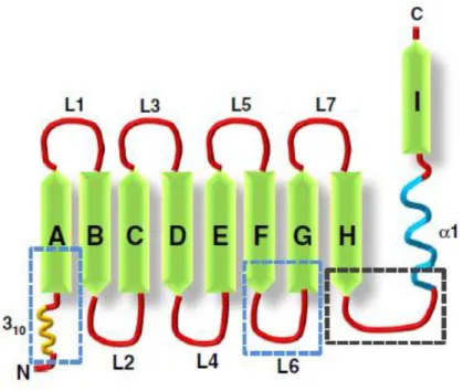
It is characterized by height β-strands that form a β-barrel defining a calyx. The calyx binds and transports low molecular weight molecules, which define the biologic activity of the lipocalin. The
beta sheets are connected to one another by seven short loops (L1–L7), of which the loop L1 forms
a lid-like structure to close the ligand binding cavity. The difference in specific amino acids within
the lipocalin fold gives rise to the wide diversity in ligands that can be bound by lipocalins (Figure
1) [3,4].
Few examples of lipocalins are retinol-binding protein that binds and transports vitamin A [5], the
lipocalin α1-microglobulin that scavenges heme [6], and nitrophorin-type lipocalins that carry heme groups complexed with nitric oxide [7].
Biological fluids contain very low levels of NGAL protein at steady state level. Serum contains
approximately 20 ng/ml NGAL which is probably derived from neutrophils and from limited
expression in liver, spleen and kidney [8]. Renal clearance is a major regulator of this steady state
level, because circulating NGAL undergoes glomerular filtration due to its low molecular weight
and positive charge. Theoretically, the kidney processes 3.4–4 mg NGAL per day (20 ng/ml×120–
140 ml/min glomerular filtration rate, GFR) as a result of filtration [9] followed by capture by the
from the apical (luminal) membrane is the most likely pathway of NGAL traffic because it appeared
in the urine when the apical megalin receptor was deleted. Biacore based binding studies confirmed
a direct interaction between NGAL and megalin fragments [10], consistent with capture of NGAL
from the glomerular filtrate rather than from the basolateral (blood) side. Similar to serum, urine
contains approximately 20 ng/ml NGAL at steady state. The origin of this protein is not clear, but
some of it may derive from serum NGAL that bypasses capture in the proximal tubule
(approximately 1/200 molecules). Alternatively, NGAL might derive from neutrophils or even from
bladder epithelia. However, it is most likely that urinary NGAL derives from a low level of
expression in the native kidney at steady state. Indeed, weak in situ RNA reactivity can be seen in
the collecting ducts of some kidneys [11].
1.1.2
NGAL, the iron binding and the bacteriostactic action
In the case of NGAL, the lipophilic compound that can be tied by the lipocalin fold is represented
by the siderophores, as demonstrated for the first time by Strong et al studies [12].
The estimated concentration of free iron in the body is estimated to be as low as 10−24 M) [13].
The exceedingly low availability of free iron is attributable to iron-binding proteins like transferrin,
ferritin and
lactoferrin which form complexes with any available free iron molecules. Bacteria have developed
special proteins called siderophores that have an affinity for iron (particularly ferric iron or Fe3+)
several times higher than that of the endogenous iron chelators. This enables siderophores to not
only bind available free iron, but also extract iron from iron-binding proteins of the host [14].
Goetz et al. [15,16] cloned NGAL in Gram−bacteria and identified a bacterial compound called enterochelin bound within the protein's calyx. Enterochelin is an organic molecule that bacteria
Figure 1. Schematic representation of the lipocalin fold. The characteristic feature of lipocalins is the “lipocalin fold” which comprises of an N-terminal 3–10 helix followed by eight beta sheets. The beta sheets are connected by loops (L1–L7). The portion of the lipocalin fold that are structurally conserved between different lipocalins is indicated by the blue boxed regions while the region that shows significant conservation in amino acid sequence is indicated by the black boxed region [4].
The interaction of enterocalin with NGAL directly blocked the capture of iron by bacteria: NGAL
delayed bacterial growth. Complexes of NGAL with enterocalin:Fe could form in the bloodstream
[17] followed by their rapid clearance by macrophages in liver and spleen and by filtration and
tubular destruction in the kidney. Hence, the binding of enterocalin to NGAL not only sequestered
the siderophore but resulted in its clearance in the kidney. Because NGAL levels can rise 100 fold
to >2000 ng/ml, as much as 300–400 mg NGAL/24 h, containing nearly 1 mg of iron can
theoretically traffic to the kidney for clearance and recycling, provided that enterocalin is fully
saturated and glomerular filtration is maintained [11].
But an oversupply of iron or the expression of siderophores [18] which do not recognize NGAL,
rescued bacterial growth. Consistently, the deletion of NGAL accelerated the growth of enterocalin
NGAL is therefore an important role in innate immune response and is expressed in particular
tissues exposed to the external environment (and therefore susceptible to infection) as the urinary or
the gastrointestinal tract (figure 2). There are also many apparent differences between known
urinary antimicrobial peptides and NGAL. For example, unlike other urinary antimicrobial proteins
[21,22,23] the cathelicidins, the defensins [24,25], Tamm–Horsfall [26-28] and Lactoferrin [29],
which are expressed constitutively, uNGAL is not likely to play a large role at steady state because
it is expressed only at very low levels (20 ng/ml). In addition, while many of these proteins are
modulated only to a small degree by infections (cathelicidin by 3–8 fold; THP and lactoferrin are
not stimulated by infections [30-34] and the AMPs and lactoferrin remain in the low ng/ml range
even after stimulation), NGAL was intensively upregulated by significant infections [35-39] as
well as aseptic stimuli. Consequently the mechanisms of expression and induction apparently differ
between NGAL and other antimicrobial peptides even though NGAL originates from the same cells
as many of these proteins.
1.1.3
NGAL, inflammation and oxidative stress
The inflammatory response is a natural defense mechanisms used by the body to clear irritants and
pathogens. NGAL has been shown to be a pro-inflammatory molecule causing some to call it a
cytokine. [40] Studies conducted in pulmonary inflammation (mouse models) for exemple have
revealed that Lcn2 (the mouse equivalent of NGAL) mRNA and protein are strongly upregulated
Figure 2 Exemple bacteriostatic action of NGAL. Gram negative bacteria (like Salmonella typhimurium) trigger an immune response characterized by the activation of antigen presenting cells (macrophages and dendritic cells) upon engulfing the bacteria. These activated cells then release cytokines like IL-18 and IL-23 that in turn activate T-lymphocytes. These activated T-cell in turn release IL-17 and IL-22. These two cytokines act on the intestinal cells and stimulate the de novo synthesis of NGAL- lipocalin 2 (Lcn2). The NGAL-Lcn2 is secreted into the intestinal lumen and binds to bacterial iron-binding proteins (siderophores) like enterochalin. Since iron is essential for the growth of bacteria, the sequestration of bacterial siderophores by NGAL-Lcn2 has a bacteriostatic effect. [4].
lungs of patients with bronchial inflammation [42]. The pro-inflammatory cytokine IL-1β is able to
induce a significant upregulation of NGAL in human lung epithelial cells [42]. This suggests a
mechanism for upregulation of NGAL in pulmonary inflammation but its functional role in this
process is still unclear. Several hypotheses have been proposed to explain it. One such hypothesis is
that acute or chronic inflammation leads to the accumulation of granulocytes at the sites of
thereby mediate local tissue injury. In support of its pro-inflammatory function, NGAL appears to
be a chemoattractant for neutrophils [43].
The importance of NGAL as an acute phase protein was demonstrated by the observation that
following an intraperitoneal injection of E. coli, NGAL levels are elevated in the serum and in liver
tissue within 4 h, and in the spleen within 6 h. The increase in serum levels of NGAL was preceded
by an increase in mRNA synthesis in the peripheral blood cells. SerumNGAL levelsrose by nearly
22-fold (from 100 ng/ml to 2200 ng/ml) within 8 h following injection (of E. coli), reached a peak
by 24 h and then gradually returned to baseline levels [44].
Oxidative stress can be defined as the imbalance between the prooxidant and antioxidantmachinery
in the cell [45]. Aberrant expression of NGAL in Chinese hamster ovary (CHO) and human
embryonic kidney (HEK293T0) cells resulted in an upregulation of the mRNA and protein levels of
the antioxidant enzymes superoxide dismutase (SOD1 and SOD2) and heme oxygenase (HO1 and
HO2) together with decrease in expression of NF-κB. Silencing of NGAL in A549 lung cancer cells
had the opposite effect on HO1 and NFκB expression but increased the expression of SOD1 and SOD2. Further, the cells ectopically expressing NGAL were more resistant to the cytotoxic effects
of hydrogen peroxide in vitro [46]. These results suggest that
NGAL by inducing the expression of antioxidant enzymes helps cells counteract oxidative damage.
Other studies have also suggested that NGAL may have a protective effect against cellular injury
mediated by reactive oxygen species (ROS) [47].
1.1.4
NGAL in kidney formation
The normal development of urinary system has three different stages: pronephros, mesonephros,
and metanephros. The development of metanephros, the permanent kidney, is the result of the
expression of many genes in the cells of bud ureter and the metanephric balstema that send each
Already at the beginning of the last century Boyden in the 1927 and later Gruenwald and Grobstein
in the 1943 demonstrated that the stimulus to the conversion from mesenchymal cells in epithelial
cells was given by the presence of the ureteric bud. In fact, surgical ablation of the ureteric bud
prevented the appearance of new epithelium from metanephric blastema. [48].
More recent studies have shown that the gene deletion of ureteric bud obtained the same results.
[49]. So it appeared clear that the ureteric bud provided the signals necessary for conversion of
mesenchymal cells in epithelial cells, but the signals wasn’t known.
In the late 1990s the Barasch and Sakuri research groups, to reveal the identity of these factors
have developed a line of cells of the ureteric bud that served as surrogate of the embryos ureteric
bud [50].These cells expressed a number of typical ureteral bud proteins including growth factors
and mesenchymal survival factors and receptors for factors produced by mesenchymal cells.In this
way were isolated numerous factors each of which can stimulate the growth of metanephric
mesenchyme and prevent its apoptosis (FGF and tissue inhibitors of metallo-proteinase) and in
1999 also the first factor able to convert mesenchymal cells in epithelial cells (LIF, leukemia
inhibitory factor) [51].
Recently several other factors belongings to the complex network of signals able to synergistically
induce mesenchymal cells into epithelial cells differentiation have been identified: FGF2, TGFβ2,
etc [52].. In 2002 Yang et al purified and recognized between these proteins also NGAL. [53].
1.1.5 NGAL as a growth factor
The differentiation-inducing properties of NGAL are not limited to the embryonic kidney.
Mounting evidence points toward growth factor effects of NGAL that modulate various cellular
responses, such as proliferation, apoptosis, and differentiation, but this is not well understood
mechanistically. [54].
In a 4T1-Ras–transformed mesenchymal tumor cell line, NGAL induces markers of epithelial cells
growth factor and promotes the organization of epithelial cells into tubular structures [56]
Antagonization of NGAL induction by expression of NGAL shRNA induces cystic structures rather
than properly assembled tubules. Therefore, in addition to inducing epithelial characteristics in non
epithelial cells, NGAL seems to affect the structure of established epithelia. It is interesting that
glycodelin, another protein of the lipocalin family, displays effects on epithelial differentiation very
similar to NGAL [57]. In vivo, NGAL protein is expressed predominantly by stimulated, growing,
dysplastic, or involuting epithelial cells, pointing to a relevance of the in vitro observations in
pathologic states [58].
Some of these effects, however, are enhanced when NGAL is associated with siderophores and
iron, raising the possibility that in the absence of bacterial infection, endogenous molecules
associate with NGAL to mediate its iron-binding properties.The cellular events differ strikingly—
at least in some biologic systems depending on whether NGAL is associated with iron [59].
Cellular uptake of NGAL is followed by distribution of the protein in endosomes [60]. Different
trafficking routes of endosomal NGAL have been proposed depending on the cell type and the
association of NGAL with its binding partners. In kidney-derived cell lines,
siderophore:iron-associated NGAL (holo-NGAL) traffics to endosomes and releases iron from the complex, which
results in regulation of iron-responsive genes, such as ferritin and transferrin receptor [61].
Similarly, in the adult mouse kidney in vivo, systemically applied holo-NGAL is taken up by
proximal tubule cells, where it delivers 55Fe [62]. The endosomal NGAL protein core is either
degraded in lysosomes [63] or recycled to the extracellular space [64]. On the basis of these
observations, siderophore:iron-associated NGAL is predicted to facilitate cytoplasmic iron delivery
into target cells. A recent report suggested that the situation may be different when NGAL is
delivered into target cells in the absence of the siderophore:iron complex. In this setting,
NGAL is proposed to scavenge intracellular iron and exit the cell via the endosomal recycling
pathway [65]. Therefore, at least some of the biologic effects of NGAL may depend markedly on its
responses to NGAL depending on the ligand. For instance, siderophore: iron-associated NGAL is
more effective than apo-NGAL in inducing epithelial characteristics in 4T1-Ras–transformed
mesenchymal tumor cells [66].
1.1.6 NGAL and pathological kidney conditions
There is an urgent need in clinical medicine for early predictive biomarkers of both acute kidney
injury (AKI) and chronic kidney disease (CKD). In both situations, early intervention can
significantly improve the prognosis. However, currently available biomarkers (such as serum
creatinine concentrations) are imprecise, and their delayed response has impaired our ability to
institute potentially effective therapies promptly. In recent years a series of kidney disease
biomarker candidates were proposed both for AKI and CKD. NGAL is widely considered one of
the most promising novel biomarkers of acute and chronic kidney damage [67].
Several investigators have used molecular techniques such as cDNA microarrays and subtractive
hybridizations combined with downstream proteomic analysis to identify novel pathways,
biomarkers and drug targets in AKI. Supavekin et al. identified NGAL as one of the most
upregulated genes in the early post-ischaemic mouse kidney [68], a finding that has now been
confirmed in several other transcriptome profiling studies following ischaemic and nephrotoxic
kidney injuries. Downstream proteomic studies have also revealed NGAL to be one of the earliest
and most robustly induced proteins in the kidney after ischaemic or nephrotoxic AKI in animal
models [69]. Importantly, NGAL protein is easily detected in the blood and urine soon after AKI in
pre-clinical studies [70]. These findings have initiated a number of translational studies to evaluate
NGAL as a novel biomarker in human AKI.In a cross-sectional study, subjects in the intensive care
unit with established acute renal failure displayed a greater than 10-fold increase in plasma NGAL
concentration and more than a 100- fold increase in urine NGAL concentration by Western blotting
when compared to normal controls [71]. Both plasma and urine NGAL concentrations correlated
accumulation of immunoreactive NGAL in 50 % of the cortical tubules. These results identified
NGAL as a widespread and sensitive response to established AKI in humans.
NGAL has also been evaluated as a biomarker of AKI in kidney transplantation. Biopsies of
kidneys obtained 1 h after vascular anastomosis revealed a significant correlation between NGAL
staining intensity and the subsequent development of delayed graft function [72]. In a prospective
multicentre study of children and adults, urine NGAL concentrations in samples collected on the
day of transplant clearly identified cadaveric kidney recipients who subsequently developed delayed
graft function and dialysis requirement (which typically occurred 2-4 days later). The ROC curve
for prediction of delayed graft function based on urine NGAL concentration at day 0 showed an
AUC of 0.9, indicative of an excellent predictive biomarker [73]. In a retrospective study of kidney
transplant patients undergoing either protocol biopsies or clinically indicated biopsies, urine NGAL
concentrations were found to be significantly increased in subjects with tubulitis or other tubular
pathologies [74]. Urine NGAL also tended to be increased in subjects with subclinical tubulitis
(p=0.06), raising the possibility of NGAL representing a non-invasive screening tool for the
detection of tubulointerstitial disease in the early months following kidney transplantation.
Several investigators have examined the role of NGAL as a predictive biomarker of nephrotoxicity
following contrast administration [75]. In a prospective study of children undergoing elective
cardiac catheterization with contrast administration, both urine and plasma NGAL predicted
contrast-induced nephropathy (defined as a 50 % increase in serum creatinine from baseline
concentration) within 2 h after contrast administration [76]. Using a cut-off value of 100 μg/L, the
AUC for prediction of contrast nephropathy was excellent for the 2-h urine NGAL (0.92) as well as
the 2-h plasma NGAL (0.91). By multivariate analysis, the 2-h NGAL concentrations in the urine
and plasma were found to be powerful independent predictors of contrast nephropathy [77].
As discussed above, NGAL is a promising biomarker of AKI. In CKD, there is a growing literature
suggesting that NGAL is also a marker of kidney disease and severity. In 45 subjects with CKD
NGAL concentrations were inversely associated with GFR [78]. As kidney function declined to <30
mL/min, NGAL outperformed cystatin C as a biomarker of kidney failure [79]. Another study [80]
in subjects with CKD (due to chronic glomerulone-phritis) demonstrated that mean urinary NGAL
concentrations were higher in CKD patients (378.28 ±111.13 μg/L vs. 7.38±3.26 μg/L in controls; p=0.01). Furthermore, urinary NGAL concentrations were significantly correlated with serum
creatinine concentrations (r=0.588, p-value=0.02), GFR (r=-0.528, p-value=0.04) and proteinuria
(r=0.294, p-value=0.01) [81]. Both urine and plasma NGAL represent biomarkers of CKD severity
in patients with autosomal dominant polycystic kidney disease [82]. In these subjects, urine and
plasma NGAL concentrations correlated with residual GFR, and those with greater severity of
cystic disease (measured as number of cysts >10) displayed the highest NGAL values [83]. Urine
NGAL has also been shown to represent an early biomarker for degree of chronic tubulointerstitial
injury in patients with IgA nephropathy [84].
1.2 IMMUNE TOLERANCE
Immune tolerance consists of two main processes, namely central and peripheral tolerance. Central
tolerance takes place in the thymus where most of the self-reactive T cells are deleted at an
immature stage of their development [85]. Despite negative selection, self-reactive T cells can
escape thymic clonal deletion, and subsequently provoke autoimmune diseases such as type 1
diabetes (T1D), multiple sclerosis (MS), and inflammatory bowel disease (IBD) unless they are
controlled by one of many peripheral mechanisms [86].
However thymic selection has been considered as an effective tolerogenic mechanism only for
widely expressed self molecules. This assumption is based on the consideration that proteins with
tissue-restricted expression would not be available for presentation in the thymus. Thus , tolerance
to such proteins could only be achieved through mechanisms of peripheral tolerance. Peripheral
anergy, ignorance and regulatory cells, and contribute to maintaining aotoreactive lymphocyte
under tight control.
Immunologic tolerance was first introduced in 1945 when Ray Owen observed that placental interchange resulted in red cell chimerism between dizygotic bovine twins [87]. In the follow
decade Medawar, McFarlane Burnet, and colleagues elaborated upon this phenomenon of acquired
immunologic tolerance with experimental models of transplantation, which awarded them the
Nobel Prize in Physiology or Medicine in 1960.
Most of the work at the time involved non-self antigen exposure in immunologically immature
hosts, until 1959 when Schwartz and Dameshek demonstrated a marked delay in the adult rabbit
immune response to iodine-labeled injections of human serum albumin when treated with
6-mercaptopurine [88]. Their descriptions of the inhibition of immune pathways in this “drug-induced
immunological tolerance” notably foreshadowed the era of pharmacologic development for tolerance induction.
The next 50 years heralded a boom in drug development and subsequent improvements in graft
survival. In contrast to 1-year graft survival in 1977 of 53 and 78% for deceased and living-related
donors, respectively [89], modern immunosuppression has enabled transplant recipients to enjoy
very favorable graft survival. One-year rates having asymptotically approached 93–96%; therefore,
short-term graft survival alone can no longer be held as the metric of success for new
immunosuppressants.
Instead, as 10-year graft survival rates still trail at 47–61%, new agents must address factors leading
to chronic rejection as well as the comorbidities associated with chronic immunosuppression. The
decisive measure of success is for a therapy to demonstrate allospecific immunosuppression while minimizing side effects and preserving immune competence to infectious pathogens and cancer
during drug administration, and permanent graft survival after its withdrawal.
The understanding of the mechanisms underlying the immune tolerance of self and non self are
1.2.1 TOLERANCE INDUCTION
Traditional and novel approaches to inducing tolerance in organ transplant are known:
MOLECULE-BASED APPROACHES 1) T CELL THERAPIES–DEPLETION
Early attempts at transplantation in humans were fraught with early graft failure due to a robust
alloimmuneresponse mediated by activated Tcells. We have since learned that the suppression of
these alloreactive Tcells permits long-term graft survival and, at times, operational tolerance [90].
Inthe1980s, [91] observed that some renal transplant patients undergoing total lymphoid irradiation acquired tolerance to their allografts after withdrawal of immunosuppression and demonstrated
donor-specific unresponsiveness in vitro. Over 30years later, the concept of eliminating alloreactive
Tcells upon induction continues to prevail, as Tcell depletion remains the most common induction
therapy in the U.S [92] Steroids, calcineurininhibitors, rapamycin, and mycophenolatemofetil
comprise essential components of most immunosuppressive manteinance regimens.
Induction strategies are instead represented by:
- Anti-thymocyteglobulin(ATG), the oldest depleting agent dat- ing back to the late 1890s, has been
a mainstay in induction therapy since the 1960s [93] (Due to its potency and markedly
heterogeneous target antigen specificities, ATG is particularly useful in high-risk recipients as well
as in preventing ischemia-reperfusion injury.
- Alemtuzumab (Campath-1H,Genzyme),a humanized monoclonal Ab to CD52 found densely
distributed on T and B lymphocytes and natural killer cells [94].
2) T CELLTHERAPIES–COSTIMULATIONBLOCKADE
Alloreactive Tcell activation requires antigen-specific engagement of the Tcell receptor with major
histocompatibility complex molecules (signal1), followed by antigen non-specific ligation of a
variety of receptor–ligand combinations, or costimulation (signal2; JenkinsandSchwartz,1987).
While costimulation blockade renders the Tcell anergic [96], the seanergic Tcells may express
inducible costimulator (ICOS) and play a regulatory role [97]. In addition, costimulation blockade
does not require radical ablation of the immune system by lymphocyte depletion or irradiation, thus
shifting the emphasis from induction to maintenance immunosuppression [98].
Costimulatory signals of the CD28:B7(CD80/86) immunoglobulin superfamily and
CD40:CD154(CD40L) tumornecrosisfactor (TNF) family are the most studied and potentially most
important activating costimulation pathways. Cytotoxiclympocyteantigen-4 (CTLA-4) shares about
30% homology with CD28, and binds with 10–20-fold higher affinity than CD28 toB7 molecules
on the antigen presentingcell(APC). Not only does this potently inhibit the Tcell, but also its
ligation with APC B7 molecules induces indoleamine 2,3-dioxygenase expression, promoting the
suppressive function sinCTLA4+ regulatoryCD4+ cells [99].
Abatacept(Orencia,Bristol-MyersSquibb) and belatacept (Nulojix,Bristol-Abatacept(Orencia,Bristol-MyersSquibb), fusion proteins composed of CTLA-4
and immoglobulinIgG1, have utilized this mechanism to confer potent inhibition of alloreactive
Tcell responses.
3) B CELLTHERAPIES
The role of B cells in operational tolerance has yet to be defined. On one hand ,an ITN-sponsored
collaboration identified a unique B cell signature associated with 25 operationally tolerant renal
transplant recipients. Not only did tolerant patients exhibit an increase in total and naïve Bcells, but
also the majority of genes that were increasingly expressed were Bcell-specific ,particularly of
transitional Bcells [100]. While these transitional B cells could represent a regulatory B cell
population based on their increased IL-10 production as discussed by Redfieldetal. [101]., no
difference in B cell subsets (total, naïve, and transitional cells) or inhibitory cytokines (IL-10 and
1.2.2 HLA-G
While both HLA-A, -B, and -C class I molecules and class II molecules play an important role in
the induction of a specific immune response by presenting peptide antigens to T cells [103] , the
non-classical major histocompatibility complex (MHC) class I molecule HLA-G has been identified
as a key mediator in immune tolerance [104]. It is characterized by :
1) A tissue-restricted distribution. Whereas expression of classical HLA class I molecules is known to be ubiquitous, HLA-G was first described on extraembryonic trophoblast tissues
that invade the maternal decidua during implantation of the embryo [105]. Recently, its
expression was also reported in endothelial cells from first trimester placental chorionic
blood vessel, in thymic epithelial cells and activated peripheral blood monocytes [106].
2) A limited polymorphism. Sixteen HLA-G alleles have been described to date, four of which may encode membrane-bound HLA-G proteins [107], plus one truncated soluble protein
which bears the ‘delC130 mutation’ [108];
3) An alternative transcription of spliced messenger RNAs (mRNAs) that encode at least six different HLA-G isoforms, namely the HLA-G1, -G2, -G3 and -G4 membrane bound and
HLA-G5 and -G6 soluble proteins [109].
The “complete” HLA-G1 molecule and its soluble counterpart HLAG5 are those that have been studied the most. They have an identical extracellular structure, which is classic HLA class I-like: a
heavy chain of 3 globular domains non-covalently bound to β2-microglobulin and a nonapeptide.
The other isoforms are likely to be of simpler structure: lacking one or 2 globular domains, they are
smaller, and should not bind β2-microglobulin and present peptides. [110].
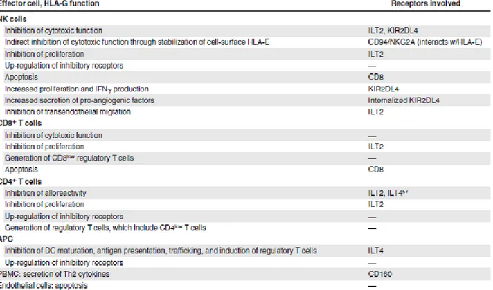
Three HLA-G receptors have been described: ILT2/CD85j/LILRB1 (ILT2), ILT4/CD85d/LILRB2
(ILT4), and KIR2DL4/CD158d (KIR2DL4) [111].
ILT2 is expressed by B cells, some T cells, some NK cells, and all monocytes/dendritic cells, [112]
KIR2DL4, its expression is mainly restricted to the CD56bright subsets of NK cells [114] which
constitute a minority of peripheral NK cells, but a majority of uterine NK cells. [115]. Binding to
CD8 has also been reported [116]. Through these differentially expressed receptors, HLA-G can
interact with B cells, T cells, NK cells, and antigen-presenting cells (APC).
Functionally, HLA-G1 inhibits the cytolytic function of uterine and peripheral blood NK cells [117]
the antigen-specific cytolytic function of cytotoxic T lymphocytes [118] the alloproliferative
response of CD4+ T cells [119] the proliferation of T cells and peripheral blood NK cells [120] and
the maturation and function of dendritic cells [121].
Soluble HLA-G5 or soluble HLA-G1, which is generated by proteasomal cleavage from the cell
membrane, has similar functions. The other HLA-G isoforms have been less well studied, and little
is known about their function except that membrane-bound HLA-G2, HLA-G3, and HLA-G4 can
inhibit NK-cell and cytotoxic T lymphocyte cytolysis in vitro [122].
Because of this broad inhibitory function, capable of targeting multiple immune cell subsets, much
effort has been put into determining whether HLA-G is pathologically relevant and whether it can
be used as a diagnostic tool or as a therapeutic tool and/or target. Transplantation and oncology are
2 particularly clear situations. In the context of transplantation, HLA-G expression might be
beneficial and promote tolerance to grafts. To date, the expression of HLA-G was studied in more
than 1000 patients after heart [123] kidney (54) liver (55) and liver-kidney(55-57) transplantation,
with those expressing HLA-G in the graft and/or the plasma exhibiting significantly better graft
acceptance. Thus, in transplanted patients, titration of HLA-G might be used as a monitoring tool to
determine and follow tolerance status, which could then be used to adjust immunosuppressive
therapies. In this context, patients with high HLA-G titers could be candidates for a reduction in
immunosuppressive treatment, whereas HLA-G–negative patients would have a comparatively
higher risk of rejection. Furthermore, HLA-G itself might be used as therapeutic tolerogenic agent,
exogenously provided to HLA-G–negative patients as complementary and/or alternative therapy
[124].
In the context of oncology, studies on more than 1000 malignant lesions confirmed Carosellaet al
first study on melanoma [125] which showed that HLA-G transcription and protein expression may
be switched on in tumor lesions and protect them from NK cytolysis. It was later shown that
HLA-G expression by tumor lesions protected against cytolysis [126] correlated with malignancy in
ovarian and breast carcinomas (61) as well as in melanocytic lesions [127] with unfavourable
outcome in chronic lymphocytic leukemia,63 and gastric and colorectal cancers [128].
High HLA-G plasma levels were also recently observed in patients with neuroblastoma and
correlated with relapse[129]. Expression of HLA-G has been evidenced in several malignant
hematopoietic diseases, particularly in acute myeloid leukemia (AML), acute lymphoblastic
leukemia (ALL), and B-chronic lymphocytic leukemia (B-CLL).
Thus, HLA-G expression would favor tumor development by impairing antitumor immunity. Here
this context, high titers of HLA-G would represent a negative factor [130]. In HLA-G–positive
patients, HLA-G itself might finally constitute a therapeutic target: if expressed as a
membrane-bound protein, as observed in some hematologic malignancies [131] HLA-G could be used as a
tumor marker to deliver therapy. Alternatively, HLA-G could be blocked or deleted as a contributor
to tumor immunosuppression and/or tumoral escape [132]. Recently, new aspects of HLA-G
biology have been reported that are critical to HLA-G pathologic relevance and should help design
HLA-G–based diagnosis and therapeutic strategies. Second is the demonstration that HLA-G is not
only a shield against immune aggression but can also have a long-term inhibitory function through
regulatory cells [133].
1.2.3 HLA-G and Regulatory T cells
Treg cellsconstitute1–10%ofthymicandperiph- eralCD4+ T cells in human sand mice, and arise
during athymic selection. They are characterized by the constitutive expression of the IL-2Rα
chain(CD25) and expression of the forkhead winged helix transcriptional regulator Foxp3The
importance of Foxp3 has been demonstrated by natural mutations of the foxp3 gene that result in a
loss of Treg cell function and the development of severe autoimmune diseases [134]. Treg cell
population can be divided in to the naturally occurring Foxp3 Treg population, generated in the
thymus and anyone of many inducible Treg cell populations that are derived in the periphery
fromCD4+Foxp3− precursors upon activation in presence of differentiating signals like TGF-β and IL-10 [135].
The functional hallmark of CD4+CD25+ Treg cells is their remarkable capacity to suppress T
effector/memory (Teff/mem) cell activation both in vitro and in vivo. Recent reviews have updated
the idea that Tregs can regulate self-reactive T cells to maintain peripheral tolerance
unrestrained proliferative responses of pathogenic, autoreactive T cells. Adoptive transfer of Treg
cells reduces the pathology of experimentally induced autoimmune diseases such as gastritis,
insulin-dependent diabetes mellitus, and colitis [137] whereas depletion of CD4+CD25+ Treg cells
results in the development of systemic autoimmune diseases [138]. Both the suppressive capacity
[139] as well as the frequency of Treg cells [140] are diminished in patients with autoimmune
diseases such as psoriasis and pemphigus vulgaris.
So it was immediately recognized the therapeutic potential of these cells, not only in autoimmune
disease, but also in certain infections and tumors. The peripheral blood of epithelial cancer patients
has elevated circulating regulatory T cells, and numerous mouse models have shown that
manipulation of this cell population can increase or decrease immune-mediated tumor rejection
[141]. Their tolerogenic effect also has been hypothesized to underlie the persistence of certain viral
infections such as hepatitis C [142].
Particular interest in their ability to determine patient tolerance to non-self antigens was augmented
by the discovery that antigen-specific CD4+ regulatory T cells were increased in mice, which
tolerated allografted tissues long-term [143]. A number of human studies have since shown that a
high number of circulating TRegs in kidney and liver transplant patients is correlated with the
stability of graft acceptance [144]. As such, considerable excitement about the clinical usage of
TRegs in organ transplantation has been drawn up in the past decade.
Foxp3 is considered to be a “master regulator” of the Treg lineage, and mutations or absence of the gene lead to a fatal, autoimmune lymphoproliferative disease in both mice and humans. [145]. The
activation of Foxp3 itself is mediated by both acetylation and phosphorylation events [146] and the
protein acts in complex with other transcription factors, including the nuclear factor of activated T
cells (NFAT), to control gene expression [147]. The interaction of Foxp3 with other transcription
factors, such as NFAT, serves to sequester these factors and thereby down-modulate expression of
genes involved in T-cell activation and effector functions [148]. Although Foxp3 is a unique marker
Regulatory T cells employ a variety of effector mechanisms to suppress immune responses [150]
through both contact dependent mechanisms as well as the secretion of soluble factors. Several
specific mechanisms have been described, including the inhibition of IL-2 secretion; release of
inhibitory cytokines; perforin- or granzyme- dependent cytolysis of APCs or responder T cells;
synthesis of immunosuppressive adenosine; and down-regulation of APC function via
co-stimulation with cytotoxic T-lymphocyte antigen 4 (CTLA-4, Figure 1).
Several studies showed that HLA-G, which is expressed by target cells, engages inhibitory
receptors on effector cells, resulting in a transient block in their functions, and so acts as a shield
against immune aggression [151]. However, it is now clear that HLA-G–related regulatory cells
exist and that some of these can have a long-lasting inhibitory effect on immune responses.
HLA-G–induced regulatory T cells were first observed after stimulation of T cells with HLA-G1–
expressing APCs [152]. These regulatory T cells arise during an antigenic stimulation, do not
respond to stimulation, and can block the alloreactivity of autologous T cells in vitro. Such
regulatory T cells can be generated by membrane-bound HLA-G [153] or soluble HLAG5 [154].
2.OBJECTIVES:
As we have seen NGAL is involved in many physiological and pathological process such as iron
delivery, bacteriostatic action, inflammation, oxidative stress and the cells growth and
differentiation in embrional period, after tissues damage and in malignancy. The physiopathological
link between this different action is still not well elucidated. It is not well elucidated yet if it
participate in to the establishment of processes useful to counterbalance a condition of “aggression”
(bacterial attack, infection disease, ischemic injury, apoptosis and necrosis), or if it’s activation promote, in the same conditions, regeneration process that may conduce to tissue repair or to the
restoring physiological conditions.
To take a step forward in the knowledge of this complex question we purpose to investigate the role
of NGAL in immune response modulation.
In particular, we evaluated:
1) NGAL effect on membrane-bound (mHLA-G) and soluble (sHLA-G) forms of HLA-G expression of PBMNCs (in vitro model of healthy subjects)
2) Relevance of iron presence on NGAL effects on HLA-G expression of PBMNCs
3. MATERIALS AND METHODS
3.1 PBMCs isolation and cell culture
In order to obtain cells for cell cultures, peripheral blood mononuclear cells (PBMCs) were
obtained from 8 healthy donors after informed consent. Blood samples of 30 ml were taken in
EDTA anticoagulant tubes. PBMCs were isolated from whole blood by Ficoll gradient (GE
Healthcare Bio-Sciences AB, Uppsala, Sweden). and resuspended in RPMI medium (Lonza, Basel,
Switzerland) with 10% fetal bovine serum (FBS) (Lonza, Basel, Switzerland), 100 U/mL penicillin
and 100 U/mL streptomycin (Sigma-Aldrich, St Louis, MO, USA). PBMCs activation was
performed by the addition of phytohaemagglutinin, PHA (Sigma Aldrich, St. Louis, MO) 5 μg/ml in cell culture.
3.2 NGAL in vitro effect on HLA-G expression in PBMCs
In order to asses the in vitro effect of NGAL on HLA-G expression, PBMCs treatment was
performed by the addition of NGAL (R&D Systems, Minneapolis, USA) conjugated with
Enterobactin:Iron (ECM Microcollection, Tuebingen, Germany). Considering NGAL concentration
used in Gwira’s study [156] we added to PBMC cell cultures 40 ng/ml of NGAL. Cells were incubated at 37° C and 5% CO2 for 72 hours. The intracytoplasmic expression of HLA-G was
evaluated through the use of the monoclonal antibody MEMG/9-FITC (Abcam, Cambridge, UK) by
cytometric analysis.
To assess the expression of the following markers on PBMCs flow cytometric analysis was made:
3.3 NGAL in vitro effect on HLA-G expression in PBMCs after treatment with scalar doses of
NGAL:Enterobactin:Iron (40-320 ng/ml)
In order to understand whether the effect of NGAL on expression of HLA-g in PBMNs was
dose-dependent, PBMCs were treated with increasing concentrations of NGAL:Enterobactin:Iron (40
ng/ml, 80 ng/ml, 160 ng/ml and 320 ng/ml). Cells were incubated at 37° C and 5% CO2 for 72
hours.
The intracytoplasmic expression of mHLA-G was evaluated through the use of the monoclonal
antibody MEMG/9-FITC (Abcam, Cambridge, UK).
3.4 NGAL:Enterobactin Iron Free effect on HLA-G expression.
In order to determinate if NGAL in vitro effect on HLA-G expression in PBMCs cultere was
influenced by iron presence, we used NGAL 40 ng/ml conjugated with Enterobactin Iron free.
Cells were incubated at 37° C and 5% CO2 for 72 hours. Intracytoplasmic expression of HLA-G
was evaluated.
3.5 NGAL:Enterobactin:Iron effect on HLA-G expression following anti-NGAL antibody in vitro
administration.
In order to evaluate if HLA-G expression depended directly by NGAL activity, Anti-NGAL
Monoclonal Antibody (200 μg; 1 mg/ml) (Thermo Scientific, Waltham, MA, USA) was used . To
evaluate NGAL activity inhibition we used 25 μl/ml of NGAL (50 ng/ml) and 50 μl/ml of anti-NGAL (100 ng/ml) to inhibit anti-NGAL 40 ng/ml and 80 ng/ml respectively.
3.6 Indirect evaluation of NGAL role on HLA-G expression after treatment with Enterobactin:Iron.
PBMCs were treated only with Enterobactin:Iron (ECM Microcollection, Tuebingen, Germany)
3.7 Soluble HLA-G evaluation trough ELISA after NGAL:Enterobactin:Iron treatment.
We evaluated sHLA-G after treatment of PBMCs with NGAL:Enterobactin:Iron 40-320 ng/ml and
with NGAL:Enterobactin 40 ng/ml trough sHLA-G ELISA (BioVendor).
3.8 NGAL effect on T regulatory cells.
T reg Detection Kit, CD4/CD25/FoxP3-PE (Miltenyi Biotech) was used to evaluate T regulatory
cells expression and to verify if NGAL is able to influence T regulatory cells expression after
treatment with NGAL 40-320 ng/ml. In order to evaluate if CD4+ FoxP3+ and CD4+ CD25+ FoxP3+
expression depended directly by NGAL activity, anti-NGAL Monoclonal Antibody (200 μg; 1
mg/ml) (Thermo Scientific, Waltham, MA, USA). To evaluate NGAL activity inhibition we used
25 μl/ml of anti-NGAL (50 ng/ml) and 50 μl/ml of anti-NGAL (100 ng/ml) to inhibit NGAL 40 ng/ml and 80 ng/ml respectively.
In order to understand whether the effect of NGAL on CD4+ FoxP3+ and CD4+ CD25+ FoxP3+
expression was dose-dependent and influenced by iron presence, PBMCs were treated with
increasing concentrations of NGAL:Enterobactin:Iron (40 ng/ml, 80 ng/ml, 160 ng/ml and 320
ng/ml) and NGAL:Enterobactin (40 ng/ml). Cells were incubated at 37° C and 5% CO2 for 72
hours.
The intracytoplasmic expression of HLA-G was evaluated through the use of the monoclonal
antibody MEMG/9-FITC (Abcam, Cambridge, UK) by flow cytometry. The expression of CD4 and
FoxP3 were evaluated through the use of antibodies anti-CD4 and anti-FoxP3 respectevely.
4. RESULTS
4.1 NGAL in vitro effect on HLA-G expression in PBMCs
We analyzed in our research intracytoplasmic HLA-G expression on healthy donors PBMCs
in the cytoplasm. After 72 hours NGAL: enterocalin:iron incubation, We observed an increase in
HLA-G expression on CD4+. This condition is not observed for CD8+. The detection of HLA-G
exptression with flow cytometry, was observed in not activated cells and also in cells activated with
phytohaemagglutinin (PHA).
Analyzing withflow cytometry PBMC cells from healthy donors, CD4+, we notice that after
incubation with NGAL: enterocalin: iron, there is an increase in the cells expressing HLA-G
percentage compared to control cells. (The data regarding two of the healthy donors are presented
in figure 3.
4.2 NGAL in vitro effect on HLA-G expression in PBMCs after treatment with scalar doses of
NGAL:Enterobactin:Iron
In the light of the results so far exposed, we continued our research trying different NGAL
concentrations to understand if CD4+ and CD8+ cells HLAG expression was dose dependent.
We then verified that increasing dose of NGAL:Enterobactin:Iron (40 ng/ml, 80 ng/ml, 160 ng/ml
and 320 ng/ml ) caused an increasing HLA-G expression on CD4+ T cells using as control PBMCs
and activated (PBMCs trated with PHA). Figure 4.
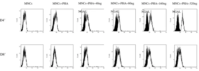
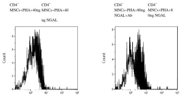
Fig. 4 HLA-G expression on CD4+ T cell: a) control PBMCs; b) PBMCs activated with PHA; c)
PBMCs activated with PHA, treated with NGAL 40 ng\ml; d) PBMCs activated with PHA, treated MNCs CD4+ MNCs+PHA MNCs+PHA+40ng NGAL MNCs+PHA+80ng NGAL MNCs+PHA+160ng NGAL MNCs+PHA+320ng NGAL CD8+
with NGAL 80 ng\ml; e) PBMCs activated with PHA, treated with NGAL 160 ng\ml; f) PBMCs
activated with PHA, treated with NGAL 320 ng\ml; isotype control in black, PBMCs at different
conditions in white.
The increase was proportional to NGAL increasing concentration: in fact for subject 4 we found a
7.3% of cell expressing HLAG in PBMCs cells, 32.8% of cell expressing HLAG in PBMCs cells
activated with PHA, 43.8% of cell expressing HLAG with NGAL 40 ng/mL, 45.8% of cell
expressing HLAG with NGAL 80 ng/mL, 61.8% of cell expressing HLAG with NGAL 160 ng/mL,
67.6% of cell expressing HLAG with NGAL 320 ng/mL; in subject 5 the trend was similar: 2.8% of
cell expressing HLAG in CD4+, 34.5% of cell expressing HLAG in CD4+ cells activated with
PHA, 44.1% of cell expressing HLAG with NGAL 40 ng/mL, 54.6% of cell expressing HLAG with
NGAL 80 ng/mL, 63.8% of cell expressing HLAG with NGAL 160 ng/mL, 71.9% of cell
expressing HLAG with NGAL 320 ng/mL (figure 4)..
The situation is different when we go to analyze CD8 + cells : in fact these cells do not exhibit a
significant increase of the expression of HLA-G if treated with increasing concentrations of NGAL.
These data demonstrate that HLA-G expression mediated by NGAL shows a different trend
among activated CD4+ and activated CD8+ T cells. In fact we found 6.5% of cell expressing
HLAG in CD8+ cells, 26.8% of cell expressing HLAG in CD8+ activated with PHA, 25% of cell
expressing HLAG with NGAL 40 ng/mL, 32.6% of cell expressing HLAG with NGAL 80 ng/mL,
33.3% of cell expressing HLAG with NGAL 160 ng/mL, 35% of cell expressing HLAG with
NGAL 320 ng/mL in subject 4 and 3.7% of cell expressing HLAG in CD8+cells, 19.5% of cell
expressing HLAG in CD8+ activated with PHA, 24.9% of cell expressing HLAG with NGAL 40
ng/mL, 33.5% of cell expressing HLAG with NGAL 80 ng/mL, 34.2% of cell expressing HLAG
Fig. 4 HLA-G expression as fluorescence intensity percentage on CD4+ T cells in control and treated groups after treatment with NGAL (40-320 ng\ml).
4.3 NGAL:Enterobactin Iron Free effect on HLA-G expression.
We also used the complex NGAL:enterocalin iron free to verify if cells response to NGAL was iron
dependent.
In PBMCs activated with PHA and treated with NGAL:Enterobactin Iron-free 40 ng/ml (g) an
increase in HLA-G expression was observed, also if not of the same concentration of
NGAL:Enterobactin:Iron complex. (Fig.xx). This was true both for CD4+ and for CD8+:36.7% and
31.3 % for subject 4 and 37.9% and 17.2 % for subject 5 respectively.
4.4 NGAL:Enterobactin:Iron effect on HLA-G expression following anti-NGAL antibody in vitro
administration.
To asses NGAL action inhibition we used anti-NGAL Monoclonal Antibody (200 μg; 1 mg/ml)
(Thermo Scientific). We found the following values of fluorescence positivity :: 1.7% in PBMCs,
13.6% in PBMCs+PHA, 28.4 % in PBMCs+PHA+NGAL:Fe 40 ng/ml, 22.2% in
PBMCs+PHA+NGAL:Fe 40 ng/ml + anti-NGAL, 39.9% in PBMCs+PHA+NGAL:Fe 80 ng/ml and
18.5 % PBMCs+PHA+NGAL:Fe 80 ng/ml + anti-NGAL.
We can also observe that the monoclonal antibody action was dose-dependent with an inhibitory
Fig. 5 HLA-G expression on PHA activated PBMCs after treatment with NGAL 40 and 80 ng/ml (black histogram) and incubation with an anti-Ngal antibody (white histogram).
Fig. 6 HLA-G expression on PBMCs PHA activated after stimulation with NGAL 40 and 80 ng/ml and
incubation with an anti-Ngal antibody.
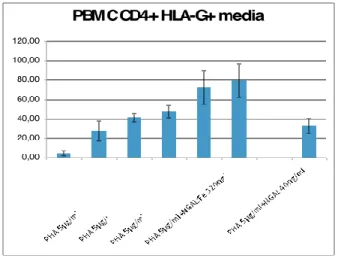
4.5 Indirect evaluation of NGAL role on HLA-G expression after treatment with Enterobactin:Iron.
Evaluation of Enterobactin:Iron 160 ng/ml effect on HLA-G expression shows that
Enterobactin:Iron without NGAL had none effect on HLA-G expression. (figure 7).
CD4
+ CD4+ MNCs+PHA+40 ng NGAL CD4+ MNCs+PHA+40ng NGAL+Ab CD4+ MNCs+PHA+8 0ng NGAL CD4+ MNCs+PHA+80ng NGAL+AbFigure 7: sHLAG values.
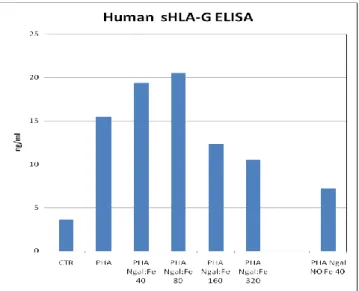
4.6 Soluble HLA-G evaluation trough ELISA after NGAL:Enterobactin:Iron treatment.
Soluble HLA-G evaluation trough ELISA after NGAL:Enterobactin:Iron treatment shows
increasing HLA-G secretion dose dependent up to NGAL:Enterobactin:Iron 80 ng/ml. In samples
treated with NGAL:Enterobactin:Iron 160 and 320 ng/ml secretion of soluble HLA-G decreases.
Stimulation with NGAL:Enterobactin 40 ng/ml induces HLA-G secretion only higher than the
control.
4,7 NGAL effect on T regulatory cells
We wanted also to evaluate NGAL action on CD4+ CD25+ Foxp3+ (regulatory t cells, Tregs) to
evaluate another important aspect of immune tolerance induction.
So we analyzed CD4+ FoxP3+ and CD4+ CD25+ FoxP3+ concentration before and after NGAL
addiction atincreasing dose and with or without iron presence (40 ng/ml with or without iron, 80
ng/ml, 160 ng/ml, 320 ng/ml). All data about Cd4+FoxP3+ cells and CD4+/CD25+/FoxP3+ cells
are reported in (figure 8).
We observed an increase in CD4+ FoxP3+ and CD4+ CD25+ FoxP3+ expression after NGAL
Indeed the percentage of Tregs increased with growing NGAL doses getting a plateau with
NGAL:Enterochelin:Iron 320 ng/ml. The lack of iron did not increased Tregs cells expression.
Comparing CD4+ and CD25+ cells FoxP3 expression confirmed a dose-dependent raise for the
treatment with NGAL (40-80-160-320ng/ml). The iron-free NGAL treatment presented less
increased expression of Tregs.
We then verified NGAL direct activity in Tregs expression adding NGAL antibody and we
observed that the presence of increasing anti-NGAL antibody determinate a significant reduction of
Tregs percentage (both CD4+/FoxP3+ and CD4+/CD25+/FoxP3+ .
Finally we evaluated NGAL effect on Tregs expression after Enterobactin:Fe 160 ng/ml treatment
and we found that Enterobactin:Fe had none effect on CD4+/FoxP3+ and CD4+/CD25+/FoxP3+
cells expression CTR PBMC CD4+ CD25+ FoxP3+ PBMC+PHA CD4+ PBMC+PHA+NGAL 40ng/ml CD4+ PBMC+PHA+NGAL 80ng/ml CD4+ PBMC+PHA+NGAL 160ng/ml CD4+ PBMC+PHA+NGAL 320ng/ml CD4+
Fig.8 Percentage CD4+/CD25+/FoxP3+ cells after treatment of PBMCs with NGAL (40 ng/ml with or without iron, 80 ng/ml, 160 ng/ml, 320 ng/ml).
5. Discussion and Conclusions
Nowadays NGAL is emerging as a mediator of various physiological and pathological conditions.
Indeed in literature is widely documented as NGAL is involved in the transport and metabolism of
iron, as well as its bacteriostatic activity is well known. The presence of NGAL molecule is also
important as growth factor and in the differentiation of immature kidney. In the last years
researchers' attention was focused on the role of NGAL as a biomarker of many diseases including
acute and chronic kidney damage, kidney transplantation, autoimmune and neoplastic disorders.
(Chakraborty)
Despite numerous studies that have investigated the physiological and pathophysiological role of
NGAL, this is not entirely elucidated. The use as biomarkers in many pathological conditions
shows that the molecule is characterized by a low specificity for pathology: this leads us to think
that there is a biological effect of the molecule that is common to these differents pathological
conditions [157].
In the light of pathologies involved, the molecule seems to be a response to different pathological
conditions that are united by inflammation and oxidative stress. [158].
With regard to the involvement of NGAL in immunity, only one study in the literature verifies the
involvement in cellular one [159], while none analyzes the role of the molecule in the humoral one.
In recent years, the interaction between NGAL and NF-kB (nuclear factor-κB) has been studied.
The expression of NGAL mRNA and protein is up-regulated in an NF-kB dependent manner in rat
and human vascular smooth muscle cells (SMCs) in response to IL-1β stimulation [160].
Recent evidence showed that there is also a possible interaction between NF-kB and NGAL gene
requiring IκB-ζ for its induction: the coexpression of IκB-ζ and the NF-κB subunits synergistically activates the transcription of NGAL genes [161]. The transcription factor NF-κB plays a key role in
the innate and adaptive immune systems. The interaction between NGAL and NF-Κb and the
involvement of NF-κB in innate and adaptative immune system suggest a possible role of NGAL in
immune tolerance.
Our study on healthy controls had the aim to individuate the possible involvement of NGAL in the
processes of immune response, focusing on the hypothesis that NGAL could promote immune
tolerannce.
By flow cytometry analysis we demonstrated that NGAL can increase the expression of mHLA-G
in PBMNCs cell cultures , particularly for CD4+ T lymphocytes. It is known as the role of HLA-g
is not only important for the maternal-fetal tolerance. He plays a significant role in adult life and
especially in some pathological conditions of Autoimmunity, cancer and tolerance in organ
transplantation [162]. As a factor able of activating the expression of HLA-G, NGAL may play the
role of mediator in acute and chronic processes with a similar anti-inflammatory meaning [163].
We also have shown that increasing NGAL concentration raises in vitro level of mHLA-G . So
we can postulate that NGAL increasing is not a random process, but it may express a biological
meaning in various pathological conditions and be modulated in favor of biological necessity. We
can also indirectly hypnotize that a decreased capability of NGAL production could be translated
in a lower reaction to inflammation and aging conditions.
The iron absence seems to have a role in NGAL action, leaving the expression of mHLA-G
unaltered. The iron deficiency may be negative not only for characterizing systemic inflammation
states by affecting the anemia conditions, but also inducing aging process and reducing tissue
renewal [164].
Use of NGAL:Ent:Fe in the presence anti-NGAL antibody is not able to modify the expression of
mHLA-G, confirming the action of NGAL molecule on mHLA-G activation.
Using of increasing concentration of NGAL conjugated with iron induces an activation of Tregs
NGAL role in FoxP3+ cells activation must be considered with extreme attention in the light of
Tregs function in autoimmune and neoplastic disease[165], and in the graft tolerance in solid organ
transplantation. Several authors indeed proposed Tregs cell therapy for graft tolerance induction.
In conclusion we believe NGAL is an extremely interesting molecule and its biological role is still
far from being fully clarified. If clinically most study in recent literature have focused on its role of
biomarker of kidney , neoplastic and inflammatory diseases we believe that other clinical interest
scenarios are possible.
These include the deep understanding of the role of NGAL in humoral immunity may lead not only
to the understanding of concerning physiopathological mechanisms but also a molecule used for
therapeutic purposes in many conditions under which the regulation of immune processes can play
6. REFERENCES
1. Kjeldsen L, Johnsen AH, Sengelov H, et al. Isolation and primary structure of NGAL a novel protein associated with human neutrophil gelatinase. J Biol Chem 268(14):1425-32, 1993.
2. Kjeldsen L, Cowland JB, Borregaard N. Human neutrophil gelatinase-associated lipocalin and homologous proteins in rat and mouse. Biochim Biophys Acta. 2000 Oct 18;1482(1-2):272-83.
3. Flower DR: Experimentally determined lipocalin structures. Biochim Biophys Acta 1482: 46 –56, 2000.
4. Chakraborty S, Kaur S, GuhaS et al. The multifaceted roles of neutrophil gelatinase associated lipocalin (NGAL) in inflammation and cancer. Biochimica et Biophysica Acta 1826: 129–169, 2012.
5. Newcomer ME, Ong DE: Plasma retinol binding protein: Structure and function of the prototypic lipocalin. Biochim Biophys Acta 1482: 57– 64, 2000.
6. Larsson J, Allhorn M, Kerstrom B: The lipocalin alpha(1)- microglobulin binds heme in different species. Arch Biochem Biophys 432: 196 –204, 2004.
7. Weichsel A, Andersen JF, Champagne DE, Walker FA,Montfort WR: Crystal structures of a nitric oxide transport protein from a blood-sucking insect. Nat Struct Biol 5:304 –309, 1998.
8. Kuwabara T., Mori K., Mukoyama M. et al. Urinary neutrophil gelatinase-associated lipocalin levels reflect damage to glomeruli, proximal tubules, and distal nephrons, Kidney Int. 75 (3): 285–294: 2009.
9. Axelsson L., M. Bergenfeldt, K. Ohlsson, Studies of the release and turnover of a human neutrophil lipocalin, Scand. J. Clin. Lab. Investig. 55: 577–588,1995.
10. V. Hvidberg, C. Jacobsen, R.K. Strong et al. The endocytic receptor megalin binds the iron transporting neutrophil-gelatinase-associated lipocalin with high affinity and mediates its cellular uptake, FEBS Lett. 579: 773–777, 2005.
11. Paragas N, Qiu A, Hollmen M. NGAL-Siderocalin in kidney disease. Biochimica et Biophysica Acta 1823: 1451–1458, 2012.
12. Strong RK, Bratt T, Cowland JB et al. Epression, purification, crystallization and
crystallographic characterization of dimeric and monomeric human neutrophil gelatinase-associated lipocalin (NGAL). Acta Crystallogra D Biol Crystallogr 54:93-5, 1998.