Acute Effects of Triiodothyronine (T
3) Replacement
Therapy in Patients with Chronic Heart Failure and
Low-T
3Syndrome: A Randomized, Placebo-Controlled
Study
Alessandro Pingitore, Elena Galli, Andrea Barison, Annalisa Iervasi, Maria Scarlattini, Daniele Nucci, Antonio L’Abbate, Rita Mariotti, and Giorgio Iervasi
Institute of Clinical Physiology (A.P., A.I., M.S., D.N., G.I.), Consiglio Nazionale delle Ricerche, 56124 Pisa, Italy; Cardiothoracic Department (E.G., A.B., R.M.), University of Pisa, 43-56126 Pisa, Italy; and Scuola Superiore Sant’Anna (A.L.), 34 56025 Pisa, Italy
Context: Low-T3syndrome is a predictor of poor outcome in patients with cardiac dysfunction. The
study aimed to assess the short-term effects of syntheticL-T3replacement therapy in patients with
low-T3syndrome and ischemic or nonischemic dilated cardiomyopathy (DC).
Design: A total of 20 clinically stable patients with ischemic (n⫽ 12) or nonischemic (n ⫽ 8) DC were enrolled. There were 10 patients (average age 72 yr, range 66 –77; median, 25–75th percentile) who underwent 3-d syntheticL-T3infusion (study group); the other 10 patients (average age 68 yr,
range 64 –71) underwent placebo infusion (control group). Clinical examination, electrocardiog-raphy, cardiac magnetic resonance, and bio-humoral profile (free thyroid hormones, TSH, plasma renin activity, aldosterone, noradrenaline, N-terminal-pro-B-Type natriuretic peptide, and IL-6) were assessed at baseline and after 3-d syntheticL-T3(initial dose: 20g/m2body surface䡠d) or
placebo infusion.
Results: After T3administration, free T3concentrations increased until reaching a plateau at 24 – 48
h (3.43, 3.20 –3.84 vs. 1.74, 1.62–1.93 pg/ml; P⫽ 0.03) without side effects. Heart rate decreased significantly after T3infusion (63, 60 – 66 vs. 69, 60 –76 beats per minute; P⫽ 0.008). Plasma
nor-adrenaline (347; 270 –740 vs. 717, 413– 808 pg/ml; P⫽ 0.009), N-terminal pro-B-Type natriuretic peptide (3000, 438-4005 vs. 3940, 528-5628 pg/ml; P⫽ 0.02), and aldosterone (175, 152–229 vs. 231, 154 –324 pg/ml; P⫽ 0.047) significantly decreased after T3administration. Neurohormonal profile
did not change after placebo infusion in the control group. After syntheticL-T3administration,
left-ventricular end-diastolic volume (142, 132–161 vs. 133, 114 –158 ml/m2body surface; P⫽ 0.02)
and stroke volume (40, 34 – 44 vs. 35, 28 –39 ml/m2body surface; P⫽ 0.01) increased, whereas
external and intracardiac workload did not change.
Conclusions: In DC patients, short-term syntheticL-T3replacement therapy significantly improved
neuroendocrine profile and ventricular performance. These data encourage further controlled trials with more patients and longer periods of syntheticL-T3administration. (J Clin Endocrinol
Metab 93: 1351–1358, 2008)
A
low T3syndrome has been documented in patients with dilated cardiomyopathy (DC); its occurrence is an inde-pendent predictor of poor outcome (1–5). The effect of decreased T3concentrations on myocyte gene expression and cardiaccon-tractility has already been documented in a model of low-T3 syndrome in which T3supplementation normalized both cardiac function and phenotype (6).
The main pathophysiological mechanism underlying low 0021-972X/08/$15.00/0
Printed in U.S.A.
Copyright © 2008 by The Endocrine Society
doi: 10.1210/jc.2007-2210 Received October 2, 2007. Accepted December 26, 2007. First Published Online January 2, 2008
Abbreviations: bs, Body surface area; CMR, cardiac magnetic resonance; CO, cardiac out-put; DC, dilated cardiomyopathy; EDV, end-diastolic volume; EF, ejection fraction; ESV, end-systolic volume; HF, heart failure; HR, heart rate; LV, left ventricular; ns, not significant; NT-proBNP, N-terminal pro-brain natriuretic peptide; PRA, plasma renin activity; SV, stroke volume; SVR, systemic vascular resistance.
E n d o c r i n e C a r e
circulating T3is the decreased activity of 5⬘-monodeiodinase, responsible for converting T4 into T3in peripheral tissues (1, 7).
The pathophysiological role of the progressive decrease in T3 that occurs in patients with heart failure (HF) has not yet been established (8). It may merely be a marker of the severity of the disease, or it could contribute to the impairment of cardiovas-cular function. The latter hypothesis is based on the key role of thyroid hormones on the homeostasis of the cardiovascular sys-tem by three different routes: 1) direct effect on cardiomyocytes; 2) peripheral effects on the vasculature; and 3) modulation of sympathetic systems (1, 9).
Although potentially promising, the usefulness of synthetic thyroid hormone administration as a new therapeutic strategy during evolution of HF is still debated (1, 10). In patients with DC and low-T3state, the short-term (a few hours) iv adminis-tration of pharmacological doses of syntheticL-T3increased car-diac output (CO) and decreased systemic vascular resistance (SVR) without changes in heart rate (HR) and arterial blood pressure (11). Administration of syntheticL-T3had no adverse effects, and, in particular, no arrhythmias were observed. How-ever, data on the effects of replacement doses ofL-T3in humans are lacking. In addition, very little information is available on the potential link between changes in thyroid hormone state and the other activated neuroendocrine/proinflammatory systems dur-ing progression of HF; however, preliminary data on humans seem promising (12).
This study aimed to evaluate the effects of 3-d iv replacement doses of syntheticL-T3on clinical status, left ventricular (LV)
function, and neuroendocrine/proinflammatory profile in pa-tients with DC and low-T3syndrome.
Patients and Methods
Patients
A total of 500 outpatients with known post-ischemic or nonisch-emic DC were screened. Post-ischnonisch-emic DC was diagnosed by angio-graphically proven coronary artery disease or by documented myo-cardial infarction; nonischemic DC was diagnosed based on the absence of coronary artery disease on angiography. Inclusion criteria were: 1) ischemic or nonischemic dilated left ventricle, i.e. end-dia-stolic diameter more than 56 mm and ejection fraction (EF) less than 40%, echocardiographically assessed; 2) optimized standard HF med-ical therapy; 3) New York Heart Association class less than III; and 3) stable thyroid function pattern with low free T3levels confirmed on
the basis of two consecutive determinations within the last month. Exclusion criteria were: 1) history of primary thyroid disease, 2) ami-odarone therapy during the past 6 months, 3) concomitant severe systemic disease, 4) complex ventricular arrhythmias, 5) severe obe-sity (body mass index⬎ 35 kg/m2), and 6) pregnant women or women
undergoing estro-progestinic therapy.
Based on the aforementioned criteria, a total of 445 patients was excluded. Of the remaining 55 patients, 35 were excluded for the fol-lowing reasons: 1) rapid, unexpected clinical worsening (n⫽ 8); 2) need for changes in medical treatment (n⫽ 11); and 3) normalization of thyroid pattern (n⫽ 5), refusal of hospitalization, and/or of synthetic L-T3infusion (n⫽ 11).
Therefore, the final population consisted of 20 patients (14 male, 6 female), with an average body mass index of 28 kg/m2(range 25–31), and
an average body surface area (bs) of 1.89 m2(range 1.81–1.92) with
post-ischemic (n⫽ 12) or nonischemic (n ⫽ 8) DC, randomly assigned TABLE 1. HR, blood pressure, and rate pressure product (RPP) at baseline and after 3 d in patients treated with syntheticL-T3or placebo infusion
Variables
Patients treated withL-T3 Patients treated with placebo
BeforeL-T3 AfterL-T3 P value Basal After 3 d P value
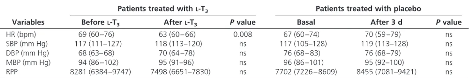
HR (bpm) 69 (60 –76) 63 (60 – 66) 0.008 67 (60 –74) 70 (59 –79) ns
SBP (mm Hg) 117 (111–127) 118 (113–120) ns 117 (105–128) 119 (113–128) ns
DBP (mm Hg) 68 (63– 68) 70 (64 –78) ns 76 (68 – 83) 76 (68 –79) ns
MBP (mm Hg) 94 (86 –102) 95 (91–96) ns 96 (86 –101) 95 (92–100) ns
RPP 8281 (6384 –9747) 7498 (6651–7830) ns 7702 (7226 – 8609) 8455 (7081–9421) ns
Data are expressed as median (25th and 75th percentiles). DBP, Diastolic blood pressure; MBP, mean blood pressure; SBP, systolic blood pressure.
TABLE 2. Effect of syntheticL-T3infusion on cardiac rhythm Patient
no.
No. of PACs No. of SVTs No. of PVCs No. of NSVTs
Before T3 After T3 Before T3 After T3 Before T3 After T3 Before T3 After T3
1 42 32 6 4 294 100 40 0 2 107 5 42 0 947 2047 6 0 3 1 3 1 0 177 104 0 0 4 35 24 1 2 924 66 0 0 5 5 1 5 0 227 355 0 1 6 2 6 2 1 230 135 0 0 7 45 31 2 2 353 121 48 2 8 8 2 4 0 272 426 0 1 9 50 38 7 5 739 53 0 0 10 86 4 34 0 663 1433 4 1
to continuous 3-d iv syntheticL-T3infusion. There were 10 patients
(mean age 72 yr, range 66 –77) who underwentL-T3infusion and
com-prised the study group. The other 10 patients (mean age 68 yr, range 64 –71) underwent continuous low-rate (100 ml/d) 3-d iv infusion of saline (placebo) solution and comprised the control group. Standardized medical therapy for HF was optimized before the study; this remained unchanged for at least 15 d before the study was initiated and remained the same throughout the entire 3-d T3infusion. Medical therapy
con-sisted of angiotensin-converting enzyme inhibitors (n⫽ 17), diuretics (n⫽ 15),-blockers (n ⫽ 16), and spironolactone (n ⫽ 10 patients). Experimental protocol
All enrolled patients were admitted to the Institute of Clinical Phys-iology in Pisa and hospitalized to perform the study protocol. All patients gave informed consent for hospitalization andL-T3infusion. The study
was approved by the local ethics review committee and conformed to the principles outlined in the Declaration of Helsinki. SyntheticL-T3was
continuously infused (initial dose 20g/m2bs䡠d diluted in 100 ml saline);
we used this dosage, which is slightly higher than the measured T3
pro-duction rate in normal humans (16⫾ 3g/m2bs䡠d, mean ⫾SD) (13), to
restore normal T3levels as rapidly as possible while avoiding potential
side effects. Starting on the first day after the beginning of infusion, on the basis of measured T3levels, the dose was adjusted to maintain T3
circulating levels within the normal range (see Thyroid function pattern throughoutL-T3infusion). Clinical signs and symptoms, bio-humoral
profile, and cardiac magnetic resonance (CMR) were evaluated at base-line and at the end ofL-T3infusion. Continuous electrocardiographic
monitoring was maintained during the entireL-T3infusion period to
detect arrhythmias. Systolic/diastolic and mean blood pressure as well as HR were measured five times per day; the reported values of these pa-rameters at baseline and after T3/placebo administration (see Results)
represent the average value of the measures. Rate pressure product was calculated as the product between HR and systolic blood pressure. Neuroendocrine and proinflammatory bio-humoral profile
Basal blood samples were taken at 0800 h from an antecubital vein after a 30-min rest in supine position. Plasma N-terminal pro-brain na-triuretic peptide (NT-proBNP) was measured with a fully automated “sandwich” electrochemiluminescence method using an Elecsys 2010 analyzer (Roche Diagnostics, Basel, Switzerland), as previously de-scribed (14). The low detection limit of the NT-proBNP assay was 4.2 pg/ml (0.50 pmol/liter), whereas the functional sensitivity was 22 pg/ml (2.60 pmol/liter). Plasma renin activity (PRA) (ng/ml䡠h) and aldosterone (pg/ml) were measured by RIA (Dia Sorin S.r.l, Saluggia, Italy); for the assay, blood samples were immediately put into ice-chilled tubes con-taining EDTA, and then plasma was rapidly separated by centrifugation at 4 C and frozen at⫺20 C (15). Serum TSH, free T3, and free T4were
measured using an AIA 21 analyzer (Eurogenetics-Tosho, Turin, Italy). The reference intervals for our laboratory were: free T3, 2.1– 4.2 pg/ml
(3.4 – 6.5 pmol/liter); free T4, 7.1–18.5 pg/ml (9.2–24 pmol/liter); and
TSH, 0.30 –3.80IU/ml. Measured functional sensitivity for the TSH assay was 0.12IU/ml. For the measurement of plasma norepinephrine (pg/ml), we used the HPLC method as previously described in detail (15). Levels of IL-6 (pg/ml) were measured by a high-sensitivity ELISA tech-nique (Diaclone Research, Besanc¸on, France).
Assessment of cardiac morphology and function
CMR imaging was performed with a 1.5 T Signa Excite Scanner (GE Medical System, Waukesha, Wisconsin) using an eight-element phased array cardiac receiver coil. To evaluate LV function, images were ac-quired in short axis views, from the mitral annulus to the ventricular apex (thickness 8 mm, no spacing) using a breath-hold gradient-echo pulse sequence triggered to electrocardiogram. For each image the myocar-dium was defined by manually tracing the endocarmyocar-dium to assess end-diastolic volume (EDV) (ml/m2bs), end-systolic volume (ESV) (ml/m2
bs), stroke volume (SV) (ml/m2bs), and EF (%). CO (liter/min) was
obtained as the product of SV and HR. SVR (dyne/sec⫻ cm) was
com-puted as the mean arterial blood pressure divided by CO. Internal and external cardiac works were calculated as follows: internal cardiac work⫽ ESV ⫻ HR ⫻ (systolic blood pressure/2); and external cardiac work⫽ SV ⫻ HR ⫻ mean blood pressure. Total cardiac work was calculated as the sum of the internal and external cardiac work. Statistical analysis
All variables are expressed as median plus 25th and 75th percentile, unless otherwise indicated. Continuous data were analyzed by the non-parametric Wilcoxon test. A P value less than 0.05 was considered sta-tistically significant. ANOVA and post hoc comparison tests for repeated measures were performed with the Friedman test and Bonferroni ad-justed Wilcoxon test to assess the differences of thyroid hormones and TSH circulating levels during the 3-dL-T3and placebo infusion. All
analyses were performed using SPSS for Windows (version 11.00; SPSS, Inc., Chicago, IL).
Results
Clinical status
The main clinical characteristics of patients are shown in Ta-ble 1. Plasma protein and albumin levels beforeL-T3infusion were normal (6.9, 6.4 –7.2 g/dl, and 4.2, 3.1– 6.2 g/dl, respec-tively). SyntheticL-T3infusion was well tolerated, and no side effects were reported. HR decreased significantly, whereas blood pressure and body weight remained unchanged. Continuous electrocardiographic monitoring showed no increase either in
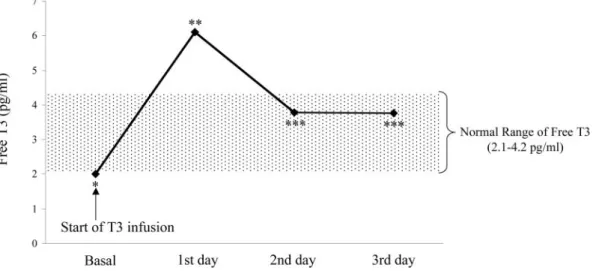
FIG. 1. Free T3levels in patients treated withL-T3(upper panel) and in patients
the number of ventricular premature beats (Table 2) or in the appearance of ischemic episodes. QT intervals did not change duringL-T3infusion [before T3437, 429 – 477 msec vs. after T3 439, 413– 478; P⫽ not significant (ns)].
Thyroid function pattern throughoutL-T3infusion
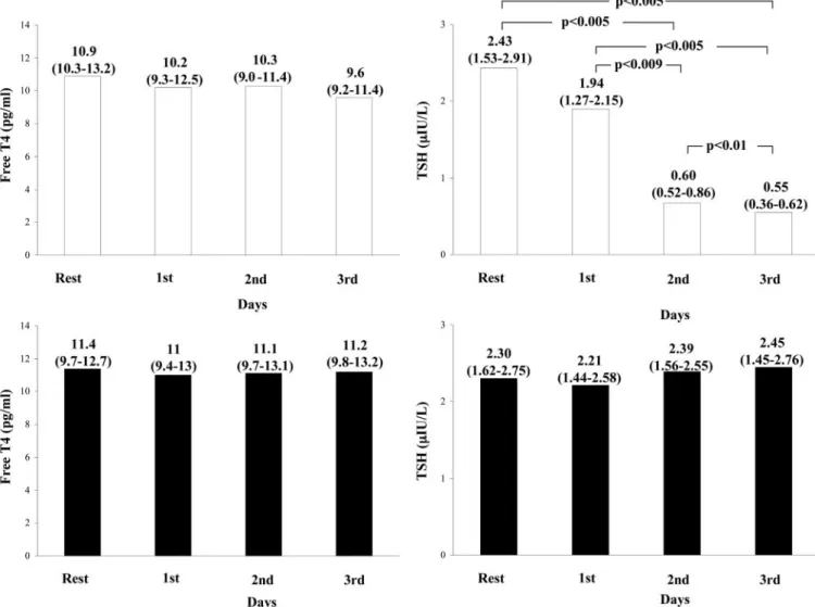
At baseline all patients showed a typical low-T3syndrome with free T3plasma levels lower than the limit of reference range. After starting T3replacement iv therapy, free T3concentrations rapidly increased until reaching the upper level of the physio-logical range, then remained stable throughout the entire infu-sion time (ANOVA P⬍ 0.001) (Fig. 1). Starting from the second day of infusion, the mean dose of administered T3was 1.10⫾ 0.11 g (mean ⫾SD) per hour (range 0.8 –1.2), which corre-sponds to 24.2g/d (range 19.2–28.8), i.e. 13.4 g/m2bs䡠d, on average. A typical example of the adjustment ofL-T3infusion rate in a patient with elevated FT3concentrations after the first day of administration is reported in Fig. 2. During treatment there was a concomitant decrease in free T4and even more in TSH levels (ANOVA P⬍ 0.001 for TSH), although the concen-trations of both hormones still remained within the normal range (Fig. 3). In the placebo-treated patients, no significant change was observed in the circulating levels of thyroid hormones and TSH.
Routine laboratory and
neuroendocrine/proinflammatory profile
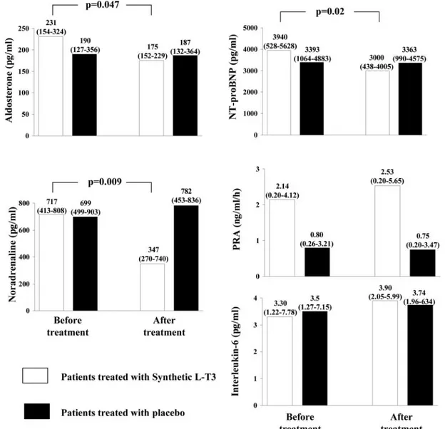
SyntheticL-T3infusion did not induce any significant change in the main routine laboratory variables. At the end of T3 infu-sion, there was a significant decrease in noradrenaline, NT-proBNP, and aldosterone plasma levels (Fig. 4), whereas PRA and IL-6 remained unchanged. Neurohormonal profile did not change after placebo infusion in the control group.
Cardiac function
End-diastolic LV volume and SV increased significantly, whereas EF, CO, SVR, external, internal, and total cardiac work-load did not change (Table 3).
Discussion
In our study we assessed the effects of the iv infusion of replace-ment doses of L-T3 on cardiac function and on the activated neuroendocrine system in patients with stable ischemic or non-ischemic LV dysfunction and low-T3syndrome. TheL-T3 infu-sion regimen adopted rapidly restored T3levels to within normal range and was associated with a significant decrease in TSH level, which still remained, however, in the normal range. A similar TSH pattern was observed in the study by Moruzzi et al. (16), in which a replacement dose ofL-T4, i.e. 0.1 mg/d, was used.
Our main finding was thatL-T3infusion induced a positive cardiac and neuroendocrine resetting characterized by improved SV of the left ventricle and deactivation of the neuroendocrine profile, resulting from the significant reduction in vasoconstric-tor/sodium retaining noradrenaline, aldosterone, and in the counterpart NT-proBNP plasma levels. The protocol we adopted for syntheticL-T3administration offers two main ad-vantages: 1) constant infusion of substitutive doses ofL-T3makes it possible to rapidly establish stable T3levels within the phys-iological range (Fig. 1), which is at variance with multiple bolus injections or an oral regimen; and 2) constant infusion of sub-stitutive doses of L-T3is more effective in promoting nuclear action of T3and T3-mediated transcription in the myocardium when compared with multiple bolus injections (14) or to an oral regimen (17–19). However, previous studies on euthyroid pa-tients with DC showed that short- and medium-term treatment
with 0.1 mg/d syntheticL-T4increased CO and reduced SVR in the absence of significant changes in HR and catecholamine cir-culating levels (16). A similar hemodynamic finding has been shown in our previous study in patients with subclinical hypo-thyroidism and without cardiac disease (20). In that study, SV, EF, and CO significantly increased after syntheticL-T4 replace-ment therapy, whereas blood pressure values did not change. On the contrary, the absence of a decrease in SVR after T3infusion observed in our low-T3cardiomyopathic patients could be re-lated to the significant decrease in HR associated with a signif-icant reduction in noradrenaline levels, neither documented in any of the previous studies cited (16), thus causing an unchanged CO despite the documented significant increase in SV.
In addition, we preferred to administer T3instead of the pro-hormone T4because in a previous study on hypothyroid animals, the restoration of serum biologically active T3by constant infu-sion of T4was unable to normalize all tissue levels of T3, includ-ing the myocardium (21). This could be even more evident in the presence of an impaired peripheral conversion of T4into T3, as observed in low-T3syndrome.
The main novel finding of this study is the evidence for a deactivation of the vasoconstrictor/sodium-retaining neuroen-docrine system that occurs afterL-T3infusion, with NT-proBNP,
noradrenaline, and aldosterone all decreasing significantly when compared with baseline values and with corresponding hor-monal levels after placebo infusion. The neuroendocrine rear-rangement may be interpreted as an indirect rather than direct T3-mediated action, very likely linked to the improved cardiac performance as documented by increased LV SV. In fact, T3“per
se” is able to increase rather than decrease catecholamines, BNP,
and aldosterone release. This effect is mediated by promoting BNP gene transcription (22) or by regulating the rate of tran-scription of the-1-adrenergic receptor gene (23). Accordingly, increased and decreased NT-proBNP levels have been observed in patients with hyperthyroidism or hypothyroidism, respec-tively (24), and parallel changes in the levels of catecholamine and catecholamine metabolites have been shown in cardiac mus-cle of rats with thyroid disorders (25). Similarly, decreased al-dosterone circulating levels have been observed in hypothyroid patients treated with synthetic thyroid hormones (26). Further evidence in favor of an indirect positive effect of T3on neuroen-docrine resetting is the observation of a decreased HR. Interest-ingly, the improvement in cardiac performance induced by T3 did not correspond to increased myocardial oxygen consump-tion, as indirectly estimated by calculation of the rate pressure product as well as total cardiac work.
Deactivation of the neuroendocrine system is a crucial goal in the therapeutic management of HF. The potential clinical relevance of T3-induced neuroendocrine deactivation in pa-tients with LV dysfunction is clearly deducible from an anal-ysis of reported data in the literature showing highly beneficial effects of aldosterone and-adrenergic antagonists in terms of survival, rate of hospitalization, symptoms, cardiac remodel-ing, and performance (27). Indeed, afterL-T3administration, in addition to neuroendocrine deactivation, we found a par-allel increase in SV and EDV in the absence of any significant change in LV EF. The increased EDV can be considered an expression of the recruitment of residual ventricular filling reserve, which is a fundamental compensatory mechanism for maintaining CO in patients with HF (28). This finding may result from the positive effects of biologically active T3 on diastolic relaxation secondary to the increase in calcium AT-Pase of the sarcoplasmic reticulum pump and to the inhibition of its counter-regulatory phospholamban (10). Studies have also shown an improved cardiac function and LV remodeling after replacement doses ofL-T3(29). Future studies are needed to clarify whether the observed positive acute changes in terms
of both neuroendocrine profile and hemodynamics will be maintained during chronicL-T3administration.
Limitations of the study
The study’s main limitation was the small number of pa-tients. Therefore, results regarding the potential safety ofL-T3 administration cannot be considered conclusive; another lim-itation is that our results cannot be extended to all patients with HF because we enrolled only highly selected and clini-cally stable patients with LV dysfunction and low-T3 syn-drome. The complexity and high cost of the protocol, includ-ing hospitalization and CMR, along with the multiplicity of inclusion and exclusion criteria adopted were the main rea-sons for the low number of patients finally enrolled forL-T3 administration. Another limitation of the study was the lack of assessment of the effects of syntheticL-T3on the diastolic function of the left ventricle. However, previous studies have clearly shown the improvement in diastolic function induced by synthetic thyroid hormone administration (30, 31). An-other limitation was that the potential total body catabolic effects of T3 were determined only indirectly by assessing FIG. 4. Aldosterone, noradrenaline, NT-proBNP, PRA, and IL-6 levels in patients treated withL-T3and in patients treated with placebo.
blood urea nitrogen levels, and not directly by calorimetry. However, in this context it has already been shown that an increased catabolic rate occurs not only when circulating T3 levels are kept within normal range, but also during supra-physiologicalL-T3administration (10), when circulating T3 exceeds the upper limit of the reference interval.
Conclusions
Altogether, our data indicate that short-term administration of substitutive doses of syntheticL-T3state reduces activation of the neuroendocrine system and improves LV SV in patients with ventricular dysfunction and low-T3syndrome. Future studies will clarify whether this approach may truly be considered a novel tool in the therapeutic strategies for managing cardiac failure.
Acknowledgments
We thank Laura Mazza for her secretarial assistance.
Address all correspondence and requests for reprints to: Giorgio Ier-vasi, M.D., Clinical Physiology Institute, Consiglio Nazionale delle Ricerche, Via Moruzzi 1 Localita` la Fontina, 56124 Pisa, Italy. E-mail: [email protected].
Disclosure Statement: The authors have nothing to disclose.
References
1. Klein I, Ojamaa K 2001 Mechanism of disease: thyroid hormone and the cardiovascular system. N Engl J Med 344:501–509
2. Hamilton MA, Stevenson LW, Luu M, Walden JA 1990 Altered thyroid hor-mone metabolism in advanced heart failure. J Am Coll Cardiol 16:91–95 3. Iervasi G, Pingitore A, Landi P, Raciti M, Ripoli A, Scarlattini M, L’Abbate A,
Donato L 2003 Low-T3 syndrome: a strong prognostic predictor of death in patients with heart disease. Circulation 107:708 –713
4. Kozdag G, Ural D, Vural A, Agacdiken A, Kahraman G, Sahin T, Ural E, Komsuoglu B 2005 Relation between free triiodothyronine/free thyroxine ra-tio, echocardiographic parameters and mortality in dilated cardiomyopathy. Eur J Heart Fail 7:113–118
5. Pingitore A, Landi P, Taddei MC, Ripoli A, L’Abbate A, Iervasi G 2005
Tri-iodothyronine levels for risk stratification of patients with chronic heart fail-ure. Am J Med 118:132–136
6. Katzeff HL, Powell SR, Ojamaa K 1997 Alterations in cardiac contractility and gene expression during low-T3 syndrome: prevention with T3. Am J Physiol 273(5 Pt 1):E951–E956
7. Utiger RD 1995 Altered thyroid function in nonthyroidal illness and surgery: to treat or not to treat? N Engl J Med 333:1562–1563
8. Utiger RD 2005 Commentary: T3 levels for risk stratification of patients with chronic heart failure. Clinical Thyroidology 17:16
9. Kahaly GJ, Dillmann WH 2005 Thyroid hormone action in the heart. Endocr Rev 26:704 –728
10. Dillmann WH, Barrieux A, Shanker R 1989 Influence of thyroid hormone on myosin heavy chain mRNA and other messenger RNAs in the rat heart. Endocr Res 15:565–577
11. Hamilton MA, Stevenson LW, Fonarow GC, Steimle A, Goldhaber JI, Child JS, Chopra IJ, Moriguchi JD, Hage A 1998 Safety and hemodynamic effects of intravenous triiodothyronine in advanced congestive heart failure. Am J Car-diol 81:443– 447
12. Pingitore A, Iervasi G, Barison A, Prontera C, Pratali L, Emdin M, Giannessi D, Neglia D 2006 Early activation of an altered thyroid hormone profile in asymptomatic or mildly symptomatic idiopathic left ventricular dysfunction. J Card Fail 12:520 –526
13. Pilo A, Iervasi G, Vitek F, Ferdeghini M, Cazzuola F, Bianchi R 1990 Thyroidal and peripheral production of 3,5,3⬘-triiodothyronine in humans by multicom-partmental analysis. Am J Physiol 258(4 Pt 1):E715–E726
14. Prontera C, Emdin M, Zucchelli GC, Ripoli A, Passino C, Clerico A 2004 Analytical performance and diagnostic accuracy of a fully-automated electro-chemiluminescent assay for the N-terminal fragment of the pro-peptide of brain natriuretic peptide in patients with cardiomyopathy: comparison with immunoradiometric assay methods for brain natriuretic peptide and atrial natriuretic peptide. Clin Chem Lab Med 42:37– 44
15. Emdin M, Passino C, Prontera C, Iervasi A, Ripoli A, Masini S, Zucchelli GC, Clerico A 2004 Cardiac natriuretic hormones, neuro-hormones, thyroid hor-mones and cytokines in normal subjects and patients with heart failure. Clin Chem Lab Med 42:627– 636
16. Moruzzi P, Doria E, Agostoni PA 1996 Medium-term effectiveness of L-thy-roxine treatment in idiopathic dilated cardiomyopathy. Am J Med 101:461– 467
17. Danzi S, Dubon P, Klein I 2005 Effect of serum triiodothyronine on regulation of cardiac gene expression: role of histone acetylation. Am J Physiol Heart Circ Physiol 289:H1506 –H1511
18. Saberi M, Utiger RD 1974 Serum thyroid hormone and thyrotropin concen-trations during thyroxine and triiodothyronine therapy. J Clin Endocrinol Metab 39:923–927
19. Danzi S, Ojamaa K, Klein I 2003 Triiodothyronine-mediated myosin heavy chain gene transcription in the heart. Am J Physiol Heart Circ Physiol 284: H2255–H2262
20. Ripoli A, Pingitore A, Favilli B, Bottoni A, Turchi S, Osman NF, De Marchi D, Lombardi M, L’Abbate A, Iervasi G 2005 Does subclinical hypothyroidism TABLE 3. CMR parameters at baseline and after 3 d in patients treated with syntheticL-T3or placebo infusion
Parameters
Patients treated withL-T3 Patients treated with placebo
BeforeL-T3 AfterL-T3 P value before L-T3vs. afterL-T3 Basal P value before L-T3vs. basal LV EDV (ml/m2bs) 133 (114–158) 142 (132–161) 0.02 130 (117–153) ns LV ESV (ml/m2bs) 103 (84–127) 108 (89–124) ns 91 (86–115) ns LV SV (ml/m2bs) 35 (28–39) 40 (34–44) 0.01 36 (29–48) ns CO (liter/min) 4.1 (3.3–5.4) 4.8 (3.4–5.4) ns 4.7 (4.0–5.3) ns CI (liter/m2bs⫻ min) 2.2 (1.7–2.8) 2.5 (1.9–2.7) ns 2.5 (2.1–2.9) ns LV EF (%) 25 (18–32) 28 (22–32) ns 27 (23–41) ns SVR (dyne/sec⫻ cm) 2.07 (1.92–3.13) 2.10 (1.87–2.48) ns 2.03 (1.86–2.36) ns Elastance 1.36 (0.93–1.63) 1.27 (0.91–1.36) ns 1.32 (0.97–2.14) ns
External cardiac work (ml⫻ mm Hg ⫻ bpm)
201,226 (161,084–3,002,307) 226,519 (169,276–266,388) ns 253,950 (190,929–306,180) ns Internal cardiac work
(ml⫻ bpm ⫻ mm Hg/2)
401,849 (348,910–534,505) 396,885 (343,080–473,613) ns 360,260 (314,153–440,763) ns Total cardiac work 626,859 (492,291–787,522) 592,085 (540,060–756,684) ns 599,945 (538,645–748,639) ns
affect cardiac pump performance? Evidence from a magnetic resonance im-aging study. J Am Coll Cardiol [Erratum (2005) 45:968] 45:439 – 445 21. Escobar-Morreale HF, del Rey FE, Obregon MJ, de Escobar GM 1996 Only
the combined treatment with thyroxine and triiodothyronine ensures euthy-roidism in all tissues of the thyroidectomized rat. Endocrinology 137:2490 – 2502
22. Liang F, Webb P, Marimuthu A, Zhang S, Gardner DG 2003 Triiodothyronine increases brain natriuretic peptide (BNP) gene transcription and amplifies en-dothelin-dependent BNP gene transcription and hypertrophy in neonatal rat ventricular myocytes. J Biol Chem 278:15073–15083
23. Bahouth SW 1991 Thyroid hormones transcriptionally regulate the beta 1-ad-renergic receptor gene in cultured ventricular myocytes. J Biol Chem 266: 15863–15869
24. Nishino M, Kimura T, Kanda T, Kotajima N, Yoshida A, Kuwabara A, Tamama K, Fukumura Y, Kobayashi I 2000 Circulating IL-6 significantly correlates to thyroid hormone in acute myocardial infarction but not in chronic heart failure. J Endocrinol Invest 23:509 –514
25. Mano T, Sakamoto H, Fujita K, Makino M, Kakizawa H, Nagata M, Kotake M, Hamada M, Uchimura K, Hayakawa N, Hayashi R, Nakai A, Itoh M, Kuzuya H, Nagasaka A 1998 Effects of thyroid hormone on catecholamine
and its metabolite concentrations in rat cardiac muscle and cerebral cortex. Thyroid 8:353–358
26. Park CW, Shin YS, Ahn SJ, Kim SY, Choi EJ, Chang YS, Bang BK 2001 Thyroxine treatment induces upregulation of renin-angiotensin-aldosterone system due to decreasing effective plasma volume in patients with primary myxoedema. Nephrol Dial Transplant 16:1799 –1806
27. Jessup M, Brozena S 2003 Heart failure. N Engl J Med 348:2007–2038 28. Colucci WS, Braunwald E 2005 Pathophysiology of heart failure. In:
Braun-wald E, Bonow R, Zipes D, Libby P, eds. Heart disease: a textbook of cardio-vascular medicine. 7th ed. Philadelphia: Elsevier Saunders; 509 –538 29. Ojamaa K, Kenessey A, Shenoy R, Klein I 2000 Thyroid hormone metabolism
and cardiac gene expression after acute myocardial infarction in the rat. Am J Physiol Endocrinol Metab 279:E1319 –E1324
30. Biondi B, Fazio S, Palmieri AM, Carella C, Panza N, Cittadini A, Bone` F, Lombardi G, Sacca` L 1999 Left ventricular diastolic dysfunction in patients with subclinical hypothyroidism. J Clin Endocrinol Metab 84:2064 –2067 31. Virtanen VK, Saha HHT, Groundstroem KWE, Salmi J, Pasternack AI 2001
Thyroid hormone substitution therapy rapidly enhances left-ventricular dia-stolic function in hypothyroid patients. Cardiology 96:59 – 64