Post-retention effects of rapid maxillary expansion on nasal cavity and on periodontal structures
9
0
0
Testo completo
(2) N AL I. IO. AZ. N. R. TE. Rapid maxillary expansion (RME) is the most effective orthopedic procedure to increase the maxillary transverse dimension in young patients by opening the midpalatal suture (1). The maxillary halves are separated by heavy forces along midpalatal suture into a triangular shape with the base of the triangle corresponding to the anterior nasal spine and the apex to posterior nasal spine with centre of rotation corresponding to third molars (2). The first clinical use of Rapid Maxillary Expansion was described over a century ago by Emerson C. Angell in 1860 that reported the possibility to gain maxillary arch and to correct marked cross-bite (3). Monson (1898) described during the same years the improvements in nasal respiration after treatment with RME; malformed palatine process form the top of the vault and floor of nasal cavity oftentimes coming in close proximity to the too distorted vomer (4). Since then numerous appliances have been designed to obtain orthopedic forces during expansion, thus limiting movement of teeth and maximizing skeletal displacement (5-7). Several researches (8-11) have studied the effects on oral respiration and on dentoskeletal structures demonstrating a lateral tipping of posterior teeth induced by RME, and this effect has been correlated to a widespread sequence of changes throughout the periodontal membrane and alveolar bone due to the presence of pressure and tension areas (8-11). A relevant concern at this regard is represented by the possible periodontal consequences of RME because of the risk of damage the buccal cortical plate of alveolar bone in correspondence of anchorage teeth and/or the development of recessions (8-11). Most investigations have analyzed the dentoskeletal effects of RME by bi-dimensional and conventional radiographic head-plates (2, 3, 5-11). Timms (1982) used for the first time Computed Tomography (CT) in studying the basal bone changes induced by rapid expansion, and since. ©. C. IC. ED. IZ. IO. N. II N. original article. Introduction. then there is very little researches based on this method of investigation (12). Podesser et al. (2007) observed the changes induced by orthopaedic treatment on nasal structures based on standardized CT scanning registration in 9 growing children with a mean age of 8.1 years. It was demonstrated a combination of skeletal change with an increase of the nasal width and a clear tipping of the dentoalveolar structures (13). Palaisa et al. (2007) studied 19 subjects with a mean age of 11.2 years by using conventional tomography and it was pointed out a gain in both area and volume of nasal cavity after orthopaedic treatment (14); Garrett et al. (2007) by using Cone Beam reported an increase in nasal width associated with a reduction of the area of maxillary sinus (15). Garib et al. (2005, 2006) used this method of investigation to evaluate dento-alveolar effects of RME. CT images of all subjects were taken before expansion and after a retention period of 3 months when the expander was definitely removed. Orthopaedic treatment produced a significant increase in all measured transverse dimensions and a buccal movement of maxillary posterior teeth by tipping and bodily translation. At the periodontal level orthopaedic forces reduced buccal plate thickness but increased lingual plate thickness of supporting teeth (16, 17). Rungcharassaeng et al. (2007) used cone-beam computed tomography to determine the factors that might affect buccal bone changes of maxillary posterior teeth after RME. A study sample of 30 patients with a men age of 13.8 years was selected and CT scans were taken before expansion and after a retention period of 3 months. Also in this research a reduction of the buccal bone plate thickness due to the tipping of anchoring teeth was observed (18). These studies were undertaken either before or after a very short period of retention (about three months). It should be noted that a retention period of at least 5 months is necessary to permit an adequate dissipation of residual loads of orthopaedic forces as well as a good mineralization of the midpalatal suture, in order to minimize the relapse tendency after rapid maxillary expansion (7, 19-. 96. ORAL & Implantology - Anno I - N. 3-4/2008.
(3) original article. ©. C. IC. ED. IZ. IO. N. N AL I. IO. AZ. N. II N. The sample investigated comprised 17 children (7 males and 10 females) with a mean age of 11.2 years (range 8-14 years) treated at the Department of Orthodontics, “Tor Vergata”. The criteria for selection of the treatment group were as follows: constricted maxillary arches, presence of uni- or bilateral posterior cross-bite, variable degree of crowding, and one or both maxillary canines presenting with palatal displacement as assessed by panoramic radiographs. The exclusion criteria were: age above 15 years, stages in cervical vertebral maturation, as assessed on lateral cephalograms, more advanced than CS4 (post-pubertal), absence of maxillary first molars, metallic restorations on the maxillary posterior teeth, previous periodontal diseases, previous orthodontic treatments, genetic diseases. Each patient underwent a standardized protocol with RME in the form of the Butterfly Palatal Ex-. R. Materials and methods. pander that follows the basic design of Haas. It’s a butterfly-shaped stainless steel framework banded on maxillary first molars that extends forward to the palatal surfaces of the deciduous molars (2224) (Fig. 1). The expansion screw was activated 2 turns per day (0.25 mm per turn) for 14 days, thus reaching the total amount of expansion of 7 mm in all subjects. Then, the screw was tied off with a ligature wire and the Butterfly Expander was kept on the teeth as a passive retainer for an average of six months. Multislices CT scans were taken before rapid palatal expansion (T0), at the end of the active expansion phase (T1) and after a retention period of six months when the expander was removed (T2). The exams were performed by a single trained radiographer at the same scanner console with the primary indication of evaluating the exact position, the three-dimensional orientation, and the spatial relationship of displaced intraosseous canines in the maxilla. Each patient was positioned horizontally on the scanner table with the Camper’s plate perpendicular to the ground. The perpendicular light beams of the machine were used to standardize the head position in the three plates, thus allowing comparison of the images achieved before, during, and after expansion. During the CT scanning, patients were biting on a piece of gauze to keep the maxillary and the mandibular teeth separated, and to avoid the overlapping of dentofacial structures. All exams were performed at the Department of Radiology, University of Rome, “Tor Vergata”, with a CT scanner equipped with a Dentascan reconstruction program used to study the maxillofacial region (Light-Speed 16, General Electric Medical System, Milwaukee, Winsconsin, USA). This machine is equipped with 16 detector rows and has a minimal rotation time of 0.5 sec, given a collimation between 0.75 and 1.5 mm with dose calibration. Subsequent scans were taken with a 1.25 mm slice thickness, 0.6 mm interval, 11.25 mm table speed/rotation, 100 mA, 13.7 cm FOV, 512x512 matrix, 0° gantry angle, and following a low dose protocol with 80 KV instead of the standard setting used for a Dentascan of 120 KV.. TE. 21). In literature there aren’t researches that have post-treatment observation after a retention period of 6 months. The aim of this study was to evaluate by means of low-dose CT the skeletal and periodontal changes associated with RME at the end of the active phase and after 6 months of retention in a relatively large group of growing patients.. Figure 1 The Butterfly Rapid Maxillary Expander.. ORAL & Implantology - Anno I - N. 3-4/2008. 97.
(4) N AL I. and distal roots of the first right and left molars (Figs. 5, 6). When tooth rotation was present, to find standardized points, those measurements were taken following the transverse diameter of the tooth.. AZ. IO. Statistical analysis. TE. R. N. All measurements were undertaken by a single operator (SV) at the scanner console to avoid interobserver method error. All measurements were repeated after a period of a month at the same console, and the mean values of the two measurements was used for final analysis as recommended by Baumrind and Frantz (25). Casual and systematic errors were calculated comparing the first and the second measurements with paired t-tests and Dahlberg’s formula (s). The correlation between the first and the second reading was calculated using Spearman’s correlation analysis and represented with the r value. Descriptive statistics included mean and standard deviation (SD). The mean differences in measurements at T0, T1 and T2 were examined with Friedman ANOVA for re-. B. C. ©. C. IC. ED. IZ. A. IO. N. II N. original article. On scanned images, measurements were performed at nasal cavity using 3 different coronal scans perpendicular to the occlusal plane and passing through ANS, midpoint between ANS and PNS, and PNS, respectively: • Anterior Nasal Width (ANW): transverse width between the most lateral point of each nasal cavity (Fig. 2); • Middle Nasal Width (MNW): transverse width between the most lateral point of each nasal cavity (Fig. 3); • Posterior Nasal Width (PNW): transverse width between the most lateral point of each nasal cavity (Fig. 4). On scanned images measurements were performed at periodontal levels on axial section at the level of right maxillary first molar furcation, according to definitions provided in previous studies by Garib and associates: • Lingual bone plate thickness (LB): width between the external aspect of the palatal cortical plate to the centre of the palatal aspect of the root of the first right and left molars; • Buccal bone plate thickness (BB): width between the external aspect of the buccal cortical plate to the centre of the buccal aspect of mesial. Figure 2 Anterior Nasal Width (ANW): transverse width between the most lateral point of each nasal cavity at T0, T1, T2 (respectively 2a, 2b, 2c).. 98. ORAL & Implantology - Anno I - N. 3-4/2008.
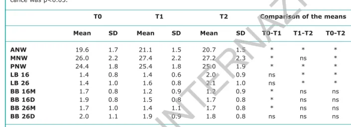
(5) original article. N AL I. N. All measurement error coefficients were found to be close to 1.00 and within acceptable limits. The midpalatal suture was opened in all subjects. In the evaluation of changes between T0-T1 nasal width resulted statistically increased in all three. IO. Results. coronal scans (ANW: +1.5mm; MNW: +1.4mm; PNW: +1.0mm). All T0-T1 increments at nasal levels showed a decrease in magnitude from the anterior to the posterior measurements. In the period between active expansion (T1) and the end of retention (T2) all measurements exhibited statistically significant decreases with the exception of MNW. In the evaluation of changes between T0 and T1, periodontal measurements were statistically significant on the buccal aspect of banded teeth, but not on the lingual side. On the buccal side a signif-. AZ. peated measures with post-hoc tests. The level of significance was p<0.05.. B. R. A. N. II N. TE. C. IZ. IO. Figure 3 Middle Nasal Width (MNW): transverse width between the most lateral point of each nasal cavity at T0, T1, T2 (respectively 3a, 3b, 3c).. B C. ©. C. IC. ED. A. Figure 4 Posterior Nasal Width (PNW): transverse width between the most lateral point of each nasal cavity at T0, T1, T2 (respectively 4a, 4b, 4c).. ORAL & Implantology - Anno I - N. 3-4/2008. 99.
(6) N AL I. lars, the mesial aspect demonstrated the greatest bone resorption, corresponding to about 0.4 mm. No root resorptions or dehiscences were present. In the evaluations of the changes between the end of active expansion (T1) and end of retention (T2), and of the overall T0-T2 changes, periodontal. T0. T2. SD. Mean. SD. Mean. 19.6 26.0 24.4 11.4 11.4 11.7 11.9 11.7 12.0. 1.7 2.2 1.8 0.8 1.0 0.8 0.8 1.0 1.1. 21.1 27.4 25.4 11.4 11.6 11.2 11.5 11.4 11.9. 1.5 2.2 1.8 0.6 0.8 0.9 0.8 1.1 0.9. 20.7 27.2 25.0 12.0 12.1 11.2 11.7 11.7 11.8. Comparison of the means. SD. N. Mean. 1.5 2.3 1.9 0.9 1.0 0.9 0.8 0.8 0.8. R. TE. ANW MNW PNW LB 16 LB 26 BB 16M BB 16D BB 26M BB 26D. T1. AZ. IO. Table 1 - Descriptive and statistical analysis for all measurements at T0, T1, T2. The mean differences in measurements at T0, T1 and T2 were examined with Friedman ANOVA for repeated measures. The level of significance was p<0.05.. II N. original article. icant reduction in alveolar bone thickness corresponding to the mesial (-0.5 mm; p<0.05) and the distal (-0.4 mm; p<0.05) roots of the right first molar, and to the mesial root of the left first molar (-0.3 mm; p<0.05) was recorded. Of the two areas investigated on the buccal aspect of the first mo-. T0-T1. T1-T2. T0-T2. * * * ns ns * * * ns. * ns * * * ns ns ns ns. * * * * * ns ns ns ns. ©. C. IC. ED. IZ. IO. N. ns: not significant; p<0.05 * statistically significant ANW: anterior nasal width: transverse width between the most lateral point of each nasal cavity; MNW: middle nasal width: transverse width between the most lateral point of each nasal cavity; PNW posterior nasal width: transverse width between the most lateral point of each nasal cavity; LB: lingual bone plate thickness: width between external of palatal cortical plane to the centre of palatal aspect of root of first molar; BB: buccal bone plate thickness: width between external of buccal cortical plane to the centre of buccal aspect of mesial and distal roots of first molar.. A. B. C. Figures 5 Lingual bone plate thickness (LB): width between the external aspect of the palatal cortical plate to the centre of the palatal aspect of the root of the first right and left molars; Buccal bone plate thickness (BB): width between the external aspect of the buccal cortical plate to the centre of the buccal aspect of mesial and distal roots of the first right and left molars at T0, T1, T2 (respectively 5a, 5b, 5c).. 100. ORAL & Implantology - Anno I - N. 3-4/2008.
(7) original article. ©. C. IC. ED. IZ. IO. N. N AL I. IO. AZ. N. II N. The aim of the present study was to evaluate skeletal and periodontal effects of RME immediately at the end of the active phase and after a 6month retention period by using a tridimensional diagnostic procedures. With regard to previous studies (12-18), the present study investigated a notably larger group of patients (17 patients), with observations recorded both after active expansion (T1) and after an adequate period for retention (T2) thus allowing for a reossification and reorganization of the midpalatal sutural tissue (19-21). The CT examination, is not a routine in orthodontic diagnosis and treatment planning, but is able to investigate craniofacial structures that would not be described adequately by traditional X-ray techniques due to superimposition of bony or dental anatomy on the same plane (26). All subjects of the study sample examined in the current study needed tri-dimensional radiographic investigations to visualize the exact position of displaced canines within the maxillary arch; further, in order to reduce exposure of patients to X-rays a low dose spiral protocol, obtained by reducing the voltage to the lowest possible level of 80 KV, was used (27). The amount of transverse gain at the level of the nasal cavity (ANS) at the end of the active phase of RME was of 1.5mm; this amount of expansion is very similar when compared with the data obtained by Podesser et al. (13) (1.2 mm) and by Garrett et al. (15) (1.9 mm) who used the same tridimensional method of investigation but can’t be compared with that reported by Wertz (19) (1.9 mm), Haas (8) (3.7 mm), da Silva Filho et al. (28) (2.1 mm) that used Postero-Anterior cephalograms. Interestingly, in the current study, a statistically. R. Discussion. significant amount of expansion of the nasal cavity was observed in all the 3 coronal scans (+1.5mm; +1.4mm; +1.0mm respectively). The findings of the present study demonstrated that the dimensional increase of the nasal cavity on the transverse plane was not limited to the anterior region but it extended to the posterior region as well. The increase in the transverse dimension of the nasal cavity was slightly greater in the anterior region than in the middle and in the posterior regions. The expansion at PNS was about 62% of that at ANS. The amount of transverse gain in the nasal cavity was stable at the post-retention observation (T2). The dimensional changes of the nasal cavity described here fully support the results of the recent study by Palaisa et al. (14) who used conventional tomography and found that RME was accompanied by stable increases in area and volume of the nasal cavity after three months. In growing patients the skeletal effects on nasal width are clinically evident with an improvement of nasal respiration to disadvantage of oral respiration. A possible undesirable orthodontic effect of RME is the compression of the periodontal ligament of supporting teeth, along with alveolar bone resorption. Several Authors (29-31) demonstrated that orthodontic and orthopaedic forces cause histological modifications, such as activation of clastic cells in the direction of the periodontal ligament, hyalinization on the pressure side; and that the lateral tipping of anchoring teeth may cause bone resorption at the dento-alveolar level. The present study assessed a reduction of the buccal bone plate thickness of 0.4 and 0.2 mm of the anchoring teeth (corresponding to the mesial and the distal root, respectively), at the end of the active phase (T1). When compared with the buccal bone loss reported by Rungcharassaeng et al. (18) (-1.24mm) our data are inferior. That could be almost a consequence of a different method of investigation that use cone beam and coronal, not axial, reconstruction images. Despite the buccal bone reduction and the absence of a corresponding compensatory bone apposition on the lingual aspect, no observation of fenestrations, dehiscences, or attachment loss were present. After the retention period of 6. TE. measurements were significant on the lingual aspects, but not on the buccal side. The lingual bone plate thickness of both first molars resulted significantly increased (+0.6 mm; p<0.05) (Table 1).. ORAL & Implantology - Anno I - N. 3-4/2008. 101.
(8) R. N. AZ. IO. N AL I. 14. Monson GS. Constricted vaults. Dental Cosmos 1898: 914-920. 15. Starnbach HK, Bayne D, Cleall JF, Subtelny DJ. Facioskeletal and dental changes resulting from rapid maxillary expansion. Angle Orthod 1966; 36: 152-164. 16. Davis MW, Kronman JH. Anatomical changes induced by splitting of the midpalatal suture. Angle Orthod 1969; 39: 126-132. 17. Haas AJ. Palatal expansion: Just the beginning of dentofacial orthopedics. American J Orthod 1970; 57: 219-255. 18. Haas AJ. Rapid expansion of the maxillary dental arch and nasal cavity by opening the midpalatal suture. Angle Orthod 1961; 31: 73-90. 19. Wertz RA. Skeletal and dental changes accompanying rapid midpalatal suture opening. American J Orthod 1970; 58: 41-64. 10. Bishara SE, Taley RN. Maxillary expansion: clinical implications. American J Orthod Dentofacial Orthop 1987; 91: 3-14. 11. Sandikcioglu M, Hazar S. Skeletal and dental changes after maxillary expansion in the mixed dentition. Am J Orthod Dentofacial Orthop 1997; 111: 321-327. 12. Timms DJ, Preston CB, Daly PF. A computed tomographic assessment of maxillary movement induced by rapid expansion – a pilot study. Eur J Orthod 1982; 4: 123-127. 13. Podesser B, Williams S, Crismani AG, Banteleon HP. Evaluation of the effects of rapid maxillary expansion in growing children using computer tomography scanning: a pilot study. Eur J Orthod 2007; 29: 37-44. 14. Palaisa J, Ngan P, Martin C, Razmus T. Use of conventional tomography to evaluate changes in the nasal cavity with rapid palatal expansion. Am J Orthod Dentofacial Orthop 2007; 132: 458-466. 15. Garrett BJ, Caruso JM, Rungcharassaeng K, Farrage JR, Kim JS, Taylor GD. Skeletal effects to the maxilla after maxillary expansion assessed with cone-beam computed tomography. Am J Orthod Dentofacial Orthop 2008; 134: 8.e1-8.e11. 16. Garib DG, Henriques JFC, Janson G, De Freitas MR, Coelho RA. Rapid maxillary expansion-tooth tissueborne versus tooth- borne expanders: a computed tomography evaluation of dentoskeletal effects. Angle Orthod 2005; 75: 548-557. 17. Garib DG, Henriques JFC, Janson G, De Freitas MR, Fernandes AY. Periodontal effects of rapid maxillary expansion with tooth-tissue-borne and tooth-borne expanders: a computed tomography evaluation. Am J Orthod Dentofacial Orthop 2006; 129: 749-758. 18. Rungcharassaeng K, Caruso JM, Kan JYK, Kim J, Taylor G. Factors affecting buccal bone changes of maxillary posterior teeth after rapid maxillary expansion. Am J Orthod Dentofacial Orthop 2007; 132: 428.e1-428.e8. 19. Wertz RA. Skeletal and dental changes accompanying. TE. original article. months (T2) statistically significant bone apposition was observed on the palatal side of both anchoring teeth (+ 0.7mm; p<0.05) because of the translation movement of the first molars. The amount of bone apposition in our study at T2 can’t be compared with the data obtained in other researches (16-18) that observe periodontal structures only after a retention period of 3 months. Indeed values reported by Garib et al. (17) show an inferior amount of bone apposition on palatal aspect. These outcomes may be in part related to the different times of observation and in part to the different skeletal maturity of the two control group in the studies. Indeed a retention protocol of adequate duration (6 months) is necessary to recovery lingual and buccal bone plate thickness.. II N. Conclusions. ED. IZ. IO. N. 1. A significant amount of expansion of the nasal cavity was observed in all the 3 coronal scans. The expansion of the nasal cavity resulted stable at the end of the retention period; 2. At the end of the active phase (T0) of expansion the buccal bone plate thickness of the supporting teeth shows a significant decrease, while no change is shown by the lingual bone plate thickness; 3. After an adequate retention period of at least 6 months (T2) a recovery of both buccal and lingual plates thickness is observed; 4. No root resorptions or dehiscences of alveolar bone were present after RME therapy.. C. IC. References. ©. 11. Haas AJ. The treatment of maxillary deficiency by opening the midpalatal suture. Angle Orthod 1965; 35: 200-217. 12. Braun S, Bottrel A, Lee Kg, Lunazzi J, legan HL. The biomechanics of rapid maxillary sutural expansion. American J Orthod Dentofacial Orthop 2000; 118: 257-261. 13. Angell EH. Treatment of irregularity of the permanent or adult teeth. San Francisco Press 1860.. 102. ORAL & Implantology - Anno I - N. 3-4/2008.
(9) original article. 24.. 25.. 26.. 30.. 31.. N AL I. IO. ©. C. IC. ED. IZ. IO. N. II N. Correspondence to: Prof.ssa Paola Cozza Via Velo, 53 - 00183 Rome E-mail: [email protected] [email protected]. 29.. AZ. 23.. 28.. N. 22.. 27.. R. 21.. High resolution multislices CT with multiplanar and 3D-reformation imaging in rapid palatal expansion (RPE). Am J Orthod Dentofacial Orthop 2007; 131: 776-781. Ballanti F, Lione R, Fiaschetti V, Fanucci E, Cozza P. Low-dose CT protocol for orthodontic diagnosis. Eur J Paediatr Dent 2008; 9: 65-70. Da Silva OG, Prado Montes LA, Torelly LF. Rapid maxillary expansion in the deciduous and mixed dentition evaluated through posteroanterior cephalometric analysis. Am J Orthod Dentofacial Orthod 1995; 107: 268-75. Reitan K. Effects of force magnitude and direction of tooth movement on different alveolar bone types. Angle Orthod 1964; 34: 244-256. Handelman CS. Nonsurgical rapid maxillary alveolar expansion in adults: a clinical evaluation. Angle Orthod 1997; 67: 291-308. Handelman CS, Wang L, BeGole AE, Haas AJ. Nonsurgical rapid maxillary expansion in adults: report on 47 cases using the Haas expander. Angle Orthod 2000; 70: 129-144.. TE. 20.. rapid midpalatal suture opening. Am J Orthod 1970; 58: 41-64. Ekstrom C, Henrickson CO, Jensen R. Mineralization in the mipalatal suture after orthodontic expansion. Am J Orthod 1977; 71: 449-455. Mew J. Relapse following maxillary expansion. A study of twenty-five consecutive cases. Am J Orthod Dentofacial Orthop 1983; 83: 56-61. Cozza P, Giancotti A, Petrosino A. Butterfly expander for use in the mixed dentition. J Clin Orthod 1999; 33: 583-587. Cozza P, Giancotti A, Petrosino A. Rapid palatal expansion in mixed dentition using a modified expander: a cephalometric investigation. J Orthod 2001; 28: 129-134. Cozza P, DeToffol L, Mucedero M, Ballanti F. Use of a modified butterfly expander to increase anterior arch length. J Clin Orthod 2003; 37: 490-495. Baumrind S, Frantz RC. The reliability of head films measurements. Landmark identification. Am J Orthod 1971; 60: 111-127. Habersack K, Karolgan A, Sommer B, Benner KU,. ORAL & Implantology - Anno I - N. 3-4/2008. 103.
(10)
Figura
Documenti correlati
Sar` a sicuramente necessario realizzare ulteriori esperimenti nello scenario delle immagini P&S per verificare le possibilit` a di effettuare un’analisi della qualit` a
O país é signatário da Declaração Universal dos Direitos do Homem, aprovada pela Assembleia Geral das Nações Unidas em 10 de dezembro de 1948; da Carta das Nações
31 Department of Clinical Microbiology and Immunology, Sackler School of Medicine, Tel Aviv University, Tel Aviv, Israel, 32 Institute of Medical Microbiology, University
Lake and Townshend (2006) define the ‘obesogenic environment’ as a model for understanding the external factors that may influence individual weight, which is to say the way in
Simili tematiche, però, non sono da considerarsi esclusive della letteratura scritta dalle donne: un esempio può proprio essere la prosa di Surendra Varmā — autore uomo,
Mutations in genes involved in heme metabolism and Fe-S cluster biogenesis cause different forms of ataxia, like posterior column ataxia and retinitis pigmentosa (PCARP),
Further work of the Hauser group demonstrated that these rationally designed aliphatic ultrashort peptides had the same assembly mechanism as short peptides from naturally
