Cronicon
O P E N A C C E S SGYNAECOLOGY
Review Article
Linda Maria Azzurra Pirollo
1, Giovanni Larciprete
1, Edoardo Valli
1, Carlotta Montagnoli
1, Gaia de Campora
2,
Giuseppe Di Pierro
31Department of Obstetrics and Gynecology, Fatebenefratelli Isola Tiberina Hospital, Rome, Italy 2Department of Pedagogy, Psychology, Philosophy, University of Cagliari, Italy
3Department of Obstetrics and Gynaecology, San Carlo Hospital, Potenza, Italy
Received: January 14, 2016; Published: January 29, 2016
*Corresponding Author: Giovanni Larciprete, Department of Obstetrics and Gynecology Fatebenefratelli Hospital, Rome, Italy.
Unusual Ectopic Pregnancy: Beyond the tubes
Abstract
Introduction
Nearly 95 percent of ectopic pregnancies are implanted in the various segments of the fallopian tube, the remaining 5 percent of non tubal ectopic pregnancies (EP) implants in the ovary, peritoneal cavity, cervix, or prior cesarean scar and are considered unusual ectopic pregnancies. Unusual EP are considered an important challenge for the gynaecologist who must recognize this condition before severe complications. The increased use of assisted reproduction techniques and the rise in cesarean deliveries have been accompanied by a rise of unusual EP, especially scar pregnancies. Diagnosis is often late because women could be asymptomatic or show unspecific pain and a severe abdominal bleeding could represent the first manifestation. Ultrasound and 3D scanning became a very useful non invasive tool for a precise diagnose. The therapeutic strategies include medical and surgical treatments but conserva-tive management is often difficult.
Citation: Giovanni Larciprete., et al. “Unusual Ectopic Pregnancy: Beyond the tubes”. EC Gynaecology 1.1S1 (2016): 12-22.
Following fertilization and fallopian tube transit, the blastocyst normally implants within the uterine cavity. Implantation elsewhere is considered ectopic and comprises 1 to 2 percent of all first-trimester pregnancies in the United States. Nearly 95 percent of ectopic pregnancies are implanted in the various segments of the fallopian tube, the remaining 5 percent of nontubal ectopic pregnancies (EP) implants in the ovary, peritoneal cavity, cervix, or prior cesarean scar [1]. When the blastocyst implants or develops in pelvic areas other than the tubes, the pregnancy is considered an unusual EP [2]. Because of the atypical symptoms and clinical history, unusual EP are considered an important challenge for the gynaecologist who must recognize this condition before severe complications. Although this incidence is low, the danger and morbility from an extratubal pregnancy is higher than normal pregnancy, with a quite high misdiagnosis rate (96.6% according to Shan et al) [2]. Moreover, the success rate for a subsequent pregnancy will be reduced after EP. The increased use of assisted reproduction techniques and the rise in Cesarean deliveries have been accompanied by a rise of unusual EP, especially scar pregnancies [3]. Occasionally it is possible to identify a heterotopic pregnancy due to an intrauterine pregnancy coexisting with an ectopic one.
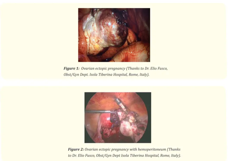
Ovarian pregnancy is a very rare and atypical ectopic pregnancy (1 in 25,000 pregnancies, of 0.5-3% of extra uterine pregnancies); there are very few reports of an accurate preoperative diagnosis utilizing sonography. The correct diagnosis is most frequently made at the surgery and requires histopathological confirmation (Figure 1 and 2) [4]. The most common risk factor associated with ovarian preg-nancy is ovulation induction and assisted reproduction techniques, but other authors [2] advise that pelvic inflammatory disease, use of
Keywords:
Ectopic pregnancy; Scar pregnancy; Ovarian pregnancy; Abdominal pregnancy; Interstitial pregnancy; Cervical pregnancyintrauterine device (IUD) and previous pelvic surgeries are significantly associated with ovarian pregnancy than other atypical sites [5-6]. Even without inflammatory history, pelvic surgeries may also cause ovarian inflammation and thicken the albuginea, which could lead to a relative lack of follicular fluid pressure. Finally, it would generate ovulation disorder. Ovum might be detained in the broken fol-licles and fertilized just in the ovary. The main clinical manifestations associated with ovarian pregnancy are amenorrhea, severe abdom-inal bleeding, secondary anemia, cervical motion pain and shock. The symptoms in common with the tubal EP are often more severe.
Figure 1: Ovarian ectopic pregnancy (Thanks to Dr. Elio Fusco,
Obst/Gyn Dept. Isola Tiberina Hospital, Rome, Italy).
Figure 2: Ovarian ectopic pregnancy with hemoperitoneum (Thanks
to Dr. Elio Fusco, Obst/Gyn Dept Isola Tiberina Hospital, Rome, Italy).
The misdiagnosis rate of extratubal pregnancies is quite high (96.6%). Ovarian and tubal pregnancies are difficult to distinguish, even by ultrasound, because their clinical manifestations are similar. Reportedly, all 24 cases of ovarian pregnancies in six hospitals were preoperatively misdiagnosed [7]. An ovarian pregnancy is differentiated from tubal pregnancy by the Spiegelberg criteria [8] which includes:
1. The gestational sac is located in the region of the ovary
2. The ectopic pregnancy is attached to the uterus by the ovarian ligament 3. Ovarian tissue in the wall of the gestational sac is proved histologically 4. The tube on the involved side is intact.
14
Positive culdocentesis results were more common in patients with ovarian pregnancies than with cornual pregnancies, probably because a patient upon admission already had her ovary ruptured and suffered from severe abdominal haemorrhage. Because the di-agnosis is often late, the main management of the ovarian pregnancies remains surgery (resection of the ovarian wedge or partial oo-phorectomy with laparotomy o laparoscopy). Ovary can be preserved since implantation is superficial. While foetus of ectopic pregnancy is typically not viable, very rarely a live baby has been delivered in cases of ovarian pregnancies. Maternal mortality and morbidity is high as attempts to remove the placenta from organs to which it is attached usually lead to uncontrollable bleeding. If placenta is attached with adnexa as seen in this case and receives blood supply from the right uterine artery or uterine fistula, that it leads to a live foetus at term [9].
The terms cornual, interstitial and angular pregnancies are indistinctly used in the literature to indicate an ectopic pregnancy with eccentric location in relation to the endometrium, closer to the uterine serosa [10]. However, according to Williams’ Obstetrics, “although used interchangeably, (these) are slightly different implantations. Cornual implantation describes those in the upper and lateral uterine cavity (Figure 3 and 4), whereas interstitial denotes those implanted within the proximal intramural portion of the tube” [11]. Still other sources in both the radiology and obstetrics literature reserve “cornual pregnancy” only for gestations in a bicornuate or septate uterus [11]. Amidst such discussion, the term “angular pregnancy” sometimes arises, defined as “implantation within the endometrium of the lateral angle of the uterus, medial to the uterotubal junction” [11]. The interstitial (or intramural) segment is approximately 1–2 cm in length, traversing the muscular myometrial layer of the uterus and opening via the inner tubal ostium into the uterine cavity [12]. Thus, by strict anatomic definition, interstitial pregnancy should refer to a pregnancy in the interstitial portion of the fallopian tube. Of tubal ectopic pregnancies, 2%–4% are reported to occur in this location [13].
The uterus is maintained in location by multiple ligaments, including the round ligament which crosses the fallopian tube at the uterotubal junction. Anatomically, the superior two-thirds of the uterus is the body; the inferior one-third is the cervix; and the supero-lateral regions of the uterine cavity where the fallopian tubes enter are the uterine horns or cornua [12].
Cornual, interstitial, and angular pregnancies
Figure 4: Angular ectopic pregnancy.
Accordingly, a normal uterus has two cornua, one on the right side and one on the left. According to these anatomical landmarks, some authors diagnosed an interstitial pregnancy when a regular endometrium without visible gestational sac or mass is visualized, and a gestational sac is located outside the endometrium and surrounded by a continuous rim of myometrium, within the interstitial area. A pregnancy is defined as cornual when it is situated in the uterine cavity but asymmetrically in the corneal region, medial to the round ligament [14]. However, with the constant use and spread of 3D ultrasound and its capability to image the coronal plane of the uterus, can have added value in the diagnosis of cornual and interstitial ectopic pregnancies [15]. So, although the terms are synony-mous in the United States, the cornual one refers to a pregnancy implanted in the interstitial segment of a unicornuate or bicornuate uterus [16].
Angular pregnancy was first defined in 1898 by the American obstetrician Howard Kelly as “implantation of the embryo just medial to the uterotubal junction, in the lateral angle of the uterine cavity” [17]. Angular pregnancy is distinguished from interstitial pregnancy, wrote Jansen and Elliot [18] years later, by its position in relation to the round ligament as seen at surgery: “The lateral uterine enlarge-ment of an angular pregnancy displaces the round ligaenlarge-ment reflection upward and outward. The swelling of an interstitial tubal preg-nancy is lateral to the round ligament”. Although there is no absolute anatomic limit distinguishing an angular pregpreg-nancy from a normal one, the closer a gestation implants to the internal uterine ostium of the fallopian tube, the greater likelihood of visual asymmetry and a symptomatic patient as the pregnancy progresses.
Traditionally, laparoscopy was the gold standard for the diagnosis of ectopic pregnancy. However, currently, the widespread avail-ability of transvaginal US and rapid assays for serum β-hCG has largely made the use of laparoscopy for diagnostic purposes an obsolete practice. According to the American College of Radiology (ACR) Appropriateness Criteria [19], the first-line imaging modality in the evaluation of patients with positive urine or serum pregnancy test presenting with first-trimester vaginal bleeding is pelvic US, utiliz-ing both a transabdominal and transvaginal approach. Although US is highly operator-dependent, its advantages include its portability, lack of ionizing radiation, relatively inexpensive cost, and the fact that it is a real-time dynamic examination. In 1992, three US criteria were proposed by Timor-Tritsch et al. [20] to diagnose an interstitial pregnancy (specificity 88%–93%, sensitivity 40%): (1) an empty uterine cavity, (2) a chorionic sac separately (>1cm) from the lateral edge of the uterine cavity, and (3) a thin (< 5 mm) myometrial layer surrounding the chorionic sac. A year later, in 1993, Ackerman et al. [21] described the “interstitial line sign” (sensitivity of 80%, speci-ficity of 98% in diagnosing interstitial pregnancy): an echogenic line in the cornual region of the uterus bordering the midportion of the gestational sac, thought to represent the interstitial portion of the tube in small interstitial pregnancies and the endometrium in larger pregnancies. The key imaging finding to highlight regarding interstitial pregnancy is that it lies outside the endometrium (extraendo-metrial). In contrast, the key imaging finding to highlight regarding angular pregnancy is that it lies within the endometrium (intraen-dometrial), and therefore, it is not an ectopic pregnancy [22]. In cases of persistent uncertainty-although not currently the standard of care or yet adequately studied-3D US and then MRI could be considered, if available, for further characterization.
With the exceedingly rare exception of six case reports of interstitial pregnancies that have achieved fetal viability and have been published in the literature [23], an interstitial ectopic pregnancy is considered nonviable because it generally cannot result in a live born baby [24]. Although increased distensibility of this segment of the fallopian tube can lead to presentation as late as the 13th week of gestation [25], if the pregnancy continues to progress, then rupture is almost universal. Thus, a ruptured interstitial ectopic pregnancy is a surgical emergency: it has a twofold mortality compared with other tubal ectopic pregnancies due to the risk of hemorrhage from uterine arteries and veins. Surgical treatment options include laparotomy, laparoscopy, cornuostomy, salpingotomy, laparoscopic cor-neal resection, cornual wedge resection (Figure 5 and Clip 1), mini-cornual excision, and hysterectomy [26]. Intraoperatively, it could be of help to get ultrasounds in order to better localize the gestational sac (Clip 2). New therapies include systemic methotrexate [27] or transvaginal sonographically guided injection of potassium chloride [28].
By this definition, an angular pregnancy is a potentially viable one. In terms of outcomes, the largest published meta-analysis to date describes 39 cases of angular pregnancy (inclusion criteria: cases that satisfy either of the first two criteria of Jansen and Elliot [29]) and reports a 38.5% rate of spontaneous or missed abortion.
Intramural pregnancy refers to a gestation completely implanted within the myometrium of the uterus with separation from the uterine cavity, fallopian tube, or round ligament. This is a very rare condition reported in less than 50 cases in literature [30] (less than 1% of all the ectopic pregnancy). These cases are usually complicated by hemorrhage and uterine rupture (with consequent hysterec-tomy) and are difficult to diagnose because they may appear similar to molar pregnancy, sarcomas, degenerating myomas, or a normal pregnancy in a congenitally abnormal uterus [31-32]. Predisposing risk factors include prior uterine trauma, caesarian section and adenomyosis [33-34]. It was also described a case of intramural pregnancy following assisted reproduction treatment in a necrotized fibroid, 8 months after uterine artery embolization [35]. Diagnostic modalities may include ultrasound, a computed tomography (CT) scan, and magnetic resonance imaging demonstrating myometrium completely surrounding the gestational sac with no communication
Prognosis and management
Intramural ectopic pregnancy
Figure 5: Cornual wedge resection. Ultrasound and operative findings. Clip 1: Cornual wedge resection using a catheter as a tourniquet.
http://youtu.be/7b6N7dPJgoc
Clip 2: Transvaginal ultrasounds during surgery show a gestational sac at the extreme corner of the uterus. Palpating the uterus with instruments helps in localizing exactly the extrauterine pregnancy.
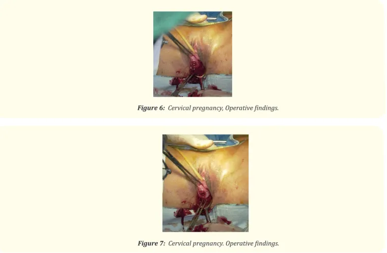
Cervical pregnancy is diagnosed when the entire gestational sac, having a well-formed shape, is demonstrated in the dilated cervix below the internal os (Figure 6 and 7). Except in the case of heterotopic pregnancy, the endometrial stripe is visualized and the uterus keeps the hourglass shape [15]. Color Doppler exam may help to differentiate a true cervical pregnancy from an inevitable miscar-riage of a gestational sac placed in the cervix. In a typical case of cervical pregnancy, the endocervix is eroded by trophoblast, and the pregnancy develops in the fibrous cervical wall so Color Doppler scanning shows a high flow velocity and low impedance, while in miscarriage the sac will be mobile, with no Doppler evidence of blood flow [14-39]. The incidence of cervical pregnancy lies between 1 in 8600 and 1 in 12,400 pregnancies, but the incidence is increasing as a result of assisted reproductive technologies (ART) [40]. Pain-less vaginal bleeding is reported by 90 percent of women with a cervical pregnancy—a third of these have massive hemorrhage [41]. Cervical pregnancy may be treated medically or surgically.
Cervical pregnancy
with the endometrial cavity. Ultrasound findings of intramural pregnancy are often difficult to distinguish because they may be similar to uterine myomas, no intramural ectopic pregnancy, missed abortion, or congenital uterine abnormality. Performing an ultrasound together with a CT scan and magnetic resonance imaging may help to make an accurate diagnosis and exclude other diagnostic possibili-ties. The use of color Doppler sonography and 3-dimensional ultrasound technologies may further add to its diagnostic ability and allow ultrasonography to be the standard diagnostic method for this type of pregnancy [36]. No documented case of term pregnancy does exist. The longest gestation reported with fetal survival in the literature is 30 weeks, but with resulting cesarean hysterectomy because of uterine rupture [37]. However, if diagnosis is made earlier, treatment may be conservative, resulting in the preservation of reproduc-tive potential. Intramural pregnancies may implant at any site within the uterus depending on the patient’s past procedures. Treatment modalities documented in the literature for intramural pregnancy include expectant management, surgical enucleation, uterine artery embolization, systemic or local methotrexate administration, hysterectomy [38], and intrafetal injection of potassium chloride [32].
Figure 6: Cervical pregnancy, Operative findings.
Scar pregnancy
Abdominal pregnancy: how did it get there?
Caesarean scar pregnancy (CSP) describes implantation within the myometrium of a prior cesarean delivery scar [1]. The etiology is still unclear and includes of course a previous caesarian section, myomectomy, adenomyosis, in vitro fertilization procedures, dilata-tion and curettage, manual removal of the placenta [47-49]. Vial et al proposed ultrasonographic criteria to diagnose a cesarean scar pregnancy that was very likely to be ruptured including (1) trophoblast between bladder and anterior uterine wall, (2) no fetal parts in the uterine cavity and (3) discontinuity of the anterior uterine wall in the sagittal plane [50]. Women with CSP usually present early, and pain and bleeding are common. However, up to 40 percent of women are asymptomatic, and the diagnosis is made during routine sono-graphic examination. The spectrum of management strategies for Cesarean scar pregnancy is broad, and has been presented in several case series and recent reviews [50-54]. Expectant management has been described, but it carries a considerable risk of uterine rupture and hemorrhage, perhaps as high as 50%. Hysterectomy is an acceptable initial choice in those desiring sterilization. It is sometimes a necessary option with heavy uncontrolled bleeding. Fertility-preserving options include systemic or locally injected methotrexate, either alone or combined with conservative surgery [55-57].
Abdominal pregnancy is defined as a gestation implanted in the peritoneal cavity except the tubes, ovaries or ligaments (Figure 8). Primary peritoneal pregnancy was first described by Studdiford as a rare form of ectopic pregnancy characterized by the following cri-teria: 1) normal tubes and ovaries, 2) absence of uteroplacental fistula, 3) attachment exclusively to a peritoneal surface early enough in gestation to eliminate the likelihood of secondary implantation [58]. Although it is possible that the zygote traverses the tube and implants primarily the peritoneal cavity, most abdominal pregnancy seems to origin from a reimplantation of a tubal rupture or abor-tion. In this case the placental re-implant can occur almost anywhere and grow as an abdominal pregnancy [2]. Many case reports in the literature describe very unusual sites of abdominal pregnancy: omentum [59-60], duodenum adjacent to the porta hepatis as a het-erotopic pregnancy [61], retroperitoneum [62], pancreas [63], appendix [64]. The placenta can be attached to the uterine wall, bowel, mesentery, liver, spleen, bladder and ligaments (Clip 3). It can be detached at any time during pregnancy leading to severe blood loss [65]. Diagnosis may be difficult. Symptoms may be absent or vague. sonographically, findings with an abdominal pregnancy may not be recognized, and the diagnosis is often missed [66]. Other clues include a fetus seen separate from the uterus or eccentrically posi-tioned within the pelvis; lack of myometrium between the fetus and the maternal anterior abdominal wall or bladder; and extrauterine placental tissue [67]. If additional anatomical information is needed, MR imaging can be used to confirm the diagnosis and provide maximal information concerning placental implantation [68-69]. In abdominal pregnancy, blastocyst may invade into maternal organs or vessels, and thus causes bleeding or rupture of maternal organs. It should be immediately treated upon diagnosis [2]. Laparoscopy, in this case, has proven to be a “gold standard” for diagnostics and therapy of ectopic pregnancy [54]. Abdominal pregnancy can be life-threatening, and management depends on the gestational age at diagnosis. Some describe waiting until fetal viability with close surveillance [70]. Abdominal pregnancy is viable exclusively with Caesarean section. Once placental implantation has been assessed, several options are available: preoperative angiographic embolization or catheters placed in the uterine arteries and multidisciplinary surgery approach as ureteral catheters, bowel preparation, assurance of sufficient blood products [71]. The most important challenge is delivery of the fetus and careful assessment of placental implantation without causing haemorrhage.
Conservative management is possible if it is diagnosed early, as described for interstitial pregnancy. In many centers methotrexate has been used as first-line therapy in stable women. Suction curettage may be especially favored in rare cases of a heterotopic preg-nancy composed of a cervical and a desired uterine pregpreg-nancy [42]. In case of cervical pregpreg-nancy it is not possible an excision of the gestational sac so, to preserve fertility, is often required an intervention to stop haemorrhage as uterine artery embolization, ligation of the descending branches of the uterine arteries, vasopressin injection or a cerclage placed at the internal cervical os to compress feeding vessels [43-46].
Figure 8: Laparoscopic and laparotomic view of the same patient with extrauterine abdominal
pregnancy. Chorionic tissue is detectable between right gross bowel and the posterior wall of the uterus. The implantation was on the anterior surface of the right colon.
Clip 3: Open surgery on the same patient with abdominal extrauterine pregnancy. Moving the colon, it is possible to see the chorionc implants.
http://youtu.be/zKs1TpXptkw
Bibliography
1. Cunningham FG., et al. “Williams Obstetrics”. (2014): 24 edn.
2. Shan N., et al. “Unusual ectopic pregnancies: A retrospective analysis of 65 cases”. Journal of Obstetrics and Gynaecology Research
40.1 (2014): 147-154.
3. Valsky V and Yagel S. “Ectopic pregnancies of unusual location: management dilemmas”. Ultrasound in Obstetrics & Gynecology
31.3 (2008): 245-251.
4. Garg MK., et al. “Primary twin ovarian pregnancy: case report and review of the literature. Journal of Clinical Ultrasound 37.1
(2009): 43-46.
5. Rimdusit P and Kasatri N. “Primary ovarian pregnancy and the intrauterine contraceptive device”. Obstetrics & Gynecology 48.1
suppl 1 (1976): 57s-59s.
6. Fernandez CM and Barbosa JJ. “Primary ovarian pregnancy and the intrauterine device”. Obstetrics & Gynecology 47.1 (1976):
9s–11s.
7. Grimes HG., et al. “Ovarian pregnancy: A series of 24 cases”. Obstetrics & Gynecology 61.2 (1983): 174-176. 8. Spiegelberg O. Zur casuistic der ovarial . Schwangerschft. Archiv für Gynaekologie 13 (1873): 73-76.
9. Zia F., et al. “Unusual ectopic-term pregnancy in the ovary; case report from Karachi”. Journal of Pakistan Medical Association
63.11 (2013): 1439-1441.
10. Dahnert W. “Radiology review manual”. 6th ed (2007).
11. Parker RA., et al. “MR imaging findings of ectopic pregnancy: a pictorial review”. Radiographics 32.5 (2012): 1445-1460. 12. Moore KL., et al. “Clinically oriented anatomy”. 6th ed (2010).
13. Eddy CA and Pauerstein CJ. “Anatomy and physiology of the fallopian tube”. Clinical Obstetrics and Gynecology 23.4 (1980):
1177-1193.
14. Jurkovic D and Mavrelos D. “Catch me if you scan: ultrasound diagnosis of ectopic pregnancy”. Ultrasound in Obstetrics &
15. Valsky DV., et al. “The use of 3D rendering, VCI-C, 3D power Doppler and B-flow in the evaluation of interstitial pregnancy with
arteriovenous malformation treated by selective uterine artery embolization”. Ultrasound in Obstetrics & Gynecology 29.3 (2007): 352-355.
16. Tulandi T and Al-Jaroudi D. “Interstitial pregnancy: results generated from The Society of Reproductive Surgeons Registry”.
stetrics & Gynecology 103.1 (2004): 47-50.
17. Kelly H. “Operative gynaecology”. Appleton (1898).
18. Jansen RP and Elliott PM. “Angular intrauterine pregnancy”. Obstetrics & Gynecology 58.2 (1981): 167-175. 19. (ACR) ACoR. ACR Appropriateness Criteria on First Trimester Bleeding.
20. Timor-Tritsch IE., et al. “Sonographic evolution of cornual pregnancies treated without surgery”. Obstetrics & Gynecology 79.6
(1992): 1044-1049.
21. Ackerman TE., et al. “Interstitial line: sonographic finding in interstitial (cornual) ectopic pregnancy”. Radiology 189.1 (1993):
83-87.
22. EK Arleo and EM DeFilippis. “Cornual, interstitial, and angular pregnancies: clarifying the terms and a review of the literature”.
Clinical Imaging 38.6 (2014): 763-770.
23. Hill AJ., et al. “A true cornual (interstitial) pregnancy resulting in a viable fetus”. Obstetrics & Gynecology 121.2pt 2 suppl 1 (2013):
427-430.
24. Doubilet PM., et al. “Diagnostic criteria for nonviable pregnancy early in the first trimester”. The New England Journal of Medicine
369.15 (2013): 1443-1451.
25. Sherer MD., et al. “Interstitial Pregnancy Undetected During Earlier First-Trimester Screening for Fetal Aneuploidy at 13 Weeks’
Gestation”. Journal of Clinical Ultrasound 37.3 (2009): 168-170.
26. Maher PJ and Grimwade JC. “Cornual pregnancy-diagnosis before rupture a report of 2 cases”. Australian and New Zealand Journal
of Obstetrics 22.3 (1982): 172-174.
27. Fernandez H., et al. “The place of methotrexate in the management of interstitial pregnancy”. Human Reproduction 6.2 (1991):
302-306.
28. Doubilet PM., et al. “Sonographically guided minimally invasive treatment of unusual ectopic pregnancies”. Journal of Ultrasound
in Medicine 23.3 (2004): 359-370.
29. Jansen RP and Elliott PM. “Angular intrauterine pregnancy”. Obstetrics & Gynecology 58.2 (1981): 167-175.
30. Bernstein HB., et al.“ Expectant management of intramural ectopic pregnancy”. Obstetrics & Gynecology 97.2 (2001): 826-827. 31. Glass T., et al. “Intramural pregnancy presenting in a patient with tuberous sclerosis”. Journal of Clinical Ultrasound 38.7 (2010):
393-396.
32. Ong C., et al. “Sonographic diagnosis and successful medical management of an intramural ectopic pregnancy”. Journal of Clinical
Ultrasound 38.6 (2010): 320-324.
33. Katano K., et al. “A case of successful conservative chemotherapy for intramural pregnancy”. Fertility and Sterility 72.4 (1999):
744-746.
34. Khalifa Y., et al. “Intramural Pregnancy following difficult embryo transfer”. Human Reproduction 9.12 (1994): 2427-2428. 35. Leyder M., et al. “Intramyometrial ectopic pregnancy in an ICSI patient following uterine artery embolization”. Reproductive
Medicine Online 20.6 (2010): 831-83 5.
36. Bannon K., et al. “Diagnosis and Management of Intramural Ectopic Pregnancy”. Journal of Minimally Invasive Gynecology 20.5
(2013): 697-700.
37. Fait G., et al. “Intramural pregnancy with fetal survival: case history and discussion of etiologic factors”. Obstetrics & Gynecology
70.3 pt 2 (1987): 472-474.
38. Fadhlaoui A., et al. “Ruptured intramural pregnancy with myometrial invasion treated conservatively”. Case Reports in Obstetrics
39. Lemus JF. “Ectopic pregnancy: an update”. Current Opinion in Obstetrics and Gynecology 16.4 (2000): 369-375.
40. Ginsburg ES., et al. “Early diagnosis and treatment of cervical pregnancy in an in vitro fertilization program”. Fertility and Sterility 61.5 (1994): 966-969.
41. Ushakov FB., et al. “Cervical pregnancy: past and future”. Obstetrical & Gynecological Survey 52.1 (1997): 45-59.
42. Moragianni VA., et al. “Management of a cervical heterotopic pregnancy presenting with first-trimester bleeding: case report and review of the literature”. Fertility and Sterility 98.1 (2012): 89-94.
43. Davis LB., et al. “Transvaginal ligation of the cervical branches of the uterine artery and injection of vasopressin in a cervical nancy as an initial step to controlling hemorrhage: a case report”. JRM-The Journal of Reproductive Medicine 53.5 (2008): 365-368. 44. De La Vega GA., et al. “Treatment of early cervical pregnancy with cerclage, carboprost, curettage, and balloon tamponade”. rics & Gynecology 109.(2 Pt2) (2007): 505-507.
45. Trojano G., et al. “Successful management of a cervical twin pregnancy: neoadjuvant systemic methotrexate and prophylactic high cervical cerclage before curettage”. Fertility and Sterility 91.3 (2009): e17-19.
46. Wang Y., et al. “An efficient conservative treatment modality for cervical pregnancy: angiographic uterine artery embolization lowed by immediate curettage”. American Journal of Obstetrics & Gynecology 204.1 (2011): e1-e31.
47. Lee CL., et al. “Laparoscopic management of an ectopic pregnancy in a previous caesarean section scar”. Human Reproduction 14.5 (1999): 1234-1236.
48. Graesslin O., et al. “Conservative treatment of ectopic pregnancy in a cesarean scar”. Obstetrics and Gynecology 105.4 (2005): 869-871.
49. Marchiole P., et al. “Intramural pregnancy embedded ina a previous cesarean section scar treated conservatively”. Ultrasound in Obstetrics and Gynecology 23.3 (2004): 305-309.
50. Maymon R., et al. “Ectopic pregnancies in a Caesarean scar: review of the medical approach to an iatrogenic complication”. Human Reproduction Update 10.6 (2004): 515-523.
51. Ash A., et al. “Caesarean scar pregnancy”. BJOG: An International Journal of Obstetrics & Gynaecology 114 (2007): 253-263. 52. Rotas MA., et al. “Cesarean scar ectopic pregnancies: etiology, diagnosis, and management”. Obstetrics and Gynecology 107.6 (2006): 1373-1381.
53. Godin PA., et al. “An ectopic pregnancy developing in a previous caesarean section scar”. Fertility and Sterility 67.2 (1997): 398-400.
54. Haimov Kochman R., et al. “Conservative management of two ectopic pregnancies implanted in previous uterine scars”. sound in Obstetrics and Gynecology 19.6 (2002): 616-619.
55. Shen L., et al. “Bilateral uterine artery chemoembolization with methotrexate for cesarean scar pregnancy”. American Journal of Obstetrics and Gynecology 207.5 (2012): 386.e1-6.
56. Timor Tritsch IE., et al. “The diagnosis, treatment, and follow-up of cesarean scar pregnancy”. American Journal of Obstetrics and Gynecology 207.1 (2012): 44.e1-13.
57. Yang XY., et al. “Uterine artery embolisation combined with local methotrexate for treatment of caesarean scar pregnancy”. BJOG: An International Journal of Obstetrics & Gynaecology 117.8 (2010): 990-996.
58. Studdiford WE. “Primary peritoneal pregnancy”. American Journal of Obstetrics and Gynecology 44 (1942): 487-491.
59. Martelli F., et al. “Neglected Primary Omental Pregnancy after Laparoscopic and Medical Treatment: A Difficult Diagnosis?” Case Reports in Obstetrics and Gynecology 2013 (2013): 1-2.
60. Akhtar MAI., et al. “An unusual haemoperitoneum--secondary abdominal pregnancy”. BMJ Case Reports (2012).
61. Honest H and Cartmill RS. “Simultaneous fallopian tube and duodenal serosal ectopic pregnancies from a spontaneous tion: a case report”. Fertility and Sterility 94.7 (2010): 2770.e1-2.
62. Martínez-Varea A1., et al. “Retroperitoneal ectopic pregnancy after intrauterine insemination”. Fertility and Sterility 95.7 (2011): 2433.e1-3.
63. Dmowski WP., et al. “Retroperitoneal Subpancreatic Ectopic Pregnancy Following In Vitro Fertilization in a Patient with Previous Bilateral Salpingectomy: How Did It Get There?” Journal of Assisted Reproduction and Genetics 19.2 (2002): 90-93.
64. Rosso R., et al. “Secondary Abdominal Appendicular Pregnancy: Case Report”. Srpski arhiv za celokupno lekarstvo 142.7-8 (2014): 484-487.
65. Ang LP., et al. “Abdominal pregnancy: a case report and literature review”. Singapore Medical Journal 41.9 (2000): 454-457. 66. Costa SD., et al. “Advanced abdominal pregnancy”. Obstetrical and Gynecological Survey 46.8 (1991): 515-525.
67. Sherer DM., et al. “Unusual maternal vasculature in the placental periphery leading to the diagnosis of abdominal pregnancy at 25 weeks’ gestation”. Journal of Clinical Ultrasound 35.5 (2007): 268-2673.
68. Bertrand G., et al. “Imaging in the management of abdominal pregnancy: a case report and review of the literature”. Journal of obstetrics and gynaecology Canada 31.1 (2009): 57-62.
69. Mittal SK., et al. “Fetal MRI in the pre-operative diagnosis and assessment of secondary abdominal pregnancy: a rare sequela of a previous caesarean section”. Diagnostic and Interventional Radiology 18.5 (2012): 496-502.
70. Gomez E., et al. “Successful expectant management of an abdominal pregnancy diagnosed at 14 weeks”. Journal of Maternal-Fetal
and Neonatal Medicine 21.12 (2008): 917-920.
71. Varma R., et al. “Successful outcome of advanced abdominal pregnancy with exclusive omental insertion”. Ultrasound in Obstetrics and Gynecology 21.2 (2003): 192-194.