A
A
l
l
m
m
a
a
M
M
a
a
t
t
e
e
r
r
S
S
t
t
u
u
d
d
i
i
o
o
r
r
u
u
m
m
–
–
U
U
n
n
i
i
v
v
e
e
r
r
s
s
i
i
t
t
à
à
d
d
i
i
B
B
o
o
l
l
o
o
g
g
n
n
a
a
_______________________________________________________________________________
DOTTORATO DI RICERCA IN
Biologia Cellulare e Molecolare
Ciclo XXVI
Settore Concorsuale di afferenza: 05/I1 Settore Scientifico disciplinare: BIO/18
TITOLO TESI
Transcriptional Dynamics of the Human DMD Locus
Presentata da:
Erriquez Daniela
Coordinatore
Dottorato Relatore
Vincenzo
Scarlato Giovanni
Perini
_______________________________________________________________________________
1 Abstract ... 3 Introduction ... 4 Duchenne and Becker Muscular Dystrophies: an Overview ... 4 Exon Skipping: a Promising Therapeutic Approach for DMD ... 5 The Human DMD Locus ... 8 The Overall Organization of the Human DMD locus ... 8 The Introns of DMD Locus ... 9 Transcriptional Dynamics of the DMD Locus... 10 The Transcription Rate of DMD Locus ... 10 The Transcriptional Regulation of Muscle Dystrophin Isoform ... 12 Non coding RNAs in Muscle Biology ... 14 Dynamics of Transcription and Co‐Transcriptional Processes: an Overview ... 19 Results ... 23 DMD‐GEx Microarray Data Analysis ... 23 Transcript Validation by Northern Blot Analysis ... 24 Full‐length Transcript Characterization by RACE PCR ... 27 Bioinformatics Analysis of Coding Potential and Secondary Structure ... 29 ncRNA Expression Profiles Mirror those of Full‐length Muscle and Brain Dystrophin Isoforms ... 30 DMD lncRNAs are Confined to the Nuclear Compartment ... 30 Sense ncRNAs Negatively Modulate Dp427m, Dp427b and Dp427p Isoforms but not Dp71 ... 34 Relative Expression of lncRNAs and Dystrophin Isoforms in Muscles of DMD Female Carriers ... 35 New Regulative Regions Within Human DMD Locus ... 37 Further Analysis of DMD Intron 52 and Exon 62 Regions ... 41 Further Analysis of DMD Intron 34 and Exon 45 Regions ... 41 Compartmentalization Dynamics of DMD Coding and Non‐Coding Transcripts after Exon Skipping Treatments ... 45 Compartmentalization of Skipped Muscular mRNA ... 49 Discussion ... 55 The DMD Locus Harbours Multiple Long Non‐Coding RNAs ... 55 Architecture, Compartmentalization and Conservation of Long Non‐coding RNAs in the DMD Gene ... 55 DMD lncRNAs Function ... 56 ChIP‐on‐chip Analyses of DMD Locus Unravel Novel Unexplored DMD Regions ... 57 DMD Intron 52 and Exon 62 Regions... 58 DMD Intron 34 and Exon 45 Regions... 60 Nuclear Compartmentalization of Muscular Dystrophin mRNA ... 61
Materials and Methods ... 65 Gene Expression Microarray Design ... 65 Sample Processing ... 65 Data Analysis ... 66 Northern Blotting ... 66 RACE PCR ... 66 Bioinformatics Prediction ... 67 lncRNAs Expression and Compartmentalization Analysis ... 67 lncRNA Expression Vectors and Transient Transfections in Cultured Human Cells ... 68 DMD Gene Micro Fluidic Card (FluiDMD v1.1) Analysis ... 68 Reporter vectors and luciferase assay ... 69 Cell culture and treatments ... 69 qChIP ... 70 ChIP‐on‐chip ... 70 Immortalized human myoblasts ... 70 Byoanalyzer analyses ... 71 Table S1 ... 71 Table S2 ... 71 Table S3 ... 72 Table S4 ... 73 Table S5 ... 74 Table S6 ... 74 Table S7 ... 75 Table S8 ... 75 References ... 76 Acknowledgements ... 86
3
Abstract
The human DMD locus encodes dystrophin protein. Absence or reduced levels of dystrophin (DMD or BMD phenotype, respectively) lead to progressive muscle wasting. Little is known about the complex coordination of dystrophin expression and its transcriptional regulation is a field of intense interest. In this work we found that DMD locus harbours multiple long non coding RNAs which orchestrate and control transcription of muscle dystrophin mRNA isoforms. These lncRNAs are tissue-specific and highly expressed during myogenesis, suggesting a possible role in tissue-specific expression of DMD gene isoforms. Their forced ectopic expression in human muscle and neuronal cells leads to a specific and negative regulation of endogenous dystrophin full lenght isoforms. An intriguing aspect regarding the transcription of the DMD locus is the gene size (2.4Mb). The mechanism that ensures the complete synthesis of the primary transcript and the coordinated splicing of 79 exons is still completely unknown. By ChIP-on-chip analyses, we discovered novel regions never been involved before in the transcription regulation of the DMD locus. Specifically, we observed enrichments for Pol II, P-Ser2, P-Ser5, Ac-H3 and 2Me-H3K4 in an intronic region of 3Kb (approximately 21Kb) downstream of the end of DMD exon 52 and in a region of 4Kb spanning the DMD exon 62. Interestingly, this latter region and the TSS of Dp71 are strongly marked by 3Me-H3K36, an histone modification associated with the regulation of splicing process. Furthermore, we also observed strong presence of open chromatin marks (Ac-H3 and 2Me-H3K4) around intron 34 and the exon 45 without presence of RNA pol II. We speculate that these two regions may exert an enhancer-like function on Dp427m promoter, although further investigations are necessary. Finally, we investigated the nuclear-cytoplasmic compartmentalization of the muscular dystrophin mRNA and, specifically, we verified whether the exon skipping therapy could influence its cellular distribution.
Introduction
Duchenne and Becker Muscular Dystrophies: an Overview
There are more than 30 different types of inherited dystrophies that are characterized by muscle wasting and weakness of variable distribution and severity, manifesting at any age from birth to middle years, resulting in mild to severe disability and even short life expectancy in the worse cases. Clinical and pathological features are generally the parameters to classify the most common type of Muscular Dystrophies (MDs). The broad spectrum of MDs arises from many different genetic mutations that reflect defects not only in structural proteins, but also in signaling molecules and enzymes. Dystrophin was the first mutant structural protein shown to cause MD. The DMD gene codes for dystrophin and mutations in the DMD gene range from single-nucleotide changes to chromosomal abnormalities (http://www.dmd.nl/). The two more common type of dystrophy, both defined as X-linked muscle disorder, are the severe Duchenne muscular dystrophy (DMD OMIM 300677) due to out-of-frame mutations, and the milder Becker muscular dystrophy (BMD OMIM 300376) caused by in-frame mutations. Some “exceptions to the reading frame rule” are associated with intermediate phenotypes. These involve both patients with BMD who carry frame-shift deletions/duplications or DMD with in-frame deletions/duplications. Moreover an increasing number of mutation in the DMD gene have been shown to result in severe dilated cardiomyopathy with no apparent skeletal muscle pathology. The term X-linked dilated cardiomyopathy (XLDC OMIM #302045) has been assigned to this clinical phenotype (Davies and Nowak, 2006; Ferlini et al., 2013). The incidence of DMD is estimated at 1 in 3500 male newborns with a prevalence of 6 in 100,000 males (Emery, 1991). DMD is characterized by weakness of leg, pelvic and shoulder girdle muscles starting in early childhood. By the early teens, the heart and respiratory muscles also are affected. The average life expectancy for patients afflicted with DMD is around 25. BMD is a milder variant than DMD with a better prognosis. Incidence of BMD is approximately 1 in 18.450 males and prevalence is 2.4 per 100,000 in the general population (Bushby et al., 1991) and the course is slower and less predictable than that of DMD. Its onset is between the age of 3 and 21 years with a mean age of onset at 11 years. Age at death is 21–89 years with an average age of about 45 years (Bushby and Gardner-Medwin, 1993; Hermans et al., 2010).
The majority of female carriers of DMD mutations are asymptomatic, nevertheless, a certain number, defined as “manifesting” or “symptomatic”, develop symptoms of the disease, which vary from a mild muscle weakness to a DMD-like clinical course. Despite intensively explored, the
5
pathogenic mechanism underlying clinical manifestation in DMD female carriers still remains a controversial issue (Brioschi et al., 2012).
The dystrophin is a large protein that acts as elastic bridge between the cell cytoskeleton and the extracellular matrix to stabilize the sarcolemma. Dystrophin belongs to the protein system involved in force transduction in muscle membrane. It is part of a multimeric protein complex called dystrophin–glycoprotein complex (DGC). Via syntrophin, a member of DGC, the enzyme neuronal Nitric Oxide Syntase (nNOS) is also localized to the membrane of muscle fibers and it regulates a sub-set of muscular gene-targets by modulation of histone deacetylases (HDACs) activity. Absence of the dystrophin protein (DMD) and reduced levels or abnormal configuration of dystrophin (BMD) leads to membrane fragility making muscle cells susceptible to damage from contraction (Petrof et al., 1993). Secondary, increase in free radicals and the impairment of nNOS-signalling are thought to further contribute to muscle degeneration. Moreover, as muscle disease advances, muscle repair and regeneration cannot adequately compensate for damage, leading to degeneration and necrosis of skeletal myofibers and cardiomyocites and gradual replacement by fibrofatty tissue (Durbeej and Campbell, 2002; Ervasti and Sonnemann, 2008; Wallace and McNally, 2009).
For this and other reasons, from over a decade, several attempts have been made in order to find a cure for DMD, but the dystrophin gene complexity, its fine regulation, the structural function of the protein as well as the large body muscle mass to be treated, including the heart, have raised many difficulties in offering an efficacious and safe treatment.
Exon Skipping: a Promising Therapeutic Approach for DMD
In the DMD patients, large deletions of one or more exons are the most common mutational event (accounting for the 65% of the dystrophin mutations) that lead to a disrupted reading frame (see Leiden Muscular Dystrophy Pages website), thus precluding the production of the muscular dystrophin protein and leading to progressive muscle weakness, cardiomyopathy and respiratory failure. Deletions are typically clustered in a hot-spot region between exons 45 and 55, and less commonly in a second deletion hot-spot towards the 5′ end of the gene (between exons 3 and 7). At present there is no effective therapy to stop the lethal progression of the disease, although several promising experimental strategies are currently under investigation (Ferlini et al., 2013).
The observation that the milder allelic variant BMD caused by in-frame mutations allows the translation of a smaller but partially functional dystrophin provided the strong rationale for the application of the exon skipping strategy to DMD. The final goal of this therapeutic strategy is overcoming an out-frame mutation in the muscular dystrophin transcript to convert the severe DMD
phenotype into the milder BMD form. Patients with BMD are able to retain ambulation into late adulthood and have a normal lifespan.
Although many different antisense oligonucleotide (AON) chemistries exist, so far, two classes are under clinical experimentation: 2′O-methylphosphorothioate oligoribonucleotides (2′OMe) and phosphorodiamidate morpholino oligomers (PMOs). Both chemistries, by direct sequence-specific steric block via hybridization to pre-mRNA, target specific exons to alter pre-mRNA splicing and causing their skipping during the splicing process. Given that most of the critical functional domains within the dystrophin protein are at the amino and carboxyl terminals and in the cysteine-rich domain, the resulting shortened dystrophin protein product retain still its structural activity, because partly deleted of internal sequences that encode spectrin-like rod repeat domains (Fairclough et al., 2013).
AONs have been extensively and successfully tested in vitro (Dunckley et al., 1998) and, more importantly, in vivo (Aoki et al., 2012; Goyenvalle et al., 2010; Yokota et al., 2012). Proof of principle has been established in Phase I and Phase II clinical trials led by groups in the Netherlands (Leiden University Medical Centre in collaboration with Prosensa–GlaxoSmithKline) and the United Kingdom (MDEX Consortium in collaboration with Sarepta Therapeutics), targeting DMD exon 51, an approach that is applicable to ~13% of patients. The 2′-O-methyl-phosphorothioate (2′OMe) and morpholino phosphorodiamidate oligonucleotide (PMO) AONs used in these trials were well-tolerated and restored dystrophin protein to variable degrees (Cirak et al., 2011; Goemans et al., 2011; Kinali et al., 2009; Mercuri and Muntoni, 2013; van Deutekom et al., 2007).
Despite the encouraging results, several hurdles limiting the therapeutic approach have to be overcome, such as the poor cellular uptake of AONs, either when delivered via intramuscular or systemic routes, and their relative rapid clearance from circulation, which means repeated administrations and probably a lifelong treatment. Another important matter to consider is the high variability in exon-skipping efficiency among different muscle types. Furthermore, the use of AON-mediated exon skipping therapy is still limited by the fact that they cannot be utilized for a significant number of DMD patients, in particular those with large deletions or with mutations in regulatory or N-/C-terminal regions of dystrophin (Aartsma-Rus et al., 2009; Benedetti et al., 2013).
7
Figure 1. A) Human DMD Locus; B) Dystrophin isoforms; C) dystrophin-glycoprotein complex; D) Out-of -frame and In-frame DMD mutation
The Human DMD Locus
The Overall Organization of the Human DMD locus
The gene encoding dystrophin (DMD) is the largest gene in the human genome and accounts for approximately 0.1% of the entire human DNA sequence; it is 2.4 Mb long and it lies on the short arm of the X chromosome (Xp21.2). It consists of 79 exons, 78 introns and at least 7 recognized promoters that give rise to 7 different dystrophin isoforms, each displaying a tissue- and temporal-specific pattern of expression. Three main promoters, the ancient Brain (B), the Muscular (M) and the Purkinje (P) promoter, drive the expression of the three full length isoforms, each contain unique first exon, spliced with a common set of 78 exons. The 14kb messenger RNA encode a protein with a molecular weight of 427 kDa. The skeletal and cardiac muscles are the main tissues where the Dp427m isoform is expressed, whereas the Dp427b (also named Dp247c) isoform is predominantly expressed in the brain (hypothalamus and cortex) and also, at low levels, in striated muscles. The Dp427p isoform is expressed in Purkinje cerebellar neurons, and, at very low concentrations, in skeletal muscle. Four internal promoters, named retinal (R), brain-3 (B3), Schwann cell (S), e general (G), give rise to shorter dystrophin proteins. Each of these promoter use a unique first exon (exons 29, 44, 55 and 62) that splices into exons 30, 45, 56, and 63 to generate protein products of 260 kDa (Dp260), 140 kDa (Dp140), 116 kDa (Dp116), and 71 kDa (Dp71), respectively. Dp260 is expressed in high concentrations in the retina, where it coexists with the full-length brain and muscle isoforms (D'Souza et al., 1995; Pillers et al., 1993). Dp140 is expressed in brain, retina, and kidney tissues (Lidov et al., 1995). Dp116 is only expressed in adult peripheral nerves (Byers et al., 1993). Dp71 is detected in most non-muscle tissues including brain, retina, kidney, liver, and lung and is present in cardiac but not in fully differentiated skeletal muscle. It is denoted as ubiquitous dystrophin isoform (Bar et al., 1990; Rapaport et al., 1992; Sadoulet-Puccio and Kunkel, 1996).
In addition to these isoforms, the dystrophin gene produces many isoforms generated through alternative splicing events. These splice variants are formed both through the exclusion of some exons from the primary transcript (exon skipping) and by subversion of the reciprocal order of exons (exon scrambling) (Sadoulet-Puccio and Kunkel, 1996; Surono et al., 1999). These events, which commonly occur in a tissue-specific way, generate further protein diversity and account for the complex expression regulation of the tissue-specific dystrophin functions (Muntoni et al., 2003).
9 The Introns of DMD Locus
The full sequencing of dystrophin introns has revealed that very large and in some cases huge introns are very common in the architecture of this gene, in particular the introns close to alternative transcription starting sites (as introns 1-Muscle, intron 1-Brain and intron 1-Purkinje, as well as intron 44) . Indeed, the intronic sequences account for more than 99% of the DMD locus length and this intriguingly aspect suggest that such genomic portion conserves sequences or structural features performing essential function or containing important regulatory domains. The DMD Leiden pages (http://www.dmd.nl/) report all the known intronic sequences and their relative accession numbers in the HGMP GeneBank. Furthermore, the detailed analysis of the large introns, emerging from the full Genome Project sequencing, have revealed interesting characteristics and focused attention on possible roles and functions they may play in gene regulation, especially transcription and splicing (McNaughton et al., 1998; McNaughton et al., 1997).
Another controversial matter regarding the DMD locus is the high occurrence of alternative splicing events occurring in this gene that raised the question why such large introns have been maintained during evolution. Their unusually large size has been asserted as one of the major causes of the high mutation rate known to occur in some regions of the gene, which give rise to two well known mutation hot-spot regions (exons 3-7 and exons 44-53). The genomic breakpoint of such hot-spots frequently lies within introns 2 and 7 or interest predominantly the intron 44 (Muntoni et al., 2003; Nobile et al., 1995). In particular, although the introns 7 and 44 experience the highest recombination rates of the dystrophin gene, they seem to be sites of positive directional selection (Nachman and Crowell, 2000). This support the idea that possible regulatory motifs are contained within. Intron 7 is adjacent to a region of “exceptions to the rule” of the Monaco open-reading-frame theory (Monaco et al., 1988); in fact a restarting dystrophin ATG located within exon 6 has been postulated as mechanism to rescue dystrophin translation in mutations located within exon 2–6 (Gurvich et al., 2009). These mutations cause a BMD phenotype despite of being out-of-frame, pointing out again that this region might be involved in regulatory processes.
To date, the co-transcriptional splicing mechanism of human huge introns is yet a controversial question. In a recent study, it has been proposed a ‘nested splicing hypothesis’ where many splice site-like sequences within the large intron could play roles in splicing events of nested introns, bringing the distant authentic splice sites into close proximity to facilitate the final splicing (Suzuki et al., 2013).
Notably, introns are the segments involved in the DNA duplication process and duplication forks formation, fact that is mechanistically linked to the gene disruption due to non-allelic homologous
recombination (NAHR), non-homologous end joining (NHEJ), and microhomology-mediated replication-dependent recombination (MMRDR) mechanisms, known to explain DNA rearrangements associated with genomic disorders (Ferlini et al., 2013). In the human dystrophin gene an unusual extended area of DNA association with nuclear matrix was been characterized and defined as an extended DNA loop anchorage region (LAR) spanning approximately 200 Kbp and covering a part of the intron 43, exon 44, and most of large intron 44. The extended LAR identified harbors the major recombination hot-spot of the dystrophin gene and also a replication origin. Because of the mechanisms determining positions of deletion hot-spots in DMD locus is not yet understood the author of this paper proposed a model where DNA topoisomerase II-mediated cleavage at the nuclear matrix may enhance recombination events within this extended LAR (Iarovaia et al., 2006).
Taken together, all these points of view enhance the idea that behind the large DMD introns are hidden several more information of that we think.
Transcriptional Dynamics of the DMD Locus
The Transcription Rate of DMD Locus
Monitoring the transcript accumulation from four different sites within the gene, it is estimated that RNA polymerase II (Pol II) takes 16 hours to transcribe the entire DMD locus at an average elongation rate of 2.4 Kb min-1.The rate of transcript accumulation seems to be reduced at the 3’ end of the gene relative to the 5’ end. This could be due, at least in part, either to the exceptional length of the dystrophin gene or the nature of internal DNA promoter sequences difficult to transcribe, likely because bound by transcriptional repressors that might interfere with the extraordinary journey of RNA polymerase II along the entire locus.
The mechanism that ensures a coordinated splicing of 79 exons is another important matter regarding the full length dystrophin transcript . Although this aspect is still unclear, lines of evidence showed that both splice site selection and splicing occur in a orderly manner, in a 5’ to 3’ direction, on the nascent dystrophin transcript and before transcription is complete (co-transcriptional splicing)(Tennyson et al., 1995). Measurements by quantitative RT–PCR indicated that mature dystrophin mRNA is 5–10 per nucleus in adult skeletal muscle tissue. This result is consistent with other studies indicating the dystrophin is a low abundance transcript and is comparable with the level of transcript expressed in myogenic cultures (Tennyson et al., 1996).
11
The precursor mRNA and mature mRNA are the main target of the most promising molecular therapies aimed to rescue a muscular full-length protein production in DMD patients. In the first case, as said above, the exon skipping approach aims to reframe the DMD transcripts redirecting pre-mRNA splicing of dystrophin. In the second one, the read-through of stop codons strategy uses compounds that force the cell to ignore premature stop codons during translation process. Both the approaches rely on the quantification of muscle dystrophin expression level and, overall, on the stability of the transcript to monitor the therapeutic potential success.
Since DMD transcripts can generally picked up in patient-derived muscles, many efforts have been made to correlate the type of mutation to amount of muscle dystrophin and the related phenotype. Mostly by microarray-analysis, several reports have attempted to quantify the DMD transcripts, concluding that the DMD gene is less expressed in DMD patients compared with controls (Chelly et al., 1990). Recently, a very appealing technology, a Custom Micro-Fluidic Exome Array (named FluiDMD), allow to profile which dystrophin isoforms are expressed in a dystrophic sample and establish changes in mRNA decay, among several pathogenic effects caused by mutations in the dystrophin gene. Both features are linked to the transcriptional dynamics of the DMD locus (Bovolenta et al., 2012b). However, latest lines of evidence focused the attention on the 5'-3' imbalance of the muscle dystrophin transcript rather than the transcriptional rate, suggesting that this is an important phenomenon that determines the dystrophin protein levels. The authors of this paper demonstrated that mutated transcript in the mdx mouse model shows a stronger 5' to 3' imbalance compared with that of its wild-type counterpart and reading frame restoration via antisense-mediated exon skipping does not correct this event. They also report significant transcript instability in human BMD samples, supporting the theory that transcript imbalance is not caused by premature nonsense mutations (Spitali et al., 2013).
Nonetheless, the export of Dp427m transcript into the cytoplasm is another important process that determine the final amount of dystrophin protein in muscle cells. The mRNA can be either committed for export to the cytoplasm or accumulates in the nucleus where it may be degraded. Formation of an export-competent messenger ribonucleoproteins (mRNP) begins at transcription. Progressively, mRNAs are channeled into the specific export pathway coordinately with their processing and assembly into mRNPs. Among the factors bound to the pre-mRNAs are also export adaptors that serve to establish a physical bridge between the mRNA molecule and its export receptors (Carmody and Wente, 2009; Köhler and Hurt, 2007). Importantly, several studies demonstrated that the factors involved in mRNA processing specify the fate of a transcript. Thus, if a transcript is not properly processed, it can be recognized by the nuclear surveillance machinery, retained in the nucleus and degraded by the nuclear exosome. This has been documented in elegant
studies of mutants that are defective in mRNA splicing, export and polyadenylation (Brodsky and Silver, 2000; Carmody and Wente, 2009; Hilleren et al., 2001; Lei and Silver, 2002; Libri et al., 2002; Zenklusen et al., 2002) For these reasons the export process should be taken into account in a dystrophic context. The availability of dystrophin mature transcripts is a key factor to determine the protein abundance and thus will influence the outcome of mRNA-targeting therapies.
The Transcriptional Regulation of Muscle Dystrophin Isoform
In terms of tissue-specific expression, the muscular dystrophin isoform is transcribed mainly in skeletal and cardiac muscle tissue and to a lesser extent in smooth muscle, kidney and brain. It accumulates as normal myoblasts differentiate in multinucleated myotubes (Chelly et al., 1988). Regulation of DMD gene expression in muscle is complex and appear to require several cis-regulatory elements and trans-acting factors that drive its transcription in a tissue- and temporal- specific pattern.
In early molecular and functional studies about muscle-specific promoter region, some of these cis-acting sequences and trans-cis-acting factors have been defined as involved in myogenic regulation of
DMD gene transcription. The upstream region of muscle dystrophin exon 1 was compared to that
of other muscle-specific gene promoters. Sequences analysis indicated that in addition to an ATA-rich region (at position -24), thought to bind RNA polymerase II, and the GC box (at position -61), this region contains most relevant conserved domains implicated in the regulation of other muscle-specific genes: a CArG box at -91 bp; myocyte-muscle-specific enhancer-binding nuclear factor 1 (MEF-1) binding site homologies at -58, -535, and -583 bp and a muscle-CAAT (MCAT) consensus sequence at -394 bp relative to the cap site. The 850 bp of 5’-flanking the transcriptional start site of muscle dystrophin isoform is capable to drive the DMD gene transcription in a cell- and developmental stage-specific manner. More specifically, the 149 bp-promoter fragment upstream of muscle exon 1 contains the cis-acting sequences required for a preferential muscle-specific activation of DMD transcription (Klamut et al., 1990). Although also a well-conserved putative E-box element has been found at -49 position in the minimal promoter fragment, this motif is not involved in the dystrophin muscle-specific transcription, because it was non-responsive to trans-activation by the known binding myogenic master regulator, MyoD (Myogenic Dyfferentiation 1 factor) (Gilgenkrantz et al., 1992; Weintraub, 1993).
In independent studies the CArG box motif was shown to be an essential functional regulatory element. This element revealed to have different trans-acting factor binding properties depending on the combined activity of several regulatory protein, which determine different transcriptional
13
outcome. Immediately downstream of CArG box motif, the partially overlapping sequence GAAACC seems necessary for a tissue-specific promoter activity. The first one is recognized by the positive serum response factor (SRF), which in turn requires the activity of the dystrophin promoter bending factor (DPBF) bound to second one. Since SRF alone is not sufficient to drive muscle-specific transcription, it has been postulated that the nuclear factor DPBF functions as an architectural component which induces a conformational change in the dystrophin promoter, likely allowing SRF to interact with other muscle-component of the transcriptional complex (Galvagni et al., 1997). Dystrophin CArG box element is also recognized by another bending factor, YY1, a zinc finger protein that, in a muscular cellular context, acts as a negative regulator of the dystrophin promoter. DPBF and YY1 compete each other to regulate the promoter activity and exert an opposite effect of bending on double helix. These results suggest that an alternative spatial organization of the dystrophin promoter could be relevant for its transcriptional regulation. Moreover, YY1 seems regulate the promoter activity in a developmental specific manner. Indeed, an up-regulation of promoter correlates with a down-regulation of the factor YY1 during muscular cell differentiation (Galvagni et al., 1998).
However, further transgenic mouse studies demonstrated that the muscle dystrophin promoter alone drives the lacZ reporter gene expression only in the right ventricle of heart. No promoter activity was reported in mature skeletal muscle, thus providing indirect evidence that additional muscle-specific regulatory control elements are necessary to target the rest of heart as well as the entire skeletal muscle tissue (Kimura et al., 1997).
Functional analysis of a 36 kb region surrounding the muscle transcription start site has identified a muscle-specific enhancer within intron one of the dystrophin gene (DME1; dystrophin muscle enhancer one) positioned 6.5 kb downstream of muscle exon one (Klamut et al., 1990). This 5kb fragment exhibits properties consistent with a muscle-specific transcriptional enhancer since it was shown to be inactive in fibroblasts and functionally independent of position and orientation.
Although this regulatory element was shown to have a positive influence on the transcriptional activity of the dystrophin muscle promoter in both immature and mature skeletal muscle, as well as in transgenic mice engineered to express lacZ reporter gene under control of mouse dystrophin muscle promoter/enhancer sequences, it seems to have higher activity in cardiac muscle-derived cells as compared to skeletal muscle-derived cells. To date the exact role of DME1 in the regulation of endogenous dystrophin gene transcription in skeletal and cardiac muscle remains unclear (Bastianutto et al., 2001; Klamut et al., 1996; Marshall et al., 2002).
The intron 1 enhancer activity and the muscle dystrophin promoter are not sufficient to fully explain the transcriptional dynamics of the DMD locus in terms of developmental and spatial regulation and
point to the existence of both additional cis-acting enhancer elements in other regions of the DMD gene and several other trans-acting factors/molecules that drive its transcriptional regulation in the skeletal and cardiac muscle compartments. However, since the enhancers can be located up to hundreds of kilobases from the promoters that they control, their identification become challenging. In fact, they are able to establish long range interactions with the promoters of regulated genes and act independently of orientation of transcription.
Without doubt, the general mechanism underlying the fine spatio-temporal transcriptional reglutation of muscle dystrophin needs many further efforts to be deeply understood.
Non coding RNAs in Muscle Biology
Muscle is a dynamic tissue that goes through many recurrent phases of degeneration and regeneration throughout an individual’s lifetime. During normal muscle development, specific molecular circuitries and signaling pathways control several events in different cell types such as activation of satellite cell proliferation, progenitor cell maintenance, myoblast differentiation, muscle cell homeostasis and immune cell recruitment. It is therefore not surprising that their deregulation heavily contributes to the degeneration of dystrophic muscles and they are the object of intense research (Marrone and Shcherbata, 2011).
There is a continuous flow of scientific reports that underpin functional links between non-coding RNA molecules (ncRNAs) and skeletal muscle biology, suggesting that these ones can play a crucial function both in physiological muscle development and in pathological muscle disorders. Transcription of the eukaryotic genome yields only 1–2% of protein coding transcripts and the remainder is classified as non-coding RNAs (ncRNAs). In other words, non-coding RNAs are the main output of the global transcription process, highlighting the idea that such an intense cellular effort cannot be just simple noise. Rather, it is reasonable to speculate that this underscored transcriptome possesses specific vital functions (Carninci et al., 2005; Kapranov et al., 2007; Mattick and Makunin, 2006).
In general, non-coding RNAs are divided into structural and regulatory RNAs. The first ones include ribosomal, transfer, small nuclear and small nucleolar RNAs (rRNAs, tRNAs, snRNAs and snoRNAs, respectively), which have been deeply characterized at the functional level. The second ones are a very broad class of RNAs whose main categorization essentially relies on their length. Small ncRNAs are defined as transcripts shorter than 200 nucleotides. The most functionally characterized are microRNAs (miRNAs), piwi-interacting RNAs (piRNAs) and small interfering RNAs (siRNAs), which are critical for the assembly and the activity of the RNA
15
interference machinery. RNAs longer than 200 nucleotides are named long non-coding RNAs (lncRNAs) and are a very heterogeneous group of molecules. Because there is not an official way to classify them, they can be placed in one or more categories depending on their genome localization and/or on their orientation (sense, antisense, bidirectional, intronic or intergeniclncRNAs) (Ponting et al., 2009; Wang et al., 2011).
The emerging studies about this intriguing category of molecules are revealing that ncRNAs are tightly interconnected with the main fundamental aspects of muscular tissue, both in physiological and in pathological contexts, revealing that they are important players in processes such as cellular lineage commitment, growth and differentiation of skeletal muscle. Since muscle differentiation and regeneration are key features that require to be considered when designing novel therapies, addressing the role of ncRNAs in MDs is of high clinical relevance. Furthermore, regulatory RNAs may serve as biomarkers, providing information on disease course, disease severity and response to therapies. Aberrant expression levels of non-coding RNAs can result in novel types of defects that cause remarkable changes in processes such as mRNA maturation, translation, signaling pathways or gene regulation. To date, it is clear that there is involvement of several miRNAs in the muscular dystrophies, on the contrary, very little is known about the role of long ncRNAs (Erriquez et al., 2013).
microRNAs control the stability and/or the translational efficiency of target messenger RNAs, thus causing post-transcriptional gene silencing. Mammalian miRNAs are transcribed as long primary transcripts (pri-miRNAs) and encode one or more miRNAs. Then, they are further processed to yield ~22 bp mature transcripts. miRNAs actively take part in the proliferation and differentiation of skeletal muscle cells as an integral component of genetic regulatory circuitries. 1, miR-133a/b and miR-206 are largely studied and defined "muscle-specific" miRNAs (myomiRs). They are regulated in muscular transcriptional networks via myogenic regulatory factors (MRFs) and via others key-regulators of the myogenic program, MEF2 (myocyte enhancer factor 2) and SRFs (serum response factors). Recently, a new regulatory pathway, the mechanistic target of rapamycin (mTOR) signaling was seen to regulate miR-1 expression and was also found responsible for MyoD stability (Liu et al., 2007; Rao et al., 2006; Rosenberg et al., 2006; Sun et al., 2010; Zhao et al., 2005). It is possible to functionally define miR-133 as enhancer of myoblast proliferation while miR-1 and miR-206 as enhancers of skeletal muscle differentiation (Chen et al., 2009; Eisenberg et al., 2009; Ge and Chen, 2011; van Rooij et al., 2008).
Interestingly, many miRNAs are defined as “non-muscle specific” (or also ubiquitously expressed), because essentially they are not exclusively expressed in muscular tissue. It has been demonstrated that they also play key-roles in modulating important pathways involved in the regulation of
muscular metabolism and cellular commitment. Some of these miRNAs counteract the differentiation process since their activity is aimed to positively regulate the proliferation phase during muscular development. In contrast to this set of miRNAs, many other “ubiquitous” miRNAs exert an active role in muscle differentiation through different mechanisms. An up-to date list of miRNAs involved in muscle biology are reported in two recent reviews providing for each miRNA the context in which they were studied and highlighting their muscular pathways/targets (Erriquez et al., 2013; Ge and Chen, 2011).
It is therefore not surprising that their deregulation heavily contributes to the degeneration of dystrophic muscles. For example muscle specific myomiR miR-1 and miR-133 and the ubiquitous miR-29c and miR-30c are down-regulated in mdx mice. It is possible to rescue wild-type levels of these miRNAs by treating animals with an exon-skipping approach. The same results are confirmed also in human DMD samples. These findings corroborate the direct correlation between miRNAs levels and dystrophin protein levels. An interesting target of muscle specific 133b and miR-206 is Utrophin (Utrn), a Dystrophin protein homolog, involved in a compensatory mechanism in DMD pathology (Basu et al., 2011; Cacchiarelli et al., 2010; Rosenberg et al., 2006). Notably, the non-muscle specific miR-31 exert its repressing activity directly targeting the 3'UTR of dystrophin transcript to regulate muscle terminal differentiation. miR-31, as well as miR-206, has a preferential localization in regenerating myoblasts, and is highly expressed in Duchenne muscles, probably due to an intensified activation of satellite cells. In both human and murine wild-type conditions its expression is detected in early phases of myoblast differentiation, supporting the idea that it contributes to avoid early expression of late differentiation markers. Its de-regulation is thought linked to the delay in the maturation program occurring in the DMD pathological context (Cacchiarelli et al., 2011a; Durbeej and Campbell, 2002).
Although the lncRNAs category is less explored than miRNAs one, in general, the biological relevance of the lncRNAs is supported by the fact that they are regulated during development and involved in almost all levels of gene expression and cellular functions, including chromosomal dosage compensation, chromatin modification, cell cycle regulation, control of imprinting, alternative splicing, intracellular trafficking, cellular differentiation, and reprogramming of stem cells (Li et al., 2013). Recently, lines of evidence linking lncRNAs to muscle are emerging both in physiological and pathological context, as well.
Key features of dystrophic muscle include central nuclei, small regenerating fibers and accumulation of connective tissue and fatty tissue. Muscle differentiation in vitro is a useful system to investigate the activity of long non-coding RNAs that show muscular specific pattern of expression. A new regulatory network involving cross-talk between several ncRNAs has been
17
identified by Cesana and colleagues. Relying on ability of myomiRs to orchestrate muscular proliferation and differentiation, the genomic region of miR-206/-133b has been analyzed in detail. Thus a novel muscle specific long non coding transcript has been identified. Because of its non-coding potential and its activated expression upon myoblast differentiation it was termed linc-MD1. More specifically, linc-MD1 is expressed in newly regenerating fibers and is abundant in dystrophic condition, however no expression is detected in mature differentiated fibers. linc-MD1 is localized in the cytoplasm and is a polyadenylated transcript. Through a series of functional studies it has been defined as competing endogenous RNA (ceRNA). linc-MD1 acts as a natural decoy for miR-133 and -135, thus interfering with miRNA repressing activity on the important targets involved in myogenic differentiation MAML1(Mastermind-like 1) and MEF2, respectively (Cesana et al., 2011).
The deeply studied lncRNA Malat1 (Metastasis associated lung adenocarcinoma transcript 1) is another example belonging to this new class of transcripts and linked to muscle biology. Metastasis associated lung adenocarcinoma transcript 1 (Malat1) is a highly conserved 8.7 kb non-coding transcript that is abundantly expressed in cancer cells and a strong predictor of metastasis . Malat1 has been proposed to regulate alternative splicing (Tripathi et al., 2010; Wilusz et al., 2012), transcriptional activation and the expression of nearby genes (Gutschner et al., 2013). Numerous experimental examples support its functional role in the regulation of cell growth, but the exact mechanism of action of Malat1 in different physiological and pathological conditions still needs to be elucidated. By a microarray data analysis obtained using skeletal muscle of mice (gastrocnemius muscle) treated with recombinant myostatin it was observed that the Malat1 expression levels are significantly decreased. Myostatin is a potent negative regulator of myogenesis that inhibits myoblast proliferation and differentiation (Langley et al., 2002; Zhang et al., 2012). Further expression analysis confirmed a persistent up-regulation of Malat1 during the differentiation of myoblasts into myotubes in C2C12 cells as well as in primary human skeletal muscle cells. Conversely, targeted knockdown of Malat1 using siRNA suppressed myoblast proliferation by arresting cell growth in the G0/G1 phase. These results reveal Malat1 as a novel downstream target of myostatin with a considerable ability to regulate myogenesis. Although Malat1 appears largely dispensable for normal mouse development, it is plausible that Malat1 has a role in the transition from the proliferative phase to differentiation in skeletal myogenesis, as well as in the commitment to muscle differentiation (Eißmann et al., 2012; Watts et al., 2013).
Many other lncRNAs have been discovered but not yet fully characterized, as for example Men ε/β lncRNAs. To date it is known that two long non-coding isoforms (Men ε/β lncRNAs) which are expressed in several human tissues, including muscle, arise from the Multiple Endocrine Neoplasia
I locus (MEN1). Experimental lines of evidence show their up-regulation upon differentiation of C2C12 myoblats, although their biological role in muscular development is not yet clear. Men ε (also known as NEAT1) and Men β are transcribed from the same RNA polymerase II promoter and are both retained in the nucleus. Suwoo and colleagues formally demonstrated that Men ε/β transcripts are critical structural/organizational components of paraspeckles, organelles localized in the nucleoplasm close to nuclear speckles, where RNA-binding proteins and Cat2-transcribed nuclear RNA (CTN-RNA) are stored. Moreover, large-scale analysis revealed that many other lncRNAs are differentially expressed in C2C12 cells upon myoblast differentiation into myotubes, although their biological functions have not been investigated (Clemson et al., 2009; Hubé et al., 2011; Sasaki et al., 2009).
Among many functions ascribed to lncRNAs, there are examples of lncRNAs modulating the activity of transcriptional activators or co-activators, directly or through the regulation of their sub-cellular localization (Li et al., 2013). Two of these have been seen also in a muscular context. The steroid receptor RNA activator (SRA) RNA is a very peculiar transcript that exists as both non-coding and non-coding RNA (yielding SRA ncRNA and protein SRAP respectively). The SRA ncRNA is highly expressed in skeletal muscle and works as a co-activator of MYOD transcription factor, a master regulator of skeletal myogenesis. To address the significance of the enigmatic bi-functional property of this transcript, Hube and colleagues performed an exhaustive analysis clarifying the opposite function of non-protein coding SRA versus ORF-containing transcripts. The balance between coding and non-coding SRA isoforms changes during myogenic differentiation in primary human cells. In particular it is shown that an increased expression of SRA ncRNA and a parallel decrease of protein SRAP occurs during myogenic differentiation in healthy muscle satellite cells. This does not happen in cells isolated from DM1 patients (Myotonic dystrophy type 1), probably because of a delay in the differentiation program. Remarkably, only the ncRNA species enhances MYOD transcriptional activity. The protein SRAP prevents this SRA RNA-dependent co-activation through interaction with its RNA counterpart. However how this is achieved is not known (Caretti et al., 2007; Caretti et al., 2006).
Non-coding repressor of NFAT (NRON) is another case of lncRNA that shows a regulatory activity on a transcription factor. NRON is not highly expressed but it has a distinct tissue specific expression. It has been found enriched in placenta, muscle, and lymphoid tissues. NFAT is a transcription factor responsive to local changes in calcium signals. It is essential for the T cell receptor–mediated immune response and plays a critical role in the development of heart and vasculature, musculature, and nervous tissue. The first study about the role of NRON showed that it regulates NFAT’s subcellular localization rather than its transcriptional activity. Sharma and
19
coworkers confirmed these data demonstrating that NRON takes part in a large cytoplasmic RNA-protein complex that acts as a scaffold for NFAT to modulate its nuclear trafficking and thus its response activity (Sharma et al., 2011; Willingham et al., 2005).
DBT-E is an example of lncRNA recently discovered within a pathological muscular context and that evidences how this type of non-coding RNAs might gain a functional role when an altered chromatin status exists. Among the many lncRNAs interacting with chromatin remodeling enzymes the most famous are Xist and HOTAIR, both acting as a negative regulators of gene expression by recruitment of PRC2 (Polycomb Repressive Complex 2) on PcG target genes (Lee, 2009; Rinn et al., 2007). Polycomb (PcG) and Trithorax (TrxG) group proteins antagonistically act in the epigenetic regulation of gene expression. Typically, TrxG counteracts PcG-mediated epigenetic gene silencing. Cabianca et al. were the first to discover an lncRNA interacting with the TrxG in the Facioscapulohumeral muscular dystrophy (FSHD, omim 158900). FSHD is an autosomal-dominant disease characterized by progressive wasting of facial, upper arm, and shoulder girdle muscles. In up to 95% of cases, the genetic defect is mapped to the subtelomeric region of chromosome 4q35 containing a macrosatellite tandem array of 3.3 Kb long D4Z4 repeats. FSHD is caused by deletions reducing copy number of D4Z4 below 11 units rather than a classical mutation in a coding-protein gene. D4Z4 deletion is associated with a loss of repressive epigenetic marks that switches from a heterochromatic/close state to a more euchromatic/open conformation of the chromatin structure. A novel long non-coding RNA, named DBT-E is produced selectively in FSHD patients. DBT-E is transcribed from D4Z4 repeats and it is a chromatin-associated lncRNA that coordinates de-repression of genes located in the 4q35 region. DBT-E recruits the Trithorax group protein Ash1L to the FSHD locus driving histone H3 lysine dimethylation and thus chromatin remodeling (Cabianca et al., 2012).
According to these lines of evidence, it would be reasonable to think that the complex regulation of dystrophin expression is carried out by a combined activity of transcription factors and lncRNAs (Erriquez et al., 2013).
Dynamics of Transcription and Co-Transcriptional Processes: an Overview
To transcribe the entire 14 Kb muscle dystrophin mRNA, Pol II acts throughout at least four internal promoter regions where many other regulative factors are bound to. In addition, Pol II elongation can be impeded in response to repressive chromatin structures (Saint-André et al., 2011; Shukla et al., 2011). As a further complication, Pol II elongation does not occur on a barrier free linear template (Larson et al., 2011; Selth et al., 2010). To achieve a efficient elongation,
nucleosomes are displaced in front of elongating Pol II and reformed in its wake in a process that is dependent on elongation rate (Bintu et al., 2011; Kristjuhan and Svejstrup, 2004; Schwabish and Struhl, 2004).
The mechanism to accomplish such remarkable task along the DMD locus is still completely unknown. Many open questions are arisen regarding this matter such as the processivity of the enzyme, the biochemical composition of Pol II along with associated auxiliary factors involved in the transcriptional regulation of the DMD locus, and how it coordinates mRNA synthesis with the splicing machinery and with the mRNA-export. Additionally, never has been investigated the chromatin context in which Pol II works to transcribe a gene containing 79 exons spread over 2.4 Mb of DNA. It would be very important to define how Pol II interacts with chromatin components and the chromatin regulatory enzymes over a journey 16 hours long.
A plethora of studies put in evidence that the transcription elongation rate and specific chromatin modifications contribute to splice site recognition during transcript processing. Rather than operating independently, these processes are tightly integrated to promote co-transcriptional pre-mRNA splicing. Surprisingly, pre-pre-mRNA splicing reciprocally influence Pol II elongation rate and chromatin structure, as well. The idea that arise is that these mechanisms fine-regulate each other and for this reason they should be examined together to provide a more detailed status on transcriptional regulation of a specific gene of interest (Shukla and Oberdoerffer, 2012).
Chromatin modifications either facilitate or hinder access of the DNA to regulate transcription expression. The basic unit of chromatin consists of an octamer of four core histones (H2A, H2B, H3 and H4) wrapped around 146 bp of DNA. Particularly histones H3 and H4 are subjected to post-translational modifications including methylation and acetylation and many of these characteristic patterns of modification are associated with distinct transcription states. Two important markers of open chromatin, histone H3-acetylation (H3ac) and methylation of lysine 4 on histone H3 (H3K4me), could be fundamental features to be considered within a so huge DNA locus like that
DMD gene. Deletion mutations either could influence the overall chromatin epigenetic marks of the
surrounding regions or remove important chromatin regions that preserve a status of DNA permissive to transcription (Eberharter and Becker, 2002; Yan and Boyd, 2006).
H3K36 methylation is considered a hallmark of transcribed DNA as well, although it has been associated with several significance and its role nowadays is not completely clear.
Cotranscriptional methylation of H3K36 by the SET2 family of methyltransferases has been implicated in control of transcription elongation, alternative splicing, and mRNA export. Several investigations have implicated H3K36me3 in alternative splice site choice. Notably, in a "splicing-affects-chromatin model" it seems that the splicing is necessary for establishment and/or
21
maintenance of normal patterns of H3K36 trimethylation. Intriguingly, intronless genes show lower levels of H3K36me3 as compared to intron-containing genes, irrespective of their expression status (de Almeida et al., 2011; Kim et al., 2011; Sims and Reinberg, 2009).
Variation in patterns of methylations and acetylation of histone tails reflects and modulates chromatin structure and function. Importantly, the post-translational, covalent modifications (PTMs) of histones are tightly coupled to Pol II and, particularly, to its major carboxil-terminal modifications. Rpb1, the largest Pol II subunit, contains a highly flexible structure at its C-terminus. The carboxil-terminal domain (CTD) consists of tandem repeats with a consensus sequences of 7 amino acids (Tyr-Ser-Pro-Thr-Ser-Pro-Ser). The number of repeats varies from 26 (all consensus) repeats in yeast to 52 (21 consensus and 31 non-consensus) repeats in the mammalian CTD. CTD modification plays a direct role in coupling transcription with co-transcriptional nuclear processes such as chromatin modification, mRNA splicing and mRNA export. CTD modification undergoes changes during transcription to recruit factors at the appropriate point of transcription cycle. Dynamic phosphorylation of serine residues on CTD heptad repeats is associated with the stages of Pol II elongation. Phospho-Ser 5 peaks near promoters and declines when the Pol II moves along the gene. In contrast, Phospho-Ser 2 gradually increases with the distance from the promoter.
The carboxil-terminal tail of Pol II is thought as a platform that ensures the correct processing factors recruitment at the appropriate phase of transcription process to coordinate the chromatin remodeling during mRNA synthesis as well as the processing of the nascent transcript. The "CTD code" guarantees the specificity of interactions between the factors involved in all these events, as by direct than by indirect mechanisms (Egloff et al., 2012).
It was demonstrated that phosphorylation of Pol II CTD on serine 5 results in recruitment of the yeast histone methyltransferase Set1, which subsequently directs methylation of H3K4 (Ng et al., 2003). Trimethylation of H3K4 peaks at promoters, whereas dimethylation extends into the 5’ region of coding regions and monomethylation persists throughout the gene. The pattern of H3K36 methylation shows a reverse gradient relative to K4, wherein trimethylation increases towards the 3’ ends of genes (Barski et al., 2007; Heintzman et al., 2007). According to these lines of evidence about the distribution of chromatin markers, the histone 3 K36 methyltransferase, Set2, binds to Pol II phosphorylated on serine 2 (Li et al., 2003).
The Neugebauer laboratory isolated chromatin associated RNA to show that Pol II pauses within terminal exons allowing sufficient time for intron excision prior to transcript release (Carrillo Oesterreich et al., 2010). Similarly, the Beggs laboratory utilized a high-resolution splicing reporter system to demonstrate splicing dependent Pol II pausing at the 3’ ends of introns coincident with splicing factor recruitment (Alexander et al., 2010). Based on these experimental data, intriguingly,
both the H3K36me3 and phospho Ser-2 marks seem linked to a kinetic regulation of co-transcriptional splicing, at least in yeast. Furthermore, these studies raise the question whether Pol II pausing represents a splicing “checkpoint” and, since H3K36me3 mark is strongly associated with splicing, whether H3K36me3 is correlated to this phenomena.
23
Results
DMD-GEx Microarray Data Analysis
To explore the possibility that long non coding RNAs might be transcribed within DMD locus the entire gene was been interrogated in both orientation (sense and antisense) through the design of two novel customised DMD specific gene-expression tiling array (DMD-Gex).
Two independent hybridisation experiments were performed with both the DMD-GEx Sense and Antisense microarrays, using poly A+ RNA from human normal brain, heart, skeletal muscle and skin. To attest the reliability of the hybridization reaction a control of probe set (Table S1) were assayed as well and the chosen control genes displayed an expression profile in agreement with the expected orientation and tissue distribution.
Data were normalized in accordance with the Agilent Quality Controls probes (Spike-in) (see
Methods and as reported in the link http://www.chem.agilent.com/library/usermanuals/public/g4140-90041_one-color_tecan.pdf).
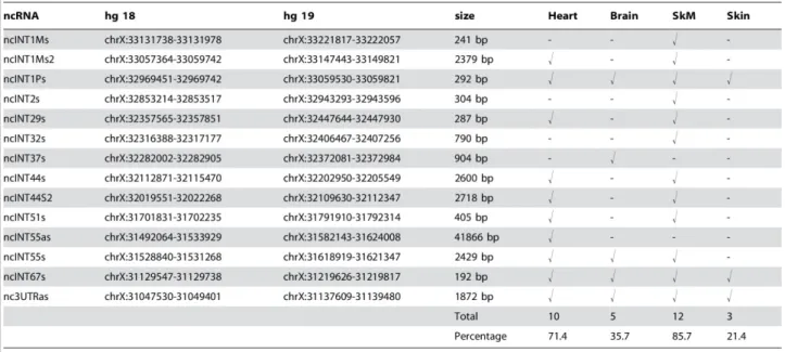
Values higher than the fluorescence intensity corresponding to 90% of all ranked probes (90th percentile ) on the array were considered as positive hybridization signals. Analysis of both DMD-GEx Sense and Antisense arrays data allowed to identify as statistically significant a total of 14 poly-adenylated transcripts which were named according to their intron of origin and orientation relative to DMD gene transcriptional direction (Figure 1B).
As it will be described below none of them contained ORFs encoding protein longer than 100 amino acids and thereby they were all considered non-coding RNAs (ncRNAs). Twelve of these ncRNAs, originating from introns 1 M (2 transcripts), 1P, 2, 29, 32, 37, 44 (2 transcripts), 51, 55 and 67, were found to be transcribed in the same orientation as the DMD gene. One ncRNA was found to correspond to the terminal exon and the 3'UTR of the Dp40 isoform (NCBI: NM_004019.2), a known coding dystrophin isoform. The remaining two ncRNAs, originating from intron 55 (ncINT55as) and from 3' UTR (nc3UTRas), were found to be transcribed in antisense orientation to the DMD gene. A significant proportion of the identified ncRNAs arose from introns harboring dystrophin isoform promoters or flanking isoform-specific first exon. In particular, three ncRNAs (ncINT1Ms, ncINT1Ms2 and ncINT1Ps) originated from intron 1 of the Dp427m and Dp427p full-length isoforms, one (ncINT29s) from intron 29 (Dp260), two (ncINT44s and ncINT44s2) from intron 44 (Dp140) and two (ncINT55s and ncINT55as) from intron 55 (Dp116). All identified transcripts were strongly expressed in at least one of the three tissues known to
express large amounts of dystrophin. A significant proportion of them were shown to be present in either the skeletal muscle (85.71%) or the heart (71.43%), and nine transcripts were identified in both tissues. Five ncRNAs were found in brain tissue, one of which (ncINT37s) was uniquely expressed in this district (Table 1) (Bovolenta et al., 2012a).
Figure 1. Example of transcripts (ncINT1Ms2) identified by the DMD-GEx array. A) a series of consecutive probes in the genome with fluorescence
intensities that rank above the 90th percentile over all probes on the array (indicated with a dashed red line) and mapping within intron 1 M (chrX:33057364-33059742). B) location of the transcripts identified from poly A+ RNA with respect to the DMD gene isoforms. Sense transcripts are represented by blue bars, whereas antisense transcripts are indicated by green bars. The transcripts marked with an asterisk and a double cross were characterized by Northern blotting, and RACE PCR. (doi:10.1371/journal.pone.0045328.g001)
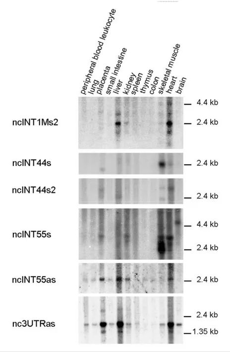
Transcript Validation by Northern Blot Analysis
To confirm and determine in a more detailed manner the pattern of expression of the identified transcripts as well as their precise size and relative amount, northern blotting analyses were performed. ncRNAs whose location within the DMD gene suggested a possible regulatory role in the expression of the many DMD gene isoforms were preferentially chosen. Nine poly A+ sense
25
ncRNAs mapping in proximity to the DMD gene promoters or nearby isoform first exons, and two antisense transcripts, mapping at the 3' end of the DMD locus were selected.
The probes identifying transcripts ncINT1Ms1, ncINT1Ps and ncINT29s revealed complex patterns of hybridisation, consisting of multiple bands or a smeared signal, in several tissue. The difficulty to obtain defined and clear bands is the reason for which we decided to not longer study these ncRNAs.
Table 1: Genomic location, length and tissue representation of the human transcriptsidentified. (doi:10.1371/journal.pone.0045328.t002)
On the contrary, Northern blotting results obtained for other four sense transcripts (ncINT1Ms2, ncINT44s, ncINT44s2 and ncINT55s) and two antisense transcripts (ncINT55as and nc3UTRas) revealed several distinct transcripts rangingfrom 1.4 to 4 kb in length and confirm their expression in at least one of the three tissues in which dystrophin is normally expressed (skeletal muscle (SkM), heart and brain) reflecting that observed in the array (Figure 2). (ncINT1Ms2, ncINT44s, ncINT44s2 and ncINT55as: SkM and heart; ncINT55s and nc3UTRas: SkM, heart and brain). Notably, the ncINT1Ms2, ncINT44s2, ncINT55as and nc3UTRas were highly represented in the heart tissue, whereas the ncINT44s and ncINT55s were found to be more intensively transcribed in skeletal muscle. Furthermore, most transcripts were also detected in the liver, placenta and/or kidney (Figure 2). Different tissue-specific isoforms were detected in four transcripts: ncINT1Ms2 presented at least four isoforms, ranging from about 1.8 to 5 Kb in size. A 2.4 Kb form is prevalent in the heart and liver, whereas it is poorly represented in the skeletal muscle and kidney; two ncINT44s2 isoforms were found in the skeletal muscle tissue, while only the larger one (about 2.7 kb) was detectable in the heart; ncINT55s was detectable in the heart and SkM in at least three
different isoforms (from 2.4 to 3 Kb), showing a specular pattern of expression in the two tissues. The larger of these isoforms was also found to be present in the kidney and placenta. An additional isoform of about 5 Kb is present exclusively in the brain. nc3UTRas show at least two isoforms (1.5 kb and 1.8 Kb). The larger form is widely represented in all tissues analysed, with the exception of the thymus and colon while both isoforms show a preferential expression in placenta, liver and heart (Figure 2). Only one form of transcript has been detected for ncINT44s and ncINT55as. ncINT44s is roughly 2.4 Kb in size and is expressed almost exclusively in the heart and skeletal muscle, being particularly abundant in the latter. In contrast, the ncINT55as transcript is about 2.4 Kb in length and displayed widespread distribution, being present in all analysed tissues except the brain and muscle (Figure 2) (Bovolenta et al., 2012a).
Figure 2. Northern blotting analyses on a 12-lane human poly A+ RNA filter using probes designed on ncRNAs originating near the first exons of
27 ncINT44s2 surround isoform Dp140. Transcripts ncINT55s and ncINT55as are located upstream the Dp116 isoform. Nc3UTRas overlaps with 39UTR in antisense direction with respect to the DMD gene. All transcripts were expressed in at least one tissue in which DMD isoforms were also expressed, but also in the liver, kidney, spleen and placenta. ncINT1Ms2, ncINT44s2, ncINT55s and nc3UTRas were found to be expressed in multiple isoforms, while one single isoform was detected for ncINT44s and ncINT55as.(doi:10.1371/journal.pone.0045328.g002)
Full-length Transcript Characterization by RACE PCR
To precisely define the size of the six DMD ncRNAs validated by Northern blotting, 3' and 5'-RACE analyses were performed on poly A+ RNA.
ncINT1Ms2: the products obtained from 3'-RACE, transcript walking and 5'-RACE were sequenced
and combined, generating a full-length sequence of 2379 bp. The ncRNA was sense-transcribed with dystrophin mRNA and was found to be colinear with the dystrophin intron 1 sequence (Figure 3 A, B and C). The 3' end of the ncINT1Ms2 transcript is located 897 bp upstream of the 5'UTR Dp427p full-length dystrophin isoform.
ncINT44s and ncINT44s2: for ncINT44s, analysis of transcript walking, 3'- and 5'-RACE products
yielded a full-length 2.6 Kb transcript entirely transcribed from intron 44 that did not undergo splicing (Figure 3 B and C). For ncINT44s2, the product obtained from 3'-RACE was combined with the two products identified by 5'-RACE, which defined two alternative transcriptional start sites (2452-bp and 2718-bp sequences) corresponding to the two full-length isoforms found in the Northern blotting experiment. Sequencing analysis revealed that these RNAs were entirely transcribed from intron 44, without undergoing splicing events (Figure 3 B and C). The two ncRNAs identified within intron 44 are located 29 Kb upstream (ncINT44s) and 61 Kb downstream (ncINT44s2) of the Dp140 dystrophin isoform promoter.
ncINT55s and ncINT55as: for ncINT55s, 3'-RACE identified two different 3' ends. After transcript
walking, 5'-RACE gave rise to two distinct PCR products corresponding to different transcriptional start sites (Figure 3 A). Nucleotide sequence analysis revealed that these transcripts originated entirely from intron 55, without splicing events (Figure 3 A, B and C). Combination of these start and polyadenylation sites potentially generates at least four transcripts of 2805, 2720, 2513 and 2428 bp, which is consistent with the Northern blotting data. The ncINT55as is transcribed in an antisense direction with respect to transcription of the dystrophin gene. 3'-RACE identified two different 3' ends, and 5'-RACE detected three isoforms with a common 5' end (Figure 3 A). Interestingly, these transcripts are alternatively spliced, generating RNAs of different sizes. A shorter isoform carrying only the common 3' and 5' ends was also identified (Figure 3 B and C). The predicted molecular weight of the three different isoforms ranges from 2642 bp to 2732 bp, being consistent with a single product detectable by Northern Blotting analysis. Notably, ncINT55s
residues within that DNA region is a part of the largest "intron" of the mature ncINT55as RNA isoforms. All of these different transcripts identified within intron 55 are located roughly 55 Kb upstream of the Dp116 promoter.
nc3UTRas: sequence analysis of the 3' and 5'-RACE products gave a full-length 1872-bp transcript
transcribed in an antisense direction from the 3'UTR region of the DMD gene. Since splicing events were not observed (Figure 3 A, B and C), this transcript overlaps (in an antisense orientation) roughly 2 Kb of the 3'UTR region of all full-length and 3'UTR region of all shorter dystrophin isoforms (Bovolenta et al., 2012a).
Figure 3. Full-length transcript characterisation by RACE PCR. A) For transcript ncINT1Ms2, a single product of 370 bp was obtained with 3'-RACE. After transcript walking, 5'-RACE was performed and identified a 396-bp PCR product. ncINT44s 3'-RACE generated a single product of 423 bp. After transcript walking, PCR, 5'-end was identified by 5'-RACE, and gave rise to a 599-bp PCR product. For transcript ncINT44s2, a single product of 552 bp was obtained by performing 3'-RACE, whereas 5'-RACE originated two distinct PCR products of 328 bp and 689 bp, corresponding to different transcriptional start sites. 3'-RACE of transcript ncINT55s revealed two products, of 150 bp and 442 bp, corresponding to different 3' ends of the transcript whereas 5'-RACE gave rise to two distinct PCR products of 673 bp and 352 bp. NcINT55as analysis by 3'-RACE identified two PCR products of 99 and 431 bp, corresponding to different 3' ends. 5'-RACE identified three PCR products of 307, 343 and 398 bp. Both 3' and 5'-RACE PCR products for transcripts nc3UTRas showed unique bands of 99 bp and 286 bp, respectively. Relevant pairs of primers used
29 are listed below each image. B) Schematic representations of the RACE PCR results for transcripts ncINT1Ms2, ncINT44s, ncINT44s2, ncINT55s, ncINT55as and nc3UTRas. Transcript orientation is indicated by the zigzag arrows. Their position with respect to adjacent dystrophin exons and isoform promoters is shown. DMD gene exons are shown as vertical red boxes, and 5' and 3' UTRs as horizontal red boxes. Lighter colours within the transcripts ncINT44s2, ncINT55s and ncINT55as boxes represent alternative starting or polyadenylation sites. The three alternative spliced isoforms of transcript ncINT55as are represented. C) Sequences of the identified transcripts are shown with reference to human genome build 18 (hg18, March 2006). Donor and acceptor splice sites are highlighted in yellow for transcript ncINT55as. Polyadenylation sites are underlined in the sequences. Curved arrows show the starting site for transcription of non-coding transcripts. (doi:10.1371/journal.pone.0045328.g003)
Bioinformatics Analysis of Coding Potential and Secondary Structure
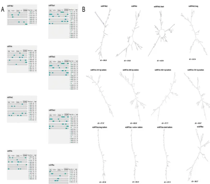
Using bioinformatic tools the six novel transcripts were tested for coding potential, ability to fold into secondary structures and for their degree of conservation.
To confirm that the ncRNAs did not possess the potential to encode for polypeptides, ORF-Finder software was employed to predict putative ORFs with a minimal length of 100 amino acids. As shown in Figure 4A, none of the three sense-frames contained any ORF fitting those criteria. The same results were obtained using the CPC tool, which identified a weak coding potential (one ORF of 51 amino acids) for the ncINT1Ms2 (Figure S2). CPC also analysed homology with UTR regions, and found 37 and 35 hits for ncINT1Ms2 and ncINT55as, respectively.
Using Mfold software, we also determined whether these transcripts could fold into higher secondary structures typical of non-coding RNAs. Interestingly all six ncRNAs can fold into secondary structures with a ΔG below -50 Kcals (Figure 4 B), thereby suggesting that these structure may be extremely stable and likely to serve as domains for interaction with other cellular components.
Conservation analysis performed using the UCSC Genome Browser (UCSC Genome Browser website. Available: http://genome.ucsc.edu/) showed that all identified transcripts are poorly conserved, with the exception of nc3UTRas, which is transcribed from the 3'UTR of the DMD gene, and transcript ncINT1Ms2, which presents a highly conserved sequence of roughly 130 bp. The UCSC Genome Browser was also employed to check for the presence of any known ESTs or mRNAs overlapping DMD ncRNAs. For some of the ncRNAs identified, predominantly small tags intersecting their sequences were detected: ncINT1Ms was found to overlap with part of a wider mRNA found in the uterus (Ota et al., 2004); ncINT1Ms2 contained two smaller ESTs found by Robertson et al. (Robertson et al., 1994) in 16-22-week foetal cochleas; ncINT1P overlapped several ESTs previously described (NCBI-CGAP website. Availble: http://www.ncbi.nlm.nih.gov/ncicgap.; NIH-MGC website. Available: http://mgc.nci.nih.gov/.) and corresponds to the TBCAP1 pseudogene (ENST00000436520); ncINT32s was found to contain a 9 bp Gallus Gallus DMD exon; and ncINT44s and ncINT55s, respectively, overlapped two smaller