Virtual Double-System Single-Box: A Nonequilibrium Alchemical
Technique for Absolute Binding Free Energy Calculations:
Application to Ligands of the SARS-CoV
‑2 Main Protease
Marina Macchiagodena, Marco Pagliai, Maurice Karrenbrock, Guido Guarnieri, Francesco Iannone,
and Piero Procacci
*
Cite This:J. Chem. Theory Comput. 2020, 16, 7160−7172 Read Online
ACCESS
Metrics & More Article Recommendations*
sı Supporting InformationABSTRACT: In the context of drug−receptor binding affinity calculations using molecular dynamics techniques, we implemented a combination of Hamiltonian replica exchange (HREM) and a novel nonequilibrium alchemical methodology, called virtual double-system
single-box, with increased accuracy, precision, and efficiency with
respect to the standard nonequilibrium approaches. The method has been applied for the determination of absolute binding free energies of 16 newly designed noncovalent ligands of the main protease
(3CLpro) of SARS-CoV-2. The core structures of 3CLproligands were
previously identified using a multimodal structure-based ligand design
in combination with docking techniques. The calculated binding free energies for four additional ligands with known activity (either for SARS-CoV or SARS-CoV-2 main protease) are also reported. The
nature of binding in the 3CLproactive site and the involved residues besides the CYS−HYS catalytic dyad have been thoroughly
characterized by enhanced sampling simulations of the bound state. We have identified several noncongeneric compounds with
predicted low micromolar activity for 3CLpro inhibition, which may constitute possible lead compounds for the development of
antiviral agents in Covid-19 treatment.
1. INTRODUCTION
As the whole world is currently plagued by the Covid-19
pandemic, the race to identify an effective antiviral agent for
SARS-CoV-2 is frantically ongoing. Among the viral functional
proteins, the main protease 3CLpro1,2 constitutes a very
attractive biomolecular target for drug design. Indeed,
inhibition of 3CLpro using small molecules is currently the
main goal of the crowdsourcing drug discovery initiative for
pandemics.3 This protein is responsible for the cleavage of
pp1a and pp1ab large polyproteins expressed by the virus RNA upon cell entry. The cleavage on multiple points of these polyproteins releases in the infected cell mature nonstructural proteins that are important for virus replication. For example, the virus replication machinery itself, the RNA-dependent
RNA polymerase (RdRp), is generated upon 3CLpro cleavage
of pp1a, pp1ab.4,5 It is hence expected that a potent and
specific inhibitor of 3CLpro can effectively block the viral
replication once the virions have entered the cell.
One of the main approaches for anti-Covid-19 drug development is based on past experience on the SARS 2003 outbreak. Indeed, the RNA of SARS-CoV-2 and of SARS-CoV
share nearly 80% genomic sequence identity.6 The main
protease of SARS-CoV7and SARS-CoV-22,5,8differs by only 12
residues, with none of these differing residues being directly
involved in the catalytic site.9 It is hence expected that
inhibitors for SARS-CoV 3CLpro bind effectively the strictly
related SARS-CoV-2 main protease. 3CLpro-based drug
discovery for SARS10was mainly directed toward the so-called
covalent Michael inhibitors11,12via electrophilic attack on the
cysteinate of the 3CLprocatalytic CYS145−HIS41 dyad. New
irreversible covalent inhibitors for SARS-CoV-2 3CLpro were
recently proposed in ref5. On the other hand, the consensus in
drug discovery leads to excluding electrophiles from drug
candidates for reasons relating to safety and adverse effects
such as allergies, tissue destruction, or carcinogenesis.13
Noncovalent inhibitors for SARS-3CLpro were first identified
in ref 14 and later characterized in ref 15, leading to the
synthesis of ML188 with a measured inhibitory activity of 2 μM.
Received: June 20, 2020
Published: October 22, 2020
Article
pubs.acs.org/JCTC
Downloaded via ENEA BOLOGNA RESEARCH CTR on March 23, 2021 at 10:20:22 (UTC).
The second arm of the current research for drug therapy of Covid-19 is focused on drug repurposing, hence testing approved compounds with little (and known) side effects
and with a minimal development cost for off-label use. In this
context, a very extensive and complete multicentric study of
the SARS-CoV-2 protein interaction map16 revealed human
targets for drug repurposing. The targets were in this case
human proteins (such as σ-receptors or bromodomains)
characterized as playing an important part in the viral interactome.
From a computational standpoint, docking studies on 3CLpro
started to appear immediately after the release in mid-February
of the PDB structure of 3CLpro.8
Docking is one of the main computational tools used in compound triaging in the cited
COVID-19 moonshot worldwide initiative.3,17 As of today,
more than 20 entries on 3CLproligand screening using docking
either alone or in combination with structure-based or data-driven approaches have been published so far, according to the Scopus database. Many more, deposited in preprint servers, are awaiting for the peer-review process to complete. We were
among the first to deposit on the arXiv server a study9 on
3CLproinhibitors using a multimodal structure-based design18
in combination with molecular docking.19 Evaluation of
docking scores is fast and docking is indeed an invaluable tool for a plausible pose prediction and for a semiquantitative assessment of the inhibitory power of a ligand. However, binding free energies solely based on docking are, in general,
considered not sufficiently reliable as this technique exhibits by
design a major weakness, that is, the partial neglect of the ligand and receptor Boltzmann-weighted conformational disorder as well as of solvent-related microsolvation phenom-ena, eliciting the crucial and elusive entropy contribution to
the binding free energy.20
Pose prediction using Docking can be assessed and refined
using molecular dynamics (MD) advanced techniques with full
atomistic detail such as free energy perturbation21 (FEP) or
thermodynamic integration (TI).22 Surprisingly, to our
knowledge, not many FEP studies combined with alchemical
transformation23,24 on 3CLpro binders appeared so far in the
literature.25 Possibly, this is due to the fact that FEP-based
modern alchemical techniques26 are costly and generally
applied to relative binding free energies on strictly congeneric series, with hence a limited value in drug discovery campaigns based on de novo design. On the other hand, FEP calculations for absolute binding free energies (ABFEs) are still quite rare as they face serious sampling problems due to the mobility of the ligand in the binding site, in general, especially for
low-coupling alchemical states.27
In the past years, we have been developing a nonequilibrium
(NE) variant28−32of alchemical transformations, whereby the
ligand is rapidly decoupled from the environment in a swarm of rapid independent trajectories producing a NE work (NEW) distribution histogram, related to the decoupling free energy via well-known NE theorems. These NE alchemical decoupling trajectories, typically lasting less than 1 ns, start from equilibrium phase space points that are sampled using
very efficient and highly parallelizable enhanced sampling
techniques, such as Hamiltonian replica exchange with
torsional tempering.33The NEW approach allows us to release
altogether the artificial conformational and orientational
restraints in the bound and unbound states that are commonly
used in FEP calculation to facilitate sampling34at the price of
focusing on a pose that can be suboptimal.35 The NEW
approach turned out to be among the top-performing
techniques assessed in recent SAMPL challenges36,37 for
blind absolute binding free energy predictions. Here, we have implemented yet a new improved variant of the NEW approach, called virtual double-system single-box (vDSSB) on the basis of the recent remark on the so-called DSSB approach
used in the latest SAMPL challenge.38
NEW-vDSSB has been applied to the calculation of the
dissociation free energy for a total of 21 3CLpro noncovalent
complexes, with some of the ligands identified in ref9and with
some of their analogues, as well as for few ligands with recently
measured activity for 3CLpro.17The common binding pattern
of these ligands in the shallow and wide binding pocket of
3CLpro is analyzed in detail using enhanced sampled
configurations, providing valuable information on drug design
against the 3CLpro target. A few compounds with predicted
submicromolar activity have been designed, hopefully
provid-ing promisprovid-ing leads for an effective medicinal chemistry
campaign for the identification of a therapeutic agent for
Covid-19.
The paper is organized as follows: wefirst lay out with some
detail the theoretical background for the NEW-vDSSB
determination of the drug−receptor dissociation free energy.
We then describe the 3CLprosystem and its function, giving a
rationale for its modelization in a drug design study. Technical details for MD, in general, as well as for enhanced sampling and nonequilibrium simulation approaches are described in Section 4. Computed dissociation free energies for the 21 scrutinized complexes using NEW-vDSSB along with a detailed
analysis of the binding pattern is reported inSection 5.Section
6presents concluding remarks.
2. THEORETICAL BACKGROUND
The NEW method has been developed in the past two decades
in the context of binding free energy calculations for drug−
receptor systems as a nonequilibrium variant of the free energy
perturbation21 method with stratification.23 In the
non-equilibrium double-system single-box approach39
(NEW-DSSB), a bound ligand is annihilated in the receptor binding site, while a second unbound ligand, kept using restraints far away from the protein, is simultaneously grown in the bulk solvent in a series n of independent alchemical trajectories,
where the alchemical parameter λ regulating the ligand−
environment coupling level23,24is varied in the range [0,1] (or
[1,0]) according to a common time schedule. The alchemical
DSSB approach was recently applied40 also in the context of
the equilibrium alchemical FEP with λ-stratification.21,41,42
Provided that the two ligands do not feel each other and the
unbound ligand is constantly surrounded by a sufficiently thick
layer of the solvent, the computed work distribution in NEW-DSSB is directly related to the dissociation free energy of the
ligand.43,44 NEW-DSSB works well if the simulation box is
large enough so that the growth and annihilation work of the two distant ligands can be assumed to be uncorrelated. Alternatively, in the single-box double-system approach
(SSDB), one can compute, in two different alchemical
processes, the decoupling and recoupling free energy for the bound ligand and for the ligand in bulk, obtaining the binding free energy as the sum of these two contributions.
In NEW-based alchemical techniques, the dissociation free
energy (except for a volume correction43,44) can be recovered
from the NE work distributions (obtained computing the work for each of the n independent alchemical trajectories) as
,
ΔGDSSB= [PDSSB(W F| )] (1)
, ,
ΔGSSDB= [PSSDB(−W Gu| )] + [PSSDB(W Ab| )] (2)
where P(W|F), P(−Wu|G), and P(Wb|A) are the work
distributions for the double system and for the unbound (growth) and bound (annihilation) states of the single system,
respectively, and,[·]is a functional of the work distribution of
the NE process, yielding the estimate of the free energy
difference, ΔG, of the process. For unidirectional processes,
,[·]corresponds to the Jarzynski estimate45
∫
∑
Δ = − = − β β − − G WP W n RT ln( d ( )e ) RT ln(1 e ) W i n Wi (3)or to the Gaussian estimate46
μ βσ
ΔG= − 1
2
2
(4) eq. 4is valid when P(W) is the normal distribution, n(W,μ,σ)
with mean and varianceμ and σ2. Wdiss=μ − ΔG corresponds
to the dissipated work in the NE process, which in the case of
the normal distribution is given by Wdiss= (1/2)βσ2. It should
be noted that, for normal distributions, the Crooks theorem47
implies that the forward distribution (PF(W)) and reverse
distribution (PR(−W) done with inverted time schedule) are
mirror-symmetric with respect to their unique crossing point,
Wc=ΔG, and that their maxima are βσ2= 2W
dissfar apart from
each other.46The inverse of the dissipated work in Gaussian
(Markovian) alchemical processes is a linear function48of the
durationτ of the NE alchemical processes. Both accuracy and
precision decline with increasing dissipation48,49 (or,
equiv-alently, faster NE processes). Note that we have assumed a forward process in NEW-DSSB that corresponds to the annihilation of the bound ligand and growth of the ligand in the bulk. The associated work for binding ligands is positive, corresponding to positive dissociation free energies. This
choice is dictated by the fact27that, in the reverse process, the
starting states of the decoupled and weakly restrained ligand in the binding pocket are characterized by a high conformational disorder, with most of the generated NE recoupling trajectories producing suboptimal poses in highly dissipative processes. This is a typical situation in NE transformations involving the
entrance into a free energy funnel.50
For non-Gaussian bidirectional processes done with
time-inverted protocols,eqs 1and2take the form of
, ΔGDSSB= [PDSSB(W F| ),PDSSB(−W R| )] (5) , , Δ = [ − | | ] + [ | − | ] G P W G P W A P W A P W G ( ), ( ) ( ), ( ) SSDB SSDB u u SSDB b b (6)
where F and G denote the forward and reverse processes and
the functional ,[·] is the Bennett acceptance ratio (BAR)
estimate.51,52Note that ineq 2, referring to the unidirectional
SSDB estimates, if the ligand bears a net charge, one must add
an analytic correction toΔG due to the annihilation of the net
charge in the two independent alchemical processes when
using particle mesh Ewald (PME)53 with a neutralizing
background plasma.54,55The correction exactly cancels out in
the unidirectional DSSB processes and in bidirectional SSDB.
At constantτ, the BAR bidirectional estimate is more accurate
than unidirectional estimates, provided that there is sufficient
overlap between the forward and reverse work
distribu-tions.48,49,56
As discussed in ref57, for the case of the NEW applied to
Gaussian processes, the DSSB/SSDB efficiency ratio R can be
shown to be given by σ = + + + + σ
(
)
R r r (1 ) 2(1 ) (1 ) 1 3 SSDB 2 2 2 SSDB (7)where r = Ls/Lb(r ∈ (0, 1]) is the ratio between the side
lengths for the optimal box of the bound and unbound
systems. According toeq 7, DSSB is more efficient than SSDB
(R < 1) when the variance of the NE work distributions (assumed to be equal for the growth and annihilation
processes) are small and r ≃ 1. The eciency gain in DSSB
becomes insignicant when the optimal box for the bound system is signicantly larger than that of the un- bound ligand (which occurs systematically in drugreceptor systems) and/or in the case of highly dissipative processes.
However, since in SSDB the work values in the two alchemical processes for bound and unbound states are independent random variables (RV), one can emulate the
DSSB by combining each value of the RV Wb(A) with each
value of the RV Wu(G), hence obtaining n2 work RV’s W =
Wb(A) + Wu(G) instead of the original n. This process
corresponds to evaluating the convolution P(W|F) = (Pb*Pu)
(W|F) = ∫ dwPb(W|A) Pu(W − w|G), thus leading to the
following equations ,
ΔGvDSSB= [(P PB* u)(W F| )] (8)
,
ΔGvDSSB= [(P PB* u)(W F| ), (P PB* u)(−W R| )] (9)
whereeqs 8and9refer to the unidirectional and bidirectional
estimates, respectively, and (Pb*Pu)(−W|R) = ∫ dwPb(−W|G)
Pu(−W + w|A). When using a bidirectional approach in
vDSSB, due care must be taken in using a time-inverted protocol for both legs (bound and unbound) of the alchemical process.
The advantages of eq 8 over eq 1 are evident. First, the
convolution of the Puand Pbdistribution boosts the statistics,
increasing significantly the resolution of the vDSSB work
distributions, P(W|F) and P(−W|R). The convolution (Pb*Pu)
(W|F) can now be computed using a sample of n2 work
outcomes at the cost of n bound and unbound trajectories. In the second instance, at variance with DSSB, where a common time protocol for the process is adopted, in vDSSB, the time protocol for the bound and unbound states alchemical simulations can be chosen independently with no violation of the Crooks or Jarzynski theorems. In particular, the alchemical process for the unbound state can be done using a much faster rate with respect to that of the bound state since the dissipation in the anisotropic environment of the binding pocket is, in general, much higher than that experienced by the ligand in the isotropic environment of the bulk solvent. Third, in vDSSB, the optimal box size can be chosen according to the physical dimension of the solute. For the ligand in bulk, the box can be chosen much smaller than that of the ligand in the
bound state, with a significant gain in the computational
3. 3CLPRO−LIGAND COMPLEXES
3.1. CLpro Structure and Function. 3CLpro acts as a
dimer.58 The monomer is in turn composed of two loosely
coupled units, the chymotrypsin-like domains I + II (residues
1−197), harboring the catalytic site, and the cluster of helices
domain III (residues 198−304), regulating dimerization via
two intertwined salt bridges involving ARG4(A)-GLU290(B) and GLU290(A)-ARG4(B) of the A and B protomers, as
shown in Figure 1a. The dimer is characterized by two
symmetric extended clefts (shown as blue arrows inFigure 1b)
for pp1a, pp1ab adhesion. Each dimer cleft ends at the
solvent-exposed catalytic site with the CYS145−HIS41 proteolytic
dyad. The two catalytic dyads, on the opposite sides of the dimer, very likely act independently for maximizing the
catalytic efficiency. Polyproteins are cleaved by betacoronavirus
SARS-CoV and SARS-CoV-2 main proteases at the glutamine
level in the general sequence X(L/F/M)Q↓(G/A/S)X, where
X is any residue.4The cleavage sites in the SARS-CoV-2 pp1ab
sequence59are reported inTable 1.
It is worth noting the hydrophobic character of most of the Q-neighboring amino acids, indicating that docking of the polyprotein at the proteolytic site is likely to occur via complementary hydrophobic interactions. In the light of the dimer peculiar structure and functional mechanism, with the solvent-exposed and distal proteolytic sites, the dissociation
constants for 3CLproligand association can be effectively and
reliably computed by modeling only the domain I + II of one
protomer for the bound state (residues 1−197). We must
nonetheless stress that the computed ΔG pertains to the
associations of the ligand with one protein, irrespective of the state of association of the protein. At free ligand concentration
equal to Kd≡ e−ΔG/RT, i.e., when half of the protein molecules
are inhibited, the probability to have both monomers inhibited on a catalytically active dimer is equal to 1/4, whatever the
dissociation constant of the dimer is,58 hence the need for
identifying nanomolar or sub-nanomolar inhibitors of 3CLpro.
3.2. 3CLpro-Tested Compounds. In the present study, we
have computed, using NEW-vDSSB, the absolute binding free
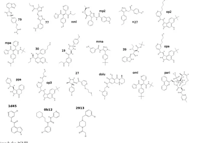
energies (ABFEs) of the 21 compounds reported inFigure 2.
Compounds 79, 27, 19, 77, and 39 were previously identified9
as probable binders (8 <ΔG < 9 kcal/mol) using a multimodal
structure-based design18 in combination with molecular
docking.19 Compound nml (ML188) is a known inhibitor15
for SARS-CoV 3CLpro with micromolar activity. Compounds
dolu and pari are Dolutegravir and Paritaprevir and are
recently identified60 using virtual screening (Docking and
Standard MD) as candidate lead compounds for SARS-CoV-2
3CLproand 2′-OMTase inhibition. All other compounds have
been designed in this study by analyzing the binding pattern from bound-state enhanced sampling trajectories (vide infra).
Among the compounds reported inFigure 2, only dolu, pari,
and oml are commercially available as reported by the ZINC
database.61The activity (IC50) of the compounds 1d45, 0b12,
and 2913 for the inhibition of the SARS-CoV-2 main protease was recently measured in the context of the Covid-19
Moonshot initiative.17
4. METHODS
4.1. Overview. We started by docking the
OpenBabel-generated623D structure of the ligands to the PDB structures
of 3CLpro(domains I + II only) and PLprousing Autodock4.19
The optimal initial docking pose was found by running 50 minimization rounds with the center of mass (COM) of the
fullyflexible ligand placed within a 15 Å side-length cubic box
centered at the protein active sites. The latter were identified
by the midpoint vectors connecting the α carbons of the
CYS145−HIS41 catalytic dyad in 3CLpro. The 3CLprotarget is
rigid to avoid sampling of unlikely nonopen conformations of the active site region.
The so-generated initial structures of the complexes were first equilibrated in a cubic box of appropriate size, filled with
TIP3P63explicit water molecules, by running short simulation
(100 ps) in the NPT ensemble. The resulting solvated
complexes were then fed to the ORAC MD program64 for
the Hamiltonian replica exchange (HREM) sampling of the bound states using a powerful torsional tempering scheme in
the binding site region engaging only eight replicas.33,65For
each complex, we collected 540 configurations sampled at
regular intervals during the 25 ns NPT simulation of the HREM target (unscaled) state (T = 300 K and P = 1 atm).
From these HREM-harvested equilibrium configurations, we
launched, on a single parallel job, a swarm of 540 independent
alchemical nonequilibrium (NE) trajectories31,32 where the
ligand−environment interactions were rapidly decoupled in
0.36 ns, eventually producing a ligand annihilation work distribution. During the HREM and NE simulations, the ligand was prevented to drift away from the active site using a weak harmonic restraint between the centers of mass (COM) of the
Figure 1.(a) Three-dimensional (3D) structure of the SARS-3CLpro
dimer.5Domains I + II and III are in yellow and gray, respectively. The catalytic sites (VdW representation) are in ocher. The salt bridges GLU290-ARG4, connecting domains III of the protomers, are in blue (GLU290) and red (ARG4), respectively. (b) Surface representation of upper and lower sides of the dimer highlighting the clefts for pp1a/pp1ab adhesion. The catalytic pocket is shown in red.
Table 1. SARS-CoV-2 pp1ab Cleavage Sites
res(Q) seq gap
3263 LQS 3263 3569 FQS 306 3922 MQG 353 3942 LQA 20 4253 LQA 311 4392 LQS 139 5324 LQA 932 5605 LQG 281 5925 LQA 320 6179 LQS 254 6452 LQS 273 6798 LQS 346
ligand and the receptors.31No orientational or conformational restraints are imposed on the ligand, which is free to explore all
of the poses and orientations within the allowance volume43,66
V = (2πRT/K)3/2set by the weak COM−COM restraint.
The recoupling work distribution of the ligand in bulk solvent was obtained using fast-growth (0.36 ns) alchemical
simulations. The starting configurations, in this case, were
generated by combining 540 solvent-decoupled conformations of the ligand, sampled in an 8 ns HREM simulation with torsional tempering of the isolated (gas-phase) molecule, with equilibrated structures of pure TIP3P water molecules in standard conditions.
The standard dissociation free energies, ΔG0, were
computed using the Jarzynski estimate45 equation (eq 8),
evaluated on the work distribution obtained by combining the negative growth work values of the ligand in bulk with the positive decoupling work values of the ligand in the bound state, and by adding a standard state binding site volume
correction.31 The 95% confidence interval of the predicted
dissociation free energies was obtained by bootstrapping with resampling on the two independent sets of growth and decoupling work values, before convoluting the data. All MD
calculations were performed using the program ORAC64 on
the CRESCO6 high-performance computing (HPC) facility
located in Portici (Italy) and managed by ENEA.67Details of
the MD settings, HREM parametrization, and NE protocols
are reported subsequently, inSections 4.2−4.4.
4.2. MD: General Settings. All simulations for the bound
and unbound states were done in the NPT isothermal−
isobaric ensemble under periodic boundary conditions on cubic or orthogonal MD boxes with explicit TIP3P water
molecules. We used the AMBER99SB-ILDN force field68 for
3CLpro. Default protonation states (pH = 7.6) of titratable
residues were used. The ligands were described using the
GAFF2 forcefield, with atom types and AM1/BCC charges
assigned using the PrimaDORAC web interface.69 The
potential parameters for all ligands of Figure 2 are provided
in theSupporting Information(SI). The external pressure was
set to 1 atm using a Parrinello−Rahman Lagrangian70 with
isotropic stress tensor. The temperature was held constant at 300 K using three Nosé−Hoover thermostats coupled to the translational degrees of freedom of the systems and the rotational/internal motions of the solute and the solvent.
Constraints were imposed only to X−H bonds, with X being a
heavy atom. The equations of motion were integrated using a
multiple time step r-RESPA scheme71 with a potential
subdivision specifically tuned for biomolecular systems in the
NPT ensemble.70,72The long-range cutoff for Lennard−Jones
interactions was set to 13 Å. Long-range electrostatics were
treated using the smooth particle mesh Ewald method,53with
anα parameter of 0.38 Å−1, a grid spacing in the direct lattice
of about 1 Å, and a fourth-order B-spline interpolation for the gridded charge array. The net charge on the system (due to proteins) was neutralized by a uniform neutralizing
back-ground plasma as it is customary when using PME.54
4.3. HREM Simulations. The HREM simulations of the bound state were run by launching, in a single parallel job, 12 batteries of independent Hamiltonian replica exchange simulation with eight replicas, for a total of 96 MPI instances
and a total simulation time on a per complex basis of≃0.2 μs
(25 ns on the target state). In each eight-replica battery, we used a torsional tempering scheme (including 14 nonbonded interactions) with a maximum scaling factor s = 0.2 corresponding to a torsional temperature of 1500 K. The
“hot” region includes all residues with at least one atom at a distance of less than 4.2 Å from any atom of the ligand, as
found in the best docking pose. The scaling factors, sm, along
the eight replica progression are computed according to the
protocol sm= s(m−1)/7. The exchange was attempted every 15 fs
(every large time step73), and the average exchange rate was, in
all cases, around 15−20% with round-trip times of around
0.3−0.4 ns.
The ligand was weakly tethered in the binding site via a harmonic restraint potential between the COM of the ligand and that of the protein, with equilibrium distance
correspond-ing to the COM−COM distance of the lowest energy docked
pose and a force constant of 0.05 kcal mol−1Å−2.
For setting up the starting configurations of the decoupled
ligand in bulk, we first harvested 540 configurations of the
isolated (gas-phase) molecule via an 8 ns (target state) HREM simulation using four replicas with torsional tempering with a minimum scaling factor of s = 0.1, corresponding to a torsional
temperature of 3000 K, and using the protocol sm= s(m−1)/3, m
= 1...4 along the four replica progression. The 540 sampled gas-phase ligand conformations, with random orientations and positions, were combined with a single equilibrated sample of about 1800 water molecules in standard conditions in a cubic
box, producing 540 starting configurations of the decoupled
(ghost) ligand in the bulk.
4.4. NE Alchemical Simulations. For the ligand in the bound state, the alchemical annihilation simulations were
performed starting from theλ = 1 (fully coupled) equilibrium
configurations collected in the preceding HREM step. NE
annihilation trajectories were run for 360 ps: in thefirst 120 ps,
the electrostatic interactions were linearly switched off; in the
following 120 ps, two-thirds of the Lennard−Jones potential
was turned off, and in the last 120 ps, the one-third residual
wasfinally switched off.
A time-inverted protocol was adopted for the ligand in the bulk state (u state); in this case, the fast-growth alchemical
simulations were started from theλ = 0 (fully decoupled) and
the NE trajectories were run for 360 ps. In thefirst 120 ps,
one-third of the Lennard−Jones potential was turned on. In
the following 120 ps, the Lennard−Jones potential was
switched on completely. In the last 120 ps, the electrostatic interactions were linearly turned on. All of the simulations for computing inhibitor constants are done using the program
ORAC.64The program is distributed under the GPL and can
be downloaded free from the website
www.chim.uni-fi.it.
5. RESULTS AND DISCUSSION
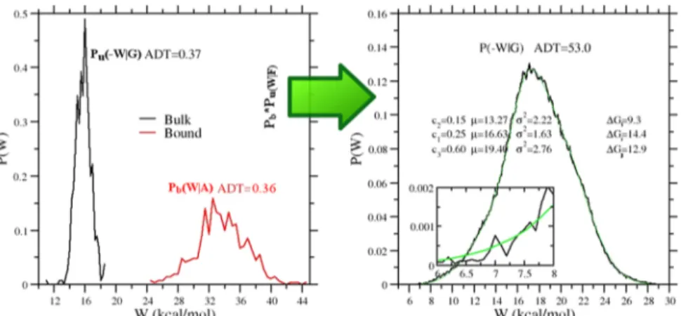
5.1. Binding Free Energy Results. In Figure 3, we
illustrate the vDSSB approach for evaluating P(W|F), referring
to the mma ligand. The annihilation (bound ligand) and growth (bulk ligand) work distributions are constructed by computing the work in a few hundreds of NE alchemical decoupling and recoupling 0.36 ns trajectories, respectively. Note that the dissipation for the growth process in bulk is much smaller than that for the annihilation in the bound state
at equal time τ of the NE processes, leading to a systematic
disparity in the histogram resolution. The mean-variance,σ2,
values for the growth and annihilation distributions Pu(−W|G)
and Pb(W|A) are 1.1 and 15.3 kcal2mol−2, respectively. In the
reported typical example for the mma ligand, both
distributions“look” Gaussian and they both amply satisfy the
Anderson Darling (AD) test for normality.74We recall that the
AD test gives only the probability for rejecting the null hypothesis (i.e., the work values are normally distributed) but does not provide, like any other normality test, any certitude on the correctness of the null hypothesis.
As a matter of fact, the convolution of the two distribution
(right panel in Figure 3), by boosting the statistics and the
resolution of the work histogram, visually reveals the
non-Gaussian character of the resulting P(W|F), which, in the case
of mma, exhibits a marked negative skewness. In this event,eq
4cannot be used. The high number (n2) of work data for the
construction of P(W|F), with good sampling also in the left tail
of the distribution, allows for a reliable estimate of the free
energy based on the Jarzynski exponential average,75 eq 3.
Alternatively, the convolution distribution can be decomposed
into c normal components, P(W|F) = ∑icw
i n(W,μi,σi), using
the expectation-maximization (EM) algorithm,76,77with∑icwi
= 1. By the Crooks theorem, it can be shown50that the free
energy functional for Gaussian mixtures is given by
∑
μ σ ΔG = −RT ln wn W( , , ) i c i i i EM Ä Ç ÅÅÅÅÅ ÅÅÅÅÅ Å É Ö ÑÑÑÑÑ ÑÑÑÑÑ Ñ (10)The EM algorithm is very efficient in fitting the convolution
histogram, as Figure 3 shows. For the assessment of the
confidence level for the estimate (whether Jarzynski or EM)
Figure 3.Left panel: growth, Pu(−W|G), and annihilation, Pb(−W|G), work distributions computed using 540 work values for the mma ligand (see
Figure 2). Right panel: convolution work distribution (Pb*Pu)(W|F) for the forward process (black) and expectation-maximization (EM) fit
based on the combination of the work data, some important
remarks are in order. Bootstrapping the n2 sample of the
combination yields an unrealistically small error. As errors in
the coarse-grained distributions Pu(−W|G), Pb(W|A) can be
propagated in the high-resolution convolution, the uncertainty of the estimate must be evaluated by bootstrapping independently the growth and annihilation work samples and then combining the data using either the Jarzynski or EM functional of the bootstrapped convolution.
In Figure 4, we show the (convolution) work distributions
P(W|F) and the COM−COM distance distribution functions
for all 21 ligands. These two probability distributions, obtained from the NE and HREM simulations, respectively, are the two ingredients used in delivering the predicted standard
dissociation free energy of the ligand−3CLpro complex. The
combined work distributions serve for the calculation of the
Jarzynski or EM functional, while the COM−COM histograms
are used for the evaluation of the correction due to the binding
site volume.55The latter is estimated as Vsite= (4/3)π (2σ)3,
where σ2 is the variance of the HREM-determined COM−
COM histogram, yielding the standard state correctionΔGvol=
RT ln(Vsite/V0) with V0= 1661 Å. All ligands are electrically
neutral at the physiological pH with the exception of 2913,
which has a positive charge on the sp3nitrogen. For the latter
ligand, we applied thefinite-size correction to the annihilation
free energy for both the bound and unbound states as
described in refs54,55.
The convolution work distributions (Figure 4, left) exhibit a
marked non-Gaussian character (see also AD tests reported in Table 2), leaving hence only eqs 3or 10for the free energy
Figure 4.Left panel: work distributions for the vDSSB approach for the ligands reported inFigure 2as obtained from the NE simulations in the bulk (growth) and in the 3CLprobound state (annihilation). Right panel: corresponding COM−COM distribution functions as obtained in the
HREM simulations of the bound state. The green circles refer to the initial pose as obtained from Autodock4 software.19
Table 2. Standard Dissociation Free Energy Estimates (in kcal/mol) for the 21 Ligands Shown inFigure 2b
ligand ΔGJ0 ΔGEM0 ΔGvol ADconv ADu ADb ΔGq ΔGexp.
pari 10.7± 0.6 8.1± 4.1 −2.4 490.8 0.40 0.37 −8.6 ± 0.9 n/a opa 9.4± 0.5 6.5± 3.1 −2.5 315.0 0.18 1.14 0.3± 0.8 n/a ml 9.0± 0.7 9.0± 1.9 −3.2 131.1 0.15 0.45 −0.4 ± 0.6 7.9a op3 8.8± 0.8 6.9± 1.8 −2.2 79.5 0.11 0.48 −2.5 ± 0.8 n/a 27 8.3± 0.5 8.1± 0.9 −2.7 165.6 0.40 0.54 −1.3 ± 0.3 n/a 39 7.6± 1.5 6.2± 2.8 −2.5 559.5 0.40 1.39 −1.8 ± 0.4 n/a mma 7.4± 0.8 6.8± 1.7 −3.4 55.6 0.37 0.36 0.2± 0.4 n/a ppa 7.3± 0.8 4.7± 2.5 −3.7 278.2 0.16 0.99 −3.6 ± 2.4 n/a op2 7.1± 2.0 6.3± 3.1 −2.6 776.2 0.39 2.03 0.2± 0.6 n/a dolu 6.7± 1.6 5.0± 3.4 −3.9 190.7 0.74 0.51 0.3± 2.0 n/a 19 6.5± 0.7 5.2± 1.5 −1.9 138.5 0.26 0.54 −3.2 ± 0.6 n/a 30 6.1± 0.9 5.1± 1.4 −3.5 116.7 0.38 0.43 −1.1 ± 0.3 n/a mp2 5.8± 0.6 3.2± 1.4 −3.4 40.7 0.82 0.36 1.8± 1.2 n/a mpa 4.3± 1.2 3.6± 2.9 −3.7 113.4 0.77 0.49 −1.5 ± 0.4 n/a 77 4.0± 0.4 2.3± 1.7 −3.0 141.7 0.17 0.53 −0.8 ± 2.4 n/a n27 3.9± 0.6 2.9± 1.3 −2.5 114.0 0.42 0.46 −1.1 ± 0.4 n/a 79 3.7± 0.9 2.4± 0.9 −2.8 142.6 1.91 0.54 −0.4 ± 0.8 n/a nml 3.1± 0.4 1.5± 0.5 −3.1 52.5 0.68 0.58 1.0± 0.9 n/a 1d45 5.4± 0.8 4.6± 1.1 −2.9 223.8 0.65 0.22 1.0± 0.9 10.0 0b12 9.3± 0.9 7.0± 1.6 −3.1 183.8 0.50 0.31 1.0± 0.9 7.46 2913 5.8± 0.8 5.5± 1.6 −2.8 201.5 0.63 0.52 1.0± 0.9 7.0
aThe experimental value refers to the SARS-CoV 3CLproinhibition.15bΔG
J,ΔEM,ΔGvol, ADconv, ADu, ADb, andΔGqrefer to the Jarzynski free
energy estimate; the EM-based free energy estimate; the volume correction; the AD normality test for P(W|F), Pu(−W|G), and Pb(W|A); and the
estimate. For ΔGEM, in all cases, we have used c = 3. The
COM−COM ligand−3CLprodistance distributions (Figure 4,
right) denote, in general, a rather large binding site volume, with distance oscillations extending for nearly 4 Å in several cases (e.g., pari, 27, op3, 19, 79), indicating either a shallow
and wide binding site pocket in 3CLpro and/or a significant
ligand conformational activity of the ligand in the binding site.
As we shall see later on, these peculiar features of 3CLpro
binding make the dissociation free energy prediction inherently less accurate. It is worth noting that, in some cases, the
docking-determined COM−COM distance, corresponding to
the initial pose, is significantly different from the most-likely
HREM-determined COM−COM distance, corresponding to
the peak of the distribution.
InTable 2, we report the results for the computed standard dissociation using the vDSSB approach. Ligands are sorted from the most powerful (pari, Jarzynski estimate), of the
predicted low nanomolar affinity (20 nM), to the weakest
(nml), of the millimolar activity (5 mM), as resulting from
ΔGJ. TheΔGJandΔGEMestimates appear, in general, strongly
correlated (see Figure 5), although the latter shows a
systematic downshift from a few fractions of kcal/mol to more than 2 kcal/mol for pari and op3. In general, the
Jarzynski estimate is more precise but less accurate thanΔGEM
based on Gaussian mixtures.55 For dissipative NE processes,
the Jarzynski estimate very likely remains biased75despite the
statistics boosting on the left tails of the work distribution obtained by combining the independent RVs corresponding to the growth and annihilation work of the ligand.
ΔGEM, on the other hand, while in principle more accurate
and unbiased,50is significantly less precise. We recall that the
error in the free energy estimates is obtained by bootstrapping independently the growth and annihilation work sample (each containing n = 540 work values) and then combining the
bootstrapped data to form the convolution P(W|F). For ΔGEM,
the error goes as [1/(n/c)]1/2, i.e., it increases with the number
of components (c = 3 in our case). EM appears to be sensitive
to bootstrapping fluctuations in the Pu(−W|G) and Pb(W|A)
distributions, producing a rather large variance for the
calculation of ΔGEM by eq 10. In general, the Jarzynski
estimate is found, with no exception, within the confidence
interval of the EM-based estimate. One could hence propose as
consensus value for the estimate and for the confidence level of
the arithmetic mean of the two estimates and errors. A comparison with the experimental results is possible for compound ML (ML188). The measured standard dissociation
free energy for the ML188-SARS-CoV 3CLpro complex was
found to be157.9 kcal/mol, which competes favorably with the
consensus vDSSB value of 9.0 kcal/mol found for the strictly related ML188-SARS-CoV-2 complex. For the ligands with known activity, i.e., 1d45, 0b12, and 2913, the consensus value agrees satisfactorily with the experimental counterpart for 0b12
(ΔGc= 8.1 kcal/mol,ΔGexp.= 7.46 kcal/mol) and 2913 (ΔGc
= 5.7 kcal/mol, ΔGexp. = 7.0 kcal/mol), while it differs
significantly for 1d45 (ΔGc= 5.0 kcal/mol,ΔGexp.= 10.0 kcal/
mol). However, while 1d45 is labeled as a noncovalent binder
of SARS-CoV-2 3CLproaccording to the Covid-19 moonshot
activity data,17the same compound was found to be a potent
covalent inhibitor with approximately the same dissociation
free energy (ΔGexp. = 10.3 kcal/mol) for the highly
homologous SARS-CoV 3CLpro.12
Covalent binding (that is not accounted for in vDSSB of FEP-based techniques) may
explain the observed difference between the experimental and
calculated dissociation free energies for 1d45-3CLpro
inter-action.
InFigure 5, we report the correlation plots of the Jarzynski
estimates with the EM and Autodock4 estimates. Jarzynski−
EM correlation is strong, as measured by the Pearson
coefficient R and the Kendall rank coefficient τ. The mean
unsigned difference (MUE) is 1.4 kcal/mol, corresponding to a
systematic underestimation ofΔGEMwith respect toΔGJ. Free
energy estimates obtained with Autodock4 exhibit a rather
unexpected significant correlation with vDSSB estimates.
The predicted dissociation free energy range for the 21 ligands goes from 11 to 5 kcal/mol with Autodock4 and from 11 to 3 kcal/mol for the vDSSB Jarzynski estimate, with a
surprising agreement for pari (highest docking affinity) and 79
(lowest docking affinity) compounds. It should be noted that,
except for pari and 79, Autodock predicts dissociation free energy in a range of less than 3 kcal/mol for all other ligands. Probably, the narrow spread in the Autodock4 prediction is due to the smoothing induced by the use of implicit solvent
along with the default Gasteiger−Marsili charges78 on polar
atoms. Absolute values of Gasteiger−Marsili charges on such
atoms are in fact significantly smaller than those of the AM1/
BCC charges and the AMBER99SB charges used in vDSSB for the ligand and the protein, respectively. Nonetheless, given the low computational cost of Docking, Autodock4 results are remarkable indeed, both in the pose prediction and estimation of the dissociation free energy.
5.2. Binding Features in 3CLpro. As discussed inSection
4.4, the alchemical protocol prescribes the turning off and on
Figure 5.Correlation diagram for the Jarzynski- and EM-based dissociation free energy estimates (left) and for the Jarzynski- and Autodock-based dissociation free energies of the 3CLproligands inFigure 2.
in the sequence of the electrostatic and Lennard−Jones
ligand−environment interactions, so that these two
contribu-tions to the dissociation free energy can be single out. InTable
2, we report the electrostatic contribution to the dissociation
free energy, computed as the sum of the discharging free energy of the ligand in the bound state and the recharging free energy of the ligand in the bulk. The estimates have been done
in all cases using eq 4 on the individual electrostatic work
samples. As can be seen, such contributions are, in general, small and often negative, with electrostatic interactions being
indifferent to or opposing the binding. As far as electrostatics is
concerned, for many ligands, the bulk water is hence a more
favorable environment than the protein-binding site.20,32,55
Since all predicted dissociation free energies are positive, the
binding contribution must come from the sum of the ligand’s
annihilation and growth Lennard−Jones contributions. The
latter is the main chemical−physical determinant for the cavity
work and hydrophobic interactions, in general. The fact that hydrophobic interactions are very often those driving the
ligand−protein association is due to the heterogeneous nature
of the receptors’ binding sites, systematically exposing a
mixture of hydrophilic and hydrophobic residues or moieties.
3CLpromakes no exception to this rule.
InTable 3, we report in detail the binding features of thefive most potent and four weakest ligands, as assessed by the
contact probability between the ligand and the protein residues
of domain I + II of 3CLpro obtained from the HREM
simulations of the bound state. A ligand is assumed to be in
contact with a given protein residue if any ligand−residue
atom−atom distance is found below 4.5 Å. Values of 1 for the
contact probabilities inTable 3imply that the ligand has been
found in contact with the given residues in all HREM-sampled
configurations during the 25 ns simulation of the target state.
The high number of vicinal residues with significant average
contact probabilities (>0.5) and their mixed character (about
half of them can be classified as hydrophobic) is again an
indication of the wideness and low specificity of the 3CLpro
proteolyitic site. Not surprisingly, years of medicinal chemistry research, after the SARS-CoV 2003 outbreak, were not
sufficient for identifying nanomolar or sub-nanomolar 3CLpro
noncovalent inhibitors, designing mostly Michael inhibitors
with an electrophilic warhead.4,10,12 As previously discussed,
the nanomolar ligand 1d4517very likely is a mild noncovalent
ligand for the SARS-CoV-2 main protease and its strength is due to a postreaction involving a covalent bond on the cysteinate, as found for the SARS-CoV highly homologous
3Clpro.12
Based on the reported data, we can attempt to propose a
common binding pattern in 3CLpro that might be of help in
designing better noncovalent inhibitors for this important viral target. All tested ligands appear to interact strongly with the
catalytic dyad H41−C145, with stronger interaction found, in
general, for the most potent binders. Persistent hydrophobic interactions in the potent ligands (left part of the table) are those referring to residues L27, M49, and H164 with the histidine residues systematically involved in stacking inter-actions with the ligand planar moieties. Weak binders (right
part of the table) show significantly smaller contact
probabilities for these nonpolar or weakly polar residues.
Remarkable differences between strong and weak binders are
also seen in correspondence to the polar residues E166, D187,
R188, and Q189, for which all of thefive best binders have
approximately unitary contact probability. Very likely, these exposed residues, located on the segment immediately preceding the loop connecting the two subunits in the
3CLpromonomer (seeFigure 1a), help to reduce or annihilate
the penalty from the electrostatic contribution to the dissociation free energy. These data, in combination with the
free energy data ofTable 2, are suggestive for an amphiphilic
pharmacophore design that is capable of interacting favorably
with the polar residues 187−189, with the catalytic dyad, and
with M49, L27, and M165.
InFigure 6, we report as an example of the two-dimensional
(2D) (generated using Ligplot79) and 3D (generated using
VMD80) NPT equilibrated structure of the binding site of the
opa−3CLprocomplex.
6. CONCLUDING REMARKS
In this contribution, we have described vDSSB, a new nonequilibrium alchemical technique that exploits enhanced
sampling and work distribution convolution to effectively
emulate the double-system single-box approach with increased
efficiency, accuracy, and precision. vDSSB, as described in the
Table 3. Residue Contact Probability (See the Text) in
3CLprofor the Some Representative Ligands Reported in
Figure 2
Figure 6.Left: 2D representation79of the binding site of the opa− 3CLprocomplex. Right: corresponding 3D representation.80
Hydro-phobic and polar residues are in blue and red, respectively. The catalytic dyad, H41−C145, is in orange.
present study, can be implemented with no need for code
modification in the most popular MD programs supporting NE
alchemical simulations (e.g., GROMACS81or AMBER82).
The collected information provides valuable clues and
indications for 3Clpro binding and, possibly, inhibition, as
they are based on extensive and advanced molecular dynamics simulations on HPC facilities involving several tens of microseconds of simulations in total using state-of-the-art
atomistic forcefields and explicit solvents. Nonetheless, when
dealing with compounds with pharmacological interest for the ongoing Covid-19 pandemic, caution is a must and some caveats regarding our results are in order.
First, in ref 9, we have shown that the protonation state of
catalytic dyad has a very limited impact (fraction of kcal/mol) on the predicted binding free energies (using Autodock4) for
about 100 tested ligand−3Clpro complexes. In this study, all
calculations have been hence done assuming both C145 and H41 in their neutral state. Although in explicit solvent atomistic simulations, the electrostatic screening at short
distance is much more effective with respect to that resulting
from an implicit solvent approach, the effect of protonation
state on binding affinity modulation cannot be ruled out.
Second, a weak point of all alchemical theories, whether equilibrium (such as FEP or TI) or nonequilibrium (vDSSB), is the computation of the standard state correction related to the binding site volume. In FEP, this correction is estimated
from the difference between the free energy of imposing the
restraint potential (usually a harmonic function involving translational, orientational, and conformational degrees of freedom of the ligand) in the binding site at full ligand coupling and the free energy of releasing that restraint at zero
coupling. In the strong restraint limit, this difference can be
shown43,83 to be equal to RT log(Vsite/V0). While the
zero-coupling contribution is computed analytically, the bound-state free energy cost of the restraint in virtually all FEP applications for absolute binding free energy determination is
inappropri-ately computed again via FEP using a stratification where the
restraints are progressively switched on, in a few windows and in a few nanoseconds in total at best, with the ligand lingering in the presumed binding site with the presumed conformation/
orientation. In NEW-vDSSB, only a COM−COM constant
restraint potential is imposed along the alchemical coordinate, with, hence, no biasing on whatsoever the ligand orientational/ conformation that is sampled (in the fully coupled initial states) using powerful enhanced sampling approaches. In this case, the binding volume correction is likely to produce fewer artifacts (related to, e.g., a wrong ligand pose) with respect to FEP in de novo absolute binding free energy predictions.
Nonetheless,ΔGvolis based on an approximated calculation of
the elusive binding site volume and standard dissociation free
energy could be hence significantly affected.
■
ASSOCIATED CONTENT*
sı Supporting InformationThe Supporting Information is available free of charge at https://pubs.acs.org/doi/10.1021/acs.jctc.0c00634.
Compressed archive containing (i) table with the SMILES codes of the 21 ligands, (ii)
PrimaDORAC-generated tpg/pmr files (orac format), (iii) NPT
equilibrated structure of the TIP3P-solvated bound
state for all ligand−receptor pairs (in pdb format), and
(iv) best-docked complexes obtained using Autodock4;
and HREM trajectories (target state, in PDB format not
including water) for all ligand−receptor complexes listed
in Table 3 that can be downloaded from the link
indicated in ref84 (ZIP)
■
AUTHOR INFORMATIONCorresponding Author
Piero Procacci− Dipartimento di Chimica “Ugo Schiff”,
Università degli Studi di Firenze, 50019 Sesto Fiorentino, Italy; orcid.org/0000-0003-2667-3847; Email:piero.procacci@ unifi.it
Authors
Marina Macchiagodena− Dipartimento di Chimica “Ugo
Schiff”, Università degli Studi di Firenze, 50019 Sesto
Fiorentino, Italy
Marco Pagliai− Dipartimento di Chimica “Ugo Schiff”,
Università degli Studi di Firenze, 50019 Sesto Fiorentino, Italy;
orcid.org/0000-0003-0240-161X
Maurice Karrenbrock− Dipartimento di Chimica “Ugo Schiff”,
Università degli Studi di Firenze, 50019 Sesto Fiorentino, Italy
Guido Guarnieri− ENEA, Portici Research Centre,
DTE-ICT-HPC P.le E. Fermi, 1, I-80055 Portici (NA), Italy
Francesco Iannone− ENEA, Portici Research Centre,
DTE-ICT-HPC P.le E. Fermi, 1, I-80055 Portici (NA), Italy Complete contact information is available at:
https://pubs.acs.org/10.1021/acs.jctc.0c00634 Notes
The authors declare no competingfinancial interest.
■
ACKNOWLEDGMENTSThe computing resources and the related technical support used for this work have been provided by CRESCO/ ENEAGRID High Performance Computing Infrastructure
and its staff.67 CRESCO/ENEAGRID High Performance
Computing infrastructure is funded by ENEA, the Italian National Agency for New Technologies, Energy and Sustainable Economic Development, and by Italian and
European research programs; see http://www.cresco.enea.it/
englishfor information.
■
REFERENCES(1) Jin, Z.; Du, X.; Xu, Y.; Deng, Y.; Liu, M.; Zhao, Y.; Zhang, B.; Li, X.; Zhang, L.; Peng, C.; Duan, Y.; Yu, J.; Wang, L.; Yang, K.; Liu, F.; Jiang, R.; Yang, X.; You, T.; Liu, X.; Yang, X.; Bai, F.; Liu, H.; Liu, X.; Guddat, L. W.; Xu, W.; Xiao, G.; Qin, C.; Shi, Z.; Jiang, H.; Rao, Z.; Yang, H. Structure of Mpro from SARS-CoV-2 and discovery of its inhibitors. Nature 2020, 582, 289−293.
(2) Osipiuk, J.; Jedrzejczak, R.; Tesar, C.; Endres, M.; Stols, L.; Babnigg, G.; Kim, Y.; Michalska, K.; Joachimiak, A. The Crystal Structure of Papain-Like Protease of SARS CoV-2, RSCB PDB, 2020; pdbode: 6W9C, 2020.
(3) Chodera, J.; Lee, A. A.; London, N.; von Delft, F. Crowdsourcing drug discovery for pandemics. Nat. Chem. 2020, 12, No. 581.
(4) Hilgenfeld, R. From SARS to MERS: Crystallographic Studies on Coronaviral Proteases Enable Antiviral Drug Design. FEBS J. 2014, 281, 4085−4096.
(5) Zhang, L.; Lin, D.; Sun, X.; Curth, U.; Drosten, C.; Sauerhering, L.; Becker, S.; Rox, K.; Hilgenfeld, R. Crystal structure of SARS-CoV-2 main protease provides a basis for design of improvedα-ketoamide inhibitors. Science 2020, 368, 409−412.
(6) Zhou, P.; Yang, X.-L.; Wang, X.-G.; Hu, B.; Zhang, L.; Zhang, W.; Si, H.-R.; Zhu, Y.; Li, B.; Huang, C.-L.; Chen, H.-D.; Chen, J.;
Luo, Y.; Guo, H.; Jiang, R.-D.; Liu, M.-Q.; Chen, Y.; Shen, X.-R.; Wang, X.; Zheng, X.-S.; Zhao, K.; Chen, Q.-J.; Deng, F.; Liu, L.-L.; Yan, B.; Zhan, F.-X.; Wang, Y.-Y.; Xiao, G.-F.; Shi, Z.-L. A pneumonia outbreak associated with a new coronavirus of probable bat origin. Nature 2020, 579, 270−273.
(7) Yang, H.; Yang, M.; Ding, Y.; Liu, Y.; Lou, Z.; Zhou, Z.; Sun, L.; Mo, L.; Ye, S.; Pang, H.; Gao, G. F.; Anand, K.; Bartlam, M.; Hilgenfeld, R.; Rao, Z. The Crystal Structures of Severe Acute Respiratory Syndrome Virus Main Protease and Its Complex with an Inhibitor. Proc. Natl. Acad. Sci. U.S.A. 2003, 100, 13190−13195.
(8) Liu, X.; Zhang, B.; Jin, Z.; Yang, H.; Rao, Z. The Crystal Structure of 2019-nCoV Main Protease in Complex with an Inhibitor N3, SCB PDB, 2020; pdbode: 6LU7, 2020.
(9) Macchiagodena, M.; Pagliai, M.; Procacci, P. Identification of potential binders of the main protease 3CLpro of the COVID-19 via structure-based ligand design and molecular modeling. Chem. Phys. Lett. 2020, 750, No. 137489.
(10) Pillaiyar, T.; Manickam, M.; Namasivayam, V.; Hayashi, Y.; Jung, S.-H. An Overview of Severe Acute Respiratory SyndromeCor-onavirus (SARS-CoV) 3CL Protease Inhibitors: Peptidomimetics and Small Molecule Chemotherapy. J. Med. Chem. 2016, 59, 6595−6628. (11) Johansson, M. H. Reversible Michael Additions: Covalent Inhibitors and Prodrugs. Mini-Rev. Med. Chem. 2012, 12, 1330−1344. (12) Ghosh, A. K.; Gong, G.; Grum-Tokars, V.; Mulhearn, D. C.; Baker, S. C.; Coughlin, M.; Prabhakar, B. S.; Sleeman, K.; Johnson, M. E.; Mesecar, A. D. Design, synthesis and antiviral efficacy of a series of potent chloropyridyl ester-derived SARS-CoV 3CLpro inhibitors. Bioorg. Med. Chem. Lett. 2008, 18, 5684−5688.
(13) Vasudevan, A.; Argiriadi, M. A.; Baranczak, A.; Friedman, M. M.; Gavrilyuk, J.; Hobson, A. D.; Hulce, J. J.; Osman, S.; Wilson, N. S. Covalent Binders in Drug Discovery. In Progress in Medicinal Chemistry; Witty, D. R.; Cox, B., Eds.; Elsevier, 2019; Vol. 58, pp 1−62.
(14) Jacobs, J.; Zhou, S.; Dawson, E.; Daniels, J. S.; Hodder, P.; Tokars, V.; Mesecar, A.; Lindsley, C. W.; Stauffer, S. R. Discovery of Non-Covalent Inhibitors of the SARS Main Proteinase 3CLpro. In Probe Reports from the NIH Molecular Libraries Program; National Center for Biotechnology Information, 2010.
(15) Jacobs, J.; Grum-Tokars, V.; Zhou, Y.; Turlington, M.; Saldanha, S. A.; Chase, P.; Eggler, A.; Dawson, E. S.; Baez-Santos, Y. M.; Tomar, S.; Mielech, A. M.; Baker, S. C.; Lindsley, C. W.; Hodder, P.; Mesecar, A.; Stauffer, S. R. Discovery, Synthesis, and Structure-Based Optimization of a Series of N-(tert-Butyl)-2-(N-arylamido)-2-(pyridin-3-yl) Acetamides (ML188) as Potent Non-covalent Small Molecule Inhibitors of the Severe Acute Respiratory Syndrome Coronavirus (SARS-CoV) 3CL Protease. J. Med. Chem. 2013, 56, 534−546.
(16) Gordon, D. E.; Jang, G. M.; Bouhaddou, M.; Xu, J.; Obernier, K.; White, K. M.; O’Meara, M. J.; Rezelj, V. V.; Guo, J. Z.; Swaney, D. L.; Tummino, T. A.; Huettenhain, R.; Kaake, R. M.; Richards, A. L.; Tutuncuoglu, B.; Foussard, H.; Batra, J.; Haas, K.; Modak, M.; Kim, M.; Haas, P.; Polacco, B. J.; Braberg, H.; Fabius, J. M.; Eckhardt, M.; Soucheray, M.; Bennett, M. J.; Cakir, M.; McGregor, M. J.; Li, Q.; Meyer, B.; Roesch, F.; Vallet, T.; Mac Kain, A.; Miorin, L.; Moreno, E.; Naing, Z. Z. C.; Zhou, Y.; Peng, S.; Shi, Y.; Zhang, Z.; Shen, W.; Kirby, I. T.; Melnyk, J. E.; Chorba, J. S.; Lou, K.; Dai, S. A.; Barrio-Hernandez, I.; Memon, D.; Hernandez-Armenta, C.; Lyu, J.; Mathy, C. J. P.; Perica, T.; Pilla, K. B.; Ganesan, S. J.; Saltzberg, D. J.; Rakesh, R.; Liu, X.; Rosenthal, S. B.; Calviello, L.; Venkataramanan, S.; Liboy-Lugo, J.; Lin, Y.; Huang, X.-P.; Liu, Y.; Wankowicz, S. A.; Bohn, M.; Safari, M.; Ugur, F. S.; Koh, C.; Savar, N. S.; Tran, Q. D.; Shengjuler, D.; Fletcher, S. J.; O’Neal, M. C.; Cai, Y.; Chang, J. C. J.; Broadhurst, D. J.; Klippsten, S.; Sharp, P. P.; Wenzell, N. A.; Kuzuoglu, D.; Wang, H.-Y.; Trenker, R.; Young, J. M.; Cavero, D. A.; Hiatt, J.; Roth, T. L.; Rathore, U.; Subramanian, A.; Noack, J.; Hubert, M.; Stroud, R. M.; Frankel, A. D.; Rosenberg, O. S.; Verba, K. A.; Agard, D. A.; Ott, M.; Emerman, M.; Jura, N.; von Zastrow, M.; Verdin, E.; Ashworth, A.; Schwartz, O.; d’Enfert, C.; Mukherjee, S.; Jacobson, M.; Malik, H. S.; Fujimori, D. G.; Ideker, T.; Craik, C. S.; Floor, S. N.; Fraser, J. S.;
Gross, J. D.; Sali, A.; Roth, B. L.; Ruggero, D.; Taunton, J.; Kortemme, T.; Beltrao, P.; Vignuzzi, M.; Garcia-Sastre, A.; Shokat, K. M.; Shoichet, B. K.; Krogan, N. J. A SARS-CoV-2 protein interaction map reveals targets for drug repurposing. Nature 2020, 1−13.
(17) COVID Moonshot.https://postera.ai/covid(accessed June 18, 2020).
(18) Skalic, M.; Sabbadin, D.; Sattarov, B.; Sciabola, S.; De Fabritiis, G. From Target to Drug: Generative Modeling for the Multimodal Structure-Based Ligand Design. Mol. Pharmaceutics 2019, 16, 4282− 4291.
(19) Morris, G. M.; Huey, R.; Lindstrom, W.; Sanner, M. F.; Belew, R. K.; Goodsell, D. S.; Olson, A. J. AutoDock4 and AutoDockTools4: Automated Docking with Selective Receptor Flexibility. J. Comput. Chem. 2009, 30, 2785−2791.
(20) Procacci, P. Myeloid Cell Leukemia 1 Inhibition: An in Silico Study Using Non-equilibrium Fast Double Annihilation Technology. J. Chem. Theory Comput. 2018, 14, 3890−3902.
(21) Zwanzig, R. W. High-temperature equation of state by a perturbation method. I. Nonpolar gases. J. Chem. Phys. 1954, 22, 1420−1426.
(22) Kirkwood, J. G. Statistical mechanics of fluid mixtures. J. Chem. Phys. 1935, 3, 300−313.
(23) Pohorille, A.; Jarzynski, C.; Chipot, C. Good Practices in Free-Energy Calculations. J. Phys. Chem, B 2010, 114, 10235−10253.
(24) Procacci, P. Alchemical determination of drug-receptor binding free energy: Where we stand and where we could move to. J. Mol. Graphics Modell. 2017, 71, 233−241.
(25) Ngo, S. T.; Pham, N. Q. A.; Le, L.; Pham, D.-H.; Vu, V. Computational Determination of Potential Inhibitors of SARS-CoV-2 Main Protease. J. Chem. Inf. Model 2020, No. 491.
(26) Wang, L.; Wu, Y.; Deng, Y.; Kim, B.; Pierce, L.; Krilov, G.; Lupyan, D.; Robinson, S.; Dahlgren, M. K.; Greenwood, J.; Romero, D. L.; Masse, C.; Knight, J. L.; Steinbrecher, T.; Beuming, T.; Damm, W.; Harder, E.; Sherman, W.; Brewer, M.; Wester, R.; Murcko, M.; Frye, L.; Farid, R.; Lin, T.; Mobley, D. L.; Jorgensen, W. L.; Berne, B. J.; Friesner, R. A.; Abel, R. Accurate and Reliable Prediction of Relative Ligand Binding Potency in Prospective Drug Discovery by Way of a Modern Free-Energy Calculation Protocol and Force Field. J. Am. Chem. Soc. 2015, 137, 2695−2703.
(27) Pal, R. K.; Gallicchio, E. Perturbation potentials to overcome order/disorder transitions in alchemical binding free energy calculations. J. Chem. Phys. 2019, 151, No. 124116.
(28) Procacci, P.; Macchiagodena, M.; Pagliai, M.; Guarnieri, G.; Iannone, F. Interaction of hydroxychloroquine with SARS-CoV2 functional proteins using all-atoms non-equilibrium alchemical simulations. Chem. Commun. 2020, 56, 8854−8856.
(29) Procacci, P.; Cardelli, C. Fast Switching Alchemical Trans-formations in Molecular Dynamics Simulations. J. Chem. Theory Comput. 2014, 10, 2813−2823.
(30) Sandberg, R. B.; Banchelli, M.; Guardiani, C.; Menichetti, S.; Caminati, G.; Procacci, P. Efficient Nonequilibrium Method for Binding Free Energy Calculations in Molecular Dynamics Simu-lations. J. Chem. Theory Comput. 2015, 11, 423−435.
(31) Procacci, P. I. Dissociation free energies of drug-receptor systems via non-equilibrium alchemical simulations: a theoretical framework. Phys. Chem. Chem. Phys. 2016, 18, 14991−15004.
(32) Nerattini, F.; Chelli, R.; Procacci, P., II Dissociation Free Energies in DrugReceptor Systems Via Nonequilibrium Alchemical Simulations: Application to the FK506-Related Immunophilin Ligands. Phys. Chem. Chem. Phys. 2016, 18, 15005−15018.
(33) Marsili, S.; Signorini, G. F.; Chelli, R.; Marchi, M.; Procacci, P. ORAC: A Molecular Dynamics Simulation Program to Explore Free Energy Surfaces in Biomolecular Systems at the Atomistic Level. J. Comput. Chem. 2010, 31, 1106−1116.
(34) Deng, Y.; Roux, B. Computations of Standard Binding Free Energies with Molecular Dynamics Simulations. J. Phys. Chem. B 2009, 113, 2234−2246.
(35) Heinzelmann, G.; Gilson, M. K. Automated docking refinement and virtual compound screening with absolute binding free energy calculations. bïoRxi ́v 2020, 22, No. 249.
(36) Rizzi, A.; Murkli, S.; McNeill, J. N.; Yao, W.; Sullivan, M.; Gilson, M. K.; Chiu, M. W.; Isaacs, L.; Gibb, B. C.; Mobley, D. L.; Chodera, J. D. Overview of the SAMPL6 host-guest binding affinity prediction challenge. J. Comput. Aided Mol. Des. 2018, 32, 937−963. (37) Rizzi, A.; Jensen, T.; Slochower, D. R.; Aldeghi, M.; Gapsys, V.; Ntekoumes, D.; Bosisio, S.; Papadourakis, M.; Henriksen, N. M.; de Groot, B. L.; Cournia, Z.; Dickson, A.; Michel, J.; Gilson, M. K.; Shirts, M. R.; Mobley, D. L.; Chodera, J. D. The SAMPL6 SAMPLing challenge: Assessing the reliability and efficiency of binding free energy calculations. bioRxiv 2019, 56, No. 1856.
(38) Rizzi, A.; Jensen, T.; Slochower, D. R.; Aldeghi, M.; Gapsys, V.; Ntekoumes, D.; Bosisio, S.; Papadourakis, M.; Henriksen, N. M.; de Groot, B. L.; Cournia, Z.; Dickson, A.; Michel, J.; Gilson, M. K.; Shirts, M. R.; Mobley, D. L.; Chodera, J. D. The SAMPL6 SAMPLing challenge: assessing the reliability and efficiency of binding free energy calculations. J. Comput.-Aided Mol. Des. 2020, 34, 601−633.
(39) Gapsys, V.; Michielssens, S.; Peters, J.; de Groot, B.; Leonov, H. Calculation of Binding Free Energies. In Molecular Modeling of Protein; Humana Press, 2015; pp 173−209.
(40) Ekimoto, T.; Yamane, T.; Ikeguchi, M. Elimination of Finite-Size Effects on Binding Free Energies via the Warp-Drive Method. J. Chem. Theory Comput. 2018, 14, 6544−6559.
(41) Jorgensen, W.; Ravimohan, C. Monte Carlo simulation of differences in free energies of hydration. J. Chem. Phys. 1985, 83, 3050−3054.
(42) Gao, J.; Kuczera, K.; Tidor, B.; Karplus, M. Hidden thermodynamics of mutant proteins: a molecular dynamics analysis. Science 1989, 244, 1069−1072.
(43) Procacci, P.; Chelli, R. Statistical Mechanics of Ligand-Receptor Noncovalent Association, Revisited: Binding Site and Standard State Volumes in Modern Alchemical Theories. J. Chem. Theory Comput. 2017, 13, 1924−1933.
(44) Gilson, M. K.; Given, J. A.; Bush, B. L.; McCammon, J. A. The Statistical-Thermodynamic Basis for Computation of Binding Affinities: A Critical Review. Biophys. J. 1997, 72, 1047−1069.
(45) Jarzynski, C. Nonequilibrium equality for Free energy differences. Phys. Rev. Lett. 1997, 78, 2690−2693.
(46) Procacci, P.; Marsili, S.; Barducci, A.; Signorini, G. F.; Chelli, R. Crooks equation for steered molecular dynamics using a Nosé-Hoover thermostat. J. Chem. Phys. 2006, 125, No. 164101.
(47) Crooks, G. E. Nonequilibrium measurements of free energy differences for microscopically reversible Markovian systems. J. Stat. Phys. 1998, 90, 1481−1487.
(48) Procacci, P. Accuracy, precision, and efficiency of non-equilibrium alchemical methods for computing free energies of solvation. I. Bidirectional approaches. J. Chem. Phys. 2019, 151, No. 144113.
(49) Procacci, P. Precision and computational efficiency of nonequilibrium alchemical methods for computing free energies of solvation. II. Unidirectional estimates. J. Chem. Phys. 2019, 151, No. 144115.
(50) Procacci, P. Unbiased free energy estimates in fast non-equilibrium transformations using Gaussian mixtures. J. Chem. Phys. 2015, 142, No. 154117.
(51) Bennett, C. H. Efficient estimation of free energy differences from Monte Carlo data. J. Comput. Phys. 1976, 22, 245−268.
(52) Shirts, M. R.; Bair, E.; Hooker, G.; Pande, V. S. Equilibrium free energies from nonequilibrium measurements using maximum like-lihood methods. Phys. Rev. Lett. 2003, 91, No. 140601.
(53) Essmann, U.; Perera, L.; Berkowitz, M. L.; Darden, T.; Lee, H.; Pedersen, L. G. A Smooth Particle Mesh Ewald Method. J. Chem. Phys. 1995, 103, 8577−8593.
(54) Darden, T.; Pearlman, D.; Pedersen, L. G. Ionic Charging Free Energies: Spherical Versus Periodic Boundary Conditions. J. Chem. Phys. 1998, 109, 10921−10935.
(55) Procacci, P.; Guarrasi, M.; Guarnieri, G. SAMPL6 host-guest blind predictions using a non equilibrium alchemical approach. J. Comput.-Aided Mol. Des. 2018, 32, 965−982.
(56) Procacci, P.; Guarnieri, G. SAMPL6 blind predictions of water-octanol partition coefficients using nonequilibrium alchemical approaches. J. Comput.-Aided Mol. Des. 2019, 34, No. 371.
(57) Procacci, P. A remark on the efficiency of the double-system/ single-box nonequilibrium approach in the SAMPL6 SAMPLing challenge. J. Comput.-Aided Mol. Des. 2020, 34, 635−639.
(58) Graziano, V.; McGrath, W. J.; Yang, L.; Mangel, W. F. SARS CoV Main Proteinase: The Monomer-Dimer Equilibrium Dissocia-tion Constant. Biochemistry 2006, 45, 14632−14641.
(59) Consortium, T. U. UniProt: a worldwide hub of protein knowledge. Nucleic Acids Res. 2018, 47, D506−D515.
(60) Khan, R. J.; Jha, R. K.; Amera, G. M.; Jain, M.; Singh, E.; Pathak, A.; Singh, R. P.; Muthukumaran, J.; Singh, A. K. Targeting SARS-CoV-2: a systematic drug repurposing approach to identify promising inhibitors against 3C-like proteinase and 2’-O-ribose methyltransferase. J. Biomol. Struct. Dyn. 2020, 1−14.
(61) Irwin, J. J.; Shoichet, B. K. ZINC-A Free Database of Commercially Available Compounds for Virtual Screening. J. Chem. Inf. Model. 2005, 45, 177−182.
(62) O’Boyle, N. M.; Banck, M.; James, C. A.; Morley, C.; Vandermeersch, T.; Hutchison, G. R. Open Babel: An Open Chemical Toolbox. J. Cheminf. 2011, 3, 33.
(63) Jorgensen, W. L.; Chandrasekhar, J.; Madura, J. D.; Impey, R. W.; Klein, M. L. Comparison of simple potential functions for simulating liquid water. J. Chem. Phys. 1983, 79, 926−935.
(64) Procacci, P. Hybrid MPI/OpenMP Implementation of the ORAC Molecular Dynamics Program for Generalized Ensemble and Fast Switching Alchemical Simulations. J. Chem. Inf. Model. 2016, 56, 1117−1121.
(65) Procacci, P. Solvation free energies via alchemical simulations: let’s get honest about sampling, once more. Phys. Chem. Chem. Phys. 2019, No. 13826.
(66) Heinzelmann, G.; Gilson, M. K. Automated docking refinement and virtual compound screening with absolute binding free energy calculations. bioRxiv 2020, 22, No. 249.
(67) Iannone, F.; Ambrosino, F.; Bracco, G.; De Rosa, M.; Funel, A.; Guarnieri, G.; Migliori, S.; Palombi, F.; Ponti, G.; Santomauro, G.; Procacci, P. CRESCO ENEA HPC clusters: a working example of a multifabric GPFS spectrum scale layout. 2019 International Conference on High Performance Computing Simulation (HPCS) 2019, 1051− 1052.
(68) Lindorff-Larsen, K.; Piana, S.; Palmo, K.; Maragakis, P.; Klepeis, J. L.; Dror, R. O.; Shaw, D. E. Improved side-chain torsion potentials for the Amber ff99SB protein force field. Proteins 2010, 78, 1950− 1958.
(69) Procacci, P. PrimaDORAC: A Free Web Interface for the Assignment of Partial Charges, Chemical Topology, and Bonded Parameters in Organic or Drug Molecules. J. Chem. Inf. Model. 2017, 57, 1240−1245.
(70) Marchi, M.; Procacci, P. Coordinates Scaling and Multiple Time Step Algorithms for Simulation of Solvated Proteins in the NPT Ensemble. J. Chem. Phys. 1998, 109, 5194−5202.
(71) Tuckerman, M.; Berne, B. J.; Martyna, G. J. Reversible Multiple Time Scale Molecular Dynamics. J. Chem. Phys. 1992, 97, 1990− 2001.
(72) Procacci, P.; Darden, T. A.; Paci, E.; Marchi, M. ORAC: A Molecular Dynamics Program to Simulate Complex Molecular Systems with Realistic Electrostatic Interactions. J. Comput. Chem. 1997, 18, 1848−1862.
(73) Sindhikara, D. J.; Emerson, D. J.; Roitberg, A. E. Exchange Often and Properly in Replica Exchange Molecular Dynamics. J. Chem. Theory Comput. 2010, 6, 2804−2808.
(74) Anderson, T. W.; Darling, D. A. A test of goodness of fit. J. Am. Stat. Assoc. 1954, 49, 765−769.
(75) Gore, J.; Ritort, F.; Bustamante, C. Bias and error in estimates of equilibrium free-energy differences from nonequilibrium measure-ments. Proc. Natl. Acad. Sci. U.S.A. 2003, 100, 12564−12569.
(76) Dempster, A.; Laird, N.; Rubin, D. Maximum Likelihood from Incomplete Data via the EM Algorithm. J. R. Stat. Soc., B 1977, 39, 1− 38.
(77) Gupta, M. R.; Chen, Y. Theory and Use of the EM Algorithm. Found. Trends Signal Process. 2011, 4, 223−296.
(78) Gasteiger, J.; Marsili, M. A new model for calculating atomic charges in molecules. Tetrahedron Lett. 1978, 19, 3181−3184.
(79) Wallace, A. C.; Laskowski, R. A.; Thornton, J. M. LIGPLOT: a program to generate schematic diagrams of protein-ligand inter-actions. Protein Eng., Des. Sel. 1995, 8, 127−134.
(80) Humphrey, W.; Dalke, A.; Schulten, K. VMDVisual Molecular Dynamics. J. Mol. Graphics 1996, 14, 33−38.
(81) Pronk, S.; Páll, S.; Schulz, R.; Larsson, P.; Bjelkmar, P.; Apostolov, R.; Shirts, M. R.; Smith, J. C.; Kasson, P. M.; van der Spoel, D.; Hess, B.; Lindahl, E. GROMACS 4.5: a High-Throughput and Highly Parallel Open Source Molecular Simulation Toolkit. Bioinformatics 2013, 29, 845.
(82) Case, D.; Belfon, K.; Ben-Shalom, I.; Brozell, S.; Cerutti, D.; Cheatham, T.; Cruzeiro, V.; Darden, T.; Duke, R.; Giambasu, G.; Gilson, M.; Gohlke, H.; Goetz, A.; Harris, R.; Izadi, S.; Izmailov, S.; Kasavajhala, K.; Kovalenko, A.; Krasny, R.; Kurtzman, T.; Lee, T.; LeGrand, S.; Li, P.; Lin, C.; Liu, J.; Luchko, T.; Luo, R.; Man, V.; Merz, K.; Miao, Y.; Mikhailovskii, O.; Monard, G.; Nguyen, H.; Onufriev, A.; Pan, F.; Pantano, S.; Qi, R.; Roe, D.; Roitberg, A.; Sagui, C.; Schott-Verdugo, S.; Shen, J.; Simmerling, C.; Skrynnikov, N. R.; Smith, J.; Swails, J.; Walker, R.; Wang, J.; Wilson, L.; Wolf, R.; Wu, X.; Xiong, Y.; Xue, Y.; York, D.; Kollman, P. AMBER; University of California: San Francisco, 2020.
(83) Deng, Y.; Roux, B. Calculation of Standard Binding Free Energies: Aromatic Molecules in the T4 Lysozyme L99A Mutant. J. Chem. Theory Comput. 2006, 2, 1255−1273.
(84) HREM Trajectories for the Target State of the Ligands Reported in Table 3 can be Downloaded from the Institutional Site of the Florence University, 2020. https://drive.google.com/drive/