Embryonic Stem Cells as a Model System to
Elucidate Early Events in Cardiac Specification
and Determination
Gabriella Minchiotti
1, Cristina D’Aniello
1, Roberto Ronca
2,
Laura Gualandi
2,3and Patrizia Dell'Era
21
Stem Cell fate Laboratory, Institute of Genetics and Biophysic “A. Buzzati-Traverso,”
Consiglio Nazionale delle Ricerche, Naples
2Unit of General Pathology and Immunology, School of Medicine, University of Brescia
3Present address: Department of Genetics and Pathology, Rudbeck Laboratory, Uppsala
University, Uppsala,
1,2
Italy
3Sweden
1. Introduction
Cardiac development is a highly ordered process that involves several steps whose features are conserved from insects to vertebrates. In the mouse, two distinct mesodermal heart fields with a common origin contribute to heart development in a temporally and spatially specific manner (Buckingham et al. 2005). Heart progenitor cells located in the primitive streak migrate anteriorly and spread laterally to form the two paired primary heart fields (FHFs). Mesodermal precursors for heart-forming cells express initially Brachyury T, a T-box transcription factor, and lately, at the precardiac stage, the mesoderm posterior 1 (Mesp1) marker (Solloway and Harvey 2003). Later in development, cardiac precursors coalesce into the linear heart tube and ultimately give rise to the left ventricle of the mature four-chambered mammalian heart. The secondary heart field (SHF) is instead derived from cells of the pharyngeal and the splanchnic mesoderm that will migrate into the developing heart and give rise to the right ventricle, the outflow tract, and portions of the inflow tract (Chien et al. 2008). FHF and SHF are characterized by the expression of specific genes: the T-box protein tbx5 and the first wave of Nkx2.5 appear to be restricted to derivatives of the FHF (Bruneau et al. 2001; Zaffran et al. 2004; Buckingham et al. 2005), whereas SHF is marked by the expression of the LIM domain homeobox gene isl1, or the second wave of Nkx2.5 (Kelly et al. 2001; Cai et al. 2003). Finally cardiomyocyte progenitor cells that contribute to the atrial and ventricular myocardium have been identified also within the epicardium, and are marked by the expression of either Wt1 or Tbx18 (Cai et al. 2008; Zhou et al. 2008). Instructions for cardiac commitment derive from neighboring embryonic tissues (Abu-Issa and Kirby 2007). Cell commitment occurs in two stages: specification and determination. While a cell is specified if it differentiates toward the definitive lineage in a neutral environment, it is determined if it differentiates in an antagonistic environment. Cardiac specification factors include activin or Transforming Growth Factor (TGF)-β while bone
morphogenetic proteins (BMPs) and FGFs are considered as determination factors (Ladd et
al. 1998). These inductive signals, summarized in Fig. 1, are released in a precisely timed and
spatially regulated fashion (Zaffran and Frasch 2002). Generally, endoderm-derived signals act as inducers of cardiac mesoderm formation, while ectoderm secretes inhibitory factors. Pro-cardiogenic molecules comprise specification factors, determination factors, which includes Wnt/b-catenin, TGF-ß family, Bone Morphogenetic Proteins (BMPs) and Cripto, Sonic Hedgehog, Crescent (Harvey 2002; Foley and Mercola 2004).
INHIBITORS INDUCERS WNT Notch Noggin Activin BMP FGF SHH Crescent Cripto
Fig. 1. Schematic diagram of cardiogenic inhibitors and inducers
At variance, myocardial differentiation is inhibited by Notch and by canonical Wnt signal through β-catenin (for a complete overview of the cardiac developmental process see (Kirby 2007). However, the scenario of the heart inducer morphogens is still incomplete, and several hints may come from in vitro study of the embryonic stem cell (ESC) differentiation process. Since ESCs possess the ability to give rise to all cell lineages (Nishikawa et al. 2007), they represent a powerful approach to elucidate the origin and the molecular identity of the cardiovascular progenitor populations. Indeed, all the cells belonging to the cardiovascular lineage have been generated in the embryoid bodies (EBs) formed during in vitro ESCs differentiation protocol and gene expression analysis suggest that their development in culture recapitulates cardiogenesis in the early embryo (Boheler et al. 2002).
ESCs are a promising tool in cellular therapy for the repair of injured myocardium. Heart failure represents a major cause of death and hospitalization in Western countries. Because of the limited capacity of mammalian heart to regenerate, the lonely therapy for CM loss is cardiac transplantation. However, the possibility to replace damaged heart tissue with cells capable of in situ differentiation and of myocardial integration is very attractive. Indeed the identification, isolation, and characterization of murine ESC (mESC)-derived cardiac progenitor cells (CPCs) on the basis of Brachyury/Flk1 (Kattman et al. 2006), Isl1/Flk1/Nkx2-5 (Moretti et al. 2006), cKit/Nkx2-5 (Wu et al. 2006), or Nkx2-5 (Christoforou
et al. 2008) expression, has been recently reported by several groups. These cells represent a promising source for heart repair because of their restricted capacity to differentiate into cardiac muscle, smooth muscle, and vascular endothelium (Kattman et al. 2006; Moretti et al.
murine heart, CPCs engrafted and differentiated into CMs, as well as contributed to neovascularization, thus improving the cardiac function of treated animals (Christoforou et
al. 2010).
As stated before, the in vitro differentiation process of mESC mostly recapitulates the embryonic development and indeed CMs derived from mESC resemble beating cells of the embryonic heart tube (Fijnvandraat et al. 2003). By combining several data, the cardiac differentiation process can be traced relying on the expression of molecular markers (Fig. 2). The cardiogenic mesoderm will give rise to a cardiovascular precursor (CVP) population characterized by the expression of Flk1, Nkx2.5, Isl1, and c-kit. From these cells cardiomyocyte progenitors, as well as precursors of endothelial and smooth muscle cell lineages, will originate. Cardiomyocyte progenitors, identified by Nkx2.5, Gata4, and Mef2c expression, will afterwards differentiate into functional mature CMs, characterized by the appearance of specific structural proteins, e.g. myosin heavy chain (MHC)-α, -β, and the ventricular myosin Myl2.
Cardiomyocyte development in ESC differentiation cultures is well established and is easily detected by the appearance of areas of spontaneously contracting cells (beating foci) that display characteristics of mature CMs (Sachinidis et al. 2003). Beating cells are absent if the genetical manipulation of ESCs abrogates the expression of a gene involved in CM differentiation. The study of knocked-out ESCs has lead to the discovery of several genes, whose contribution to cardiogenesis could not be assessed in mice because of the early embryonic lethality. Indeed, the absence of EphB4 (Wang et al. 2004), Cripto-1 (Xu et al. 1998), Shp-2 (Qu and Feng 1998), FGFR1 (Dell'Era et al. 2003), Sik1 (Romito et al. 2010)., JSAP1 (Sato et al. 2005), and the overexpression of a constitutively active Rac (Puceat et al. 2003), severely impaired the appearance of beating foci. Due to the complexity of events, the blockade of cardiac differentiation can occur either in different step of CM differentiation or in cell lineages that secrete cardiac specification/determination peptides. Indeed, has been recently shown that the absence of CM differentiation in EphB4-/- mESC can be rescued by EphB4+, CD31+ endothelial cells (Chen et al. 2010).
In this review we will focus on two selected membrane receptor systems, FGFR1 and Cripto, both of them involved in cardiac mesoderm formation and patterning at different levels, whose contribution to murine early cardiac development has been established by studying mESC differentiation cultures.
2. Cripto
Cripto is the original member of a family of vertebrate signaling molecules, the EGF-CFC family (Ciccodicola et al. 1989) which includes human, mouse and chick Cripto; human and mouse Cryptic, Xenopus FRL-1 and Zebrafish OEP (one eyed-pinhead) (Shen and Schier 2000). Initially described as secreted molecules, members of this family are extracellular membrane proteins, anchored to the lipid bilayer through a glycosilphosphatidylinositol (GPI) moiety (Minchiotti et al. 2000).
Early studies of cripto were focused on its possible role in cell transformation and tumor progression (Salomon et al. 1999; Persico et al. 2001). Cripto expression was first found in human and mouse embryonal carcinoma cells and male teratocarcinomas and was demonstrated to be over-expressed in breast, cervical, ovarian, gastric, lung, colon and pancreatic carcinomas, in contrast to normal tissues where cripto expression was invariably absent (Strizzi et al. 2005).
In mouse embryos, cripto is expressed early in the ICM and the throphoblast cells of the blastocyst (Johnson et al. 1994). At 6.5 dpc cripto is expressed in the epiblast and at the primitive streak stage in the forming mesoderm. Later on, cripto expression is associated with the developing heart structures; its expression is restricted to the myocardium of the developing heart tube at 8.5 dpc and in the outflow region of the heart at 9.5 dpc (Dono et al. 1993). This expression pattern suggests that cripto may play a role in the early events leading to heart morphogenesis. Mouse embryos deficient for the cripto gene die around day 7.5 of embryogenesis due to defects in mesoderm formation and axial organization (Ding et al. 1998; Liguori et al. 2003). Notably, mouse cripto mutants exhibit defects in myocardial development as evidenced by the absence of expression of terminal myocardial differentiation genes such as MHC and MLC2v (Ding et al. 1998; Xu et al. 1999a).
PRECARDIAC MESODERM CARDIOGENIC MESODERM CARDIO-VASCULAR PROGENITOR CARDIO-MYOCYTE PROGENITOR CARDIOMYOCYTE Brachyury c-kit Mesp1 Mesp2 Nkx2.5 GATA4 Mef2c Flk-1 Nkx2.5 Isl-1 c-kit Nkx2.5 GATA4 Structural proteins
Fig. 2. Schematic illustration of cardiomyocyte development. Adapted from Chen et al., 2008 Given the early lethality of cripto-/- mice, ESCs have been a powerful tool to shed light into the functional role of cripto in mammalian cardiomiogenesis. Indeed, by using EBs derived from Cripto-/- ES cells, it has been demonstrated that genetic ablation of cripto block cardiac differentiation (Xu et al. 1998) and that forced expression of wild-type cripto fully rescues the cardiac phenotype, thus providing experimental evidence of a functional role of this gene in mammalian cardiomyogenesis. Interestingly, a structure function analysis using different deletion mutant derivatives of cripto cDNAs showed that i) a secreted form of Cripto, which lacks the C-terminus, is capable of retaining its biological activity and efficiently induces cardiogenesis and ii) the EGF-CFC domain represents the minimal functional domain, which is sufficient per sè to restore cardiac differentiation of Cripto-/- ES cells (Parisi et al. 2003). Worth noting, the EGF-like domain, which was previously shown to be mitogenic on mammary cell lines (Salomon et al. 1999), although essential, it is not sufficient per sè to promote cardiac differentiation, suggesting that there may be divergent Cripto signaling pathways depending both on different domains of the protein and/or on specific cell types, an issue that still remains undefined.
Another important issue was the timing of initiation, the strength and duration of Cripto signaling in the commitment of ESCs to a cardiac fate, which was experimentally addressed by using a recombinant secreted Cripto protein. Infact, kinetic experiments performed by adding recombinant Cripto to the culture medium of Cripto-/- ES cells indicate that Cripto is
lineage. Moreover, a transient presence of Cripto protein is inefficient and a sustained Cripto signaling is strictly required to promote cardiogenesis; thus providing evidence that timing and strength of the signaling are critical parameters for correct specification and differentiation of the cardiac lineage (Parisi et al. 2003).
Both the functional data and the expression profile of Cripto, which is expressed early during cardiac differentiation of ESCs (i.e., day 0-4) while is absent at stages where contracting cardiomyocites appear, indicate that Cripto is required for cardiac commitment of ESCs rather than for terminal differentiation of cardiomyocytes in culture.
Intriguingly, disruption of cripto leads to spontaneous neuronal differentiation of ESCs in the presence of serum, and in the absence of either specific inducers or defined culture conditions. Again, kinetic experiments indicate that the timing of Cripto signaling required for priming ESCs to cardiomyocytes resembles the competence window those cells to acquire a neural fate (Parisi et al. 2003). Indeed, addition of effective doses of Cripto protein to Cripto-/- ESCs in the 0-2 day interval of differentiation rescues the cardiac phenotype and results in a dramatic inhibition of neural differentiation. Conversely, addition of recombinant Cripto at later time points (i.e 3-6 day interval) results in progressive impairment of cardiac differentiation, and in increased competence of the cells to acquire a neural fate; thus suggesting that different timing of Cripto signaling induces different fates in ESCs.
2.1 Cripto/Smad2 signaling pathway in cardiomyogenesis
Cripto is involved in the modulation of several signaling pathways in development and tumorigenesis (Strizzi et al. 2005). Genetic studies and cell-based assays provide evidence for a role of Cripto and, more generally of the EGF-CFC factors, in the activation of the TGFβ-family member Nodal or related ligands GDF1 and -3 (Chen et al. 2006; Tanaka et al. 2007) through Activin type IB (ALK-4 and Alk7) and Activin type IIB serine/threonine kinase (ActRIIB) receptors (Reissmann et al. 2001). Upon receptor activation, the intracellular kinase domain of the type I receptor phosphorylates Smad2 and/or Smad3, which form a hexameric complex with the common Smad4, and translocate into the nucleus to regulate gene expression in conjunction with other transcription factors, such as FoxH1 (Massague and Chen 2000; Adkins et al. 2003; Gray et al. 2003; Harrison et al. 2005).
Notably, acute stimulation of Cripto-/- ESCs with recombinant Cripto protein rapidly induces Smad2 phosphorylation; thus although competent in activating Smad2, transient stimulation with Cripto is insufficient to achieve proper terminal cardiac differentiation, again highlighting the importance of Cripto signalling duration for cardiomyogenesis (Parisi
et al. 2003).
Intracellular activation of Smad2 upon stimulation with Cripto, requires assembly of an active activin type I (ALK4) and type II receptor complex. In fact, forced overexpression of the activated forms of ALK-4 receptor are able to compensate for the lack of Cripto in cardiac differentiation. Moreover, loss-of-function experiments performed using Nodal antagonist, Cerberus-S, provide direct evidence that the TGF-β family member Nodal is required to support Cripto-regulated cardiac induction and differentiation in ES cells. Infact, addition of Cerberus Short protein, which specifically blocks Nodal by direct binding to the ligand (Piccolo et al. 1999) results in a strong inhibition of Cripto activity in ESCs (Parisi et al. 2003).
Besides the above mentioned data, several other line of evidence support the idea that temporal and spatial regulation of the Smad pathway is important for normal cardiac
development from initial cardiomyocyte differentiation to terminal cardiac morphogenesis in pluripotent cells. In fact, data on P19 cells indicate that the Smad pathway is indispensable for normal cardiomyocyte differentiation (Monzen et al. 2001). Moreover, more recent data pointed for a key role of Nodal/Cripto for the early activation of Smad2, which was indispensable for mesendodermal induction and the subsequent cardiac differentiation of ESCs (Kitamura et al. 2007).
2.2 Downstream targets of Cripto signaling in cardiomyogenesis
It is now well accepted that Cripto/Smad2 is one of the key signaling pathway which regulates cardiac specification in mammals; however, little is yet known about the mechanisms of action and the identity of the factors downstream of this pathway in mammalian cardiomyogenesis. Very recently, two genes, the Angiotensin Type-I Like Receptor (AGTRL-1/APJ/msr1) and its ligand apelin, have been identified as previously undescribed downstream targets of Cripto-Smad2 pathway in cardiogenesis (D'Aniello et al. 2009).
Apj was identified and characterized in 1993 as a seven transmembrane receptor associated with G-proteins (O'Dowd et al. 1993); it shows a high sequence homology (30%) with angiotensin II type 1 receptor (AT-1), although APJ does not bind angiotensin II (AngII). Apj was kept “orphan” until 1998 when Tatemoto et al. identified Apelin as its selective endogenous ligand. Apelin is a prepropeptide of 77 aminoacids but its biological activity resides in the C-terminus (apelin-36 and apelin-13) (Tatemoto et al. 1998).
In the adult, Apelin and Apj are abundantly expressed in the heart, the central nervous system (CNS) and the lungs (Kawamata et al. 2001; Medhurst et al. 2003). The wide distribution of Apj and apelin in several organs correlates with multifunctional activities, such as the regulation of gastrointestinal and immune functions, the modulation of the hypothalamus-hypophysis axis activity and the regulation of vascular tone, cardiac contractile function and fluid balance (Kleinz and Davenport 2005).
Most remarkably, growing evidence indicate that apelin and APJ play an important role in cardiac development both in Xenopus (Inui et al. 2006; Cox et al. 2006) and in Zebrafish (Scott
et al. 2007; Zeng et al. 2007). Indeed, under or over expression of apelin-APJ signalling results
in a reduction in cardiomyocyte numbers and abnormal cardiac morphology (Scott et al. 2007; Zeng et al. 2007).
Notably, the expression of Apj and apelin i) correlates with that of cripto both in ESC differentiation and in gastrulating embryos and ii) is regulated by Cripto/Smad2 pathway in ESCs. Indeed, Apj and apelin expression is dramatically reduced in the absence of cripto (D'Aniello et al. 2009) both in ESC cardiac differentiation and in vivo. Most remarkably, APJ overexpression is capable of redirecting the neuronal fate of cripto knockout ESCs, restoring mesendodermal patterning and the cardiogenic program, although it fails to induce beating EBs. Finally, both apelin and Apj silencing blocks cardiac differentiation, thus pointing out a central role of APJ/Apelin in the gene regulatory cascade promoting cardiac specification and differentiation in ESCs (D'Aniello et al. 2009).
One of the major transduction pathways activated by Apelin depends on the interaction with a Gi-protein coupled to the Apj receptor with the subsequent interaction with the protein Kinase C (PKC) (Masri et al. 2004). Morever, apelin activates the phosphorylation of the intracellular kinase p70S6K through a pertussis toxin (PTX) sensitive G protein (Masri et
al. 2002). Data on primary endothelial cells indicate that stimulation of p70S6K by Apelin
2004). In line with these findings, Apelin induces mammalian cardiomyogenesis via a PTX-sensitive GTP binding protein associated to Apj receptor, through the activation of ERK-dependent p70S6K signaling pathway (D'Aniello et al. 2009).
3. FGFR1
The FGF/FGFR system has been implicated in a variety of physiological and pathological conditions, including embryonic development, tissue growth and remodeling, inflammation, tumor growth and vascularization (Powers et al. 2000; Presta et al. 2005). FGFR1 is one of the four member of the FGFR family, whose amino acid sequence is highly conserved between members and throughout evolution (Itoh and Ornitz 2004). FGFRs differ from one another in their ligand affinities and tissue distribution. A full-length representative protein consists of an extracellular region, composed of three immunoglobulin-like domains (D1-D3), a single hydrophobic transmembrane region and an intracellular tyrosine kinase (TK) domain (Beenken and Mohammadi 2009). The ligand binding site for FGFs is located in the D2-D3 domains and the linker that connects them (Plotnikov et al. 2000), whereas the D1 domain is involved in receptor autoinhibition (Olsen
et al. 2004). Ligand specificity is achieved primarily through splicing events in which the
alternative exons IIIb and IIIc encode the carboxyl terminal portion of the third Ig-like loop. Indeed, alternative splicing of FGFR1 results in isoforms FGFR1-IIIb and FGFR1-IIIc with distinct FGF binding characteristics: FGFR1-IIIb binds efficiently to FGF1, FGF3 and FGF 10, whereas FGFR1-IIIc binds to FGF1, FGF2, FGF4, FGF6, FGF8 and FGF9 (Ornitz et al. 1996). A variety of other alternative spliced receptor molecules have been described, including the β isoforms that lack the first Ig-like domain whereas the isoforms identify the full-length receptors (Wang et al. 1995).
m R NA expres si o n ratio
Days of differentiation Days of differentiation Days of differentiation Days of differentiation
Fig. 3. Mesodermal marker expression by qPCR analysis in fgfr1+/- and fgfr1-/- EBs
FGFR1 signaling plays important functions in mesoderm formation and development (Xu et
al. 1999b). Heterozygous animals develop normally but fgfr1-/- mice dye during gastrulation, displaying defective mesoderm patterning with reduction in the amount of paraxial mesoderm and lack of somite formation (Deng et al. 1994; Yamaguchi et al. 1994). Studies on chimeric embryos using FGFR1-deficient mESC revealed an early defect in the mesodermal and endodermal cell movement through the primitive streak, followed by deficiencies in contributing to anterior mesoderm, including heart tissue (Ciruna et al. 1997; Deng et al. 1997). We analyzed early mesodermal marker expression during the first days of mESC differentiation and indeed, Brachyury, Mesp1, Mesp2, and Mef2c genes are strongly
upregulated in fgfr1-/- compared to fgfr1+/- mESC (Fig. 3). Although we still don’t know the significance of this upregulation, we can confirm a bias in mesodermal lineage development also in the mESC differentiation model.
The pivotal contribution of FGF signaling in heart formation has been demonstrated in different animal models: in C. intestinalis, FGF signaling delineates the cardiac progenitor field (Davidson 2007); in Drosophila, mesoderm spreading depends upon the expression of
heartless, homologous to vertebrate fgfr1 (Beiman et al. 1996; Gisselbrecht et al. 1996), and heartless mutant is characterized by the absence of the heart (Frasch 1995; Beiman et al. 1996);
in chicken, FGF signaling activated by FGF8 contributes to the heart-inducing properties of the endoderm (Alsan and Schultheiss 2002); in zebrafish, induction and differentiation of the heart requires FGF8 (Reifers et al. 2000); in mice, Fgf8neo/– mutants show complex cardiac defects (Abu-Issa et al. 2002).
FGFR1 has been implicated in cardiac development also during mESC differentiation. When
fgfr1-/- mESC were differentiated with the “hanging drop” protocol, no beating foci were seen in EB cultures within the first fourteen days, whereas contracting areas were observed microscopically in more than 90% of the heterozygous EBs at day 8th. To verify the morphological data, total RNA was extracted at different time point of differentiation, and then subjected to retrotranscription, and semiquantitative polymerase chain reaction (PCR) for cardiac marker expression. In parallel, EBs were fixed, paraffin included, cut in 7μM sections, and analyzed by immunofluorescence for the presence of the structural protein MHC-α. Both methods confirmed the presence of cardiac markers lonely in fgfr1+/- mESC, thus indicating that the beating areas indeed correspond to CMs, whose differentiation depends upon FGFR1 expression (Dell'Era et al. 2003).
m R NA expres si o n ratio
Days of differentiation Days of differentiation Days of differentiation Days of differentiation Fig. 4. Heart field marker expression by qPCR analysis in fgfr1+/- and fgfr1-/- EBs
Nkx2.5 is considered the earliest cardiac transcription factor because of its expression in cardiovascular precursors and the mRNA analysis of fgfr1-/- EBs showed that the absence of FGFR1 does not allow Nkx2.5 upregulation (Dell'Era et al. 2003; Ronca et al. 2009). This result suggest that fgfr1-/- EBs cannot make the transition to develop cardiac lineage from cardiogenic mesoderm, and indeed, Mesp1, Mesp2, and Mef2c accumulation suggests a flooding of mesodermal precursors. As mentioned, Nkx2.5+ CVP can give rise to CMs, endothelial, and smooth muscle cells (Chen et al. 2008). However, CD31 and α-smooth muscle actin immunostaining of EBs showed that differentiation of endothelial and smooth muscle cells is not affected by the lack of FGFR1 (Magnusson et al. 2004). Indeed, the vascular plexus in fgfr1-/- EBs is more abundant to that observed in heterozygous EBs, thus suggesting that the cells that cannot become CMs are forced toward parallel lineages, such
as endothelial cells. FGFR1 was first isolated by a endothelial cell cDNA library due to its homology with the tyrosine kinase receptor Fms (Dionne et al. 1990), and both the prototype FGFs (FGF1 and FGF2) are considered as angiogenic growth factors (Presta et al. 2005). Then, it was really surprising to realize that the receptor was not really involved in endothelial development. However, as suggested by immunostaining of EBs with an antibody specific for the activated receptor, FGFR-1 is phosphorylated in a subpopulation of proliferating endothelial cells (Magnusson et al. 2005), thus confirming its role in endothelial cell proliferation rather than differentiation.
It has previously shown that FGFR-1 is required by epicardium-derived cells for myocardial invasion (Pennisi and Mikawa 2009), and that mature CMs expressing FGFR1 proliferate upon receptor stimulation (Seyed and Dimario 2008). Then, the open question if FGFR1 is expressed by cardiogenic precursors and/or by other mesodermal cells and why is it needed during mESC CM differentiation is still controversial. To add a little piece to the puzzle, we analyzed FHF and SHF markers in fgfr1-/- EBs and the results are reported in Fig. 4.
When compared with heterozygous EBs, both nkx2.5 and FGF10 genes do not show any upregulation during fgfr1-/- mESC differentiation, while a smaller increase can be seen for Tbx5; at variance, the SHF marker Isl1 seem to be comparable between the two populations, thus suggesting that the absence of FGFR1 results in a bias in primary heart field development. It should be pointed out that one of the limitation of the mESC model is that it is impossible to dissect anatomical structures of the EBs; this fact imply that cells anatomically distant can be found in close proximity, thus leading to artificial paracrine stimulations affecting the “normal” development. Taking together, our data suggest that FGFR1, probably present on cardiomyocyte precursor cell surface, may mediate cardiomyocyte differentiation by activating Nkx2.5 in Mef2c-cardiogenic mesodermal cells. Indeed, in other models FGF signaling has been shown to be sufficient to induce cardiac transcription factor expression: FGF8-soaked beads induce nkx2.5 in chick (Alsan and Schultheiss 2002) and zebrafish (Reifers et al. 2000), whereas an ectopic FGF signaling results in a surplus of CMs prior to terminal differentiation (Marques et al. 2008).
The interaction of FGFR1 with a ligand leads to a cascade of downstream signals, ultimately influencing mitogenesis and differentiation. Seven tyrosine residues become phosphorylated in human FGFR1 (hFGFR1): Y653/654 are critical for TK activity (Mohammadi et al. 1996), Y463 is involved in endothelial cell proliferation by binding to Src homology (SH)2/SH3 domain-containing adaptor protein Crk (Larsson et al. 1999), and phosphorylated Y766 has been shown to bind phospholipase C-γ(PLC- γ) in L6 myoblasts, Shb in endothelial cells, and Grb14 in MDA-MB-231 human breast cancer cells (Mohammadi
et al. 1991; Cross et al. 2002; Cailliau et al. 2005). Also, FGFR1 activation leads to FRS2
phosphorylation (Kouhara et al. 1997) followed by Grb2 and Shp-2 interactions (Hadari et al. 1998). Frs2, Crk, and Shb binding to FGFR1 affect the classical Ras/Raf-1/MEK/ERK/Jun proliferation pathway activated by TK receptors, while PLCγ1 activates PKC (Hug and Sarre 1993), whose role in CM differentiation has been demonstrated (Zhou et al. 2003).
Previous observation had shown that the FGFR1 TK inhibitor SU 5402 (Mohammadi et al. 1997), the MEK1/2 inhibitor U0126 (Favata et al. 1998), and the classical/novel protein kinase C (PKC) inhibitor GF109203X (Kuchera et al. 1993) were all able to hamper beating foci formation in EBs originated by fgfr1+/- mESC (Dell'Era et al. 2003). In order to define the requirements for FGFR1 signaling in CM development, we transduced fgfr1-/- mESC via a lentiviral vector system with the IIIc isoform of either wt receptor (v-wt-hFGFR1), or hFGFR1 mutants in different tyrosine autophosphorylation sites: the tyrosine kinase
defective (TK-) Y653/654F-hFGFR1 mutant, and the two TK+ Y463F-, and Y766F-h-FGFR1. Resulting cell lines were analyzed to confirm that the receptors were correctly exposed on cell surface, by 125I-FGF2 binding assay and, at least for TK+ receptors, were able to upregulate the downstream signaling molecule ERK1/2. Then, we evaluated the presence of cardiomyocyte during the differentiation process by looking microscopically to the appearance of beating foci, and by expression analysis of both early and late cardiac markers. We first observed that transduction of the human receptor molecule fully reconstitutes cardiomyocyte differentiation, while, in agreement with previous data obtained by using pharmacological inhibitors, TK- receptor does not. The analysis of Y463F and Y766F mutant EBs demonstrate that v-Y766F-hFGFR1 ES cells are able to support cardiomyocyte differentiation in a manner undistinguishable from v-wt-hFGFR1 ES cells and fgfr1+/- ES cells. In contrast, transduction of fgfr1-/- ES cells with the Y463F-hFGFR1 mutant results in a loss of rescue of cardiomyocyte formation, as assessed by the absence of beating foci and early and late cardiomyocyte markers in the corresponding EBs (Ronca et al. 2009). At present, the signaling cascade triggered by the autophosphorylation of Y463 in FGFR1 and its cross-talk with PKC- and ERK-mediated signaling during cardiomyocyte differentiation of mESC remains to be elucidated.
v- wt-hFGFR1 v-Y463F-hFGFR1 v-Y653/654F-hFGFR1 fgfr 1 -/-IB anti p-FRS2α IB anti p-CrkII IB anti CrkII IB anti FRS2
fgfr1-/- , v-wt-hFGFR1, v-Y463F-hFGFR1, and v-Y653/654F-hFGFR1 ES cells were subjected to standard
differentiation protocol. At day 9th of differentiation, cell extracts were analyzed for activated signaling molecules.
Fig. 4. Characterization of FGFR1 signalling during ES differentiation.
Indeed, Western blot analysis of total EB protein extract at day 9 of differentiation does not show any difference in CrkII phosphorylation levels among fgfr1-/-, v-wt-hFGFR1, v-Y463F-hFGFR1 and v-Y653/654F-v-Y463F-hFGFR1 EBs, thus indicating that the overall activation of these signaling molecules in differentiating EBs is not restricted to FGFR1 activity. Also, FRS2
appears to be phosphorylated in both v-Y463F-hFGFR1 and v-wt-hFGFR1 EBs but not in TK -v-Y653/654F-hFGFR1 and fgfr1-/- EBs, indicating that its activation depends on the TK activity of FGFR1 but not on Y463 phosphorylation. Again, only the isolation of the different cardiomyocyte progenitor populations from these EBs will allow the identification of the FGFR1-dependent signalling pathway(s) involved in cardiomyocyte differentiation in mESC.
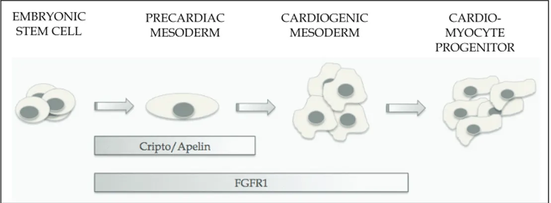
4. Conclusions
Although ESCs represent a viable source of specific cellular subtypes for drug discovery and transplantation, the successful use of ES-derived donor cells would require the generation of essentially pure cultures of specific cell types. Although further analysis will be required to get insight into the regulatory networks that involve Cripto/APJ and FGFR1 in cardiac differentiation (Figure 5), these membrane receptors may represent valuable targets for the generation of pure cardiomyocyte populations from ESCs.
PRECARDIAC MESODERM CARDIOGENIC MESODERM CARDIO-MYOCYTE PROGENITOR EMBRYONIC STEM CELL
Fig. 5. Cripto/Apelin and FGFR1 influence the early phases of cardiomyocyte differentiation
5. References
Abu-Issa, R. and M. L. Kirby (2007) Heart field: from mesoderm to heart tube. Annu Rev Cell
Dev Biol. 23, 45-68.
Abu-Issa, R., G. Smyth, I. Smoak, K. Yamamura and E. N. Meyers (2002) Fgf8 is required for pharyngeal arch and cardiovascular development in the mouse. Development. 129, 4613-25.
Adkins, H. B., C. Bianco, S. G. Schiffer, P. Rayhorn, M. Zafari, A. E. Cheung, O. Orozco, D. Olson, A. De Luca, L. L. Chen, K. Miatkowski, C. Benjamin, N. Normanno, K. P. Williams, M. Jarpe, D. LePage, D. Salomon and M. Sanicola (2003) Antibody blockade of the Cripto CFC domain suppresses tumor cell growth in vivo. J Clin
Invest. 112, 575-87.
Alsan, B. H. and T. M. Schultheiss (2002) Regulation of avian cardiogenesis by Fgf8 signaling. Development. 129, 1935-43.
Beenken, A. and M. Mohammadi (2009) The FGF family: biology, pathophysiology and therapy. Nat Rev Drug Discov. 8, 235-53.
Beiman, M., B. Z. Shilo and T. Volk (1996) Heartless, a Drosophila FGF receptor homolog, is essential for cell migration and establishment of several mesodermal lineages.
Genes Dev. 10, 2993-3002.
Boheler, K. R., J. Czyz, D. Tweedie, H. T. Yang, S. V. Anisimov and A. M. Wobus (2002) Differentiation of pluripotent embryonic stem cells into cardiomyocytes. Circ Res. 91, 189-201.
Bruneau, B. G., G. Nemer, J. P. Schmitt, F. Charron, L. Robitaille, S. Caron, D. A. Conner, M. Gessler, M. Nemer, C. E. Seidman and J. G. Seidman (2001) A murine model of Holt-Oram syndrome defines roles of the T-box transcription factor Tbx5 in cardiogenesis and disease. Cell. 106, 709-21.
Buckingham, M., S. Meilhac and S. Zaffran (2005) Building the mammalian heart from two sources of myocardial cells. Nat Rev Genet. 6, 826-35.
Cai, C. L., X. Liang, Y. Shi, P. H. Chu, S. L. Pfaff, J. Chen and S. Evans (2003) Isl1 identifies a cardiac progenitor population that proliferates prior to differentiation and contributes a majority of cells to the heart. Dev Cell. 5, 877-89.
Cai, C. L., J. C. Martin, Y. Sun, L. Cui, L. Wang, K. Ouyang, L. Yang, L. Bu, X. Liang, X. Zhang, W. B. Stallcup, C. P. Denton, A. McCulloch, J. Chen and S. M. Evans (2008) A myocardial lineage derives from Tbx18 epicardial cells. Nature. 454, 104-8. Cailliau, K., D. Perdereau, A. Lescuyer, H. Chen, C. Garbay, J. P. Vilain, A. F. Burnol and E.
Browaeys-Poly (2005) FGF receptor phosphotyrosine 766 is a target for Grb14 to inhibit MDA-MB-231 human breast cancer cell signaling. Anticancer Res. 25, 3877-82.
Chen, C., S. M. Ware, A. Sato, D. E. Houston-Hawkins, R. Habas, M. M. Matzuk, M. M. Shen and C. W. Brown (2006) The Vg1-related protein Gdf3 acts in a Nodal signaling pathway in the pre-gastrulation mouse embryo. Development. 133, 319-29.
Chen, K., H. Bai, M. Arzigian, Y. X. Chen, J. Bao, W. S. Wu, L. Wu and Z. Z. Wang (2010) Endothelial cells regulate cardiomyocyte development from embryonic stem cells. J
Cell Biochem. 111, 29-39.
Chen, K., L. Wu and Z. Z. Wang (2008) Extrinsic regulation of cardiomyocyte differentiation of embryonic stem cells. J Cell Biochem. 104, 119-28.
Chien, K. R., I. J. Domian and K. K. Parker (2008) Cardiogenesis and the complex biology of regenerative cardiovascular medicine. Science. 322, 1494-7.
Christoforou, N., R. A. Miller, C. M. Hill, C. C. Jie, A. S. McCallion and J. D. Gearhart (2008) Mouse ES cell-derived cardiac precursor cells are multipotent and facilitate identification of novel cardiac genes. J Clin Invest. 118, 894-903.
Christoforou, N., B. N. Oskouei, P. Esteso, C. M. Hill, J. M. Zimmet, W. Bian, N. Bursac, K.W. Leong, J. M. Hare and J. D. Gearhart (2010) Implantation of mouse embryonic stem cell-derived cardiac progenitor cells preserves function of infarcted murine hearts. PLoS One. 5, e11536.
Ciccodicola, A., R. Dono, S. Obici, A. Simeone, M. Zollo and M. G. Persico (1989) Molecular characterization of a gene of the 'EGF family' expressed in undifferentiated human NTERA2 teratocarcinoma cells. EMBO J. 8, 1987-91.
Ciruna, B. G., L. Schwartz, K. Harpal, T. P. Yamaguchi and J. Rossant (1997) Chimeric analysis of fibroblast growth factor receptor-1 (Fgfr1) function: a role for FGFR1 in morphogenetic movement through the primitive streak. Development. 124, 2829-41.
Cox, C. M., S. L. D'Agostino, M. K. Miller, R. L. Heimark and P. A. Krieg (2006) Apelin, the ligand for the endothelial G-protein-coupled receptor, APJ, is a potent angiogenic factor required for normal vascular development of the frog embryo. Dev Biol. 296, 177-89.
Cross, M. J., L. Lu, P. Magnusson, D. Nyqvist, K. Holmqvist, M. Welsh and L. Claesson-Welsh (2002) The Shb adaptor protein binds to tyrosine 766 in the FGFR-1 and regulates the Ras/MEK/MAPK pathway via FRS2 phosphorylation in endothelial cells. Mol Biol Cell. 13, 2881-93.
D'Aniello, C., E. Lonardo, S. Iaconis, O. Guardiola, A. M. Liguoro, G. L. Liguori, M. Autiero, P. Carmeliet and G. Minchiotti (2009) G protein-coupled receptor APJ and its ligand apelin act downstream of Cripto to specify embryonic stem cells toward the cardiac lineage through extracellular signal-regulated kinase/p70S6 kinase signaling pathway. Circ Res. 105, 231-8.
Davidson, B. (2007) Ciona intestinalis as a model for cardiac development. Semin Cell Dev
Biol. 18, 16-26.
Dell'Era, P., R. Ronca, L. Coco, S. Nicoli, M. Metra and M. Presta (2003) Fibroblast growth factor receptor-1 is essential for in vitro cardiomyocyte development. Circ Res. 93, 414-20.
Deng, C., M. Bedford, C. Li, X. Xu, X. Yang, J. Dunmore and P. Leder (1997) Fibroblast growth factor receptor-1 (FGFR-1) is essential for normal neural tube and limb development. Dev Biol. 185, 42-54.
Deng, C. X., A. Wynshaw-Boris, M. M. Shen, C. Daugherty, D. M. Ornitz and P. Leder (1994) Murine FGFR-1 is required for early postimplantation growth and axial organization. Genes Dev. 8, 3045-57.
Ding, J., L. Yang, Y. T. Yan, A. Chen, N. Desai, A. Wynshaw-Boris and M. M. Shen (1998) Cripto is required for correct orientation of the anterior-posterior axis in the mouse embryo. Nature. 395, 702-7.
Dionne, C. A., G. Crumley, F. Bellot, J. M. Kaplow, G. Searfoss, M. Ruta, W. H. Burgess, M. Jaye and J. Schlessinger (1990) Cloning and expression of two distinct high-affinity receptors cross-reacting with acidic and basic fibroblast growth factors. Embo J. 9, 2685-92.
Dono, R., L. Scalera, F. Pacifico, D. Acampora, M. G. Persico and A. Simeone (1993) The murine cripto gene: expression during mesoderm induction and early heart morphogenesis. Development. 118, 1157-68.
Evans, M. J. and M. H. Kaufman (1981) Establishment in culture of pluripotential cells from mouse embryos. Nature. 292, 154-6.
Favata, M. F., K. Y. Horiuchi, E. J. Manos, A. J. Daulerio, D. A. Stradley, W. S. Feeser, D. E. Van Dyk, W. J. Pitts, R. A. Earl, F. Hobbs, R. A. Copeland, R. L. Magolda, P. A. Scherle and J. M. Trzaskos (1998) Identification of a novel inhibitor of mitogen-activated protein kinase kinase. J Biol Chem. 273, 18623-32.
Fijnvandraat, A. C., A. C. van Ginneken, P. A. de Boer, J. M. Ruijter, V. M. Christoffels, A. F. Moorman and R. H. Lekanne Deprez (2003) Cardiomyocytes derived from embryonic stem cells resemble cardiomyocytes of the embryonic heart tube.
Cardiovasc Res. 58, 399-409.
Foley, A. and M. Mercola (2004) Heart induction: embryology to cardiomyocyte regeneration. Trends Cardiovasc Med. 14, 121-5.
Frasch, M. (1995) Induction of visceral and cardiac mesoderm by ectodermal Dpp in the early Drosophila embryo. Nature. 374, 464-7.
Gisselbrecht, S., J. B. Skeath, C. Q. Doe and A. M. Michelson (1996) heartless encodes a fibroblast growth factor receptor (DFR1/DFGF-R2) involved in the directional migration of early mesodermal cells in the Drosophila embryo. Genes Dev. 10, 3003-17.
Gray, P. C., C. A. Harrison and W. Vale (2003) Cripto forms a complex with activin and type II activin receptors and can block activin signaling. Proc Natl Acad Sci U S A. 100, 5193-8.
Hadari, Y. R., H. Kouhara, I. Lax and J. Schlessinger (1998) Binding of Shp2 tyrosine phosphatase to FRS2 is essential for fibroblast growth factor-induced PC12 cell differentiation. Mol Cell Biol. 18, 3966-73.
Harrison, C. A., P. C. Gray, W. W. Vale and D. M. Robertson (2005) Antagonists of activin signaling: mechanisms and potential biological applications. Trends Endocrinol
Metab. 16, 73-8.
Harvey, R. P. (2002) Patterning the vertebrate heart. Nat Rev Genet. 3, 544-56.
Hug, H. and T. F. Sarre (1993) Protein kinase C isoenzymes: divergence in signal transduction? Biochem J. 291, 329-43.
Inui, M., A. Fukui, Y. Ito and M. Asashima (2006) Xapelin and Xmsr are required for cardiovascular development in Xenopus laevis. Dev Biol. 298, 188-200.
Itoh, N. and D. M. Ornitz (2004) Evolution of the Fgf and Fgfr gene families. Trends Genet. 20, 563-9.
Johnson, S. E., J. L. Rothstein and B. B. Knowles (1994) Expression of epidermal growth factor family gene members in early mouse development. Dev Dyn. 201, 216-26. Kattman, S. J., T. L. Huber and G. M. Keller (2006) Multipotent flk-1+ cardiovascular
progenitor cells give rise to the cardiomyocyte, endothelial, and vascular smooth muscle lineages. Dev Cell. 11, 723-32.
Kawamata, Y., Y. Habata, S. Fukusumi, M. Hosoya, R. Fujii, S. Hinuma, N. Nishizawa, C. Kitada, H. Onda, O. Nishimura and M. Fujino (2001) Molecular properties of apelin: tissue distribution and receptor binding. Biochim Biophys Acta. 1538, 162-71. Kelly, R. G., N. A. Brown and M. E. Buckingham (2001) The arterial pole of the mouse heart
forms from Fgf10-expressing cells in pharyngeal mesoderm. Dev Cell. 1, 435-40. Kirby, M. L. (2007) Cardiac development, Oxford University Press, Oxford ; New York
Kitamura, R., T. Takahashi, N. Nakajima, K. Isodono, S. Asada, H. Ueno, T. Ueyama, T. Yoshikawa, H. Matsubara and H. Oh (2007) Stage-specific role of endogenous Smad2 activation in cardiomyogenesis of embryonic stem cells. Circ Res. 101, 78-87. Kleinz, M. J. and A. P. Davenport (2005) Emerging roles of apelin in biology and medicine.
Pharmacol Ther. 107, 198-211.
Kouhara, H., Y. R. Hadari, T. Spivak-Kroizman, J. Schilling, D. Bar-Sagi, I. Lax and J. Schlessinger (1997) A lipid-anchored Grb2-binding protein that links FGF-receptor activation to the Ras/MAPK signaling pathway. Cell. 89, 693-702.
Kuchera, S., H. Barth, P. Jacobson, A. Metz, C. Schaechtele and D. Schrier (1993) Anti-inflammatory properties of the protein kinase C inhibitor, 3-[1-[3-(dimethylamino)propyl]-1H-indol-3-yl]-4-(1H-indol-3-yl)-1H- pyrrole-2,5-dione monohydrochloride (GF109203X) in the PMA-mouse ear edema model. Agents
Ladd, A. N., T. A. Yatskievych and P. B. Antin (1998) Regulation of avian cardiac myogenesis by activin/TGFbeta and bone morphogenetic proteins. Dev Biol. 204, 407-19.
Larsson, H., P. Klint, E. Landgren and L. Claesson-Welsh (1999) Fibroblast growth factor receptor-1-mediated endothelial cell proliferation is dependent on the Src homology (SH) 2/SH3 domain-containing adaptor protein Crk. J Biol Chem. 274, 25726-34.
Liguori, G. L., D. Echevarria, R. Improta, M. Signore, E. Adamson, S. Martinez and M. G. Persico (2003) Anterior neural plate regionalization in cripto null mutant mouse embryos in the absence of node and primitive streak. Dev Biol. 264, 537-49.
Magnusson, P., C. Rolny, L. Jakobsson, C. Wikner, Y. Wu, D. J. Hicklin and L. Claesson-Welsh (2004) Deregulation of Flk-1/vascular endothelial growth factor receptor-2 in fibroblast growth factor receptor-1-deficient vascular stem cell development. J
Cell Sci. 117, 1513-23.
Magnusson, P. U., R. Ronca, P. Dell'Era, P. Carlstedt, L. Jakobsson, J. Partanen, A. Dimberg and L. Claesson-Welsh (2005) Fibroblast growth factor receptor-1 expression is required for hematopoietic but not endothelial cell development. Arterioscler
Thromb Vasc Biol. 25, 944-9.
Marques, S. R., Y. Lee, K. D. Poss and D. Yelon (2008) Reiterative roles for FGF signaling in the establishment of size and proportion of the zebrafish heart. Dev Biol. 321, 397-406.
Martin, G. R. (1981) Isolation of a pluripotent cell line from early mouse embryos cultured in medium conditioned by teratocarcinoma stem cells. Proc Natl Acad Sci U S A. 78, 7634-8.
Masri, B., H. Lahlou, H. Mazarguil, B. Knibiehler and Y. Audigier (2002) Apelin (65-77) activates extracellular signal-regulated kinases via a PTX-sensitive G protein.
Biochem Biophys Res Commun. 290, 539-45.
Masri, B., N. Morin, M. Cornu, B. Knibiehler and Y. Audigier (2004) Apelin (65-77) activates p70 S6 kinase and is mitogenic for umbilical endothelial cells. FASEB J. 18, 1909-11. Massague, J. and Y. G. Chen (2000) Controlling TGF-beta signaling. Genes Dev. 14, 627-44. Medhurst, A. D., C. A. Jennings, M. J. Robbins, R. P. Davis, C. Ellis, K. Y. Winborn, K. W.
Lawrie, G. Hervieu, G. Riley, J. E. Bolaky, N. C. Herrity, P. Murdock and J. G. Darker (2003) Pharmacological and immunohistochemical characterization of the APJ receptor and its endogenous ligand apelin. J Neurochem. 84, 1162-72.
Minchiotti, G., S. Parisi, G. Liguori, M. Signore, G. Lania, E. D. Adamson, C. T. Lago and M. G. Persico (2000) Membrane-anchorage of Cripto protein by glycosylphosphatidylinositol and its distribution during early mouse development.
Mech Dev. 90, 133-42.
Mohammadi, M., I. Dikic, A. Sorokin, W. H. Burgess, M. Jaye and J. Schlessinger (1996) Identification of six novel autophosphorylation sites on fibroblast growth factor receptor 1 and elucidation of their importance in receptor activation and signal transduction. Mol Cell Biol. 16, 977-89.
Mohammadi, M., A. M. Honegger, D. Rotin, R. Fischer, F. Bellot, W. Li, C. A. Dionne, M. Jaye, M. Rubinstein and J. Schlessinger (1991) A tyrosine-phosphorylated carboxy-terminal peptide of the fibroblast growth factor receptor (Flg) is a binding site for the SH2 domain of phospholipase C-gamma 1. Mol Cell Biol. 11, 5068-78.
Mohammadi, M., G. McMahon, L. Sun, C. Tang, P. Hirth, B. K. Yeh, S. R. Hubbard and J. Schlessinger (1997) Structures of the tyrosine kinase domain of fibroblast growth factor receptor in complex with inhibitors. Science. 276, 955-60.
Monzen, K., Y. Hiroi, S. Kudoh, H. Akazawa, T. Oka, E. Takimoto, D. Hayashi, T. Hosoda, M. Kawabata, K. Miyazono, S. Ishii, Y. Yazaki, R. Nagai and I. Komuro (2001) Smads, TAK1, and their common target ATF-2 play a critical role in cardiomyocyte differentiation. J Cell Biol. 153, 687-98.
Moretti, A., L. Caron, A. Nakano, J. T. Lam, A. Bernshausen, Y. Chen, Y. Qyang, L. Bu, M. Sasaki, S. Martin-Puig, Y. Sun, S. M. Evans, K. L. Laugwitz and K. R. Chien (2006) Multipotent embryonic isl1+ progenitor cells lead to cardiac, smooth muscle, and endothelial cell diversification. Cell. 127, 1151-65.
Nishikawa, S., L. M. Jakt and T. Era (2007) Embryonic stem-cell culture as a tool for developmental cell biology. Nat Rev Mol Cell Biol. 8, 502-7.
O'Dowd, B. F., M. Heiber, A. Chan, H. H. Heng, L. C. Tsui, J. L. Kennedy, X. Shi, A. Petronis, S. R. George and T. Nguyen (1993) A human gene that shows identity with the gene encoding the angiotensin receptor is located on chromosome 11. Gene. 136, 355-60. Olsen, S. K., O. A. Ibrahimi, A. Raucci, F. Zhang, A. V. Eliseenkova, A. Yayon, C. Basilico, R.
J. Linhardt, J. Schlessinger and M. Mohammadi (2004) Insights into the molecular basis for fibroblast growth factor receptor autoinhibition and ligand-binding promiscuity. Proc Natl Acad Sci U S A. 101, 935-40.
Ornitz, D. M., J. Xu, J. S. Colvin, D. G. McEwen, C. A. MacArthur, F. Coulier, G. Gao and M. Goldfarb (1996) Receptor specificity of the fibroblast growth factor family. J Biol
Chem. 271, 15292-7.
Parisi, S., D. D'Andrea, C. T. Lago, E. D. Adamson, M. G. Persico and G. Minchiotti (2003) Nodal-dependent Cripto signaling promotes cardiomyogenesis and redirects the neural fate of embryonic stem cells. J Cell Biol. 163, 303-14.
Pennisi, D. J. and T. Mikawa (2009) FGFR-1 is required by epicardium-derived cells for myocardial invasion and correct coronary vascular lineage differentiation. Dev Biol. 328, 148-59.
Persico, M. G., G. L. Liguori, S. Parisi, D. D'Andrea, D. S. Salomon and G. Minchiotti (2001) Cripto in tumors and embryo development. Biochim Biophys Acta. 1552, 87-93. Piccolo, S., E. Agius, L. Leyns, S. Bhattacharyya, H. Grunz, T. Bouwmeester and E. M. De
Robertis (1999) The head inducer Cerberus is a multifunctional antagonist of Nodal, BMP and Wnt signals. Nature. 397, 707-10.
Plotnikov, A. N., S. R. Hubbard, J. Schlessinger and M. Mohammadi (2000) Crystal structures of two FGF-FGFR complexes reveal the determinants of ligand-receptor specificity. Cell. 101, 413-424.
Powers, C. J., S. W. McLeskey and A. Wellstein (2000) Fibroblast growth factors, their receptors and signaling. Endocr Relat Cancer. 7, 165-97.
Presta, M., P. Dell'Era, S. Mitola, E. Moroni, R. Ronca and M. Rusnati (2005) Fibroblast growth factor/fibroblast growth factor receptor system in angiogenesis. Cytokine
Growth Factor Rev. 16, 159-78.
Puceat, M., P. Travo, M. T. Quinn and P. Fort (2003) A dual role of the GTPase Rac in cardiac differentiation of stem cells. Mol Biol Cell. 14, 2781-92.
Qu, C. K. and G. S. Feng (1998) Shp-2 has a positive regulatory role in ES cell differentiation and proliferation. Oncogene. 17, 433-9.
Reifers, F., E. C. Walsh, S. Leger, D. Y. Stainier and M. Brand (2000) Induction and differentiation of the zebrafish heart requires fibroblast growth factor 8 (fgf8/acerebellar). Development. 127, 225-35.
Reissmann, E., H. Jornvall, A. Blokzijl, O. Andersson, C. Chang, G. Minchiotti, M. G. Persico, C. F. Ibanez and A. H. Brivanlou (2001) The orphan receptor ALK7 and the Activin receptor ALK4 mediate signaling by Nodal proteins during vertebrate development. Genes Dev. 15, 2010-22.
Romito, A., E. Lonardo, G. Roma, G. Minchiotti, A. Ballabio and G. Cobellis (2010) Lack of sik1 in mouse embryonic stem cells impairs cardiomyogenesis by down-regulating the cyclin-dependent kinase inhibitor p57kip2. PLoS One. 5, e9029.
Ronca, R., L. Gualandi, E. Crescini, S. Calza, M. Presta and P. Dell'Era (2009) Fibroblast growth factor receptor-1 phosphorylation requirement for cardiomyocyte differentiation in murine embryonic stem cells. J Cell Mol Med. 13, 1489-98.
Sachinidis, A., B. K. Fleischmann, E. Kolossov, M. Wartenberg, H. Sauer and J. Hescheler (2003) Cardiac specific differentiation of mouse embryonic stem cells. Cardiovasc
Res. 58, 278-91.
Salomon, D. S., C. Bianco and M. De Santis (1999) Cripto: a novel epidermal growth factor (EGF)-related peptide in mammary gland development and neoplasia. Bioessays. 21, 61-70.
Sato, T., K. Hidaka, A. Iwanaga, M. Ito, M. Asano, Y. Nakabeppu, T. Morisaki and K. Yoshioka (2005) Impairment of cardiomyogenesis in embryonic stem cells lacking scaffold protein JSAP1. Biochem Biophys Res Commun. 338, 1152-7.
Scott, I. C., B. Masri, L. A. D'Amico, S. W. Jin, B. Jungblut, A. M. Wehman, H. Baier, Y. Audigier and D. Y. Stainier (2007) The g protein-coupled receptor agtrl1b regulates early development of myocardial progenitors. Dev Cell. 12, 403-13.
Seyed, M. and J. X. Dimario (2008) Fibroblast growth factor receptor 1 gene expression is required for cardiomyocyte proliferation and is repressed by Sp3. J Mol Cell Cardiol. 44, 510-9.
Shen, M. M. and A. F. Schier (2000) The EGF-CFC gene family in vertebrate development.
Trends Genet. 16, 303-9.
Solloway, M. J. and R. P. Harvey (2003) Molecular pathways in myocardial development: a stem cell perspective. Cardiovasc Res. 58, 264-77.
Strizzi, L., C. Bianco, N. Normanno and D. Salomon (2005) Cripto-1: a multifunctional modulator during embryogenesis and oncogenesis. Oncogene. 24, 5731-41.
Tanaka, C., R. Sakuma, T. Nakamura, H. Hamada and Y. Saijoh (2007) Long-range action of Nodal requires interaction with GDF1. Genes Dev. 21, 3272-82.
Tatemoto, K., M. Hosoya, Y. Habata, R. Fujii, T. Kakegawa, M. X. Zou, Y. Kawamata, S. Fukusumi, S. Hinuma, C. Kitada, T. Kurokawa, H. Onda and M. Fujino (1998) Isolation and characterization of a novel endogenous peptide ligand for the human APJ receptor. Biochem Biophys Res Commun. 251, 471-6.
Wang, F., M. Kan, G. Yan, J. Xu and W. L. McKeehan (1995) Alternately spliced NH2-terminal immunoglobulin-like Loop I in the ectodomain of the fibroblast growth factor (FGF) receptor 1 lowers affinity for both heparin and FGF-1. Journal of
Wang, Z., K. Cohen, Y. Shao, P. Mole, D. Dombkowski and D. T. Scadden (2004) Ephrin receptor, EphB4, regulates ES cell differentiation of primitive mammalian hemangioblasts, blood, cardiomyocytes, and blood vessels. Blood. 103, 100-9.
Wu, S. M., Y. Fujiwara, S. M. Cibulsky, D. E. Clapham, C. L. Lien, T. M. Schultheiss and S. H. Orkin (2006) Developmental origin of a bipotential myocardial and smooth muscle cell precursor in the mammalian heart. Cell. 127, 1137-50.
Xu, C., G. Liguori, E. D. Adamson and M. G. Persico (1998) Specific arrest of cardiogenesis in cultured embryonic stem cells lacking Cripto-1. Dev Biol. 196, 237-47.
Xu, C., G. Liguori, M. G. Persico and E. D. Adamson (1999a) Abrogation of the Cripto gene in mouse leads to failure of postgastrulation morphogenesis and lack of differentiation of cardiomyocytes. Development. 126, 483-94.
Xu, X., C. Li, K. Takahashi, H. C. Slavkin, L. Shum and C. X. Deng (1999b) Murine fibroblast growth factor receptor 1alpha isoforms mediate node regression and are essential for posterior mesoderm development. Dev Biol. 208, 293-306.
Yamaguchi, T. P., K. Harpal, M. Henkemeyer and J. Rossant (1994) fgfr-1 is required for embryonic growth and mesodermal patterning during mouse gastrulation. Genes
Dev. 8, 3032-44.
Zaffran, S. and M. Frasch (2002) Early signals in cardiac development. Circ Res. 91, 457-69. Zaffran, S., R. G. Kelly, S. M. Meilhac, M. E. Buckingham and N. A. Brown (2004) Right
ventricular myocardium derives from the anterior heart field. Circ Res. 95, 261-8. Zeng, X. X., T. P. Wilm, D. S. Sepich and L. Solnica-Krezel (2007) Apelin and its receptor
control heart field formation during zebrafish gastrulation. Dev Cell. 12, 391-402. Zhou, B., Q. Ma, S. Rajagopal, S. M. Wu, I. Domian, J. Rivera-Feliciano, D. Jiang, A. von Gise,
S. Ikeda, K. R. Chien and W. T. Pu (2008) Epicardial progenitors contribute to the cardiomyocyte lineage in the developing heart. Nature. 454, 109-13.
Zhou, X., E. Quann and G. I. Gallicano (2003) Differentiation of nonbeating embryonic stem cells into beating cardiomyocytes is dependent on downregulation of PKC beta and zeta in concert with upregulation of PKC epsilon. Dev Biol. 255, 407-22.