U
NIVERSITÀ DEGLI
S
TUDI DI
S
ALERNO
Dipartimento di Medicina, Chirurgia ed Odontoiatria
Dottorato di Ricerca in Medicina Traslazionale dello
Sviluppo e dell’Invecchiamento Attivo XXXI ciclo
Curriculum Medicina Traslazionale e Clinica
Tesi di dottorato
DEVELOPMENT AND VALIDATION OF ANALYTICAL
METHODS FOR THERAPEUTIC DRUG MONITORING
Coordinatore:
Ch.mo Prof. Palmiero Monteleone
Relatore: Prof.ssa Viviana Izzo
Candidato/a:
Marine Pingeon
Matricola:8800900012
Summary
1.1 Chapter I. Introduction. Going toward Personalized Medicine: the need of therapeutic drug
monitoring ... 1
1.2 Variability in drug response ... 3
1.2.1 Biomarkers ... 3
1.2.2 Pharmacogenomics and pharmacogenetics ... 4
1.2.3 Pharmacokinetics and Pharmacodynamics ... 5
1.2.4 Pharmacokinetic and Pharmacodynamic variability ... 7
1.3 Therapeutic Drug Monitoring... 12
1.3.1 Analytical Methods in Therapeutic Drug Monitoring ... 13
2.1 Chapter II. Hydroxychloroquine monitoring in the treatment of patients with Systemic Lupus Erythematosus. Introduction... 16
2.1.1 Systemic Lupus Erythematosus ... 16
2.1.2 Genetics and epidemiology ... 16
2.1.3 Pathophysiology ... 17
2.1.4 Clinical manifestations ... 18
2.1.5 Clinical management ... 19
2.1.6 Hydroxychloroquine ... 19
2.1.7 Project aim ... 21
2.2 Materials and methods ... 22
2.2.1 Patients ... 22
2.2.2 Chemicals and reagents ... 23
2.2.3 Sample preparation ... 23
2.2.4 Preparation of standard calibration curves and quality control samples ... 24
2.2.5 Chromatography ... 24
2.2.6 Method validation ... 25
2.2.7 Mass spectrometry ... 26
2.2.8 Statistical analysis ... 26
2.3 Results and Discussion ... 27
2.3.1 Sample preparation and extraction ... 27
2.3.2 Optimization of HPLC-FL analysis... 27
2.3.3 Method validation... 29
2.3.4 Comparison between HPLC-FL and LC-MS/MS on a subset of clinical samples... 32
2.3.5. Relationship between blood concentration of HCQ and QoL in SLE patients with inactive disease 34 2.4 Discussion ... 37
3.1 Chapter III. Ruxolitinib monitoring in the treatment of patients with Myeloproliferative Disorders. Introduction ... 41
3.1.1 Definitions ... 41
3.1.3 Polycythemia Vera ... 43
3.1.4 JAK-STAT signaling pathway ... 43
3.1.5 Ruxolitinib ... 44
3.1.6 Project aim ... 45
3.2 Materials and methods... 46
3.2.1. Chemicals and reagents ... 46
3.2.2 Chromatography ... 46
3.2.3 Sample preparation ... 46
3.2.4 Detection and quantification limits ... 47
3.2.5 Linearity and recovery ... 47
3.2.6 Intra- and inter-day variability ... 48
3.2.7 Stock solutions and sample stability... 48
3.2.8 Method robustness ... 48
3.3 Results and discussion ... 49
3.3.1 Optimization of chromatographic conditions ... 49
3.3.2 Pre-analytical treatment ... 50
3.3.3 Method validation... 50
3.4 Conclusions ... 54
4.1 Chapter IV. 5-Fluorouracil monitoring of oncologic patients in course of chemotherapy treatment. Introduction ... 56
4.1.1 TDM applied to chemotherapeutic agents ... 56
4.1.2 5-Fluorouracil ... 56
4.1.3 -Therapeutic drug monitoring of 5-Fluorouracil ... 58
4.1.4 Project aim ... 58
4.2 Materials and methods... 60
4.2.1 Patients ... 60
4.2.2 Chemicals and reagents ... 60
4.2.3 Solution preparation ... 60
4.2.4 5-FU extraction... 60
4.2.5 Preparation of calibration standards and quality controls ... 61
4.2.6 LC-MS/MS analysis ... 61
4.2.7 Method validation... 62
4.3 Results and discussion ... 63
5.1. Chapter V. Neonatal monitoring of phenobarbital and caffeine. Introduction ... 71
5.1.1. Neonatal TDM ... 71
5.1.2. Phenobarbital ... 73
5.1.3. Caffeine ... 74
5.1.4. Project aim ... 75
5.2.2. Chemicals and reagents ... 76
5.2.3. Patients ... 76
5.2.4. Preparation of stock and working solutions ... 76
5.2.5. LC-MS/MS conditions ... 76
5.2.6. Samples preparation ... 78
5.2.7. Method validation ... 78
5.2.8. Comparison between DBS and plasma ... 80
5.3. Results and discussion ... 81
6. Chapter VI. Conclusions……… 89
“Le bon sens de la vie humaine nous montre que la vie humaine est courte, et qu'il faut mieux faire de notre court passage sur terre quelque chose d'utile
pour soi et pour les autres.”
“Il buon senso ci indica che la vita umana è breve, e che è meglio trasformare il nostro fugace passaggio sulla Terra in qualcosa di utile, per noi e per
gli altri".
Abstract
The U.S. Food and Drug Administration defines as Precision or Personalized Medicine (PM) an innovative therapeutic approach that tailors therapy and prevention on patients based on inter-individual variabilities in molecular or environmental features and in lifestyles. The major goals of PM are to maximize treatment efficacy and to reduce cost, toxicities and therapy failure rates by early identification of patients who might benefit or not of a specific treatment. In this scenario, therapeutic drug monitoring (TDM) is an important laboratory tool for PM because of the possibility to measure several drugs and bioactive molecules in human biological matrices. TDM is based on the hypothesis that in the majority of drugs, there is a relationship between administered dose and circulating concentration of unbound fraction - and between this concentration and observed pharmacological effects. TDM is recommended for drugs with significant inter-individual pharmacokinetic variability and an established relationship between blood concentrations and clinical efficacy and/or toxicity. Moreover, TDM is also advisable in special populations such as pregnant women and children. To date, liquid chromatography and immunometric assay are still considered the standard for molecule measurement in biological fluids; however, in recent years, LC tandem mass spectrometry (LC-MS) is gaining popularity because of the possibility of in-depth and multiplexed analysis with high selectivity and specificity.
During this Ph.D. program, we developed several high performance LC (HPLC)- and LC-MS/MS-based approaches for TDM of different drugs measured in various types of body fluids and validated according to EMA and FDA guidelines. In particular, we focused on:
1) TDM of hydroxychloroquine (HCQ) blood concentration, a drug with a wide therapeutic window. Our method was validated on a cohort of patients with Systemic Lupus Erythematosus treated with HCQ and blood concentrations were correlated to several clinical parameters, such quality of life. Moreover, TDM of HCQ was also used to monitor treatment adherence in those subjects.
2) TDM of a commonly used chemotherapeutic agent, the 5-fluorouracil (5-FU), which is known to have a narrow therapeutic window and a high toxicity
3) TDM of a new kinase inhibitor, Ruxolitinib, approved for the treatment of myeloproliferative hematologic disorders.
4) TDM of several drugs, such as caffeine and phenobarbital, in newborns who are at particular risk of uncorrected drug dosage. Due to the need to carry out analyses on very-small volume samples, we validated an analytical method using micro-sampling techniques such as the dried blood spot (DBS) sampling combined with LC-MS/MS analysis.
Riassunto
La U.S. Food and Drug Administration definisce con il termine Medicina di Precisione o Personalizzata (MP) un approccio terapeutico innovativo che identifica strategie terapeutiche e di prevenzione sul singolo paziente in base alla presenza di variazioni interindividuali in fattori biologici, ambientali e dello stile di vita. Gli obiettivi principali della MP sono quelli di aumentare l’efficacia terapeutica e di ridurre i costi, le tossicità e il tasso di fallimento terapeutico individuando precocemente i pazienti che possono beneficiare o meno di una specifica terapia. Il monitoraggio terapeutico dei farmaci (TDM) è quindi un importante strumento diagnostico per la MP perché permette di misurare farmaci e molecole bioattive in varie matrici biologiche. Il TDM si basa sull’ipotesi che per la maggior parte dei farmaci esiste un rapporto diretto tra dose somministrata e concentrazione circolante – e tra queste e l’effetto terapeutico. Per questo motivo, il TDM è raccomandato in tutti quei casi in cui sussiste una significativa variabilità farmacocinetica interindividuale ed una relazione nota tra concentrazioni plasmatiche ed effetto terapeutico o tossicità. Inoltre, il TDM è indicato anche in particolari popolazioni come le donne in gravidanza e i bambini. Ad oggi, la cromatografia liquida (LC) e le tecniche immunometriche sono ancora considerate lo standard per il dosaggio di molecole in fluidi biologici; tuttavia, negli ultimi anni, la LC accoppiata alla spettrometria di massa (LC-MS) sta rapidamente affiancando -e in alcuni casi soppiantando- le altre due tecniche grazie alla possibilità di effettuare analisi approfondite su numerose molecole in contemporanea, con alta selettività e specificità.
Nell’ambito del presente progetto di dottorato, sono stati sviluppati numerosi metodi analitici con analisi in high performance LC (HPLC)- o LC-MS/MS per il TDM di varie molecole in fluidi biologici e validati seguendo le linee guida della EMA e della FDA. In particolare, l’attenzione è stata focalizzata su:
1) TDM dell’idrossiclorochina (HCQ), un farmaco con un’ampia finestra terapeutica. Il metodo sviluppato è stato validato su una coorte di pazienti affetta da Lupus Eritematoso Sistemico in trattamento con HCQ e le concentrazioni plasmatiche sono state correlate con vari parametri clinici e con la qualità della vita. Inoltre, il TDM dell’HCQ è stato anche utilizzato per monitorare l’aderenza al trattamento di tali soggetti.
2) TDM di un chemioterapeutico largamente utilizzato quale il 5-fluorouracile, caratterizzato da una stretta finestra terapeutica e da grave tossicità.
3) TDM di un nuovo inibitore di chinasi, il Ruxolitinib, approvato per il trattamento di malattie ematologiche mieloproliferative.
4) TDM di varie molecole quali caffeina e fenobarbital in neonati che sono particolarmente a rischio di dosaggi terapeutici non corretti. In particolare, data la necessità di lavorare su
piccoli volumi, è stato messo a punto il TDM con metodica di micro-campionamento quale il dried blood spot accoppiato ad analisi in LC-MS/MS.
Chapter I
1 1.1 Going toward Personalized Medicine: the need of therapeutic drug monitoring
Genetic and epi-genetic differences between individuals may induce significant variability in drug response [1]. Consequently, a therapeutic treatment based on the “one-size-fits-all” concept, which ignores the widely inter-individual variation that exists in response to medication, can be ineffective or even damaging. Nowadays medicine strives towards personalized therapy and “personalized medicine” (PM), which stands for therapy adaptation to the molecular characteristics of patient and its disease (Figure 1) [2].
PM challenges are to identify and classify subgroups of patients who differ in their susceptibility to a particular disease or their response to a specific treatment. Preventing or therapeutic intervention can then be foreseen for those who will actually benefit, sparing expense and side effects for those who will not. [3]. PM definition, commonly accepted and taken over by the European Medicine Agency (EMA), consists in administrating “the right dose of the right drug to the right person at the right time” [4]. From Ayurvedic medicine to Hippocrates, the concept of PM has long been known [5-7].
Even if the PM idea is not new, its current application is quite recent. Since 1990s technical advances are part of a historical evolution of biology and biomedicine, leading to a new turning point in personalized medicine. Today, -omics technologies offer an access to different molecular profiles and disturbances of cellular signaling pathways which seeks to understand all the molecular underpinnings of drug response or posology [1,2]. PM may thus be considered as an extension of traditional approaches to understand and treat diseases, but with a greater precision [8]. Several other important aspects, such as the decreasing rate of new medical product development and the increasing of healthcare costs generated by both aging population who lives longer and the increasing hospitalization due to adverse drug reactions (ADRs), encouraged manufacturers, authority and government to invest on PM.
ADRs are an important cause of morbidity and mortality worldwide, with a significant and increasing health and economic burden. Several meta-analysis studies conducted in the USA and in European countries found that the frequency of serious ADRs leading to hospitalization ranged, respectively, from 1.0 % to 16.8% and from 0.5% to 12.8%. Among hospitalized patients, the global incidence of serious ADRs was 6.7% and that of fatal ADRs was of 0.32% [9,10]. From 1999 to 2006, national USA Vital Statistics System data have shown that the rate of ADR-related deaths increased from 0.08 to 0.12 per 100,000 persons [11]. In the UK, an historical study on 20,000 hospitalized patients found that ADRs resulted in an average of eight days of hospital stay and were
associated with approximately € 706 million per year, including ADRs judged potentially avoidable. [12].
Another problem that healthcare system faces is the limited effectiveness of numerous treatments, especially those regarding chronic diseases. A substantial inter-variability to drug response in oncology field as well as for common conditions like hypertension, heart failure, depression, hypercholesterolemia and asthma, has indeed been observed [13,14]. As a result, the ability to rapidly discriminate between patients who will benefit from a given treatment and those who are at risk of significant adverse events could result in significant cost savings for the entire health system.
In this scenario, therapeutic drug monitoring would be a cheap and widely-applicable armored PM arm because, by using common simple instrumentations, such as liquid chromatography, or more complex apparatus, such as mass spectrometers, clinicians can rapidly adjust theoretical drug doses
Figure 1. Personalized medicine. Personalized medicine promises to transform the delivery of healthcare to patients.
Its aim is to evolve from a reactive “one-size-fits-all” system towards a system of predictive, preventive, and precision care. This picture depicts how personalized medicine could classify people into smaller subsets based on the therapy response from one large disease group. Genetic tests can help stratify patients in those who would respond effectively to a specific drug and those who would experience an ADR.
3
in single patients in order to maintain an effective and safe serum/plasma trough concentration by maximizing efficacy while minimizing ADR incidence.
The need of PM tailored on each patient is of growing interest in scientific and non-scientific communities: the creation of several large-scale international projects such as the "Personalized Medicine Coalition", the "Genomics and Personalized Medicine Act" as well as ministerial commissions throughout Europe, demonstrate the growing interest of physicians, researchers, industry and government for this topic. However, we are still far from a real PM as many different fields need to deeply collaborate in order to combine epidemiology, genomics, molecular biology, proteomics, and metabolomics fingerprints which all might affect drug response in patients.
1.2 Variability in drug response
1.2.1 Biomarkers
The individual’s response to a drug depends on the complex interplay between environmental and genetic factors. There are multiple contributory factors which play a role in the drug response variability such as gender, age, diet, body mass, health status, epigenetic factors, concurrent therapy or environmental exposure to certain chemicals or toxins (e.g. cigarette smoke) [1,15]. To identify the ideal treatment and to adapt it to each patient it is therefore necessary to use specific tools to measure accurately and reproducibly the clinical manifestations of patient's health status. It was in 1998 that the word biomarker was defined for the first time by the National Institute of Health as “a characteristic that is objectively measured and evaluated as an indicator of normal biological processes, pathogenic processes, or pharmacologic responses to a therapeutic intervention.” Even if biomarker use has been known for long time, recent advances in new high-speed technologies as genomic platforms, high-performance biochips or new generation of mass spectrometry, increase possibilities to identify new biomarkers for precocious diagnosis, prognosis and therapeutic monitoring [16].
Biomarkers are physiological, biochemical or molecular parameters that can be detected in tissue or biological fluids such as blood, urine, etc. Today, biomarkers represent a vast field that covers different areas of application ranging from pharmaceutical development to clinical medicine. [17,18]. They can be classified into three categories: prognostic, used to predict the natural course of a confirmed disease; predictive, used to predict the response of patient to a drug; pharmacodynamic, used to evaluate efficacy or toxicity to treatment and to modulate drug dosing. Predictive biomarkers are mainly used in hematology and oncology and consider both conventional environmental factors and dose selection. Indeed, immunohistochemistry tests such as Her2 protein in breast cancer or
pharmacogenomics tests as the dihydropyrimidine dehydrogenase (DPYD) test for 5-fluorouracil make it possible to identify patients with a pharmacokinetic/pharmacodynamic (PK/PD) risk such as ADRs or inefficient treatment [19].
1.2.2 Pharmacogenomics and pharmacogenetics
Genetic variations can affect how the body responds to certain medications [20]. Two main types of inter-individual variations have been found in the genome i.e. variable number tandem repeats (VNTRs) which occur in the non-coding DNA and is used for paternity testing, forensic science or DNA fingerprinting and single nucleotide polymorphisms (SNPs), which are the most important source of genome variation. These latter contribute for 0.1% of differences retrieved in gene sequences and are at the origin of interindividual variabilities; SNPs can lead to protein modifications that may be responsible of alteration of their stability, cell concentrations, metabolic activity or signaling properties [21].
Pharmacogenetics takes its first steps in the ‘50s and brings genetics, biochemistry, and pharmacology together. The first person to have mentioned that genetic variants can modulate drug action is the physiologist Garrod. Thereafter, a tide of reports suggested that ADRs could be genetically determined variations in enzyme activity. For example, hemolysis caused by antimalarials was recognized as being caused by inherited variants of glucose-6-phosphate dehydrogenase. Similarly, inherited changes in DPYD gene leading to low enzyme activity produce a greater bioavailability of the antimetabolite 5-fluorouracile, thus causing serious hematological, neurological and gastrointestinal toxicities. However, it was only after 1959 that F. Vogel proposed the term pharmacogenetics, which is defined as the study of the influence of genome variability in drug response [22]. Pharmacogenetics is aimed at determining the genetic differences in metabolic pathways that can affect individual responses to drugs [23]. In pharmacogenetics, the analysis of a specific gene, or group of genes, can be used to predict responses to a specific drug or class of drugs. However, rarely a single gene directly controls drug response or ADRs and advances in pharmacogenetics have remained limited. Emergence and development of Human Genome Project and genome science in 1990s [24] have then spawned a newer field, named pharmacogenomics [25]. The main aim of pharmacogenomics is to understand all of the molecular underpinnings of drug response. Pharmacogenomics carries the idea that variable drug response may reflect sets of variants within an individual or across a population. Pharmacogenomics is a broader application of genomic technologies for development of new drug and / or further categorization of existing drugs [26]. Even if current uses of pharmacogenomics are limited, it still offers new perspectives for the next years
5
including i) a better medication selection, predicting patients with risk of ADRs and those who will be likely to respond successfully, ii) a safer dosing option, since in some cases parameters approval of FDA is not sufficient, iii) improvements in drug development by reducing time and cost of R&D, thus preventing unsuccessful clinical trials and targeting a specific population for one specific medication.
Today, the boundary between pharmacogenetics and pharmacogenomics remains unclear and the debate in the scientific community is still very active. Indeed, both terms are often used interchangeably, and a unanimous and precise definition of either remains elusive [27].
1.2.3 Pharmacokinetics and Pharmacodynamics
Pharmacokinetic (PK) and Pharmacodynamic (PD) determine the clinical effect of drug therapy and can be resumed in what the organism does to the drug and what the drug does to the organism, respectively. Once a drug is administered, it is absorbed and distributed to its site of action, where it interacts with the target(s). Intensity and duration of drug response is determined not only by the PK processes, but also by the PD ones. Therefore, the variability in drug response is a result of the variability in either PK or PD processes, or a combination of both [28].
PK depends on the relationship between drug effective concentration in the body and its dose. Molecular mechanisms leading to a specific PK were summarized in the acronym L.A.D.M.E. that includes liberation phases, absorption, distribution, metabolism and excretion of the drug. Today, several evidences demonstrated the key role played by the patient’s compliance, which should be considered as an additional parameter, thus leading to the more complete current acronym C.L.A.D.M.E.
Drug action begins with its administration and depends on its absorption pathway, which may be oral, intramuscular, subcutaneous or topical/transdermal. Obviously, the patient has an active role in the correct application of the therapeutic regimen; compliance concept describes the patient adherence degree with respect to medical prescriptions, and consequently underlines patient's responsibility in the success of any therapy [29]. Following drug administration, the bioactive compound must be liberated from its release system. Pharmaceutical formulation, physiochemical features of the drug - such as molecular weight and hydrophilicity/lipophilicity-, and the type of environment at the site of administration strongly influence the liberation process. Drug absorption mechanisms may be different before reaching the bloodstream: passive diffusion (para-cellular or facilitated), active transport and transcytosis. After absorption, the drug reaches the circulation and is distributed at the various compartments of the organism. Again, drug-related factors (e.g. molecular
size, acid dissociation constant…), the presence and location of drug transporters, protein binding (albumin and acid α1-glycoprotein), systemic pH, and tissue perfusion influence the distribution. Other parameters, as body composition in fat or water, nutritional and health status or age, have some effects on the drug distribution volume. Consequently, the way in which the drug will be distributed will affect drug concentration and consequently the PD. Within organism, a drug undergoes various metabolic processes, mainly occurring in the liver. These processes have the role to enzymatically transform the drug making it more hydrophilic and facilitating its excretion. Drug metabolism is mainly performed in two phases. Phase I enzymes introduce reactive or polar groups and are catalyzed by the superfamily of cytochrome (CYP)-450 monooxygenases. These enzymes catalyze reactions as oxidation, reduction, hydrolysis or hydroxylation. Phase II enzymes perform conjugation reactions such as glucuronidation, sulfonation, methylation. Phase II are synthetic reactions catalyzed by several enzymes, and are aimed at transforming metabolites in more polar compounds that can be more easily excreted. Phase I and phase II processes are also responsible for the so called "first pass effect" and the enterohepatic circulation. The ultimate step of C.L.A.D.M.E. consists of elimination phase, which is composed of two main routes: the renal route –characterized by glomerular filtration and tubular secretion - and the hepatic route. Elimination phase regulates the half-life of each drug, which corresponds to the time required to observe a decrease of fifty percent in the maximum plasmatic concentration obtained for that specific drug.
All these processes contribute in the definition of the bioavailability concept, which is the drug amount (expressed as percentage) in its active form that reaches the systemic circulation and can, then, reach the action site. After a single drug administration, monitoring of plasma concentration over the time allows to obtain PK parameters important to define the bioavailability concept such as the maximum concentration (Cmax) corresponding to a maximum time (Tmax), and the area under the curve (AUC) (Figure 2) [30].
7
Once undergone the different PK processes, the active principle reaches the target and can carry out its action on the biological systems thus determining the PD events. PD describes the relationship between drug concentration and the pharmacological action, including ADRs. Some drugs exert their function almost exclusively due to their chemical and physical characteristics; nevertheless, they generally interact with specific macromolecular systems like receptors, target proteins or ion channels, modulating or inhibiting normal biochemical processes. Drug-receptor interaction generate biochemical changes or physiological modifications that strongly influence drug response [31].
1.2.4 Pharmacokinetic and Pharmacodynamic variability
The large interindividual variability in drug response represents a major challenge in drug therapy, particularly for drugs with a narrow therapeutic index. Genetic factors represent the most important source of interindividual variability in drug response. Unlike non-genetic factors, genetic factors generally remain stable throughout a person’s lifetime. Given the high SNP density, it is not surprising to see that virtually every protein involved in the drug PK and PD, such as drug-metabolizing enzymes, transporters and receptors will be eventually found to have genetic variation. Although genetic polymorphisms in drug metabolizing enzymes have generally been known for more than 50 years, genetic polymorphisms for transporters and receptors have only recently received attention [32-35].
PHARMACOKINETIC VARIABILITY. Many factors can contribute to the marked variability
among patients in their plasma profiles following a fixed oral dose, including genetic and non-genetic factors. The former includes genetic variability in drug-metabolizing enzymes and drug transporter systems and the latter comprise gastrointestinal physiology and diet.
Genetic Variability in Drug-Metabolizing Enzymes. Genetic polymorphisms of
drug-metabolizing enzymes may significantly contribute to the interindividual and interethnic differences in drug disposition and represents a major challenge in drug development. However, it is important to point out that although polymorphisms of drug metabolizing enzymes are generally considered undesirable and might be problematic, they are manageable in most cases. In this context many members of the superfamily of cytochrome P450 are involved such asCYP2D6, which has the largest phenotypic variation among the P450 enzymes, CYP2C19, whose impact has been highlighted by several studies and a meta-analysis in patients with coronary artery disease treated with the antiplatelet agent clopidogrel, CYP2C9, which is responsible for the metabolism of several drugs with narrow therapeutic indices (i.e. phenytoin and warfarin), and CYP3A4/CYP3A5 that, although accounting for approximately 30% of hepatic P450 content and being involved in over 50% of drug
metabolism, does not appear to have polymorphisms that result in absence of functional protein. Noteworthy, wide variability in CYP3A4 activity seems to be due in part to the large number of substrates capable of inhibiting or inducing the enzyme.
Genetic variability in drug transporters. With few exceptions such as anticoagulant drugs
that act directly in the blood compartment, most drugs have to be transported from blood circulation to the action site in target tissue(s) in order to exert their pharmacologic activity. Once a drug enters the systemic circulation, it readily crosses several cell membranes and reaches the intracellular fluids of almost every organ and tissue. There are many processes by which drugs cross cell membranes, including passive (simple) diffusion and transporter-mediated influx and efflux transport. Kinetically, cellular distribution of drugs can be viewed as a two-step process arranged in series by influx (uptake) and efflux transport. Drug concentration in cells is thus determined by the difference between the rate of influx and efflux transport [36]. The uptake transporters include organic anion transporting polypeptide (OATP), organic anion transporter (OAT), and organic cation transporter (OCT) subfamilies, while the efflux transporters include multidrug resistance protein (P-glycoprotein; MDR1), multidrug resistance-related protein (MRP), and the breast cancer resistance protein (BCRP) families [37-42]. In the past 10 years, genetic polymorphisms have been identified for many uptake and efflux transporters, and there is increasing evidence that genetic polymorphisms in transporters may also contribute to drug absorption, disposition and response.
Although in general the effect of transporter polymorphisms on drug pharmacokinetics appears to be quantitatively less significant compared to that of drug metabolizing enzymes, the impact of transporter polymorphisms might be underestimated if the sole plasma concentration is monitored. An important lesson learned from the kinetic studies of transgenic animals is that genetic polymorphisms have a much greater impact on tissue distribution of drugs than on plasma concentration of drugs [43,44]. Therefore, together with the genetic polymorphisms of drug metabolizing enzymes, one should carefully assess the impact of transporter polymorphisms on drug pharmacokinetics to better predict drug efficacy and toxicity.
Non-genetic Pharmacokinetic Variability. Although genetic polymorphisms of both drug
metabolizing enzymes and transporters are the major sources responsible for the observed interindividual variability in pharmacokinetics of drugs, many non-genetic factors, such as physiological, pathological and environmental factors, may also contribute significantly to the interindividual variability in ADME processes. Being absorbed primarily from the upper part of the small intestine, oral absorption of drugs is often affected by the gastric emptying time and small intestinal motility, which vary considerably between individuals, even within the same individual on different occasions [45,46]. Therefore, it is expected to see significant variations in oral absorption of drug at same dose level, and same formulation in the same subject during multiple drug dosing.
9
Both the rate and extent of drug absorption can vary considerably between individuals and within the same individual. Intraindividual variation in oral absorption is best exemplified by the study of azathioprine [47]. In a clinical study, the intraindividual (day-to-day) variability in the pharmacokinetics of azathioprine was determined in 10 renal transplant patients on two consecutive days after oral administration. In some patients, a 2- to 3-fold intraindividual difference in the AUC of azathioprine between the two days was retrieved. In this study, there was a 6-fold difference in the AUC between patients. Similarly, there was a 2- to 3-fold intraindividual variation in the AUC of cyclosporine in seven healthy volunteers after single oral doses on two occasions at 2-week interval [48]. Significant inter- and intra-individual variability in the oral AUC has also been reported for other drugs, such as nifedipine, doxorubicin, furosemide and chlorambucil [49-52]. Food intake has also a significant impact on drug absorption. Since the diet is so different among individuals, even within the same individual on daily basis, food intake is an important source responsible for the intra- and inter-individual variability in oral absorption of drugs. The effect of food on drug absorption is largely unpredictable. Depending on the type and size of a meal and the physicochemical properties of drugs, oral absorption of drugs may be reduced, delayed, increased, accelerated, or not affected by concomitant food intake [53,54]. Mechanisms related to food effects include direct effect on food-drug interactions and indirect effect on gastrointestinal physiology. The degree of food-food-drug interactions are determined mainly by the composition of food and the physicochemical properties of drugs. Dietary components may influence the dissolution and solubility of drugs and subsequent absorption. In some cases, irreversible interactions between food components and drugs, such as complexation and chelation interactions between drugs and metal ions in meals, may occur and decrease absorption. In addition, food may act as a physical barrier, preventing drug access to the absorptive surface of the intestinal tract. For drugs that are highly soluble and highly permeable, oral absorption is usually less sensitive to food effects, particularly when given with rapidly dissolving and immediate-release formulations [55]. Food can influence both the rate and extent of drug absorption by indirectly altering gastrointestinal physiology. Although food generally tends to delay gastric emptying, it has a stimulating effect on intestinal motility [50,56]. Delayed gastric emptying will delay absorption of drugs that are absorbed rapidly from small intestine. On the other hand, delayed gastric emptying might increase systemic availability of drugs that have relatively poor solubility by permitting more material to dissolve in the stomach before passing into small intestine, while drugs that are unstable in acidic pH are likely to be degraded because of prolonged residence in the stomach. In addition, food intake increases the gastric secretion of hydrochloric acid, intestinal secretion of digestive enzymes and bile secretion. If a drug’s solubility is pH- or bile-dependent, hydrochloric acid and bile secretion will have significant effect on drug absorption as exemplified by indinavir and rufinamide [57,58]. In addition, food intake may increase the splanchnic blood flow
leading to an increase in the bioavailability of drugs by decreasing first-pass metabolism. The indirect effect of splanchnic blood flow on drug absorption is best exemplified by food effect on the absorption of propranolol. The splanchnic blood flow-mediated food effects have also been reported for other drugs as well [59-61]. Food may have direct effect on drug metabolism also by inducing and inhibiting drug metabolizing enzymes. The metabolic clearance of antipyrine and theophylline was increased in healthy subjects by a high-protein low-carbohydrate diet for 2 weeks, suggesting an increase in enzyme activity [62]. Certain vegetables, including brussel sprouts, cabbage, broccoli and cauliflower contain chemicals that induce drug metabolizing enzymes and decrease drug bioavailability [63]. The lower plasma concentrations of antipyrine and theophylline after digestion of charcoal-broiled meat is believed to be due to the induction of CYP1A1/2 through the contamination of polycyclic hydrocarbons resulting from the incomplete combustion of meat drippings [64]. On the other hand, food may inhibit drug metabolism enzymes. Drinking grapefruit juice is known to increase the oral bioavailability of some drugs in humans by decreasing intestinal CYP3A4 protein expression [65]. Recently, it has been reported that fexofenadine absorption was reduced by ingestion of grapefruit juice [66]. The reduction of fexofenadine absorption might be due to the inhibition of uptake transporter by grapefruit juice.
In addition to food intake, environmental factors and pathophysiological changes, such as deterioration of renal function or progression of a chronic disease, could also contribute to the pharmacokinetic variability.
PHARMACODYNAMIC VARIABILITY. Unlike the dose-concentration relationship, a clear
concentration-response relationship cannot always be obtained, due to the complexity of disease pathogenesis and the difficulty in evaluating the effective pharmacological response. Therefore, the knowledge of the genetic causes responsible for the interindividual variability in pharmacological response is not as advanced as that described above for drug-metabolizing enzymes.
Genetic Variability in Receptors. In spite of a large body of information on genetically
polymorphic variations of receptor systems, our understanding about the pharmacologic consequences of these polymorphisms is still at early stage. There are still large gaps in our knowledge about how to utilize the information obtained from pharmacogenetic studies to explain the interindividual variability in drug response. First, many pharmacogenetic studies of the effect of receptor polymorphisms on drug response showed conflicting and inconsistent results. One of the reasons for the inconsistence is that most diseases have complex genetic traits, with multiple genetic and environmental components contributing to susceptibility. The complexity of gene-disease relationship can be best illustrated by a recent survey of genetic association studies. More than 600 positive associations between genes and diseases were reported; 166 had been studied more than 3 times, but only 3 had been consistently replicated [67]. These results strongly suggest that if the
11
disease-causing allele is not prevalent, it might not be easier to find a good relationship between a single genetic variant and drug response. Recent evidence suggests that determining haplotypes may be more informative than genotyping single variant in relating genetic factor to drug response [68]. The haplotypes of ß2-andrenergic receptor is a good example for predicting drug response [69]. While there was no association between the response to albuterol and any individual SNP of the ß2-andrenergic receptor in isolation, the presence of 5 haplotypes predicted well the albuterol response (increase in FEV1) in patients with asthma. Other possible reasons for the conflicting reports on the effect of polymorphisms of drug receptors may be related to the methods of measuring drug response and the power of sample size of study. With the advance of bioanalytical technologies, the effect of polymorphism of drug metabolizing enzymes on the plasma concentration of drugs can be very accurately measured. In contrast, the measurement of pharmacologic response is generally less accurate. Therefore, the inaccurate measurement of drug response could contribute significantly to the conflicting reports. In addition, the number of patients needed to detect drug-gene interactions is highly dependent on the type of trait (discrete or continuous) and the precision of drug response measurement [70]. Complex traits require a large number of patients to establish the relationship of drug-gene interactions.
Non-genetic Pharmacodynamic Variability. Although genetic polymorphisms of drug targets
(receptors and enzymes) are the major sources responsible for the interindividual variability in pharmacodynamics, many nongenetic factors, such as psychological, pathological and environmental factors, may also contribute significantly to the interindividual variability in drug response. Among these non-genetic factors, psychological factor is probably the most intriguing aspect in drug treatment and represents a major source of non-genetic pharmacodynamic variability. In clinical trials, the pharmacologic response of a test drug is often evaluated against inactive substance known as placebo. Although the phenomenon underlying the placebo effect is not fully understood by science, it is believed that the placebo effect is psychological, due to a belief in the treatment. In contrast, a patient who disbelieves in a treatment may experience a worsening of symptoms, the so-called “nocebo effect” [71]. The nocebo effect may compromise the drug action when active drug is given. Clearly, depending on the patient’s attitude towards the treatment, the effectiveness of the drug prescribed could be in more positive or negative direction. The notion that the placebo-induced pain reduction is mediated by an endogenous opiate-related mechanism is supported by a recent study on a total of 1183 participants by Sauro and Greenberg [72]. Placebo administration was associated with a decrease in self-report of pain, and injection of naloxone, an opiate receptor antagonist, reversed placebo-induced analgesia in those individuals experiencing placebo analgesia but had no effect in those who had no placebo pain reduction. The hypothesis that placebo-induced pain reduction is mediated via opiate receptor mechanism is further supported by the finding that proglumide, an
antagonist blocking cholecystokinin-induced excitation of midbrain dopaminergic neurons, enhanced the placebo analgesia [73]. Similar to the inhibitory effect of naloxone on placebo analgesia, placebo potentiation by proglumide occurred only in placebo responders, but not in non-responders.
1.3 Therapeutic Drug Monitoring
Therapeutic drug monitoring (TDM) is a clinical tool that allows to measure the concentration of a large variety of drugs, or more generally of different bioactive molecules in human biological matrices such as blood, plasma and urine, but also fluids such as serum, saliva and cerebral fluid and cell and tissue extracts [74].
TDM is based on the hypothesis that there is a definable relationship between the dose of drug administered and its concentration in blood and/or plasma - or more generally in biological fluids - and between this concentration and the observed pharmacological effects. However, the correlation between dose and concentration of a specific drug is not always as linear as it might seem and is generally characterized by several variables associated with its pharmacokinetics: mode of absorption, distribution, metabolism, excretion, concomitant pathologies, drug-drug interactions, pharmacogenetics factors etc [75]. Therefore, TDM is not a simple measurement and its output is not a mere numerical datum, but it consists of both the accurate evaluation of the drug concentration in the selected matrix, and the rational interpretation of the different factors for which this concentration has been reached [76].
In that light, a multidisciplinary approach is required to carry out a TDM study, and it has to be performed with the help of different professional orchestrating figures (doctors, pharmacists, nurses, biologists and chemists). This teamwork allows establishing the appropriate conclusions from clinical studies and optimizing therapeutic treatment, thus increasing its effectiveness and decreasing adverse effects [77].
TDM is now considered fully part of the new frontier of medicine, a personalized medicine in which the therapy is tailored taking in account all possible variables affecting a patient health status. Noteworthy, not every administrated drug is susceptible to benefit from concentration-based dosage adjustment in terms of improvement of therapeutic effect and/or of tolerance profile. The main criteria making a drug a suitable candidate to TDM are a significant inter-individual pharmacokinetic variability and an established relationship between blood concentrations and clinical efficacy and/or toxicity. TDM is generally considered for drugs that have to be administered on the long term (chronic conditions, e.g. new oral targeted anticancer drugs, antiviral agents against HIV infection, etc.), or for drugs constituting last resort treatments administered to critically ill patients, such as in intensive care units where appropriate exposure is key for maximizing efficacy while minimizing toxicity [78].
13
Some of the therapeutic classes presently recognized to benefit from TDM are listed in Table 1 [74].
Table 1: Example of therapeutic drug classes currently subjected to TDM.
However, there are conditions suggesting the use of TDM, regardless of the used drug. In particular, when clinical evidences suggest a less than expected clinical response to therapy, or if over-dosage is suspected in the presence of clinical signs of toxicity. Moreover, when special patients (pregnant, children, newborns…) are subjected to drug therapy, TDM is always advisable. For these populations, the available data on drug effects, toxicity and metabolism are generally reduced because of the limited number of treatments and/or for the lack of clinical experimentation. Therefore, there is an increasing occurrence of TDM use in pediatrics, also in the case of therapies involving drugs with a quite wide therapeutic window [79]. Analogously, TDM has to be carried out for patients showing a borderline pharmacogenetic profile, suggesting a possible hyper-susceptibility to specific drugs, when it is not possible to use a different active compound [80].
1.3.1 Analytical Methods in Therapeutic Drug Monitoring
Different analytical approaches have been used so far to measure the concentration of a drug and/or its metabolites in biologic fluids or tissues. Although approaches as Surface Enhanced Raman Spectroscopy [81] or electrochemical biosensors have been proposed [82], generally TDM studies are carried out using immunoassay-based or chromatography-based techniques. However, the immunoassay approach suffers from the known limitations of antibodies-based methods (high costs, cross-reactivity, need of a specific antibody for each analyte), and on the other hand chromatography coupled with UV or fluorescence detection is poorly selective, requires complex sample treatments
Therapeutic classes Selected examples
Immunosuppressants ciclosporin, tacrolimus, everolimus, sirolimus, mycophenolate Antiepileptics phenobarbital, phenytoin, carbamazepine, valproate, lamotrigine, topiramate,
levetiracetam, lacosamide, zonisamide
Anti-HIV drugs darunavir, atazanavir, lopinavir, efavirenz, nevirapine, rilpivirine, maraviroc, raltegravir, elvitegravir, dolutegravir
Antivirals ribavirin, ganciclovir, acyclovir
Antifungals voriconazole, posaconazole, itraconazole, hydroxy-itraconazole, fluconazole, caspofungin, aniludafungin, micafungin, flucytocin
Antibiotics
gentamicin, amikacin, tobramycin, vancomycin, teicoplanin, meropenem, imipenem, meropenem, cefepime, piperacillin/tazobactam, amoxicillin, flucloxacillin, ceftazidime, ceftriaxone, cefuroxime, daptomycin, ciprofloxacin,
levofloxacin, linezolid, tigecycline Tuberculostatic drugs isoniazid, rifampicin, pyrazinamide, ethambutol
Antimalarials Quinine
Anticancer drugs
methotrexate, busulfan, mitotane, azathioprine, mercaptopurine, thioguanine, imatinib, nilotinib, dasatinib, bosutinib, sunitinib, sorafenib, erlotinib, gefitinib,
lapatinib, vemurafenib, regorafenib, tamoxifen/endoxifen.
Antipsychotics, antidepressants
lithium, amisulpride, asenapine, mirtazapine, iloperidone, olanzapine, paliperidone, quetiapine, risperidone, citalopram, fluoxetine, fluvoxamine,
and long analytical times, and is not always very sensitive.
Liquid chromatography (HPLC) and gas chromatography (GC) have been considered for years the "gold standard", in parallel with immunometric techniques, for the measurement of specific substances in biological fluids such as plasma/blood and urine. Since 1960s, HPLC has gained popularity because of its ability to separate with exceptional resolving power and quantitatively analyze complex samples in a rapid and cheap manner. In recent years, the ever-increasing use of methods based on mass spectrometry in liquid phase (LC-MS) has opened new horizons in clinical analysis. However, even though LC-MS allows deep analysis with high selectivity and specificity, this technique requires an initial money investment for very sophisticated instrumentations which need skilled staff and dedicated laboratory areas.
In the last 15 years the LC-MS/MS technology emerged for its impressive performances and has now became a key analytical tool for all modern clinical laboratories. High Performance and Ultra Performance Liquid Chromatography (HPLC and UPLC., respectively) coupled to MS/MS has transformed the way most drugs are now analyzed, accelerating the rate of analytical developments and hence playing a major role for the current deployment of TDM [83]. This technique (or better, this family of techniques) allows identification, structural characterization and quantitation of molecules that range in size from tens of daltons to hundreds of thousands of daltons. Qualitative analysis is based on the ability of a mass spectrometer to “weigh on the molecular scale” by determining the mass-to-charge ratio (m/z) of quasi-molecular-ions and fragment ions obtained by the ionization of the investigated compounds.
The LC-MS methods are characterized by a high versatility, allowing to analyze substances ranging from small hydrophobic molecules, to biomacromolecules, good sensitivity and a high speed of analysis. The high selectivity of LC-MS/MS makes it possible to simultaneously analyze many structurally unrelated analytes, within short times; therefore, it is possible to set-up numerous multiplex assays for the quantification of drugs from the same therapeutic class in a single run. Thanks to their sensitivity and selectivity, mass-based approaches can be easily applied to poorly purified samples, such as water-diluted urines.
The setting-up of assays by MS has become greatly facilitated by the availability of stable isotopically-labelled Internal Standards (IS) (deuterium, 13C, 15N) that compensates for deleterious matrix effects variably affecting biological samples, which may otherwise compromise the accuracy of the analytical method. For the assay of drugs in peculiar biological matrices, such as dried blood spots, the use of isotopically-labelled IS assures that unidentified, possibly variable, matrix effects are compensated for.
15
Chapter II
Hydroxychloroquine monitoring in the
treatment of patients with systemic
2.1
Introduction
2.1.1 Systemic Lupus Erythematosus
The term “lupus erythematosus” was first introduced at the beginning of the 19th century to
describe skin lesions. It took almost 100 years to understand that the disease was systemic and caused by an aberrant autoimmune response. Indeed, the immune system, which normally functions to protect against foreign invaders, is hyperactive during disease and antibodies against normal autologous tissues and organs are produced. Usually, immune responses are directed against skin, joints, kidneys, brain, heart, lungs, and blood [84]. To date, systemic lupus erythematosus (SLE) is defined as a chronic autoimmune syndrome characterized by a broad spectrum of clinical manifestations, ranging from periods of illness, or flares, to periods of wellness, or remission. Because of the lack of curative therapeutic strategies, early detection and treatment remain the key points for achieving a good clinical outcome and for reducing disease severity and the probability of progression.
SLE affects individuals of all ethnical groups especially Americans, Hispanics and Africans with a prevalence of 20-150 cases per 100,000 population. In addition, SLE has an increased prevalence in women of childbearing age with a female/male ratio of 9:1 [85,86].
Lupus is a complex disease of unknown etiology whose pathophysiology has been proposed to be due to a combination of genetic, environmental and hormonal factors, such as sun exposure, stress, some drugs and infectious agents.
2.1.2 Genetics and epidemiology
Female hormones may play an important role in lupus pathogenesis given that 90% of patients are women. Evidence shows that menopausal women with lupus who received hormone therapy have a greater risk to develop flares than women who received placebo [87]. A protective role of male hormones has been suggested; however, in trials with dehydroepiandrosterone (DHEA) the presence of flares was observed only in 16% of cases [88]. Therefore, the real impact of hormones on lupus pathophysiology is still unclear.
Genetic factors might confer a predisposition in SLE development [89]. Studies have shown an increased incidence in monozygotic twins compared to heterozygotic twins (25 % vs 2%, respectively) [90,91]. Genome-wide association study (G-WAS) analysis of lupus patients and their relatives have highlighted several potential genetic markers involved in the pathophysiology of the disease. In rare cases, SLE might be related to a single gene mutation. For example, the lack of C4 or C1q, proteins involved in the complement system, decrease the elimination of self-reactive B cells
17
[92], or necrotic materials [93]. However, the contribution of these single-gene hits minimally affects the probability to develop SLE, which is instead greatly improved by increasing the number of predisposing factors. The analysis of SNPs has revealed the presence of SLE-associated polymorphisms in intronic DNA regions encoding for immune–related genes [94], such as the immunosuppressive cytokine IL-10, transcription factor STAT4, or TNIP1 gene encoding for the cellular inhibitor A20-binding protein. Several of these SNPs are also associated with other autoimmune diseases, such as rheumatoid arthritis or type I diabetes. [95-97].
DNA accessibility to transcription factors, and thus gene expression, is regulated by DNA methylation and histone modifications (acetylation and methylation), regulated by various epigenetic factors, such as deacetylation of IL2 promoter by histone deacetylase 1 [98]. During SLE, regulatory regions of many lupus-related genes, such as CD40LG or CD70, are hypomethylated and subsequently their expression is upregulated, which may favor the hyperactivation of the immune system.
Few cases of SLE are reported to be drug-induced, the so-called drug-induced lupus (DIL); however, tissue and organ damage occurs similarly to SLE. Among drugs, procainamide, hydralazine, and quinidine are the most involved in the pathogenesis of this type of lupus. DIL symptoms are different from common SLE with only mild or few lupus-like symptoms, such as fever, malaise, weight loss, polyarticular arthralgia and symmetric myalgia. These symptoms worsen if the treatment is continued, otherwise a rapid resolution occurs after drug discontinuing. Therefore, this action provides a key (although retrospective) diagnostic tool [99].
Studies have described that viruses have epitopes similar to self-antigens; for example, EBNA-1 protein of Epstein-Barr virus can cross-react with the self Ro antigen, a common target of antibodies involved in the genesis of SLE. By contrast, some endemic infectious agents (malaria and some parasites) could play a protective role in the development of autoimmune diseases [100,101].
Among environmental factors, ultraviolet radiations are well-known lupus-related factors: indeed, photosensitive rash is a disease classification criterion of the American College of Rheumatology for diagnosis of SLE [102,103].
2.1.3 Pathophysiology
The hypothesis of a multifactorial pathophysiology of SLE is supported by the presence of immunological derangement in both humoral and cellular immune responses.CD4+ T lymphocytes have a crucial role in the pathogenesis of the disease contributing to B cell over-activation through an excessive production of IL-2, a key cytokine in regulation of immune cell proliferation and in
maintenance of self-tolerance [104]. In addition, pro-inflammatory Th17 cells by producing IL-6 and IL-17contribute to B cell activation and maturation in germinal centers [105].
In 2003, an American study performed on 130 subjects has highlighted the presence of specific antibodies in the blood of patients before the onset of clinical manifestations. The presence of different types of autoantibodies is a characteristic of SLE: antibodies against nuclear antigens (ANA or ANF) are the most common and react against native double or single stranded DNA (dsDNA or ssDNA), nucleoproteins, histones and others. Additional antibodies found in the blood of SLE patients are directed against cytoplasmic antigens, coagulation factors and several tissue antigens, such as liver or kidney. Moreover, the presence of autoantibodies is one of the main evidences of the autologous activation of B cells in the pathophysiology of SLE: it has been suggested that circulating autoantibodies are produced by B cells that lost their tolerance due to derangement of immune responses driven by dendritic and T cell activation [106].
The formation of immune complexes by aggregation of circulating autoantibodies is responsible of cellular damage because of their deposition on membranes, such as glomerular basement membranes or capillary endothelium, leading to complement activation and cellular necrosis [107]. In some cases, the presence of circulating anti-RH antibodies can cause hemolytic anemia by humoral response against red blood cells carrying the Rh factor on their surface. Likewise, antibodies against coagulation factors can interfere with hemostatic functions determining bleeding or thrombotic events [108].
2.1.4 Clinical manifestations
In early stages, clinical manifestations are non-specific and include fever, asthenia, anorexia, weight loss and generalized malaise. Clinical course of SLE may extremely vary based on severity or cycles of remission or activation, thus being a main challenge for SLE diagnosis for physicians [109,110].
The characteristic clinical feature is the “malar” or butterfly rash, an acute, erythematous and edematous eruption extending from nasolabial folds to the cheekbones. Other frequent skin/mucosal lesions are ulcers, alopecia, urticaria, Raynaud's phenomenon, purpura and acrocyanosis. However, these manifestations are also present in several autoimmune diseases, such as rheumatoid arthritis and connective tissue diseases. Musculoskeletal manifestations can occur in 90% of patients as symmetric inflammatory arthralgia with swelling and functional limitation of small and medium joints. In addition to joint involvement, myalgia and myositis often occur with non-specific muscle pain, sometimes associated with strength deficit and high serum concentrations of muscle enzymes.
19
Among all clinical manifestations, renal damage is a life-threating complication, and in early stage, damage is asymptomatic and can be accidentally discovered by routinely laboratory test. The presence of circulating anti-dsDNA autoantibodies and reduced levels of complement factors (C3, C4) can guide the differential diagnosis of Lupus nephritis. However, the goal standard for diagnosis remains the renal biopsy with histopathology for grading and evaluation of disease activity and / or the presence of irreversible damage [109-112]. Cardiovascular, enterogastric, and hepatic manifestations can also be observed in SLE patients.
2.1.5 Clinical management
To date, there are no curative therapies for SLE, even though progresses have been made in the management of symptoms and complications, improving both quality and life expectancy. Treatments are tailored based on disease severity and include non-steroidal anti-inflammatory drugs, HCQ, low or high doses of corticosteroids and several immunosuppressive agents (e.g. azathioprine, cyclophosphamide). Some selected subjects can benefit of biologic agents, such as rituximab or abatacept used as off-label drugs. Only in 2011, FDA approved the first biologic drug for SLE, Belimumab, an anti-Blys monoclonal antibodies [113]. However, HCQ represents a milestone in the treatment of SLE and is still recommended for prevention of flares and thromboembolic events, management of lipid and glucose metabolism, and to reduce tissue damage over time [114].
2.1.6 Hydroxychloroquine
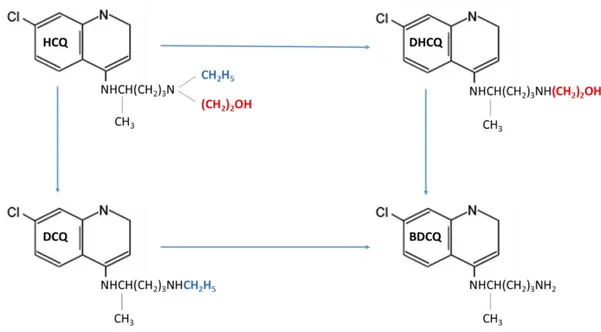
Antimalarial drugs with a 4‐ aminoquinoline scaffold, such as quinine, or chloroquine (CQ), are still essential pharmacological tools in prevention and treatment of malaria [115,116]. Hydroxychloroquine (HCQ), first synthesized in 1946, was proposed as a safer alternative to CQ in 1955 [117]. Later, both CQ and HCQ were suggested as effective alternative for treatment of autoimmune rheumatic diseases [118]. To date, HCQ is one of the most prescribed drugs in patients affected by SLE. HCQ exerts its pharmacological action by preventing the occurrence of both flares and antiphospholipid antibody-dependent thrombotic manifestations, thus improving quality of life and survival of patients [114,118-121]. This drug is commonly administrated orally, as racemic sulfate salt, at a loading dose of 400mg/day followed by a maintenance dosage of 200–400mg/day [122]. Liver metabolizes HCQ into three active metabolites (Figure 3), i.e. desethylchloroquine (DCQ), desethylhydroxychloroquine (DHCQ) and bisdesethylchloroquine (BDCQ) [123].
DHCQ, the major metabolite, is produced through the N‐ desethylation pathway, catalyzed by the enzymatic activity of the cytochromes (CYP) 2D6, 3A4, 3A5 and 2C8. Previous pharmacokinetic studies have shown that HCQ has a long elimination half‐ life of up to 40 days. Hence, the onset of action after initiating the therapy is slow and the steady state may be obtained only after 6months of treatment [124,125]. Moreover, the efficacy of this drug is strictly related to its trough concentration and maintaining plasma concentration above 1000ng/mL can reduce the frequency of flares, as described in the PLUS study [126]. High-responder subjects usually have increased blood concentration of HCQ >1250 ng/mL; while low responder patients show lower concentrations(100-750ng/mL) [127]; and non-responders very low blood HCQ levels (0–129ng/mL) also related to poor adherence to therapy [128,129]. For these reasons, monitoring blood concentration of HCQ is highly suggested for drug dose adjustment in order to increase responsiveness to therapy in SLE patients. Because the fine-tuned dosage of HCQ and its metabolites is emerging as one of the most critical points for an effective therapy, the availability of sensitive, fast and inexpensive analytical methods for qualitative and quantitative measurements of these compounds in body fluids is required for a good clinical management of SLE. Previously published methodologies although valid and well-designed show several drawbacks [130-137]. First, they rarely evaluate HCQ major metabolites, and thus lack the ability to perform comprehensive pharmacokinetic studies of all active molecules deriving from HCQ metabolism. In other cases, chromatograms did not display an optimal separation of all species of interest. Finally, methods providing the most
Figure 3. Molecular structure of hydroxychloroquine (HCQ) and its major metabolites:
21
accurate results often require complex and very skilled analytical techniques (i.e. LC-MS/MS or sequential achiral–chiral HPLC), thus limiting their use in routinely laboratory practice.
2.1.7 Project aim
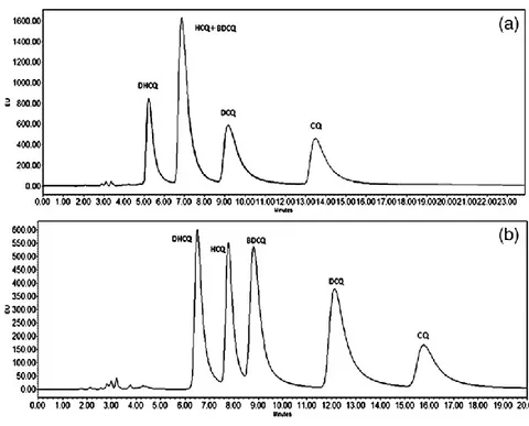
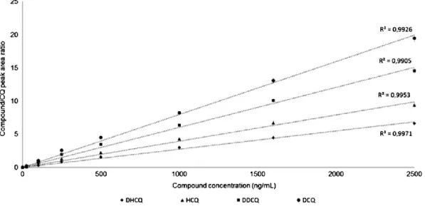
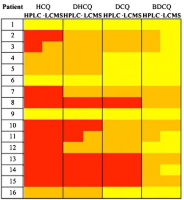
An ion‐ pairing high‐ performance liquid chromatography coupled with fluorescence detector (HPLC‐ FL) methodology was developed and optimized to efficiently separate and quantify HCQ, DHCQ, BDCQ and DCQ in peripheral blood. The method was validated and used for the TDM of SLE patients enrolled at the Rheumatology Unit of the University “Luigi Vanvitelli” of Naples (Italy). The same blood samples were also analyzed using a previously validated LC-MS/MS method and data were compared to those obtained from our HPLC technique [133].
After method validation, we correlated HCQ blood concentrations to quality-of-life (QoL) in SLE patients. Indeed, despite increasing interest, the extent of poor adherence to HCQ treatment, its main originating factors and the relationship with disease progression have not been yet sufficiently investigated in SLE patients with prolonged inactive disease. Therefore, this study was designed in collaboration with the Rheumatology Unit of the University “Luigi Vanvitelli” of Naples (Italy) to estimate the extent of and the main demographic, clinical and laboratory factors associated with HCQ non-adherence, and the relationship between outcomes and HCQ blood concentration in SLE patients with prolonged (>1 year) inactive disease.
2.2 Materials and methods
2.2.1 Patients
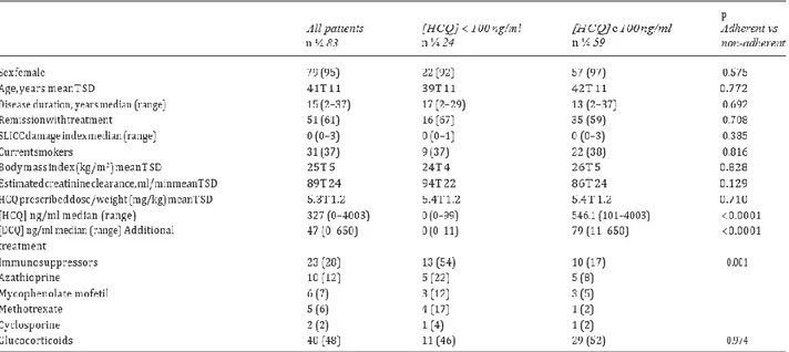
To evaluate circulating levels of HCQ and its metabolites, patients were enrolled at the Rheumatology Unit of the University of Campania ‘‘Luigi Vanvitelli’’, Naples (Italy) from November 2014 to April 2016, after written informed consent was obtained. The study was approved by the Ethics Committee of the University of Campania ‘‘Luigi Vanvitelli’’. Each patient had to satisfy the following inclusion criteria: diagnosis according to the 2012 classification criteria of the Systemic Lupus International Collaborating Clinics (SLICC); complete or clinical remission (with or without treatment) according to the preliminary Definitions of Remission in SLE (DORIS) criteria for at least one year; treatment with a stable dose of oral HCQ during previous six months. If taken, immunosuppressants and glucocorticoids had to be prescribed at a stable dose during the previous month. Exclusion criteria were: concomitant fibromyalgia; psychiatric disorders; or pregnancy.
On admission, medical history, physical examination and laboratory investigations were performed to assess disease activity according to the Safety of Estrogens in Lupus Erythematosus National Assessment SLE Disease Activity Index (SELENA-SLEDAI), and disease damage by the Systemic Lupus International Collaborating Clinics/American College of Rheumatology Damage Index of SLE. Creatinine clearance was measured based on the Modification of Diet in Renal Disease (MDRD). Remission was defined according to the DORIS proposal as follows: complete remission without treatment, i.e. absence of clinical (SLE-related) manifestations, and/or serologic abnormalities (low C3 and/or C4, increasing dsDNA titer), only antimalarials allowed; complete remission on treatment, i.e. absence of clinical manifestations and/or serologic abnormalities, therapy with antimalarials, prednisone 5 mg/day and/or immunosuppressants allowed; clinical remission without treatment, i.e. absence of clinical manifestations, presence of serologic abnormalities, only antimalarials allowed; clinical remission on treatment, i.e. no clinical manifestations with serologic abnormalities, therapy with antimalarials, prednisone 5 mg/day and/or immunosuppressants allowed. The following parameters were also collected: height, weight, body mass index, time of the HCQ tablet intake, daily dose of HCQ, smoke status (current/past smoker). In addition, each patient was examined to assess the co-existence of fibromyalgia according to published diagnostic criteria, and the concomitant use of drugs for psychiatric problems was evaluated.
Each patient completed various tests: a visual analogue scale (VAS) for pain, fatigue and self-assessment of disease activity ranging from 0 (none) to 100 mm (very severe/active); a VAS for global health (GH) status ranging from 0 (poorest) to 100 mm (the best); the Italian version of Short-Form 36 (SF-36), summarized in two composite summary scores, the physical component summary