Amelanotic metastasing melanoma mimicking lung cancer
Daniela Xhemalaj¹*,Fatmir Caushi¹, Linda Ulazzi², Rosa Rinaldi², Giovani Lanza², Hasan Hafizi¹, Mehdi Alimehmeti³, Gisela Pumo³, Asela Hasa³.
1-University Hospital ”Shefqet Ndroqi”- Tirana, Albania 2-University of Ferrara - Italy
3-University Hospital Center “ Mother Teresa ”- Tirana, Albania
*Corresponding author: Daniela Xhemalaj, Spitali Universitar” Shefqet Ndroqi”-Tirana,Albania, Tel.: +664012636; Email: [email protected]
Abstract
Lung is one of the most common site of metastasis in patients with malignant melanoma. We report a 64 year old man, who was submitted to our hospital. He complained of increasing intensity of shortage of breath, cough, and loss of weight within the last weeks. Serum analysis was normal. X-ray and CT images displayed a circumscribed density in the lower left lung, suspicious for lung cancer. Bronchoscopy and biopsy indicated a non differentiated malignancy which could not be further classified. Excision of the lower left lobe and of mediastinal lymph nodes was performed. The post surgical definite histological diagnosis revealed an amelanocytic epithelioid melanoma (positive for HMB-45, S-100). Pathologists, pulmonologists and thoracic surgeons should be aware of different cell types of intrapulmonary malignancies although primary lung cancer contributes to the outstanding majority of large and solid lung lesions.
Keywords: Malignant Melanoma, Lung metastasis, Immunohistochemistry, Lobectomy.
Virtual Slides: www.diagnosticpathology.eu/vs/2015_1_86/
Introduction
Melanoma accounts to one of the most common malignancies in women and in men. Its world-wide incidence has gained to place no 6 in women and no 5 in man [1, 2]. Patients with metastasizing malignant melanoma present with a poor prognosis in general. In 1999, approximately 7,300 patients died of metastasizing malignant melanoma in the USA [6]. The median survival rates of all patients varied from 6 to 15 months [3, 4, 5]; that of patients undergoing a resection of metastases lived for 18 – 40 months (median survival), i.e. considerably longer [7, 8, 9, 10, 11], and only 7.9 % of patients presenting with intra-pulmonary metastases lived for five years or longer [12]. The statistics of the Amecican Cancer Society report that melanoma contribute to 5% of all malignancies [13], and amelanocytic melanomas, which exhibit rare or even no
melanin pigment, to only 7% of all melanomas [14, 15]. Metastases are not necessarily at the same degree pigmented or amlenanocytic when compared to their cutaneous origin. Thus, metastases of an amelanocytic melanoma are a rare and sometimes difficult to diagnose malignancy and a challenge for diagnosis and treatment as well.
Case report:
This 64 years old man had not visited a physician since at least 20 years. He complained of shortness of breath, cough, loss of weight, and sweating. The intensity of his symptoms increased in the last weeks. He was a heavy smoker for at least 25 years and consumed 30 cigarettes / day (90 packyears).
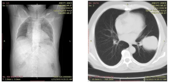
Serum and blood examinations were in the normal range. A dense and circumscribed mass of the lower left lung were detected by X-ray and CT-scans (Figures 1, 2).
Figure 1 (left): X-ray showing a central shadow in the left lung with elevated diaphragm.
Figure 2 (right): CT showing a circumscribed central located density, suspicious for lung cancer.
A bronchoscopy was performed. Representative tissue that would permit a defifnite diagnosis could not be taken from. Therefore, the patient underwent lobectomy with medistinal lymph node dissection.
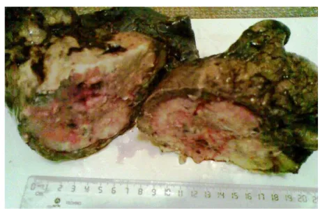
The excised tissue displayed with a whitish cut surface, and asolid mass of increased consistency. It measures 8,0 × 5,7 x 7 cm in maximum diameter. (Figure 3).
Figure 3: Excised tumor with hemorrhages, necrotic areas, multiple growth centers, and distinct intra-pulmonary boundary.
Microscopy (Biopsy number 286-295/2015) of the tumor and lung parenchyma revealed an extended tumor mass composed of epithelioid cells with pleomorphic nuclei and abundant cytoplasm, necrotic areas and hemorrhage clustered around the blood vessels, some palisading, and scattered inflammatory infiltrates (HE stain). These findings excluded a common lung carcinoma and can be considered suggestive for metastasis.
Immunohistochemistry findings: Cytokeratine CK7 negative, Vimentin, HMB-45 and S-100 positive. Melanin pigment could not be noted. Molecular biology investigations revealed a BRAF V600E mutation.
(×10, HE)
(×40 HE)
Figures 4, 5: Both figures show necrotic hemorrhages, some palisading grouped around necrotic vessels, densely packed epithelioid tumor cells with broad nuclear variance, scattered lymphocytes.
The postoperative definite diagnosis of a metastasis of an amelanocytic melanoma is based upon the palisading structures, hemorrhage and necrotic areas, and, most characteristic the expression of HMB-45, S-100 as well as the BRAF V600 mutation. The thorough search for the underlying primary was not successful. No suspicious
shoulder was noted. The patient explained that a skin lesion has been removed years ago. This lesion has not been microscopically examined, and its diagnosis remains unclear. The patient refused a post surgical treatment with Vemurafenib. Until today, ten months after surgery, he is in good condition and doing well. No indication for additional tumor lesions.
Discussion:
Melanoma is a common skin neoplasm, and less than 5% derive in organs other than skin, for example brain or lung. Melanoma that are detected in the lungs are nearly always metastases, and only a few primary lesions have been reported [11, 16, 17]. Pigmented melanomas usually pose no diagnostic difficulties in contrast to their amelanocytic counterparts, that can be mistaken for a broad variety of soft tissue tumors.
The diagnosis becomes even more challenging when the primary lesion remains unclear and the neoplasm is partly or completely undifferentiated.
The prognosis of patients with a metastatic melanoma is poor, even though new trials of a targeted therapy are under investigations.
In conclusion, we want to draw attention to pathologists, clinicians, and surgeons that rare diseases should always to be taken into consideration, and that a definite diagnosis is crucial to exactly classify a malignant disease, especially to initiating the most effective and promising therapy, such as targeted therapy..
Conflicts of interests:
The authors declare that there is no conflict of interests regarding the publication of this paper.
Disclosure:
There is no financial support either for this study or for the authors.
References:
1. Miller AJ, Mihm MC. Melanoma. N Engl J Med 2006;355:51–65.
2. Siegel R, Naishadham D, Jemal A. Cancer statistics. CA Cancer J Clin 2012; 62:10–29.
3. Balch CM, Gershenwald JE, Soong SJ, et al. Final version of 2009 AJCC melanoma staging and classification. J Clin Oncol 2009;27(36):6199-6206.
4. Landy SH, Murray T, Bolden S, et al: Cancer statistics, 1999. CA Cancer J
Clin; 49: 8-31.
5. Barth A, Wanek LA, Morton DL. Prognostic factors in 1,521 melanoma patients with distant metastases. J Am Coll Surg 1995;181:193-201.
6. Manola J, Atkins M, Ibrahim J, Kirkwood J Prognostic factors in metastatic melanoma: a pooled analysis of Eastern Cooperative Oncology Group trials. J Clin Oncol 2000;18:3782-3793.
7. Ollila DW, Hsueh EC, Stern SL, Morton DL. Metastasectomy for recurrent stage IV melanoma. J Surg Oncol 1999; 71: 209–213.
8. Neuman H, Patel A, Hanlon C et al. Stage-IV melanoma and pulmonary metastases: factors predictive of survival. Ann Surg Oncol 2007; 14: 2847–2853. 9. Pastorino U, Buyse M, Friedel G et al. Long-term results of lung
metastasectomy: prognostic analyses based on 5206 cases. J Thorac Cardiovasc Surg 1997;113: 37–49.
10. Tafra L, Dale PS, Wanek LA et al. Resection and adjuvant immunotherapy for melanoma metastatic to the lung and thorax. J Thorac Cardiovasc Surg 1995; 110: 119–129.
11. Petersen RP, Hanish SI, Haney JC et al. Improved survival with pulmonary metastasectomy: an analysis of 1720 patients with pulmonary metastatic; J Thorac Cardiovasc Surg. 2007 Jan;133(1):104-10.
12. Balch CM, Buzaid AC, Soong SJ, Atkins MB, Cascinelli N, Coit DG, et al. Final version of the American Joint Committee on cancer staging system for cutaneous melanoma. J Clin Oncol 2001;19:3635-48.
13. American Cancer Society.Skin Cancer—Melanoma Detailed Guide. Available at:http://www.cancer.org/Cancer/SkinCancer-Melanoma/DetailedGuide/ index. Accessed August 20, 2010.
14. Busam KJ, Hester K, Charles C, et al. Detection of clinically amelanotic malignant melanoma and assessment of its margins by in vivo confocal
15. Koch SE, Lange JR. Amelanotic melanoma: the great masquerader. J Am Acad Dermatol. 2000;42:731–734.
16. Scolyer RA, Bishop JF, Thompson JF. Primary Melanoma of the lung. In: Raghavan D, Brecher ML, Johnson DH, Meropol NJ, Moots PL, et al., eds. Textbook of Uncommon Cancer. 3rd ed. West Sussex: John Wiley & Sons Ltd, 2006; pp 293–8.
17. K Kayser, Analytical Lung Pathology, 1992, Springer, Berlin, Heidelberg, New York.