A.A. 2016/17
Università degli Studi della Tuscia di Viterbo
in convenzione con CIHEAM- Istituto Agronomico Mediterraneo di Bari Corso di Dottorato di Ricerca in
Scienze delle Produzioni Vegetali e Animali - XXIX Ciclo
TITOLO TESI DI DOTTORATO DI RICERCA
D
EVELOPMENT OF ISOTHERMAL DETECTION METHODOLOGYFOR
P
LASMOPARA VITICOLA ANDP
HYTOPHTHORA INFESTANS(S.S.D. AGR/12)
Tesi di dottorato di:
Dott. MELISSA SI AMMOUR Coordinatore del corso
Prof. STEFANIA MASCI
Tutor
Prof. LEONARDO VARVARO Dott. THAER YASEEN
Co-tutor
DEVELOPMENT OF ISOTHERMAL DETECTION METHODOLOGY
FOR PLASMOPARA VITICOLA AND PHYTOPHTHORA INFESTANS
MELISSA SI AMMOUR
Thesis submitted to the Università degli Studi della Tuscia of Viterbo,
Department of Agriculture and Forestry Science, for the degree of Doctor of Philosophy
i First and foremost, I wish to express my cordial thanks to the International Center for Advanced Mediterranean Agronomic Studies (CIHEAM) and the Mediterranean Agronomic Institute of Bari (MAIB), in particular, along with the Università degli Studi della Tuscia of Viterbo for giving me this precious opportunity and for their scientific and financial contributions.
I sincerely thank my supervisors Dr. Thaer Yaseen and Prof. Leonardo Varvaro for their support and valuable guidance during my PhD study.
Deep gratitude must be expressed to Prof. Stefania Masci for her continuous availability and help.
I am heartily thankful to Dr. Odile Carisse for taking me under her wing. It was an honour to be part of her research team. I will never forget her patience, continuous motivation, and vast knowledge.
My sincere thanks also goes to Dr. Ilaria Pertot and Dr. Guillaume Bilodeau who provided me the opportunity to join their teams as intern, and who gave access to their respective laboratories and research facilities. Without they precious support it would not be possible to conduct this research.
I am indebted to all the friends and colleagues I met during this path, and particularly a big “Thank you!” for the great help I received from: D. M. Tremblay, A. Levasseur, A. Lefebvre, and Dr. L. M. Fall in Saint-Jean-sur-Richelieu Research Center; M. Newton, D. Shearlaw and E. Tremblay in the Pathogen Identification Research Lab, CFIA, Ottawa; H. Van Der Heyden and T. Wallon in Phytodata, Sherrington; Dr. M. Perazzolli, O. Giovannini, C. Sicher and D. Ress in the Edmund Mach Foundation, San Michele all’Adige; and finally G. Cavallo, Dr. M. Gallo, S. Fodil and I. Drais in the MAIB, Valenzano.
Tender and special thoughts to Dr. Ahmed Abdelfattah who has stood by me and believed in my potential during all those years.
Last but not least, I owe my success to my parents, brother and sisters. Their unconditional support and encouragements gave me the strength and motivation to carry forward my projects in life.
SI AMMOUR Mélissa March 2017
ii This research concerns the development and application of isothermal amplification detection techniques to two important plant pathogenic Oomycete: Plasmopara viticola causing grapevine downy mildew and Phytophthora infestans, the causal agent of potato late blight. Real-time Loop-Mediated Isothermal Amplification (LAMP) assay was applied for the early detection of Plasmopara viticola from infected grapevine plants. A rapid crude plant extract (CPE) preparation method from infected leaves was developed for on-site testing. The LAMP assay targeting the large ribosomal subunit gene (LSU) was specific to P. viticola and sensitive to 10 sporangia/ml. In the inoculation experiments of greenhouse plants and leaf discs samples, LSU primers detected downy mildew infections at a concentration of 103
sporangia/ml after 24h of inoculation. In addition, Real-time LAMP and Recombinase Polymerase Amplification (RPA) assays were developed targeting the ITS2 region of the ribosomal DNA of P. infestans. Both LAMP and RPA assays showed specificity to P. infestans and the closely related species P. andina, P. mirabilis, P. phaseoli and P. ipomoeae. No cross-reaction occurred with the potato pathogens tested. LAMP and RPA assays detected DNA at 50 fg/ul and showed to be insensitive to CPE inhibition. The isothermal assays were validated with inoculated potato plants using a portable Smart-DART device. The LAMP and RPA assays effectively detected P. infestans DNA in symptomless leaf tissue 24 h and 72 h post-inoculation, respectively. Finally, the suitability of the latter LAMP assay was tested for a rapid detection and quantification of airborne inoculum of P. infestans. Standard curves of P. infestans sporangia and ITS copy were constructed. The quantitative LAMP (qLAMP) assay was validated in the laboratory with silicon-coated rods containing a known number of sporangia. The analysis was performed using a regression procedure. A linear relationship between the number of sporangia deposited onto the rods estimated with microscopy and the number of sporangia estimated with the qLAMP assay was obtained. A rapid and accurate on-site detection of P. infestans and P. viticola in plant material and spore samplers will contribute to improved disease diagnosis, early detection of first infections and facilitate prompt management decisions.
iii time detection, crude plant extract, on-site diagnostics.
iv La ricerca svolta riguarda lo sviluppo e l’applicazione di tecniche di amplificazione isotermica per il rilevamento di due importanti oomyceti fitopatogeni: Plasmopara viticola, agente causale della peronospora della vite e Phytophthora infestans, agente causale della peronospora della patata. Per il rilevamento precoce di P. viticola in piante di vite infette, é stata utilizzata la tecnica Real-time Loop-Mediated Isothermal Amplification (LAMP). Per il saggio in campo, é stato sviluppato un metodo di preparazione rapida di un estratto di pianta crudo (CPE) a partire da foglie infette. Il saggio LAMP é stato specifico per il gene per la sub-unità ribosomiale (LSU) di P. viticola, e sensibile a 10 sporangi/ml. Negli esperimenti di inoculazione su piante in serra e su dischi fogliari campionati, i primers LSU sono stati in grado di rilevare infezioni di peronospora a concentrazioni di 103 sporangi/ml dopo 24 ore
dall’inoculazione. Inoltre, saggi di Real-time LAMP e Recombinase Polymerase Amplification (RPA) sono stati sviluppati per il rilevamento della regione ITS2 del DNA ribosomiale di P. infestans. Entrambi i saggi LAMP e RPA hanno dimostrato specificità per P. infestans e per le specie strettamente correlate P. andina, P. mirabilis, P. phaseoli e P. ipomoeae. Non sono state rilevate reazioni incrociate con gli altri patogeni della patata saggiati. I saggi LAMP e RPA hanno rilevato DNA a 50 fg/ul e non hanno mostrato alcuna inibizione al CPE. I saggi isotermici sono stati validati con piante di patate inoculate utilizzando uno strumento portatile Smart-DART. I saggi LAMP e RPA hanno rilevato DNA di P. infestans nei tessuti fogliari di piante asintomatiche a 24 e 72 ore dall’inoculazione, rispettivamente. In fine, l’utilizzo del saggio LAMP è stato testato per il rapido rilevamento e quantificazione dell’inoculo airborne di P. infestans. Sono state ottenute le curve standard degli sporangi e del numero di copie ITS2 di P. infestans. Il saggio LAMP quantitativo (qLAMP) è stato validato in laboratorio con barrette rivestite di silicone contenenti un numero noto di sporangi. L'analisi è stata effettuata tramite regressione lineare, con cui è stata ottenuta una relazione lineare tra il numero di sporangi presenti sulle barrette, valutato mediante conteggi al microscopio, e il numero di sporangi stimato dal saggio qLAMP. Un rapido e accurato rilevamento in campo di P. infestans e P. viticola contribuirà a migliorare
v Parole chiave: Patologia delle piante, peronospora della vite, peronospora della patata, Loop-Mediated Isothermal Amplification, Recombinase Polymerase Amplification, rilevamento Real-time, estratto di piante crudo, diagnosi in campo.
vi
Acknowledgments ... i
Abstract ... ii
Riassunto ... iv
List of figures ... ix
List of tables ... xii
I General introduction ... 1
Integrated disease management ... 1
Oomycete plant pathogens ... 2
Plasmopara viticola ... 2
Phytophthora infestans ... 4
Molecular diagnostics of plant pathogens ... 7
Isothermal amplification detection methods ... 8
Loop Mediated Isothermal Amplification ... 9
Recombinase Polymerase Amplification ... 12
On-site detection of plant pathogens ... 16
Thesis objectives ... 17
References ... 18
II Real-time LAMP for the early detection of Plasmopara viticola from infected plants 26 Introduction ... 26
Materials and methods ... 28
II.1.1 Design of LAMP primers ... 28
vii
II.1.5 Specificity of the LAMP assays ... 31
II.1.6 Sensitivity of the LAMP assays ... 31
II.1.7 Plants inoculation and experimental design ... 31
Results ... 34
II.1.8 Specificity of the LAMP assays ... 34
II.1.9 Sensitivity of the LAMP assays ... 34
II.1.10 Inoculation experiments ... 35
Discussion ... 37
References ... 40
III Development of real-time isothermal amplification assays for on-site detection of Phytophthora infestans in potato leaves ... 42
Introduction ... 42
Materials and methods ... 45
III.1.1 Fungal isolates. ... 45
III.1.2 Design of LAMP primers and assimilating probe. ... 45
III.1.3 Design of RPA primers and Exo-probe. ... 45
III.1.4 Design of RPA primers and Exo-probe. ... 49
III.1.5 LAMP reaction. ... 49
III.1.6 RPA reaction. ... 49
III.1.7 Specificity of the isothermal assays. ... 51
III.1.8 Sensitivity of the isothermal assays. ... 51
III.1.9 Crude plant extract preparation method. ... 51
III.1.10 Inoculum preparation. ... 52
viii
Results ... 55
III.1.14 Specificity of the LAMP and RPA assays. ... 55
III.1.15 Sensitivity of LAMP and RPA assays. ... 55
III.1.16 Testing on inoculated plants. ... 57
III.1.17 Field samples. ... 59
Discussion ... 59
References ... 64
IV Towards the application of a real-time quantitative LAMP assay for the detection of Phytophthora infestans airborne inoculum. ... 69
Introduction ... 69
Materials and Methods ... 71
IV.1.1 Real-time LAMP assay ... 71
IV.1.2 Preparation of DNA solutions for P. infestans ITS2 copy standard curve 71 IV.1.3 Preparation of DNA solutions for P. infestans sporangia standard curve 73 IV.1.4 Estimation of the number of sporangia from the number of ITS2 copies 73 IV.1.5 DNA extraction procedure from spore trap rods ... 74
IV.1.6 Laboratory validation of the qLAMP assay ... 75
Results ... 76
IV.1.7 Estimation of the number of spores from the number of ITS2 copies 76 IV.1.8 Laboratory validation of the qLAMP assay ... 76
ix
List of figures
Figure I-1. Disease cycle of downy mildew of grapes caused by Plasmopara viticola (Agrios, 2005). ... 3 Figure I-2 Disease cycle of late blight of potato caused by Phytophthora infestans (Agrios, 2005). ... 6
Figure I-3 Principle of loop-mediated isothermal amplification (LAMP) method. ... 10 Figure I-4 Principle of assimilating probe with LAMP amplification process. .... 13 Figure I-5 Schematic outline of the recombinase polymerase amplification (RPA) ... 14 Figure I-6 Schematic outline of the TwistAmp® exo probe for RPA real-time detection. Exonuclease cuts the THF residue during RPA amplification. Source: TwistDx-Limited 2013 ... 16
Figure II-1 Sporangia-bearing grapevine leaves used for the preparation of Plasmopara viticola inoculum ... 30 Figure II-2 Sensitivity of the LSU LAMP assay. Real time amplification curve generated in the LAMP assay using 10 fold serial dilutions of P. viticola A) pure sporangia suspension ranging from 104 to 1 sporangia/ml and B) sporangia suspension serial dilutions extracted with healthy grape leaf disc. ... 35
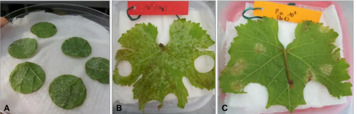
Figure II-3 Plasmopara viticola sporulation on the inoculated A) leaf discs, B) Greenhouse, and C) field plants. ... 35
Figure III-1 Potato leaf samples collected for the reliability test of the LAMP and RPA assays. Samples were collected from A, B) potato fields without late blight infections showing symptoms similar to P. infestans lesions, and from C, D) potato fields with late blight infections. ... 54
x DNA ranging from 0.5ng/ul to 5fg/ul and B) DNA serial dilutions incorporated into healthy crude plant extract. Standard curve obtained in a real-time machine by plotting C) P. infestans pure DNA concentration, and D) DNA serial dilutions incorporated into healthy crude plant extract against LAMP Ct values. R2 values of standard curve obtained from LAMP assay are indicated. ... 56
Figure III-3 Sensitivity of RPA assay. Smart- DART amplification curve generated in the RPA assay using 10 fold serial dilutions of P. infestans A) pure DNA ranging from 0.5ng/ul to 5fg/ul and B) DNA serial dilutions incorporated into healthy crude plant extract. Standard curve obtained in a real-time machine by plotting C) P. infestans pure DNA concentration, and D) DNA serial dilutions incorporated into healthy crude plant extract against RPA Ct values. R2 values of standard curve
obtained from RPA assay are indicated. ... 56 Figure III-4 Lesions progression of Phytophthora infestans on inoculated potato plants and LAMP and RPA detection over incubation time. ... 57
Figure IV-1 Preparation of spore trap rods for the laboratory validation of the qLAMP assay. A) Droplets of P. infestans sporangia solutions with variable concentration are deposited in the centre of the silicon-coated rod before microscope count and qLAMP assay. B) Example of a rotating arm spore sampler placed in a potato field for P. infestans sporangia monitoring. ... 75
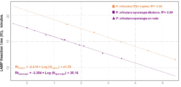
Figure IV-2 Real-time LAMP standard curves obtained in a real-time PCR machine by plotting the reaction time (Rt) value with the log number of Phytophthora infestans sporangia, and the Rt value with the log number of internal transcribed spacer 2 (ITS2) copies. ... 77
Figure IV-3 Double regression analysis of the qLAMP assay predictions of the number of P. infestans sporangia against the predicted number of ITS2 copies, based on Rt values interval of 11.5 to 18 minutes. ... 77
Figure IV-4 Real-time LAMP standard curves obtained in Smart-DART instrument by plotting the reaction time (Rt) value with the log number of Phytophthora infestans
xi Figure IV-5 Relationship between estimates of the number of Phytophthora infestans sporangia deposited on silicon-greased rods, based on the P. infestans qLAMP assay in a real-time PCR machine and estimates based on microscope counts. ... 78 Figure IV-6 Relationship between estimates of the number of Phytophthora infestans sporangia deposited on silicon-greased rods, based on the P. infestans qLAMP assay in a Smart-DART instrument and estimates based on microscope counts. ... 79
xii Table II-1 LAMP primers used in this study for Plasmopara viticola isothermal detection ... 29
Table II-2 Experimental design of Plasmopara viticola inoculation on leaf discs, greenhouse plants and field plants. ... 33
Table II-3 Results of the inoculation experiments of Plasmopara viticola. ... 36 Table III-1 Isolates of Phytophthora species and other fungal species used for sensitivity and specificity tests of the isothermal assays and tests on infected plant material. The LAMP and RPA specificty test results are indicated as amplified (+) and not-amplified (-). ... 46
Table III-2 LAMP and RPA primers and probes used in this study for Phytophthora infestans isothermal detectiona ... 50
Table III-3 Results of LAMP and RPA detection of Phytophthora infestans infections on inoculated potato plants ... 58
Table III-4 Contingency table of the LAMP and RPA testing on potato leaf samples collected from fields with and without late blight (LB) infections. ... 60
1
Chapter I
I G
ENERAL INTRODUCTIONThis chapter relates the background and the main hypothesis of the study. As a first step, we will discuss the main concepts of integrated plant disease management. We will also define some aspects related to Oomycete plant pathogens, especially the pathogens studied: Plasmopara viticola and Phytophthora infestans. We will then address the contribution of molecular methods in the detection of plant pathogens and the recent advances in isothermal detection methods that account for this thesis. Finally, we will conclude this chapter by covering the objectives of this thesis. Integrated disease management
Plant diseases cause enormous crop losses worldwide. Damages can occur in the field as early as seed sowing to harvesting, but also in storage. Major historical consequences of plant disease epidemics are the Irish great famine (1845) due to late blight of potato and Bengal famine (1943) due to brown spot of rice. Plant pathology addresses the cause, biology, epidemiology, subsequent losses and management of the plant diseases.
Foremost, a correct disease diagnostic is essential in order to identify the right causal agent. Disease management practices rely essentially on preventing the occurrence of disease, and target critical stages of the pathogen in the disease cycle. Integrated disease control strategy or Integrated Disease Management (IDM) is a resultant concept from the popular Integrated Pest Management (IPM) systems developed by entomologists for insect and mite control. IDM consists of disease and pathogen scouting with proper application of a combination of control strategies. This approach integrates the adaptation of cultural practices, the pathogen and disease survey, and monitoring the environmental factors. Based on established economic thresholds, fungicide treatments are usually applied with the support of disease forecasting models (Maloy 2005).
An integrated disease control program aims to (1) eradicate or reduce the initial inoculum, (2) reduce the effectiveness of initial inoculum, (3) increase the host
2 resistance, (4) delay the disease onset, and (5) slow the secondary cycles (Agrios 2005).
Oomycete plant pathogens
The oomycetes are a group of fungal-like organisms that represent some of the most destructive plant pathogens and considered as persistent threats in agriculture. Oomycetes are different from true fungi and more related to diatoms and seaweeds (Thines 2014). The main features distinguishing oomycetes from fungi are: their cell wall contains cellulose and have glucans instead of chitin derivatives, their mycelium is coenocytic (non-septate), and many species produce wall-less, biflagellate swimming zoospores (Bartnicki-Garcia 1968; Burki et al. 2007). They cause important diseases that include seedling blights, damping-off, root rots, foliar blights and downy mildews (Fry and Grünwald 2010), such as, late blight of potato, sudden oak death, downy mildew of grape vine, root and stem rot of soybean, and root rot and gummosis of citrus (Kamoun et al. 2015).
Plasmopara viticola
Plasmopara viticola (Berk et Curt.) Berlese et de Toni is the causal agent of grapevine downy mildew, an important disease in all grape-growing areas with frequent rains (Lafon and Clerjeau 1988). This pathogen has been the subject of study since its introduction into European vineyards in 1878 (Gessler et al. 2011). P. viticola is a heterothallic diploid oomycete and obligate biotrophic parasite native to North America (Dick 2002; Wong et al. 2001), and consequently more aggressive on European Vitis vinifera than the American Vitis varieties (Agrios 2005).
P. viticola overwinters in fallen leaves as sexual dormant structures called oospores or as mycelium in dormant twigs (Fig.I-1). The primary infections usually start when the oospores germinate producing sporangia that release zoospores.
3 Figure I-1. Disease cycle of downy mildew of grapes caused by Plasmopara viticola
(Agrios, 2005).
The sporangia or their zoospores are transported by wind or water, and can penetrate through stomata in grapevine wet green tissues (Agrios 2005). Early infections are recognized as yellowish lesions on leaves upper surface, known as “oil spots”. After 5 to 10 days of infection, sporangiophores and sporangia are produced forming a white-cottony mildew in lower leaf surface (Caffi et al. 2012), hence the name of the disease. Sporangia become airborne and release clonal zoospores as secondary inoculum. A sexual secondary infection cycles occur usually in humid nights (Caffi et al. 2012).
The most effective fungicides for the control of downy mildew are copper-based products such as the Bordeaux mixture and some broad-spectrum protective fungicides usually applied in combination with several systemic fungicides (Agrios 2005). Downy mildew control has been thoroughly reviewed by Gessler et al. (2011). Currently, the disease can be controlled with properly timed fungicide applications
4 that aim to control primary infections in spring and to limit the spread of secondary inoculum during the summer. In Italy for instance, during the growing season 2009-2010, an average of 12.3 treatment per hectare of area treated were used to protect vineyards (Istat 2010). Across the entire Piedmont region, in particular, the annual cost for downy mildew control typically ranges from 8 to 16 million Euros, depending on weather conditions (Salinari et al. 2006). Moreover, downy mildew control remains difficult as the disease cycle, including the production of primary inoculum, occurs throughout the growing season (Gessler et al. 2011). In addition to seasonal weather variations that can lead to unexpected disease outbreaks.
The relationship between the environmental factors, host susceptibility, and P. viticola has been established and numerous disease prediction models for downy mildew have been developed following different modelling approaches (Rossi et al. 2013). Those models are mostly weather-driven and aim to improve the fungicide schedule application while reducing the number of applications and associated costs. Two Decision Support Systems, namely “Vitebio.net™” for organic growers (Caffi et al. 2011) and “Vite.net™” for growers following IPM guidelines (Rossi et al. 2014) are currently used in several areas of Italy.
Phytophthora infestans
Phytophthora infestans (Mont.) de Bary. is the causal agent of late blight, the most devastating disease of potato (Solanum tuberosum). It had been introduced into Europe and North America from the Toluca Valley in central Mexico (Grunwald and Flier 2005), which triggered disastrous blight epidemics in the 1840s in Europe. Besides potato, the blight pathogen is also destructive to tomatoes (S. lycopersicum), and affects other hosts including hairy nightshade (S. sarrachoides), petunia (Petunia hybrida) and bittersweet nightshade (S. dulcamara) (Dorrance and Inglis 1997; Knapova and Gisi 2002; Platt 1999). P. infestans is considered a heterothallic oomycete, although the existence of self-fertile isolates has been reported (Prakob and Judelson 2007; Zhu et al. 2016). These genetic recombination lead to the emergence of new clonal lineages that bear different aggressiveness and fungicide resistance (Cooke et al. 2012; Fry et al. 2015), thus causing severe outbreaks worldwide (Chowdappa et al. 2015; Fry et al. 2013). Late blight pathogen receives, to this day, a constant attention among oomycete plat pathogens (Kamoun et al. 2015).
5 P. infestans is a hemibiotroph organism that exhibits distinct phases of its life cycle: an early asymptomatic biotrophic phase and a late necrotrophic stage that is characterized by tissue degradation and disease symptoms (Lee and Rose 2010). The oospores of P. infestans may survive in the soil for 3 to 4 years. They germinate by means of a germ tube that produces a sporangium (Fig.I-2). In the absence of a sexual stage, sporangia can also be produced from infected tubers or infected shoots. When mature, the sporangia are dispersed by air or by rain and cause new infections either by direct germination or indirectly by the release of zoospores (Agrios 2005). Foliar blight symptoms appear at first as water-soaked spots that turn into black/brown lesions, and expand rapidly to become necrotic. In moist weather, P. infestans produces sporangia and sporangiophores on the surface of infected tissue. This sporulation appears as a white mildew at the edge of the lesions on the lower leaf surface (Schumann and D’Arcy 2000). In favorable conditions, foliar epidemics may cause total destruction of all plants in a field within a week or two (Agrios 2005). On the other hand, tuber blight infections happen in the field, during rainy weather, when sporangia are washed from the leaves into the soil. The released zoospores germinate and penetrate the tubers through lenticels or through wounds (Agrios 2005). Infected tuber tissues are copper brown, reddish or purplish in color. Most of the infected tubers rot in the ground or during storage (Schumann and D’Arcy 2000).
The first step in integrated control of late blight is reducing the primary source of inoculum by the elimination of infected seed tubers and volunteer plants, but also cull piles in potato fields should be destroyed.
6 Figure I-2 Disease cycle of late blight of potato caused by Phytophthora infestans
(Agrios, 2005).
The use late blight resistant varieties has been widely investigated in order to reduce fungicide application rates, or extend intervals between applications (Bain et al. 2009; Fry 1978; Nærstad et al. 2007; Nielsen and Bødker 2001). Conversely, these cultivars are not grown on a large scale as they do not possess the commercially important characteristics such as quality, yield and earliness (Cooke et al. 2011). Chemical control of late light includes the use of broad-spectrum and systemic fungicides, preferably applied with a previous knowledge of the prevailing pathogen population and fungicide resistance trait (Saville et al. 2015). Various late blight forecasting systems are used for well-timed chemical sprays. These Decision Supports Systems (DSSs) are regularly updated with weather information and late blight scouting inputs. Several DSSs have been developed and validated in a number of European countries (Hansen et al. 2002). The so-called EuroBlight network has made available a free online platform that provides an overview of the existing and new European DSSs
7 (http://euroblight.net). The Euroblight tool allows testing and comparing the weather based late blight sub-models, from different European regions, in order to improve their quality (Hansen et al. 2010). Likewise, in the United States, DSS BlightPro is a web platform that integrates considerable information useful to the management of late blight (http://usablight.org). In addition to site-specific weather data and weather forecasts, it incorporates disease reports containing information about the pathogen genotype, fungicide resistance, the host, and management technologies (Small et al. 2015).
Molecular diagnostics of plant pathogens
It is crucial to accurately detect and identify pathogens to initiate preventive disease control measures. An essential key in disease management is the early detection of pathogens, particularly in seeds, mother plants and propagative plant material but also in the early stages of the infection to avoid the introduction and further dispersal of the inoculum (Narayanasamy 2011). Continuous advances in DNA-based detection methods have provided fast, sensitive and reliable detection and quantification of fungal pathogens, when compared to culture-based identification methods (Capote et al. 2012). Most of these techniques rely on polymerase chain reaction (PCR) and real-time PCR assays and have been extensively applied to plant pathology from soil, water, air samples and plant material (Böhm et al. 1999; Carisse et al. 2009; Lievens et al. 2006; Schaad and Frederick 2002; Schena et al. 2013; van de Graaf et al. 2003; West et al. 2008). Moreover, PCR-based techniques provided highly specific assays that can discriminate between species isolates and genotypes (Abbott et al. 2010; Kroon et al. 2004).
In the aim for on-site testing of plant pathogens, the equipment for such systems is quite expensive and is not mobile to achieve testing closer to the point of sampling. Research efforts have been made to move real-time PCR technology from the laboratory to the field using portable thermocycler (Almassian et al. 2013; De Boer and Lopez 2012; Mavrodieva et al. 2004; Schaad et al. 2002). Despite some successful applications, these technologies have not been widely adopted as the portable thermocyclers are expensive, but predominantly, the assays require laborious modifications of DNA extraction protocols in order to adapt to field conditions (Hughes et al. 2006; Tomlinson et al. 2005). Recently, insulated isothermal PCR
8 (iiPCR) method, has been described for sensitive and specific detection of both DNA and RNA (Tsai et al. 2012). In addition, iiPCR can be performed in relatively simple and inexpensive device when compared thermocyclers (Lin et al. 2016).
Isothermal amplification detection methods
Isothermal amplification detection methods have been developed to overcome the use of PCR thermocyclers for a possible on-site testing. As the name suggests, isothermal amplification of DNA (or RNA) occurs at a constant temperature, which confers to some of these methods the potential to be used in the field using portable instruments (Chang et al. 2012). A number of reviews have fully described these methods for isothermal amplification (Gill and Ghaemi 2008; Li and Macdonald 2015; Niessen 2014; Yan et al. 2014).
Isothermal amplification methods rely on several approaches to generate single-stranded primer binding sites, facilitate primer annealing and template replication using DNA polymerase (or RNA polymerase) without thermal cycling. These methods can be based on:
- Transcription of RNA, such as nucleic acid sequence-based amplification (NASBA) and signal-mediated amplification of RNA technology (SMART) methods (Compton 1991; Wharam et al. 2001);
- Additional enzymes for the separation of the target nucleic acid and primer annealing, such as, helicase-dependent amplification (HDA) and recombinase polymerase amplification (RPA) methods (Piepenburg et al. 2006; Vincent et al. 2004);
- Strand displacement DNA polymerase for the replication of linear or circular targets such as, rolling circle amplification (RCA) and loop mediated isothermal amplification (LAMP) methods (Fire and Xu 1995; Notomi et al. 2000); - Restriction enzyme nicking of primer extension products and strand
displacement DNA polymerase, namely, strand displacement amplification (SDA) method (Walker et al. 1992).
9 Loop Mediated Isothermal Amplification
Loop-mediated isothermal amplification (LAMP) is an isothermal nucleic acid amplification method offering rapid, accurate, and cost-effective diagnosis of diseases. It has been described as a novel method by Notomi et al. (2000), and has been applied to produce highly specific and sensitive amplification of DNA or RNA. The addition of reverse transcriptase makes it possible to amplify DNA from RNA sequences (RT-LAMP) (Curtis et al. 2008). LAMP method has attracted much attention, in fact, numerous reports have been recorded to evaluate its efficiency in recognizing bacterial, viral, fungal and parasitic diseases worldwide (Damhorst et al. 2015; Fu et al. 2011; Kurosaki et al. 2016; Parida et al. 2008; Temple and Johnson 2011). In the initial phase of development, LAMP has been applied to many kinds of pathogens causing food-borne diseases, such as many LAMP kits for detecting Salmonella, Legionella, Listeria, verotoxin-producing Escherichia coli, and Campylobacter have been commercialized (Mori and Notomi 2009). Recently, a growing interest in this method has been also observed in the detection of plant pathogenic agents, especially with the possible on-field application through portable devices (Harper et al. 2010; Keremane et al. 2015; Moradi et al. 2012; Thiessen et al. 2016; Tomlinson et al. 2010a; Villari et al. 2016).
The mechanism of the LAMP reaction can be explained through two steps: i) a starting structure producing step, i.e.: the double stem-loop (Fig.I-3b) and ii) a cycling amplification step (Fig.I-3c). LAMP does not require initial template denaturation (Nagamine et al. 2001) and employs a DNA polymerase with strand-displacement activity, along with two forward and backward inner primers (FIP, BIP) and outer primers (F3, B3) which recognize six separate regions (Fig.I-3a) within a target DNA (Tomita et al. 2008).
10 Figure I-3 Principle of loop-mediated isothermal amplification (LAMP) method.
(a) LAMP outer primers (F3 and B3) and inner primers (FIP and BIP) targeting six distinct regions within a target DNA; (b) Starting structure producing step: FIP primer anneals to the targeted sequence and initiates DNA synthesis. F3 primer carries out the strand-displacement DNA synthesis and produces a single stranded DNA that forms a DNA loop structure at 5’ end. This structure works as a template for B3 and BIP primers that anneal to the 3’ end. A double stem-loop structure is formed; (c) Cycling amplification step: FIP and BIP primers bind to the loops and synthesize new DNA strands. The extension of each primer opens the loop end and elongated structures are continuously produced. Source: Tomita et al. 2008.
11 The loop primers (LF, LB) are additional primers designed to anneal at the loop structure in LAMP amplicons, can accelerate and enhance the sensitivity of the LAMP reaction (Nagamine et al. 2002). An animation of the amplification process is available on the Eiken Chemical website for better understanding of the LAMP principle (EikenChemical).
The LAMP assay is highly specific as the amplification reaction occurs only when the primers correctly recognize all six regions, within a target DNA. The procedure is rapid and is able to amplify from a single copy to 109 in one hour at constant temperature; typically in the range of 60–70°C (Notomi et al. 2000). Moreover, it was reported that adding in the loop primers (LF, LB) can reduce the amplification time from the previous one hour to less than 30 minutes (Ushikubo 2004). It is robust, with reagents stable at ambient temperature for up to 2 hours (Thekisoe et al. 2009), and consistently insensitive to extraneous nucleic acids or interference from sample or media components that are problematic for other detection methods (Kaneko et al. 2007; Niessen 2014). However, the main limitation of the LAMP method is the primers design that is complicated and different from the usual PCR primers design, making it difficult for beginners. Even though, software for LAMP primer design are available (e.g. Primer Explorer and LAMP Designer), optimal primers performance is not certain and many primers set candidates should be evaluated when developing an assay.
Several methods have been reported to detect LAMP products: the existence of ladder-like bands on agarose gel, the use of lateral-flow-devices, visual observation of turbidity derived from magnesium pyrophosphate formation, real-time measurement of turbidity, as well as colorimetric LAMP assays using DNA or metallic ions binding dyes (Parida et al. 2008; Tomita et al. 2008; Tomlinson et al. 2010b, a). The peculiarity of the LAMP reaction is that a large amount of magnesium pyrophosphate is precipitated as a by-product of the reaction that enables visual assessment of amplification (Mori et al. 2001). Moreover, these methods generally use Bst DNA polymerase (Bacillus stearothermophilus) (Hafner et al. 2001) and different dyes as fluorescent indicators for positive or negative results. These dyes can be added after the reaction is complete, for example, SYBR Green I as DNA binding dye (Notomi et
12 al. 2000), or pre-added in the reaction mix, such as calcein and hydroxynaphthol blue as metallic ions binding dyes (Wastling et al. 2010).
Since LAMP reaction generates tremendous amount of self-replicating amplicons, it is often strongly advised not to open completed LAMP reaction tubes to detect LAMP products (Keremane et al. 2015; Tomita et al. 2008). Therefore, monitoring the isothermal LAMP reaction in real-time has been made available using a LAMP master mix that contains an engineered GspSSD LF DNA Polymerase with strand displacement and reverse transcriptase activities and a double-strand DNA binding dye (OptiGeneLimited). This reagent contains also a pyrophosphatase that hydrolyses magnesium pyrophosphate and therefore does not allow visual observation of the LAMP amplification. Moreover, a fluorescence resonance energy transfer (FRET)-based probes technology was described by Kubota et al. (2011), namely, assimilating probes. These probes are designed in a particular architecture, where a quenching strand is displaced from a partially complementary fluorescent strand, during the process of DNA synthesis (Fig.I-4). Assimilating probes have been used for sequence-specific detection of the LAMP amplicon, but also for quantification and multiplexing purposes (Kubota and Jenkins 2015; Tanner and Evans 2014; Villari et al. 2016). Recombinase Polymerase Amplification
Piepenburg et al. (2006) first introduced recombinase polymerase amplification (RPA) as another isothermal technique, similar to HDA. The RPA process employs three enzymes – a recombinase, a single-stranded DNA-binding protein (SSB) and strand-displacing polymerase. The RPA reaction initiates with the combination of the recombinase with each of the two primers prior to their annealing to specific sequences in the target (Fig.I-5). After the primer annealing, the recombinase detaches from the primers making their 3’ end accessible to the Sau polymerase (Staphylococcus aureus). This form a D-loop structure, which is stabilized by the SSB protein to keep the DNA open as the DNA polymerase carries on the amplification.
13 Figure I-4 Principle of assimilating probe with LAMP amplification process.
(a) the inner primer and assimilating probe anneal to the double stem-loop structure and new DNA strands are synthetized; (b) secondary products from self-primed double stem-loop, extension of inner primer and assimilating probe; (c) assimilating probe extended sequence separated from the double stem-loop and annealed to inner primer BIP; and (d) quench strand displaced by synthesis of new strand from BIP, resulting in fluorescence emission. Source: Kubota et al. 2011.
14 Figure I-5 Schematic outline of the recombinase polymerase amplification (RPA)
(A) Recombinase integrates with primers to form recombinase-primer complexes and target specific DNA sequences. (B) Strand exchange occurs and single stranded binding proteins (SSB) bind to the DNA to form a D-loop. (C) DNA polymerase initiates DNA amplification. (D) Displaced D-loop stabilized by SSB as amplification continues. (E) Two dsDNA molecules form and the entire cycle starts again. Source: TwistDx-Limited 2013.
15 RPA is very efficient, as billions of DNA copies can be generated within 40 to 60 minutes at incubation temperatures between 37 °C and 42 °C (Piepenburg et al. 2006). Since primers form a complex with the recombinase to target the homologous sequences, RPA primer design is simple without consideration of annealing temperature. However, the primers required for RPA are longer than PCR primers (30 to 35 nucleotides), and are often reported to generate high background noise in low temperature RPA reactions (Yan et al. 2014). Methods that are used to detect PCR or HDA products can be applied to detect RPA products. In order to improve amplification and detection efficiency, the use of lateral flow devices and real-time RPA detection are made possible through the design of unique probes: specifically, TwistAmp® LF probe for lateral flow detection, and TwistAmp® fpg probe and TwistAmp® exo probe for real-time detection (TwistDx-Limited 2016). Exo probes carry internal fluorophore and quencher linked to thymine bases and separated by an abasic site mimic (tetrahydrofuran or THF), that is localized approximately 15 nucleotides upstream from the 3’ end of the probe (45 to 55 nucleotides). In addition, a suitable 3’ modification group blocks probes from polymerase extension (Fig.I-6). The THF residue presents a substrate for Exonuclease III present in the TwistAmp® exo kit, which will cleave the exo probe at the THF position, thereby separating the fluorophore and the quencher and generating a fluorescent signal for real-time detection (TwistDx-Limited 2013).
RPA method has been extensively reported in clinical applications (Abd El Wahed et al. 2015; Abd El Wahed et al. 2016; Boyle et al. 2014; Daher et al. 2016; Euler et al. 2013; Hill-Cawthorne et al. 2014). Besides, a number of RPA assays have been described to detect plant pathogens. Reverse transcriptase RPA (RT-RPA) has been applied for the detection of important plant viruses, such as plum pox virus, little cherry virus, and tomato yellow leaf curl virus, and has proven to be more cost effective and sensitive than RT-PCR and ELISA (Londoño et al. 2016; Mekuria et al. 2014; Silva et al. 2015; Zhang et al. 2014).
16 Figure I-6 Schematic outline of the TwistAmp® exo probe for RPA real-time detection.
Exonuclease cuts the THF residue during RPA amplification. Source: TwistDx-Limited 2013
Miles et al. (2015) recently used RPA approaches to develop a genus-specific assay for detection of Phytophthora spp., and other assays for P. ramorum and P. kernoviae species detection from crude plant extract.
On-site detection of plant pathogens
Early detection of plant pathogens is crucial for timely disease management. On-site nucleic acid-based methods that can be performed with minimal equipment, rapidly and at low cost are meeting a growing interest in plant pathology. Isothermal amplification detection methods have a greater portability potential over PCR-based methods (LaBarre et al. 2011). For instance, LAMP method combined with lateral flow strips or portable fluorometers have been developed to enable field detection of plant pathogens (Bühlmann et al. 2013; Keremane et al. 2015; Mekuria et al. 2014; Rigano et al. 2014; Thiessen et al. 2016). More recent handheld instruments are available for real-time isothermal detection, such as: Genie III (Optigene Ltd., Horsham, UK), T8 isothermal device (TwistDx Ltd., Cambridge, UK), and Smart-DART V3.0 device (Diagenetix Inc., Honolulu, HI, USA). Most of these instruments consist of simple heating block, with a testing capacity of eight standard 0.2 ml tubes, and usually include dual channel fluorescence measurement to allow the use of internal controls and multiplexed assays. Some of them also provide positional information through GPS in addition to wireless connectivity via Bluetooth and Wi-Fi.
17 Surely, phytopathology can benefit from the use of affordable and robust on-site assays as plantations can be distant from diagnostic laboratories, in particular the interval of time between sampling and diagnosis, and in some cases it would be highly recommended to perform testing in the sampling site (e.g. quarantine plant pathogens). On-site molecular testing requires not only a portable platform and suitable assay but also a simple and robust alternative DNA extraction method, which can be performed in the field. Preparation of a sample has traditionally been difficult and time consuming. It is particularly known that plant tissue samples require DNA extraction methods that are able to efficiently wash away any chemical compound that can inhibit the DNA amplification reaction (Porebski et al. 1997). However, isothermal assays showed to efficiently detect plant pathogenic DNA from crude extract samples (Fukuta et al. 2004; Keremane et al. 2015; Londoño et al. 2016; Miles et al. 2015; Tomlinson et al. 2005), following simple and short sample preparation methods. Thesis objectives
The aim of this research project was to investigate the use of isothermal amplification detection methods for the detection of Oomycete plant pathogens, by developing assays potentially suitable for on-site deployment.
Specific goals were:
1- to develop a real-time LAMP assay for the early detection of Plasmopara viticola from infected grapevine plants (Chapter II);
2- to develop real-time LAMP and RPA assays for on-site detection of Phytophthora infestans in potato leaves (Chapter III);
3- to investigate the application of the real-time LAMP assay for the detection and quantification of airborne inoculum of Phytophthora infestans (Chapter IV).
18 References
Abbott, C. L., Gilmore, S. R., Lewis, C. T., Chapados, J. T., Peters, R. D., Platt, H. W., Coffey, M. D., and Lévesque, C. A. 2010. Development of a SNP genetic marker system based on variation in microsatellite flanking regions of Phytophthora infestans. Canadian Journal of Plant Pathology 32:440-457. Abd El Wahed, A., Patel, P., Faye, O., Thaloengsok, S., Heidenreich, D.,
Matangkasombut, P., Manopwisedjaroen, K., Sakuntabhai, A., Sall, A. A., Hufert, F. T., and Weidmann, M. 2015. Recombinase polymerase amplification assay for rapid diagnostics of dengue infection. PloS one 10:e0129682.
Abd El Wahed, A., Sanabani, S. S., Faye, O., Pessoa, R., Patriota, J.-V., Rodrigues-Giorgi, R., Patel, P., Boehlken-Fascher, S., Landt, O., Niedrig, M., Zanotto, P. M. d. A., Czerny, C.-P., Sall, A. A., and Weidmann, M. 2016. Rapid molecular detection of Zika virus in urine using the recombinase polymerase amplification assay. bioRxiv.
Agrios, G. N. 2005. Plant pathology. 5th eds ed. Elsevier Academic Press, San Diego, California, USA.
Almassian, D. R., Cockrell, L. M., and Nelson, W. M. 2013. Portable nucleic acid thermocyclers. Chemical Society Reviews 42:8769-8798.
Bain, R. A., Ritchie, F., and Bradshaw, N. J. 2009. Matching fungicide inputs to cultivar resistance for the control of Phytophthora infestans on potato. Pages 283–289 in: PPO-Special Report no. 13, S. HTAM, ed.
Bartnicki-Garcia, S. 1968. Cell wall chemistry, morphogenesis, and taxonomy of fungi. Annual Reviews in Microbiology 22:87-108.
Böhm, J., Hahn, A., Schubert, R., Bahnweg, G., Adler, N., Nechwatal, J., Oehlmann, R., and Obszwald, W. 1999. Real-time quantitative PCR: DNA determination in isolated spores of the mycorrhizal fungus Glomus mosseae and monitoring of Phytophthora infestans and Phytophthora citricola in their respective host plants. Journal of Phytopathology 147:409-416.
Boyle, D. S., McNerney, R., Teng Low, H., Leader, B. T., Perez-Osorio, A. C., Meyer, J. C., O'Sullivan, D. M., Brooks, D. G., Piepenburg, O., and Forrest, M. S. 2014. Rapid detection of Mycobacterium tuberculosis by recombinase polymerase amplification. PloS one 9:e103091.
Bühlmann, A., Pothier, J. F., Rezzonico, F., Smits, T. H. M., Andreou, M., Boonham, N., Duffy, B., and Frey, J. E. 2013. Erwinia amylovora loop-mediated isothermal amplification (LAMP) assay for rapid pathogen detection and on-site diagnosis of fire blight. Journal of Microbiological Methods 92:332-339. Burki, F., Shalchian-Tabrizi, K., Minge, M., Skjæveland, Å., Nikolaev, S. I., Jakobsen,
K. S., and Pawlowski, J. 2007. Phylogenomics reshuffles the eukaryotic supergroups. PloS one 2:e790.
Caffi, T., Salinari, F., and Rossi, V. 2011. A decision support system for management of organic vineyards against downy mildew. Phytopathology 101:24-24. Caffi, T., Gilardi, G., Monchiero, M., and Rossi, V. 2012. Production and release of
asexual sporangia in Plasmopara viticola. Phytopathology 103:64-73.
Capote, N., Pastrana, A. M., Aguado, A., and P., S.-T. 2012. Molecular tools for detection of plant pathogenic fungi and fungicide resistance. in: Plant Pathology. C. C. J., ed., InTech.
19 Carisse, O., Tremblay, D. M., Lévesque, C. A., Gindro, K., Ward, P., and Houde, A. 2009. Development of a TaqMan real-time PCR assay for quantification of airborne conidia of Botrytis squamosa and management of Botrytis leaf blight of onion. Phytopathology 99:1273-1280.
Chang, C.-C., Chen, C.-C., Wei, S.-C., Lu, H.-H., Liang, Y.-H., and Lin, C.-W. 2012. diagnostic devices for isothermal nucleic acid amplification. Sensors (Basel, Switzerland) 12:8319-8337.
Chowdappa, P., Nirmal Kumar, B. J., Madhura, S., Mohan Kumar, S. P., Myers, K. L., Fry, W. E., and Cooke, D. E. L. 2015. Severe outbreaks of late blight on potato and tomato in South India caused by recent changes in the Phytophthora infestans population. Plant Pathology 64:191-199.
Compton, J. 1991. Nucleic acid sequence-based amplification. Nature 350:91-92. Cooke, D. E. L., Cano, L. M., Raffaele, S., Bain, R. A., Cooke, L. R., Etherington, G.
J., Deahl, K. L., Farrer, R. A., Gilroy, E. M., Goss, E. M., Grünwald, N. J., Hein, I., MacLean, D., McNicol, J. W., Randall, E., Oliva, R. F., Pel, M. A., Shaw, D. S., Squires, J. N., Taylor, M. C., Vleeshouwers, V. G. A. A., Birch, P. R. J., Lees, A. K., and Kamoun, S. 2012. Genome analyses of an aggressive and invasive lineage of the Irish potato famine pathogen. PLoS Pathog 8:e1002940.
Cooke, L. R., Schepers, H. T. A. M., Hermansen, A., Bain, R. A., Bradshaw, N. J., Ritchie, F., Shaw, D. S., Evenhuis, A., Kessel, G. J. T., Wander, J. G. N., Andersson, B., Hansen, J. G., Hannukkala, A., Nærstad, R., and Nielsen, B. J. 2011. Epidemiology and integrated control of potato late blight in Europe. Potato Research 54:183-222.
Curtis, K. A., Rudolph, D. L., and Owen, S. M. 2008. Rapid detection of HIV-1 by reverse-transcription, loop-mediated isothermal amplification (RT-LAMP). Journal of Virological Methods 151:264-270.
Daher, R. K., Stewart, G., Boissinot, M., and Bergeron, M. G. 2016. Recombinase polymerase amplification for diagnostic applications. Clinical chemistry. Damhorst, G. L., Duarte-Guevara, C., Chen, W., Ghonge, T., Cunningham, B. T., and
Bashir, R. 2015. Smartphone-imaged HIV-1 reverse-transcription loop-mediated isothermal amplification (RT-LAMP) on a chip from whole blood. Engineering (Beijing, China) 1:324-335.
De Boer, S. H., and Lopez, M. M. 2012. New grower-friendly methods for plant pathogen monitoring. Annual Review of Phytopathology 50:197-218.
Dick, M. W. 2002. Towards and understanding of the evolution of the downy mildews. Pages 1-57 in: Advances in downy mildew research. P. T. N. Spencer-Phillips, U. Gisi and A. Lebeda, eds. Springer Netherlands, Dordrecht.
Dorrance, A. E., and Inglis, D. A. 1997. Assessment of greenhouse and laboratory screening methods for evaluating potato foliage for resistance to late blight. Plant Disease 81:1206-1213.
EikenChemical. Eiken Chemical. Eiken genome site.
http://loopamp.eiken.co.jp/e/lamp/anim.html . Access date Dec. 2016.
Euler, M., Wang, Y., Heidenreich, D., Patel, P., Strohmeier, O., Hakenberg, S., Niedrig, M., Hufert, F. T., and Weidmann, M. 2013. Development of a panel of recombinase polymerase amplification assays for detection of biothreat agents. Journal of Clinical Microbiology 51:1110-1117.
Fire, A., and Xu, S. Q. 1995. Rolling replication of short DNA circles. Proceedings of the National Academy of Sciences 92:4641-4645.
20 Fry, W. 1978. Quantification of general resistance of potato cultivars and fungicide effects for integrated control of potato late blight. Phytopathology. 68:1650-1655.
Fry, W. E., and Grünwald, N. J. 2010. Introduction to Oomycetes. The Plant Health Instructor. DOI:10.1094/PHI-I-2010-1207-01
Fry, W. E., Birch, P. R., Judelson, H. S., Grunwald, N. J., Danies, G., Everts, K. L., Gevens, A. J., Gugino, B. K., Johnson, D. A., Johnson, S. B., McGrath, M. T., Myers, K. L., Ristaino, J. B., Roberts, P. D., Secor, G., and Smart, C. D. 2015. Five reasons to consider Phytophthora infestans a reemerging pathogen. Phytopathology 105:966-981.
Fry, W. E., McGrath, M. T., Seaman, A., Zitter, T. A., McLeod, A., Danies, G., Small, I. M., Myers, K., Everts, K., Gevens, A. J., Gugino, B. K., Johnson, S. B., Judelson, H., Ristaino, J., Roberts, P., Secor, G., Seebold, K., Snover-Clift, K., Wyenandt, A., Grünwald, N. J., and Smart, C. D. 2013. The 2009 late blight pandemic in the eastern United States – causes and results. Plant Disease 97:296-306.
Fu, S., Qu, G., Guo, S., Ma, L., Zhang, N., Zhang, S., Gao, S., and Shen, Z. 2011. Applications of loop-mediated isothermal DNA amplification. Applied Biochemistry and Biotechnology 163:845-850.
Fukuta, S., Ohishi, K., Yoshida, K., Mizukami, Y., Ishida, A., and Kanbe, M. 2004. Development of immunocapture reverse transcription loop-mediated isothermal amplification for the detection of tomato spotted wilt virus from chrysanthemum. Journal of Virological Methods 121:49-55.
Gessler, C., Pertot, I., and Perazzolli, M. 2011. Plasmopara viticola: A review of knowledge on downy mildew of grapevine and effective disease management. Phytopathologia Mediterranea 50:3-44.
Gill, P., and Ghaemi, A. 2008. Nucleic acid isothermal amplification technologies— A review. Nucleosides, Nucleotides and Nucleic Acids 27:224-243.
Grunwald, N. J., and Flier, W. G. 2005. The biology of Phytophthora infestans at its center of origin. Annual Review of Phytopathology 43:171-190.
Hafner, G., Yang, I., Wolter, L., Stafford, M., and Giffard, P. 2001. Isothermal amplification and multimerization of DNA by Bst DNA polymerase. Biotechniques 30:852-867.
Hansen, J. G., Kessel, G., Nærstad, R., Schepers, H., Nielsen, B. J., and Lassen, P. 2010. EuroBlight tool for the comparison of late blight sub-models - Status and perspectives. Pages 67-74 Praktijkonderzoek Plant & Omgeving, PPO, Lelystad.
Hansen, J. G., Kleinhenz, B., Jörg, E., Wander, J., Spits, H., Dowley, L., Rauscher, E., Michelante, D., Dubois, L., and Steenblock, T. 2002. Results of validation trials of Phytophthora DSSs in Europe, 2001. Pages 231-242 in: Proceedings of the sixth workshop on the European network for development of an integrated control strategy of potato late blight. PPO special report.
Harper, S., Ward, L., and Clover, G. 2010. Development of LAMP and real-time PCR methods for the rapid detection of Xylella fastidiosa for quarantine and field applications. Phytopathology 100:1282-1288.
Hill-Cawthorne, G. A., Hudson, L. O., El Ghany, M. F., Piepenburg, O., Nair, M., Dodgson, A., Forrest, M. S., Clark, T. G., and Pain, A. 2014. Recombinations in staphylococcal cassette chromosome mec elements compromise the
21 molecular detection of methicillin resistance in Staphylococcus aureus. PloS one 9:e101419.
Hughes, K. J. D., Giltrap, P. M., Barton, V. C., Hobden, E., Tomlinson, J. A., and Barber, P. 2006. On-site real-time PCR detection of Phytophthora ramorum causing dieback of Parrotia persica in the UK. Plant Pathology 55:813-813. Istat. 2010. The use of phytosanitary products in wine grape
http://www.istat.it/en/archive/20219 . Access date Jan. 2017.
Kamoun, S., Furzer, O., Jones, J. D., Judelson, H. S., Ali, G. S., Dalio, R. J., Roy, S. G., Schena, L., Zambounis, A., Panabieres, F., Cahill, D., Ruocco, M., Figueiredo, A., Chen, X. R., Hulvey, J., Stam, R., Lamour, K., Gijzen, M., Tyler, B. M., Grunwald, N. J., Mukhtar, M. S., Tome, D. F., Tor, M., Van Den Ackerveken, G., McDowell, J., Daayf, F., Fry, W. E., Lindqvist-Kreuze, H., Meijer, H. J., Petre, B., Ristaino, J., Yoshida, K., Birch, P. R., and Govers, F. 2015. The Top 10 oomycete pathogens in molecular plant pathology. Molecular Plant Pathology 16:413-434.
Kaneko, H., Kawana, T., Fukushima, E., and Suzutani, T. 2007. Tolerance of loop-mediated isothermal amplification to a culture medium and biological substances. Journal of Biochemical and Biophysical Methods 70:499-501. Keremane, M. L., Ramadugu, C., Rodriguez, E., Kubota, R., Shibata, S., Hall, D. G.,
Roose, M. L., Jenkins, D., and Lee, R. F. 2015. A rapid field detection system for citrus huanglongbing associated ‘Candidatus Liberibacter asiaticus’ from the psyllid vector, Diaphorina citri Kuwayama and its implications in disease management. Crop Protection 68:41-48.
Knapova, G., and Gisi, U. 2002. Phenotypic and genotypic structure of Phytophthora infestans populations on potato and tomato in France and Switzerland. Plant Pathology 51:641-653.
Kroon, L. P. N. M., Verstappen, E. C. P., Kox, L. F. F., Flier, W. G., and Bonants, P. J. M. 2004. A rapid diagnostic test to distinguish between American and European populations of Phytophthora ramorum. Phytopathology 94:613-620. Kubota, R., and Jenkins, D. M. 2015. Real-time duplex applications of loop-mediated amplification (LAMP) by assimilating probes. International Journal of Molecular Sciences 16:4786-4799.
Kubota, R., Alvarez, A. M., Su, W. W., and Jenkins, D. M. 2011. FRET-based assimilating probe for sequence-specific real-time monitoring of loop-mediated isothermal amplification (LAMP). Biological Engineering Transactions 4:81-100.
Kurosaki, Y., Magassouba, N., Oloniniyi, O. K., Cherif, M. S., Sakabe, S., Takada, A., Hirayama, K., and Yasuda, J. 2016. Development and evaluation of reverse transcription-loop-mediated isothermal amplification (RT-LAMP) assay coupled with a portable device for rapid diagnosis of Ebola virus disease in Guinea. PLoS Negl Trop Dis 10:e0004472.
LaBarre, P., Hawkins, K. R., Gerlach, J., Wilmoth, J., Beddoe, A., Singleton, J., Boyle, D., and Weigl, B. 2011. A simple, inexpensive device for nucleic acid amplification without electricity-toward instrument-free molecular diagnostics in low-resource settings. PloS one 6.
Lafon, R., and Clerjeau, M. 1988. Downy mildew Pages 11-13 in: Compendium of grape diseases. R. C. Pearson and A. C. Goheen, eds. APS Press, St. Paul, MN, USA.
22 Lee, S.-J., and Rose, J. K. C. 2010. Mediation of the transition from biotrophy to necrotrophy in hemibiotrophic plant pathogens by secreted effector proteins. Plant Signaling & Behavior 5:769-772.
Li, J., and Macdonald, J. 2015. Advances in isothermal amplification: Novel strategies inspired by biological processes. Biosensors & bioelectronics 64:196-211. Lievens, B., Brouwer, M., Vanachter, A. C. R. C., Cammue, B. P. A., and Thomma,
B. P. H. J. 2006. Real-time PCR for detection and quantification of fungal and oomycete tomato pathogens in plant and soil samples. Plant Science 171:155-165.
Lin, Y.-H., Lin, Y.-J., Chang, T.-D., Hong, L.-L., Chen, T.-Y., and Chang, P.-F. L. 2016. Development of a TaqMan probe-based insulated isothermal polymerase chain reaction (iiPCR) assay for detection of Fusarium oxysporum f. sp. cubense Race 4. PloS one 11:e0159681.
Londoño, M. A., Harmon, C. L., and Polston, J. E. 2016. Evaluation of recombinase polymerase amplification for detection of begomoviruses by plant diagnostic clinics. Virology Journal 13:48.
Maloy, O. C. 2005. Plant disease management. DOI: 10.1094/PHI-I-2005-0202-01. Mavrodieva, V., Levy, L., and Gabriel, D. W. 2004. Improved sampling methods for
real-time polymerase chain reaction diagnosis of Citrus canker from field samples. Phytopathology 94:61-68.
Mekuria, T. A., Zhang, S., and Eastwell, K. C. 2014. Rapid and sensitive detection of Little cherry virus 2 using isothermal reverse transcription-recombinase polymerase amplification. Journal of Virological Methods 205:24-30.
Miles, T. D., Martin, F. N., and Coffey, M. D. 2015. Development of rapid isothermal amplification assays for detection of Phytophthora spp. in plant tissue. Phytopathology 105:265-278.
Moradi, A., Nasiri, J., Abdollahi, H., and Almasi, M. 2012. Development and evaluation of a loop-mediated isothermal amplification assay for detection of Erwinia amylovora based on chromosomal DNA. Eur J Plant Pathol 133:609-620.
Mori, Y., and Notomi, T. 2009. Loop-mediated isothermal amplification (LAMP): A rapid, accurate, and cost-effective diagnostic method for infectious diseases. Journal of Infection and Chemotherapy 15:62-69.
Mori, Y., Nagamine, K., Tomita, N., and Notomi, T. 2001. Detection of loop-mediated isothermal amplification reaction by turbidity derived from Magnesium pyrophosphate formation. Biochemical and Biophysical Research Communications 289:150-154.
Nærstad, R., Hermansen, A., and Bjor, T. 2007. Exploiting host resistance to reduce the use of fungicides to control potato late blight. Plant Pathology 56:156-166. Nagamine, K., Hase, T., and Notomi, T. 2002. Accelerated reaction by loop-mediated isothermal amplification using loop primers. Molecular and Cellular Probes 16:223-229.
Nagamine, K., Watanabe, K., Ohtsuka, K., Hase, T., and Notomi, T. 2001. Loop-mediated isothermal amplification reaction using a nondenatured template. Clinical Chemistry 47:1742-1743.
Narayanasamy, P. 2011. Detection of fungal pathogens in plants. Pages 5-199 in: Microbial plant pathogens-detection and disease diagnosis:: Fungal pathogens, Vol.1. Springer Netherlands, Dordrecht.
23 Nielsen, B. J., and Bødker, L. 2001. Strategies for control of late blight (Phytophthora infestans) integrating variety resistance, intervals, fungicide doses and weather forecast. Pages 207-214 in: Proceedings of the Workshop on the European Network for Development of an Integrated Control Strategy of Potato Late Blight, München.
Niessen, L. 2014. Current state and future perspectives of loop-mediated isothermal amplification (LAMP)-based diagnosis of filamentous fungi and yeasts. Applied Microbiology and Biotechnology 99:553-574.
Notomi, T., Okayama, H., Masubuchi, H., Yonekawa, T., Watanabe, K., Amino, N., and Hase, T. 2000. Loop-mediated isothermal amplification of DNA. Nucleic Acids Research 28:e63-e63.
OptiGeneLimited. GspSSD Isothermal Mastermix (ISO-001).
http://www.optigene.co.uk/gspssd-isothermal-mastermix-iso-001/. Access
date Dec. 2016.
Parida, M., Sannarangaiah, S., Dash, P. K., Rao, P., and Morita, K. 2008a. Loop mediated isothermal amplification (LAMP): A new generation of innovative gene amplification technique; Perspectives in clinical diagnosis of infectious diseases. Reviews in Medical Virology 18:407-421.
Piepenburg, O., Williams, C. H., Stemple, D. L., and Armes, N. A. 2006. DNA detection using recombination proteins. PLoS Biol 4:e204.
Platt, H. W. 1999. Response of solanaceous cultivated plants and weed species to inoculation with A1 or A2 mating type strains of Phytophthora infestans. Canadian Journal of Plant Pathology 21:301-307.
Porebski, S., Bailey, L. G., and Baum, B. R. 1997. Modification of a CTAB DNA extraction protocol for plants containing high polysaccharide and polyphenol components. Plant Molecular Biology Reporter 15:8-15.
Prakob, W., and Judelson, H. S. 2007. Gene expression during oosporogenesis in heterothallic and homothallic Phytophthora. Fungal Genetics and Biology : FG & B 44:726-739.
Rigano, L. A., Malamud, F., Orce, I. G., Filippone, M. P., Marano, M. R., do Amaral, A. M., Castagnaro, A. P., and Vojnov, A. A. 2014. Rapid and sensitive detection of Candidatus Liberibacter asiaticus by loop mediated isothermal amplification combined with a lateral flow dipstick. BMC Microbiology 14:86. Rossi, V., Caffi, T., and Gobbin, D. 2013. Contribution of molecular studies to botanical epidemiology and disease modelling: Grapevine downy mildew as a case-study. European Journal of Plant Pathology 135:641-654.
Rossi, V., Salinari, F., Poni, S., Caffi, T., and Bettati, T. 2014. Addressing the implementation problem in agricultural decision support systems: The example of vite.net®. Computers and Electronics in Agriculture 100:88-99.
Salinari, F., Simona, G., Francesco Nicola, T., Andrea, R., Vittorio, R., Federico, S., Cynthia, R., and Maria Lodovica, G. 2006. Downy mildew (Plasmopara viticola) epidemics on grapevine under climate change. Global Change Biology 12:1299-1307.
Saville, A., Graham, K., Grünwald, N. J., Myers, K., Fry, W. E., and Ristaino, J. B. 2015. Fungicide sensitivity of U.S. genotypes of Phytophthora infestans to six oomycete-targeted compounds. Plant Disease 99:659-666.
Schaad, N. W., and Frederick, R. D. 2002. Real-time PCR and its application for rapid plant disease diagnostics. Canadian Journal of Plant Pathology 24:250-258.
24 Schaad, N. W., Opgenorth, D., and Gaush, P. 2002. Real-Time polymerase chain reaction for one-hour on-site diagnosis of Pierce's disease of grape in early season asymptomatic vines. Phytopathology 92:721-728.
Schena, L., Li Destri Nicosia, M. G., Sanzani, S. M., Faedda, R., Ippolito, A., and Cacciola, S. O. 2013. Development of quantitative PCR detection methods for phytopathogenic fungi and Oomycetes. Journal of Plant Pathology 95:7-24. Schumann, G. L., and D’Arcy, C. J. 2000. Late blight of potato and tomato. The Plant
Health Instructor. DOI: 10.1094/PHI-I-2000-0724-01.
Silva, G., Bomer, M., Nkere, C., Kumar, P. L., and Seal, S. E. 2015. Rapid and specific detection of Yam mosaic virus by reverse-transcription recombinase polymerase amplification. Journal of Virological Methods 222:138-144. Small, I. M., Joseph, L., and Fry, W. E. 2015. Development and implementation of the
BlightPro decision support system for potato and tomato late blight management. Computers and Electronics in Agriculture 115:57-65.
Tanner, N. A., and Evans, T. C., Jr. 2014. Loop-mediated isothermal amplification for detection of nucleic acids. Current Protocols in Molecular Biology 105:Unit 15 14.
Temple, T. N., and Johnson, K. B. 2011. Evaluation of loop-mediated isothermal amplification for rapid detection of Erwinia amylovora on pear and apple fruit flowers. Plant Disease 95:423-430.
Thekisoe, O. M., Bazie, R. S., Coronel-Servian, A. M., Sugimoto, C., Kawazu, S.-i., Inoue, N., and 井上昇. 2009. Stability of loop-mediated isothermal amplification (LAMP) reagents and its amplification efficiency on crude trypanosome DNA templates.
Thiessen, L. D., Keune, J. A., Neill, T. M., Turechek, W. W., Grove, G. G., and Mahaffee, W. F. 2016. Development of a grower-conducted inoculum detection assay for management of grape powdery mildew. Plant Pathology 65:238-249.
Thines, M. 2014. Phylogeny and evolution of plant pathogenic Oomycetes—A global overview. European Journal of Plant Pathology 138:431-447.
Tomita, N., Mori, Y., Kanda, H., and Notomi, T. 2008. Loop-mediated isothermal amplification (LAMP) of gene sequences and simple visual detection of products. Nature Protocols 3:877-882.
Tomlinson, J., Dickinson, M., and Boonham, N. 2010a. Rapid detection of Phytophthora ramorum and P. kernoviae by two-minute DNA extraction followed by isothermal amplification and amplicon detection by generic lateral flow device. Phytopathology 100:143-149.
Tomlinson, J., Dickinson, M., and Boonham, N. 2010b. Detection of Botrytis cinerea by loop‐mediated isothermal amplification. Letters in Applied Microbiology 51:650-657.
Tomlinson, J. A., Boonham, N., Hughes, K. J. D., Griffin, R. L., and Barker, I. 2005. On-Site DNA extraction and real-time PCR for detection of Phytophthora ramorum in the field. Applied and Environmental Microbiology 71:6702-6710.
Tsai, Y.-L., Wang, H.-T. T., Chang, H.-F. G., Tsai, C.-F., Lin, C.-K., Teng, P.-H., Su, C., Jeng, C.-C., and Lee, P.-Y. 2012. Development of TaqMan probe-based insulated isothermal PCR (iiPCR) for sensitive and specific on-site pathogen detection. PloS one 7:e45278.