Research Article
Audiomotor Integration in Minimally Conscious State:
Proof of Concept!
Antonino Naro,
1Antonino Leo,
1Antonino Cannavò,
1Antonio Buda,
1Rocco Bruno,
2Carlo Salviera,
2Placido Bramanti,
1and Rocco Salvatore Calabrò
11IRCCS Centro Neurolesi “Bonino-Pulejo”, Contrada Casazza, SS113, 98124 Messina, Italy
2Otorhinolaryngoiatry Unit, University of Messina, Messina, Italy
Correspondence should be addressed to Rocco Salvatore Calabr`o; [email protected] Received 9 June 2015; Revised 18 August 2015; Accepted 19 August 2015
Academic Editor: Preston E. Garraghty
Copyright © 2015 Antonino Naro et al. This is an open access article distributed under the Creative Commons Attribution License, which permits unrestricted use, distribution, and reproduction in any medium, provided the original work is properly cited. Patients suffering from chronic disorders of consciousness (DOC) are characterized by profound unawareness and an impairment of large-scale cortical and subcortical connectivity. In this study, we applied an electrophysiological approach aimed at identifying the residual audiomotor connectivity patterns that are thought to be linked to awareness. We measured some markers of audiomotor integration (AMI) in 20 patients affected by DOC, before and after the application of a repetitive transcranial magnetic stimulation protocol (rTMS) delivered over the left primary motor area (M1), paired to a transauricular alternating current stimulation. Our protocol induced potentiating of the electrophysiological markers of AMI and M1 excitability, paired to a clinical improvement, in all of the patients with minimally conscious state (MCS) but in none of those suffering from unresponsive wakefulness syndrome (UWS). Our protocol could be a promising approach to potentiate the functional connectivity within large-scale audiomotor networks, thus allowing clinicians to differentiate patients affected by MCS from UWS, besides the clinical assessment.
1. Introduction
Patients suffering from chronic disorders of consciousness (DOC) show dissociation between the two main components of consciousness, that is, awareness and wakefulness. Indeed, the unresponsive wakefulness syndrome (UWS) patients do not show signs of awareness (with preservation of wakeful-ness) whereas the minimally conscious state (MCS) individ-uals show some purposeful behaviors [1, 2]. DOC differential diagnosis relies on awareness assessment through ad hoc behavioral scales, such as the Coma Recovery Scale-Revised (CRS-R) [3]. Behavioral impairment could be related to an extensive connectivity disruption within complex corticotha-lamocortical networks [4–6]. Nevertheless, some patients could be unable to properly react to stimuli for other reasons, such as poor cooperation or cognitive impairment [7]. Hence, specific paradigms aimed at objectifying a possible correla-tion between wide brain disconnectivity and motor output failure should be fostered. To thisend, there is growing evi-dence regarding auditory-motor integration processes (AMI)
in DOC patients, showing residual preservation of the audi-tory processing, also involving the associative areas [8–12].
In addition, it has been shown that some noninvasive neurostimulation protocol could unmask residual covert connectivity patterns in some DOC patients, including UWS [13]. Recently, paired associative stimulation (PAS) protocol has been employed in shaping the AMI in healthy individuals [14]. PAS is an electrophysiological technique that pairs con-ditioning stimuli (e.g., visual, sensory, and auditory stimuli, motor imagery, or movements) with transcranial magnetic stimuli (TMS) over the motor cortex [15–17], thus inducing a long-lasting change in cortical excitability probably by means of Hebbian long-term potentiation or depression-like process (LTP, LTD) [18]. Concerning AMI, conditioning auditory stimuli affect the motor cortex excitability [14], whereas acoustic stimuli paired with TMS over the auditory cortex induce tonotopically specific and tone-unspecific auditory cortex plasticity [19]. In addition, speech perception can modulate the motor cortical excitability within hand, lips, and tongue area representation [20–22].
Volume 2015, Article ID 391349, 12 pages http://dx.doi.org/10.1155/2015/391349
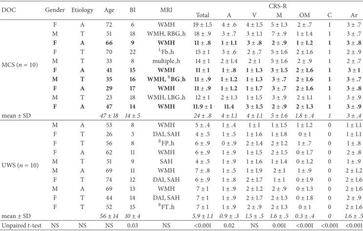
Table 1: The clinical and demographic characteristics of the whole sample. We reported the monthly individual and group CRS-R scores±
SD (the CRS-R was daily administered for 30 consecutive days before protocol enrollment), with the unpaired𝑡-test values. The MCS patients
who ameliorated at the auditory function (𝑛. 3, 6, 7, 8, and 10) at 𝑇postfollowing the real protocol are marked in bold.
DOC Gender Etiology Age BI MRI CRS-R
Total A V M OM C Ar MCS (𝑛 = 10) F A 72 6 WMH 19± 1.5 4± .6 4± 1.5 5 ± 1.3 2± .7 1 3± .7 M T 51 18 WMH, RBG h 18± .9 3± .7 3± 1.1 7± .9 1± 1.4 1 3± .7 F A 66 9 WMH 11 ± .8 1 ± 1.1 3 ± .8 2 ± .9 1 ± 1.2 1 3 ± .8 F T 70 22 LFb h 15± 1 3± .6 2± .7 5± 1.6 2± 1.6 1 2± .9 M T 33 8 multiple h 14± 1 2± 1.4 2± 1 5± 1.6 2± .9 1 2± .7 F A 41 15 WMH 11 ± 1 1 ± .8 1 ± 1.3 3 ± 1.5 2 ± 1.6 1 3 ± 1 M T 35 16 WMH,RBG h 11 ± .9 1 ± 1.2 1 ± 1.3 3 ± .7 2 ± 1.6 1 3 ± .7 F A 29 17 WMH 11 ± .9 1 ± 1.2 1 ± 1.7 3 ± .7 2 ± 1.6 1 3 ± .8 M T 23 18 WMH, LBG h 12± 1 2± 1.3 1± 1.5 3± .9 2± 1.1 1 3± .9 F A 47 14 WMH 11.9 ± 1 11.4 3 ± 1.5 2 ± .9 2 ± 1.3 1 3 ± .9 mean± SD 47± 18 14 ± 5 24± .8 4± 1.1 4± 1.1 5 ± 1.6 1.8 ± .4 1 3± .4 UWS (𝑛 = 10) M A 53 8 WMH 5± .4 1± .4 1± 1 1± 1.5 1± 1.2 0 1± 1.1 F T 26 3 DAI, SAH 4± .5 1± .5 1± 1.6 1± 1.8 0± 1 0 1± 1.1 F T 56 8 RFP h 6± .9 0± .9 2± 1.4 2 ± 1.2 1± .7 0 1± .8 F A 62 11 WMH 6± .9 1± .9 1± 1.5 2± 1.5 0± 1.7 0 2± .8 M T 51 9 SAH 4± .5 1± .9 1± 1.6 1± 1.4 0± 1.2 0 1± .9 M A 69 11 WMH 7± .8 1± .5 1± 1.9 2± 1 1± .9 0 2± 1.2 F T 74 12 DAI, SAH 6± .9 1± .8 2± 1.7 1± 1 0± 1.9 0 2± 1.6 M A 69 13 WMH 7± 1 1± .9 2± 1.2 2± .9 0± 1.3 0 2± 1.6 F T 44 14 DAI, SAH 7± 1 1± .9 2± 1.7 2 ± 1.3 0± 1.8 0 2± .9 F T 52 15 RFT h 7± 1 1± .9 2± .9 2± 1.3 0± 1 0 2± 1.6 mean± SD 56± 14 10 ± 4 5.9± 1.1 0.9 ± .3 1.5 ± .5 1.6 ± .5 0.3 ± .4 0 1.6± .5 Unpaired𝑡-test NS NS NS 0.03 NS <0.001 0.02 NS 0.001 <0.001 <0.001 <0.001
Age in years; BI: brain injury onset in months; CRS-R: Coma Recovery Scale-Revised including auditory (A), visual (V), motor (M), oromotor (OM), communication domain (C), and arousal induction (Ar); etiology: A, postanoxic; T, posttraumatic brain injury; MRI: structural patterns including WMH (white matter hyperintensity), h (hemorrhagic lesion),RFP (right frontopolar),RBG (basal ganglia),LFb (left frontobasal), SAH (subarachnoid hemorrhage), and DAI (diffuse axonal injury); SD: standard deviation.
Hence, aim of the current study was to investigate whether it was possible to induce plasticity within the motor system by applying an audiomotor PAS protocol in DOC patients. To this end, we paired a 5 Hz repetitive TMS (rTMS) over the left M1 with a transauricular repetitive electric stim-ulation (rES) of the right acoustic nerve in a DOC sample and in healthy individuals (HC). We hypothesized that such paired protocol could induce a M1 excitability increase through the recruitment of residual audiomotor pathways, thus allowing us to differentiate MCS (that should show residual connectivity properties) from UWS individuals (who should lack of such properties), besides the clinical assess-ment.
2. Materials and Methods
2.1. Subjects. Of the 47 chronic DOC subjects who attended
over two years to the Neurorehabilitation Unit of the IRCCS Centro Neurolesi “Bonino-Pulejo” (Messina, Italy), we en-rolled 20 patients who met the criteria for vegetative state and MCS diagnosis [2, 23, 24] and the following inclusion criteria:
a DOC condition lasting more than 3 months after the brain injury; no other severe neurological or systemic diseases; no critical conditions (i.e., inability to breathe indepen-dently and hemodynamic instability); no cortical excitability-modifying drugs assumption beyond L-DOPA and baclofen; absence of epileptic history, pace-maker, aneurysms clips, neurostimulator, brain/subdural electrodes or other elec-tromechanical devices; absence of electroencephalographic (EEG) burst-suppression pattern; presence of long-latency auditory evoked potentials (LLAEP); no lesion of eardrum or external meatus. In addition, we included 10 HC (6 females and 4 males, mean age: 45.3± 6.2 years) as control group in the study.
We resumed the clinical and demographic characteris-tics in Table 1. DOC etiology consisted of postanoxic or posttraumatic brain damage. The neurological examination mainly showed a pattern of spastic tetraparesis. Two neurolo-gists, skilled in DOC diagnosis, independently evaluated the patients through the JFK CRS-R, which was daily adminis-tered for 30 days consecutively, at different times, in order to steadily establish the level of consciousness impairment. EEG
Tpre MEP AMI LLAEP Auditory CRS-R MEP AMI LLAEP Auditory CRS-R MEP AMI LLAEP Auditory CRS-R ES ES ES ES ES ES TMS TMS TMS TMS TMS TMS
Auditory function scale (4) Consistent movements to command (3) Reproducible movements to command (2) Localization to sound
(1) Auditory startle (0) None
Auditory function scale (4) Consistent movements to command (3) Reproducible movements to command (2) Localization to sound
(1) Auditory startle (0) None
Auditory function scale (4) Consistent movements to command (3) Reproducible movements to command (2) Localization to sound (1) Auditory startle (0) None rTMS rTMS rES rES Conditioning protocol real_protocol rTMS_alone rES_alone sham_rES sham_rTMS Tpost T+30
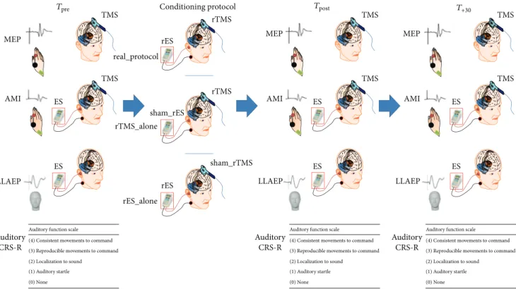
Figure 1: Resuming the experimental design. We measured before (𝑇pre) and after (𝑇postand𝑇+30) each conditioning protocol (real protocol,
rTMS alone, and rES alone) the motor evoked potential (MEP) amplitude, the audiomotor integration (AMI) strength, the long-latency auditory evoked potentials (LLAEP) latency and amplitude, and the CRS-R auditory function.
examination evidenced continuous slowing in theta and/or delta frequency ranges.
Our Research Institute Ethics Committee approved the present study and either the HC or the legal guardian of each patient gave their written informed consent.
2.2. Experimental Design. HC were seated on a comfortable
reclining chair, in a mild-lighted room during the entire experimental procedure, whereas the patients were lying in their bed. At baseline (𝑇pre), we assessed the audiomotor domain score of the CRS-R (in DOC patients), the resting motor threshold (RMT), the motor evoked potential (MEP) peak-to-peak amplitude, the LLAEP latency and amplitude, and the strength of audiomotor interaction (AMI). Then, each participant underwent three different protocols, admin-istered in a random scheme at one-day interval: (i) a real protocol (rTMS paired to rES); (ii) a rTMS alone (i.e., rTMS paired to a sham rES); and (iii) a rES alone (i.e., rES paired to a sham rTMS). We repeated the aforementioned baseline measures immediately (𝑇post) and 30 minutes after (𝑇+30) the application of each conditioning protocol. The experimental design is summarized in Figure 1. The experimenters who analyzed the data were blinded on the scheme procedure.
2.3. Clinical Assessment. The JFK CRS-R is a reliable and
standardized scale that integrates neuropsychological and clinical assessment; it includes the current diagnostic criteria for coma, VS, and MCS and allows the clinician to assign
a patient to the most appropriate diagnostic category. Hence, the CRS-R represents a good approach for characterizing the level of consciousness and for monitoring the neurobehav-ioral function recovery [24].
2.4. Motor Evoked Potentials. We positioned the coil over
the optimum position (hot-spot) to elicit a stable MEP of 0.5 mV peak-to-peak amplitude in the right first dorsal interosseous (FDI) muscle at rest. The hot-spot was identified by moving the coil in 0.5 cm steps around the presumed hot-spot. The coil was held tangentially to the scalp, with the handle pointing backwards and laterally to 45∘ from the midline (approximately perpendicular to the line of the central sulcus). We thus estimated the RMT, which was defined as the minimum intensity able to evoke a peak-to-peak MEP amplitude of 50𝜇V in at least five-out-of-ten consecutive trials in the relaxed FDI muscle [25]. Therefore, fifteen MEPs were recorded from the right FDI muscle at rest (using a stimulation intensity of 120% of RMT) at baseline (𝑇pre), immediately (𝑇post), and 30 minutes after (𝑇+30) the application of each conditioning protocol. The peak-to-peak amplitude of each MEP was measured offline, and the mean amplitude was calculated. MEP amplitude changes were calculated as percent of the baseline MEP (𝑇pre).
We used a high-power Magstim 200 stimulator (Magstim, Whitland, Dyfed, UK) and a standard figure-of-eight coil, with external loop diameters of 9 cm. The magnetic stimuli had monophasic pulse configuration and a rise-time of ∼100 𝜇s, decaying back to zero over ∼800 𝜇s. The coil current
during the rising phase of the magnetic field flowed toward the handle. Thus, the induced current in the cortex flowed in a posterior-to-anterior direction.
2.5. Long-Latency Auditory Evoked Potentials. Since a
stan-dard AEP assessment in DOC patients is extremely challeng-ing owchalleng-ing to the low and inconsistent cooperation, we chose a rES approach [26] in order to elicit LLAEP. We used a battery-driven stimulator (Brain Stim, E.M.S., Bologna, Italy) with a couple of silver electrodes. The stimulation electrode (a silver ball) was placed in the right external auditory meatus near the eardrum (after having flushed the external auditory meatus with physiologic saline solution) and the reference electrode (a silver disk) on the skin of the patient’s neck (near the right mastoid). We delivered two consecutive trains of 200 electric stimuli (500 Hz sine tones at an intensity of 500𝜇A, at 5 Hz). The intertrain interval was 30 sec. The stimulation procedure induced a hearing sensation of intermediate loud-ness in the HC. Each participant wore an earplug in the left ear. During the stimulation, we recorded the EEG from electrode Cz referring to the right mastoid using Ag/AgCl electrodes. An electrode at the centre of forehead served as ground. Two additional channels were employed for the electrooculogram (active electrode on the left supraorbital position and the reference electrode on the left infraorbital position). Impedance was ≤10 kΩ. Signals were digitized (A/D = 1000 Hz), amplified (1000 times), and filtered (0.15– 100 Hz, 50 Hz-notched) through a 1401 plus AD laboratory interface (Cambridge Electronic Design, Cambridge, UK) and a Digitimer D360 (Digitimer Ltd., Welwyn Garden City, UK) and stored on a personal computer for offline analysis (Signal software, Cambridge Electronic Design, UK). Then, data were processed by artifact rejecting (±100 𝜇V and by subtracting ocular artifacts), epoch from −100 to 500 ms, filtered (1–30 Hz, 12 dB/octave) and averaged. Hence, we registered a cortical triphasic positive-negative-positive potential (P1-N1-P2), starting at around 50 ms in the HC, in analogy to previous LLAEP findings [27, 28]. We measured the component latencies and the baseline-peak amplitude of N1. Latencies were determined by using a modified box-plot method known as the median rule.
2.6. Audiomotor Integration. In analogy to a previous work
[14], we applied pairs of stimuli consisting of a conditioning stimulus (500 Hz sine tone burst) followed by a magnetic test (90% of AMT), with an interstimulus interval of subject’s N1 peak-latency +50 ms [20, 29, 30]. Although it has been reported that speech sounds topographically activate the motor cortex (e.g., [21]), others suggest that the motor cortex might be also nontopographically activated by nonspeech sounds [31]. We registered 15 MEP (test MEP) intermingled with 15 electric-magnetic pairs of stimuli interactions (con-ditioned MEP) in a single trial, delivered at a frequency of 0.2 Hz at baseline (𝑇pre) and immediately (𝑇post) and 30 min-utes after (𝑇+30) the application of each conditioning protocol. We measured the mean amplitude of the conditioned MEP as percentage of the amplitude of the unconditioned MEP (test MEP), which was taken as a measure of the strength of AMI.
2.7. rTMS and rES. rTMS was employed in either the real
protocol or the rTMS alone. We delivered 600 stimuli at a frequency of 5 Hz (3 blocks of 200 pulses in 40 seconds, intertrain interval of 10 seconds). The intensity of magnetic stimulation was set at 90% of RMT. For the sham rTMS, we used the same abovementioned set-up, but with a sham coil. Each rTMS protocol was carried out in accordance with published safety recommendations [32].
Repetitive magnetic stimuli were delivered through a figure-of-eight coil connected to a Magstim Rapid stimulator (Magstim Company, Whitland, Dyfed, UK), with a biphasic waveform of the magnetic stimulus and a pulse width of ∼300 𝜇s. The coil was positioned over the hot-spot for the right FDI muscle. During the first phase of the biphasic stimulus, the current flowed in the coil toward the handle and induced a posterior-anterior current within the brain. EMG activity of the right FDI muscle was continuously monitored through loudspeakers throughout the entire rTMS session.
rES was employed in either the real protocol or the rES alone. It consisted of 600 bursts of 500 Hz sine tone at 5 Hz (3 blocks of 200 pairs in 40 seconds, intertrain interval of 10 seconds) in the right ear, delivered through the aforementioned battery-driven stimulator. With regard to the sham rES, the electric stimulator was switched off after 30 sec.
2.8. Conditioning Protocols. Each participant underwent
three different conditioning protocols, administered in a ran-dom scheme (i, ii, and iii) and in different sessions, at one-day interval:
(i) The real protocol, which consisted of rTMS paired to rES, thus delivering 600 pairs of electric-magnetic stimuli at a frequency of 5 Hz, with an interstimulus interval of subject’s N1 latency +50 ms (as in AMI). (ii) The rTMS alone (i.e., rTMS paired to a sham rES),
in which the electric stimulator was switched off after 30 sec (thus 600 pairs of sham electric stimuli and real TMS pulses).
(iii) The rES alone (i.e., rES paired to a sham rTMS), in which we used a sham rTMS coil (thus 600 pairs of real electric stimuli and sham TMS pulses).
2.9. Statistical Analysis. We compared the baseline
clini-cal and electrophysiologiclini-cal parameters among HC, MCS patients, and UWS patients, through unpaired𝑡-tests (calcu-lated on the mean of the three𝑇prevalues). We thus evaluated the effects of the conditioning protocols on each electrophys-iological variable (RMT%, MEP amplitude, AMI strength, and LLAEP latency and amplitude) through separated three-way repeated-measure analyses of variance (rmANOVA), implying time (three levels:𝑇pre,𝑇post, and𝑇+30) and protocol (three levels: real protocol, rTMS alone, and rES alone), as within-subject factors, and group (three levels: MCS patients, UWS patients, and HC) as between-subject factor. The effect of the conditioning protocols on audiomotor CRS-R was measured through a Wilcoxon test. The Greenhouse-Geisser method was used if necessary to correct for nonsphericity.
Conditional on a significant𝐹 value, we performed post hoc 𝑡-tests (Bonferroni) to explore the strength of main effects and the patterns of interaction between the experimental factors. All statistical tests were applied two-tailed. A significant𝑝 value was <0.05. All data are given as means or percent changes ±se. We calculated a Spearman correlation test in order to assess an eventual correlation among clinical and electrophysiological parameters.
3. Results
We did not observe any side effect in both the patients and HC, either during or after the entire experimental procedure.
3.1. DOC/HC Clinical and Electrophysiological Differences at Baseline. We resumed the DOC sample demographic
char-acteristics and the monthly CRS-R scores in Table 1. There were no significant MCS-UWS differences concerning the demographic characteristics, except for slightly longer dis-ease duration in the MCS than the UWS patients. Instead, the monthly and daily CRS-R scores were significantly higher in the MCS than the UWS individuals (≤7). Daily CRS-R scores in each patient showed a relatively low variability during the 30-day observation period. The auditory CRS-R score at each𝑇pre was superimposable to the monthly CRS-R score in each patient. Similarly, the baseline electrophysiological parameters were similar and stable during the three days of experimentation. We reported the raw values of the electrophysiological parameters at𝑇pre(calculated as mean of the three𝑇prevalues) for each participant in Table 2. RMT and MEP amplitudes were similar in the three groups. The LLAEP amplitude was slightly reduced only in the UWS individuals, whereas LLAEP latency was significantly increased in the DOC participants (more in the UWS than the MCS patients). The stimulation set-up we used to elicit AMI induced clear inhibitory effects on MEP amplitude in the HC, but such effects were reduced in the MCS patients and nearly absent in the UWS patients.
3.2. Conditioning Protocol’s Effects on Clinical Assessment.
The Wilcoxon test showed a statistically significant increase of the audiomotor CRS-R score only in the MCS patients after the real protocol at𝑇post (𝑝 = 0.04). Indeed, five MCS patients (numbers 3, 6, 7, 8, and 10) upgraded from a𝑇pre “auditory startle” response (1 point at the CRS-R auditory function scale) to a “localization to sound” (2 points) at𝑇post (Table 1).
3.3. Conditioning Protocol Electrophysiological Effects. We
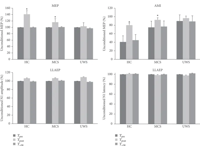
resumed in Table 2 and in Figure 2 the time course of elec-trophysiological parameters following each protocol. We summarized the data statistical analysis in Table 3. The RMT and LLAEP latency and amplitude did not significantly vary after each conditioning protocol. MEP and AMI amplitude significantly increased only in the HC and MCS patients after the real protocol at𝑇post. Instead, the𝑇+30values were com-parable to𝑇pre(Figure 2). Notably, none of the UWS patients showed any protocol-induced effect (Figure 2). There were no
significant differences concerning the protocol posteffects in relation to the clinical and demographic characteristics. Inter-estingly, we observed a correlation trend between audiomotor CRS-R amelioration and AMI modulation at𝑇post(𝑟 = 0.576, 𝑝 = 0.07).
4. Discussion
For the first time ever, we assessed the presence of residual audiomotor functional plasticity in a DOC sample by means of an audiomotor PAS. Only the real protocol (rTMS + rES) induced strengthening of the M1 excitability (MEP ampli-tude increase) and a modification of audiomotor functional connectivity (weakening of inhibitory AMI) in the HC and MCS patients. Such posteffects were paralleled by a transient audiomotor CRS-R score improvement in some MCS patients (i.e., from “auditory startle” to “sound localization”). On the contrary, the UWS patients did not show any clear posteffect. The clinical and electrophysiological ameliorations in HC and MCS patients mainly depended on the type of the condi-tioning protocol that was employed, as also previously shown in healthy individuals [14, 19]. In fact, neither the rTMS alone nor the rES alone induced any significant posteffect. Indeed, PAS has been suggested to induce associative LTP or LTD-like neuronal synapses via mechanisms of spike-timing depen-dent synaptic plasticity [18]. Therefore, in our patients, the real-protocol modulated the audiomotor connectivity prob-ably through time-locked neural activity encompassing the primary auditory area and M1. It has been hypothesized that plasticity and connectivity recovery in individuals suffering from DOC might depend on the modulation of postischemic LTP, the production of specific neurotrophins, and the reg-ulation of excitatory/inhibitory dynamics within corticotha-lamocortical circuits [33–37]. Thereby, it is conceivable that one or more of these mechanisms may have been triggered by the real protocol and could have favored the recruitment of silent or stunned residual corticothalamocortical projections, thus enhancing the behavioral output in some of our patients. To this end, we could hypothesize the enrolment of a wide audiomotor network including multiple and interconnected cortical areas (encompassing primary auditory cortex, motor areas, and prefrontal cortex) and probably other cortical and subcortical areas (maybe the cerebellum and the basal ganglia) [38–40]. Such network could hierarchically organize different audiomotor processes, thus allowing a repertoire of audiomotor responses ranging from protective reflex motor activations to complex feedback and feedforward processes regarding purposeful motor responses [38, 41–49].
We can therefore argue that the enhancement of the audiomotor clinical responses in the MCS patients could express a functional upgrading, although transient, of the residual brainstem-thalamocortical and corticocortical net-works supporting AMI processes, so as to get a higher and more complex motor behavior. On the other hand, our data further confirm the connectivity impairment affecting UWS individuals within audiomotor integration pathways [1, 8]. Nevertheless, the presence of residual functional connectivity in some UWS patients has been evidenced within other
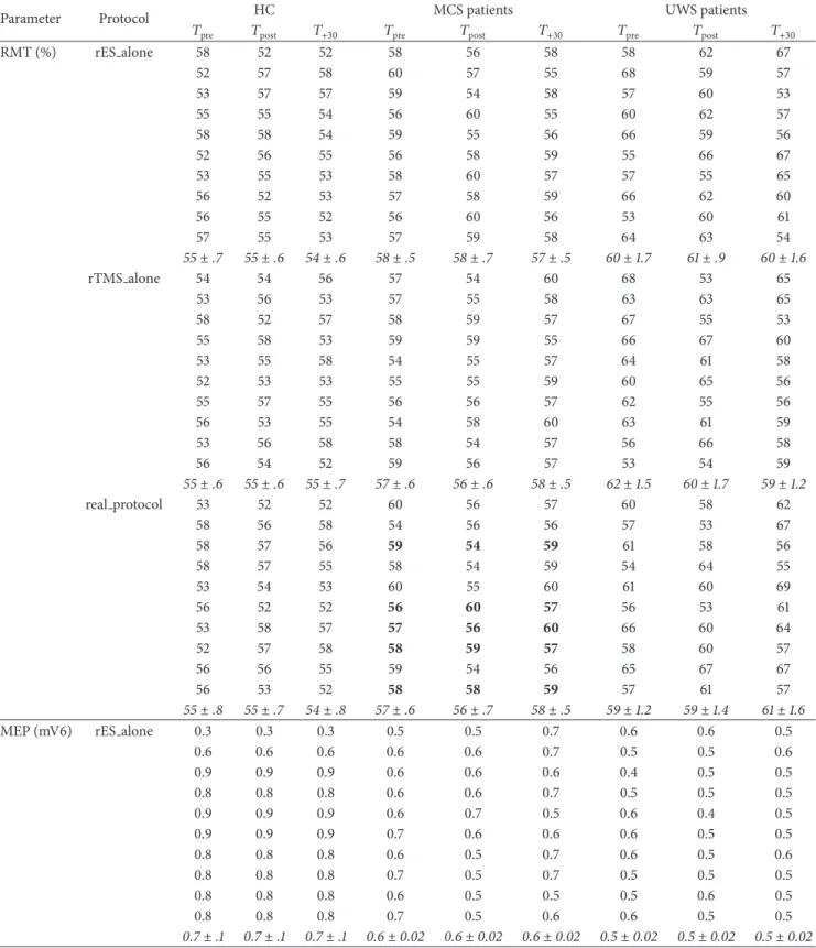
Table 2: The time course of the individual raw values of the electrophysiological parameters in the HC and MCS and UWS participants.
At𝑇pre, there were not significant differences among the three groups concerning RMT% and MEP amplitude. MCS displayed partially
preserved audiomotor connectivity, although weakened (i.e., increased AMI). The UWS showed instead markedly weakened AMI and a global impairment of cortical excitability, connectivity, and LLAEP N1 latency and amplitude (increased and decreased, resp.). Data are
summarized as mean± se (in italic). The MCS patients who ameliorated at the auditory function (𝑛. 3, 6, 7, 8, and 10) at 𝑇postfollowing the
real protocol are marked in bold.
(a)
Parameter Protocol HC MCS patients UWS patients
𝑇pre 𝑇post 𝑇+30 𝑇pre 𝑇post 𝑇+30 𝑇pre 𝑇post 𝑇+30
RMT (%) rES alone 58 52 52 58 56 58 58 62 67 52 57 58 60 57 55 68 59 57 53 57 57 59 54 58 57 60 53 55 55 54 56 60 55 60 62 57 58 58 54 59 55 56 66 59 56 52 56 55 56 58 59 55 66 67 53 55 53 58 60 57 57 55 65 56 52 53 57 58 59 66 62 60 56 55 52 56 60 56 53 60 61 57 55 53 57 59 58 64 63 54 55± .7 55± .6 54± .6 58± .5 58± .7 57± .5 60± 1.7 61± .9 60± 1.6 rTMS alone 54 54 56 57 54 60 68 53 65 53 56 53 57 55 58 63 63 65 58 52 57 58 59 57 67 55 53 55 58 53 59 59 55 66 67 60 53 55 58 54 55 57 64 61 58 52 53 53 55 55 59 60 65 56 55 57 55 56 56 57 62 55 56 56 53 55 54 58 60 63 61 59 53 56 58 58 54 57 56 66 58 56 54 52 59 56 57 53 54 59 55± .6 55± .6 55± .7 57± .6 56± .6 58± .5 62± 1.5 60± 1.7 59± 1.2 real protocol 53 52 52 60 56 57 60 58 62 58 56 58 54 56 56 57 53 67 58 57 56 59 54 59 61 58 56 58 57 55 58 54 59 54 64 55 53 54 53 60 55 60 61 60 69 56 52 52 56 60 57 56 53 61 53 58 57 57 56 60 66 60 64 52 57 58 58 59 57 58 60 57 56 56 55 59 54 56 65 67 67 56 53 52 58 58 59 57 61 57 55± .8 55± .7 54± .8 57± .6 56± .7 58± .5 59± 1.2 59± 1.4 61± 1.6
MEP (mV6) rES alone 0.3 0.3 0.3 0.5 0.5 0.7 0.6 0.6 0.5
0.6 0.6 0.6 0.6 0.6 0.7 0.5 0.5 0.6 0.9 0.9 0.9 0.6 0.6 0.6 0.4 0.5 0.5 0.8 0.8 0.8 0.6 0.6 0.7 0.5 0.5 0.5 0.9 0.9 0.9 0.6 0.7 0.5 0.6 0.4 0.5 0.9 0.9 0.9 0.7 0.6 0.6 0.6 0.5 0.5 0.8 0.8 0.8 0.6 0.5 0.7 0.6 0.5 0.6 0.8 0.8 0.8 0.7 0.5 0.7 0.5 0.5 0.5 0.8 0.8 0.8 0.6 0.5 0.5 0.5 0.6 0.5 0.8 0.8 0.8 0.7 0.5 0.6 0.6 0.5 0.5 0.7± .1 0.7± .1 0.7± .1 0.6± 0.02 0.6± 0.02 0.6± 0.02 0.5± 0.02 0.5± 0.02 0.5± 0.02
(a) Continued.
Parameter Protocol HC MCS patients UWS patients
𝑇pre 𝑇post 𝑇+30 𝑇pre 𝑇post 𝑇+30 𝑇pre 𝑇post 𝑇+30
rTMS alone 0.3 0.3 0.3 0.7 0.5 0.7 0.5 0.5 0.6 0.6 0.6 0.6 0.6 0.6 0.5 0.4 0.5 0.5 0.9 0.9 0.9 0.7 0.5 0.7 0.6 0.5 0.5 0.8 0.8 0.8 0.7 0.6 0.6 0.6 0.5 0.5 0.9 0.9 0.9 0.6 0.6 0.6 0.6 0.6 0.6 0.9 0.9 0.9 0.5 0.6 0.6 0.6 0.6 0.4 0.8 0.8 0.8 0.7 0.6 0.5 0.4 0.6 0.4 0.8 0.8 0.8 0.6 0.5 0.7 0.5 0.4 0.4 0.8 0.8 0.8 0.6 0.6 0.5 0.5 0.6 0.4 0.8 0.8 0.8 0.7 0.6 0.6 0.5 0.5 0.5 0.7± .1 0.7± .1 0.7± .1 0.6± .1 0.6± .1 0.6± .1 0.5± 0.02 0.5 ± 0.02 0.5 ± 0.02 real protocol 0.3 0.7 0.3 0.8 0.8 0.8 0.5 0.6 0.5 0.6 0.8 0.6 0.9 0.9 0.9 0.5 0.5 0.5 0.9 1.2 0.9 0.6 0.8 0.6 0.4 0.6 0.4 0.8 1.0 0.8 0.8 0.9 0.8 0.6 0.4 0.4 0.9 1.2 0.9 0.8 0.8 0.8 0.4 0.5 0.5 0.9 1.2 0.9 0.9 1 0.9 0.4 0.5 0.5 0.8 1.1 0.8 0.8 1 0.8 0.5 0.5 0.5 0.8 1.1 0.8 0.3 0.9 0.3 0.6 0.5 0.5 0.8 1.2 0.8 0.8 0.8 0.8 0.5 0.5 0.4 0.8 1.1 0.8 0.9 1 0.9 0.5 0.5 0.6 0.7± .1 1.1± .1 0.7± .1 0.7± .1 0.9± .05 0.7± .1 0.5± 0.02 0.5 ± 0.02 0.5 ± 0.02
AMI (mV) rES alone 0.5 0.4 0.3 0.6 0.5 0.5 0.6 0.5 0.5
0.3 0.5 0.4 0.4 0.5 0.5 0.4 0.5 0.5 0.4 0.5 0.3 0.4 0.5 0.5 0.4 0.5 0.5 0.5 0.3 0.4 0.5 0.5 0.5 0.5 0.5 0.5 0.5 0.5 0.3 0.4 0.6 0.5 0.4 0.6 0.5 0.5 0.3 0.4 0.5 0.4 0.5 0.5 0.4 0.5 0.3 0.3 0.4 0.4 0.5 0.6 0.4 0.5 0.6 0.5 0.4 0.3 0.5 0.5 0.4 0.5 0.5 0.4 0.3 0.4 0.3 0.5 0.6 0.4 0.5 0.6 0.4 0.5 0.4 0.3 0.5 0.4 0.4 0.5 0.4 0.4 0.4± 0.02 0.4 ± 0.02 0.4 ± 0.02 0.5 ± 0.02 0.5 ± 0.02 0.5 ± 0.02 0.5 ± 0.02 0.5 ± 0.02 0.5 ± 0.02 rTMS alone 0.5 0.3 0.4 0.5 0.4 0.5 0.5 0.4 0.5 0.5 0.4 0.4 0.5 0.5 0.6 0.5 0.5 0.6 0.4 0.3 0.3 0.5 0.6 0.5 0.5 0.6 0.5 0.3 0.3 0.4 0.5 0.4 0.5 0.5 0.4 0.5 0.3 0.5 0.4 0.6 0.4 0.6 0.6 0.4 0.6 0.3 0.3 0.5 0.5 0.5 0.5 0.5 0.5 0.5 0.3 0.3 0.4 0.5 0.5 0.4 0.5 0.5 0.4 0.4 0.3 0.3 0.4 0.5 0.5 0.4 0.5 0.5 0.4 0.3 0.5 0.6 0.5 0.5 0.6 0.5 0.5 0.4 0.4 0.5 0.6 0.5 0.5 0.6 0.5 0.5 0.4± 0.02 0.4 ± 0.01 0.4 ± 0.02 0.5 ± 0.02 0.5 ± 0.02 0.5 ± 0.02 0.5 ± 0.02 0.5 ± 0.02 0.5 ± 0.02 real protocol 0.5 0.9 0.4 0.6 0.3 0.5 0.6 0.4 0.6 0.4 1 0.3 0.4 0.6 0.4 0.5 0.4 0.5 0.3 1 0.4 0.6 0.9 0.5 0.5 0.4 0.4 0.3 0.8 0.5 0.5 0.8 0.5 0.6 0.6 0.6 0.4 0.9 0.5 0.4 0.9 0.4 0.5 0.6 0.5
(a) Continued.
Parameter Protocol HC MCS patients UWS patients
𝑇pre 𝑇post 𝑇+30 𝑇pre 𝑇post 𝑇+30 𝑇pre 𝑇post 𝑇+30
0.5 0.8 0.3 0.5 0.9 0.6 0.6 0.5 0.4 0.4 0.8 0.4 0.4 0.8 0.5 0.4 0.5 0.5 0.4 0.9 0.4 0.5 0.8 0.6 0.5 0.5 0.5 0.5 1 0.4 0.4 0.8 0.6 0.5 0.5 0.5 0.4 0.8 0.5 0.4 0.8 0.5 0.5 0.6 0.6 0.4± .03 0.9 ± .05 0.4 ± .02 0.5 ± 0.5 0.7 ± .1 0.5 ± 0.02 0.5 ± 0.02 0.5 ± 0.02 0.5 ± 0.02
LLAEP amplitude (𝜇V) rES alone 16 3 7 7 11 11 5 0 2
6 4 2 11 11 7 4 2 5 3 6 7 10 10 13 0 7 1 14 1 8 11 9 6 7 6 2 2 3 6 6 12 5 2 0 0 9 10 15 5 11 8 6 7 2 16 8 15 12 7 12 6 7 6 10 12 11 12 5 5 5 1 4 1 5 16 5 13 9 6 6 2 9 3 16 8 6 10 5 2 6 9± 1 6± 2 10± 2 9± 1 10± 1 9± 1 5± 1 4± 0.4 3± 1 rTMS alone 16 0 2 12 8 7 2 1 7 7 10 7 13 13 6 3 1 4 7 6 8 12 11 8 7 7 6 16 15 10 5 11 5 2 7 4 14 1 0 8 9 11 4 7 6 16 8 4 12 8 7 4 6 2 5 10 11 6 11 13 5 7 4 5 16 13 5 13 10 6 1 5 15 9 5 6 9 10 3 4 0 1 2 2 7 13 13 2 1 4 10± 2 8± 2 6± .4 9± 1 11± 1 9± 1 4± 1 4± 1 4± 1 real protocol 12 3 13 9 9 11 0 0 2 16 5 6 9 9 7 0 1 4 6 6 6 12 12 10 7 4 5 3 16 2 13 11 8 6 2 2 12 10 2 7 9 12 7 3 6 4 7 16 11 6 5 2 4 3 6 11 10 10 5 9 4 5 6 14 15 8 13 11 13 3 2 2 3 3 14 10 11 6 3 5 6 4 14 6 9 5 7 5 6 4 8± 2 9± 1 8± 1 10± .4 9± 1 9± 1 4± .4 3± 1 4± 1
LLAEP latency (ms) rES alone 89 124 100 111 118 114 131 124 123
81 95 90 98 117 126 162 135 125 95 127 133 95 115 131 117 118 106 84 97 110 110 147 135 162 163 121 89 96 127 111 145 114 161 161 119 95 105 126 110 130 151 164 126 122 82 110 88 109 140 140 139 138 110 79 84 103 102 107 115 130 140 107 91 109 105 108 132 143 138 133 106 86 94 103 101 136 128 116 148 108 87± 2 104± 4 109± 5 106± 2 129 ± 4 130± 4 142± 3 138± 5 115± 6
(a) Continued.
Parameter Protocol HC MCS patients UWS patients
𝑇pre 𝑇post 𝑇+30 𝑇pre 𝑇post 𝑇+30 𝑇pre 𝑇post 𝑇+30
rTMS alone 89 109 97 113 111 133 126 123 124 81 86 82 103 98 111 171 125 157 95 130 106 103 95 105 135 106 146 84 107 89 148 110 126 132 121 153 89 95 117 122 111 111 166 119 129 95 119 104 110 110 115 142 122 147 82 95 107 132 109 117 146 110 134 79 104 84 131 102 114 131 107 127 91 113 105 138 108 128 145 106 126 86 109 100 135 101 108 113 108 110 87± 2 107± 4 99± 4 123± 2 106± 5 117± 3 141± 3 115± 6 135± 5 real protocol 89 116 117 155 119 111 128 137 159 81 97 90 124 122 98 106 146 139 95 96 114 102 110 95 133 110 121 84 86 96 135 107 110 147 126 128 89 120 111 139 106 111 142 140 119 95 121 124 135 122 110 132 146 168 82 108 106 111 110 109 132 153 125 79 86 94 103 107 102 103 119 109 91 97 116 116 106 108 150 123 110 86 105 97 117 108 101 126 115 132 87± 2 103± 4 106± 4 124± 2 112± 5 106± 5 130± 2 132± 5 131± 6 (b) Parameter 𝑝 HC/DOC 𝑝 MCS/UWS RMT (%) NS NS MEP (mV) NS NS AMI (%) 0.002 0.002 N1 latency (ms) <0.001 0.005 N1 amplitude (𝜇V) NS 0.05
AMI: audiomotor integration; LLAEP: long-latency auditory potential; MEP: motor evoked potential; NS: nonsignificant; rES: repetitive electric stimulation; RMT: resting motor threshold; rTMS: repetitive transcranial magnetic stimulation.
Table 3: We observed significant real protocol posteffects in the HC and MCS patients concerning MEP amplitude and AMI strength at
𝑇post, whereas the UWS patients did not show any significant posteffect. The rTMS alone and the rES alone did not induce any significant
posteffect. The nonsignificant interactions, factors, and time intervals are not shown.
Time× group × protocol
interaction𝐹(8,216),𝑝
Time× protocol interaction 𝐹(4,36),𝑝 Time effect
𝐹(2,18),𝑝 𝑡(1,9),𝑝 MEP amplitude 22,<0.001 HC 90,<0.001 90,<0.001 3.5, 0.001 MCS 53,<0.001 real protocol 12,<0.001 𝑇 0 3.6, 0.001 AMI % 13,<0.001 HC 78,<0.001 78,<0.001 3.5, 0.002 MCS 6.3, 0.006 9.4, 0.001 2.4, 0.02
sensory-motor modalities (e.g. [13]), thus allowing us to sup-pose a condition of functional locked-in syndrome [7, 50]. Hence, such issue needs to be further clarified in more detailed audiomotor integration studies.
Notably, we have to highlight other issues concerning the physiological effects of our combined real protocol:
(1) Maladaptive plasticity phenomena could play an im-portant role in limiting the range of our posteffects in all of the UWS and in some MCS patients [51]. (2) In the pioneering work of Sowman and coworkers
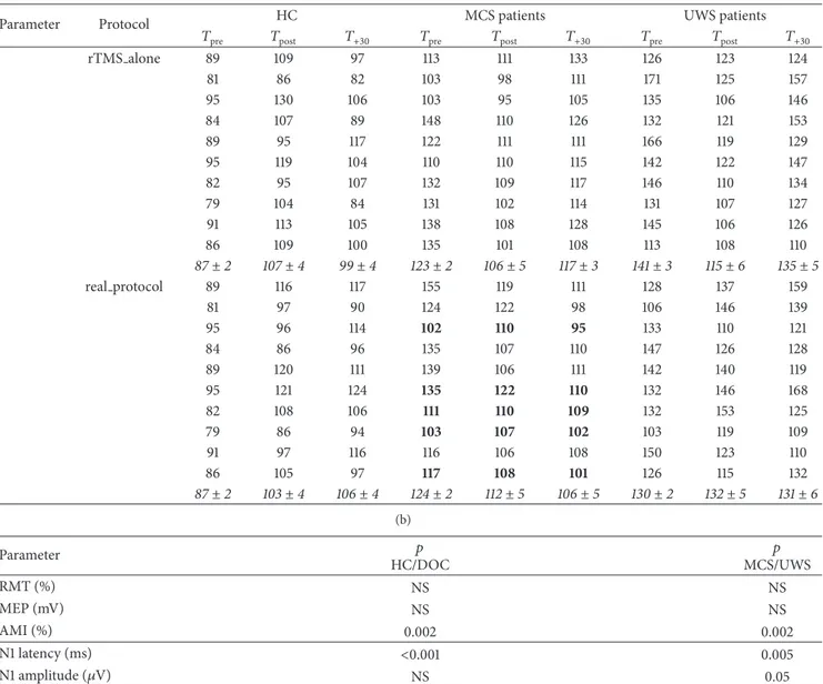
0 20 40 60 80 100 120 HC MCS UWS AMI 0 20 40 60 80 100 120 140 160 HC MCS UWS U n co ndi tio ned MEP (%) U n co ndi tio ned MEP (%) MEP 0 20 40 60 80 100 HC MCS UWS U n co ndi tio ne d N1 la te nc y (%) LLAEP 0 20 40 60 80 100 120 HC MCS UWS U n co ndi tio ne d N1 a m p li tude (%) LLAEP ∗ ∗ ∗ ∗ Tpre T+30 Tpost Tpre T+30 Tpost
Figure 2: It shows the electrophysiological posteffects induced by the real protocol. We observed a significant, although short lasting, increase of MEP and AMI amplitude in the HC and MCS patients, whereas the UWS patients did not show any significant effect. The latency and amplitude of LLAEP were affected in none of the groups. The values are reported as mean of the percent change in comparison to the baseline
value. The error bars refer to the se.∗ indicates a significant change (𝑝 < 0.05).
to TMS, being therefore the posteffects potentially dependent on phonological motor resonance and tonotopic-topographic specificity [20, 29], as also suggested by a recent study employing 1–4 kHz tones paired to primary auditory area rTMS [19]. Instead, we triggered brain networks with different tonotopic specificity, whereas the topographic specificity should be more deeply investigated (e.g., by studying the muscle involved in articulation).
(3) Since RMT, LLAEP, and MEP amplitude were not substantially different at baseline between HC and DOC and RMT and LLAEP did not vary after the conditioning protocols, we can exclude the possibility that baseline cortical excitability or LLAEP differ-ences could have influenced our posteffects.
(4) We may exclude differences in the attentive level in the HC participants in reason of their blinded condition concerning the different experimental sessions [52]. (5) The lack of rTMS alone posteffects on MEP
ampli-tude confirms the findings of a previous high-fre-quency PAS study in healthy individuals, in which
600 magnetic stimuli failed in producing a significant corticospinal excitability modulation [53]. Therefore, the heterologous sensory stimulation we employed (rTMS + rES) boosted up the cortical effects of rTMS, similarly to previous rapid PAS reports [53, 54]. The relatively small sample size and the consequent mixed etiology represent the main limiting factor in our study. Nonetheless, it is difficult to study a large sample of patients with DOC, since the negative outcome of such patients is still unfortunately high.
5. Conclusions
In our opinion, the present study shows a promising approach in an attempt to identify residual patterns of AMI in patients affected by severe DOC. Indeed, our data further support the importance of diagnostic approaches that are independent from patient’s cooperation, aimed at assessing the brain connectivity patterns, whose impairment is proportionally related to the awareness impairment. In addition, the pos-sibility to identify such partially preserved corticocortical and corticosubcortical networks in DOC may be useful in
the selection of candidate patients for therapeutic and reha-bilitative trials by means of noninvasive neurostimulation approaches.
Conflict of Interests
The authors declare that there is no conflict of interests regarding the publication of this paper.
References
[1] S. Laureys, M.-E. Faymonville, C. Degueldre et al., “Auditory processing in the vegetative state,” Brain, vol. 123, no. 8, pp. 1589–1601, 2000.
[2] J. T. Giacino, S. Ashwal, N. Childs et al., “The minimally con-scious state: definition and diagnostic criteria,” Neurology, vol. 58, no. 3, pp. 349–353, 2002.
[3] K. Kalmar and J. T. Giacino, “The JFK coma recovery scale— revised,” Neuropsychological Rehabilitation, vol. 15, no. 3-4, pp. 454–460, 2005.
[4] S. Dehaene, J.-P. Changeux, L. Naccache, J. Sackur, and C. Sergent, “Conscious, preconscious, and subliminal processing: a testable taxonomy,” Trends in Cognitive Sciences, vol. 10, no. 5, pp. 204–211, 2006.
[5] A. Del Cul, S. Baillet, and S. Dehaene, “Brain dynamics underly-ing the non-linear threshold for access to consciousness,” PLoS
Biology, vol. 5, no. 10, article e260, 2007.
[6] A. Vanhaudenhuyse, Q. Noirhomme, L. J.-F. Tshibanda et al., “Default network connectivity reflects the level of consciousness in non-communicative brain-damaged patients,” Brain, vol. 133, no. 1, pp. 161–171, 2010.
[7] M.-A. Bruno, A. Vanhaudenhuyse, A. Thibaut, G. Moonen, and S. Laureys, “From unresponsive wakefulness to minimally conscious PLUS and functional locked-in syndromes: recent advances in our understanding of disorders of consciousness,”
Journal of Neurology, vol. 258, no. 7, pp. 1373–1384, 2011.
[8] M. Boly, M.-E. Faymonville, P. Peigneux et al., “Auditory pro-cessing in severely brain injured patients: differences between the minimally conscious state and the persistent vegetative state,” Archives of Neurology, vol. 61, no. 2, pp. 233–238, 2004. [9] M. Boly, M. I. Garrido, O. Gosseries et al., “Preserved
feedfor-ward but impaired top-down processes in the vegetative state,”
Science, vol. 332, no. 6031, pp. 858–862, 2011.
[10] A. M. Owen, M. R. Coleman, D. K. Menon et al., “Residual auditory function in persistent vegetative state: a combined PET and fMRI study,” Neuropsychological Rehabilitation, vol. 15, no. 3-4, pp. 290–306, 2005.
[11] F. Perrin, C. Schnakers, M. Schabus et al., “Brain response to one’s own name in vegetative state, minimally conscious state, and locked-in syndrome,” Archives of Neurology, vol. 63, no. 4, pp. 562–569, 2006.
[12] M. Cavinato, C. Volpato, S. Silvoni, M. Sacchetto, A. Merico, and F. Piccione, “Event-related brain potential modulation in patients with severe brain damage,” Clinical Neurophysiology, vol. 122, no. 4, pp. 719–724, 2011.
[13] A. Naro, R. S. Calabr`o, M. Russo et al., “Can transcranial direct current stimulation be useful in differentiating unrespon-sive wakefulness syndrome from minimally conscious state patients?” Restorative Neurology and Neuroscience, vol. 33, no. 2, pp. 159–176, 2015.
[14] P. F. Sowman, S. S. Dueholm, J. H. Rasmussen, and N. Mra-chacz-Kersting, “Induction of plasticity in the human motor cortex by pairing an auditory stimulus with TMS,” Frontiers in
Human Neuroscience, vol. 8, article 398, 2014.
[15] M. N. Thabit, Y. Ueki, S. Koganemaru, G. Fawi, H. Fukuyama, and T. Mima, “Movement-related cortical stimulation can induce human motor plasticity,” The Journal of Neuroscience, vol. 30, no. 34, pp. 11529–11536, 2010.
[16] N. Mrachacz-Kersting, S. R. Kristensen, I. K. Niazi, and D. Farina, “Precise temporal association between cortical poten-tials evoked by motor imagination and afference induces corti-cal plasticity,” The Journal of Physiology, vol. 590, no. 7, pp. 1669– 1682, 2012.
[17] A. Suppa, A. Biasiotta, D. Belvisi et al., “Heat-evoked exper-imental pain induces long-term potentiation-like plasticity in human primary motor cortex,” Cerebral Cortex, vol. 23, no. 8, pp. 1942–1951, 2013.
[18] K. Stefan, E. Kunesch, R. Benecke, L. G. Cohen, and J. Classen, “Mechanisms of enhancement of human motor cortex excitabil-ity induced by interventional paired associative stimulation,”
The Journal of Physiology, vol. 543, no. 2, pp. 699–708, 2002.
[19] M. Schecklmann, G. Volberg, G. Frank et al., “Paired associative stimulation of the auditory system: a proof-of-principle study,”
PLoS ONE, vol. 6, no. 11, Article ID e27088, 2011.
[20] L. Fadiga, L. Craighero, G. Buccino, and G. Rizzolatti, “Speech listening specifically modulates the excitability of tongue mus-cles: a TMS study,” European Journal of Neuroscience, vol. 15, no. 2, pp. 399–402, 2002.
[21] A. Fl¨oel, T. Ellger, C. Breitenstein, and S. Knecht, “Language per-ception activates the hand motor cortex: implications for motor theories of speech perception,” European Journal of
Neuro-science, vol. 18, no. 3, pp. 704–708, 2003.
[22] K. E. Watkins, A. P. Strafella, and T. Paus, “Seeing and hearing speech excites the motor system involved in speech production,”
Neuropsychologia, vol. 41, no. 8, pp. 989–994, 2003.
[23] The Multi-Society Task Force on PVS, “Medical aspects of the persistent vegetative state,” The New England Journal of
Medicine, vol. 330, no. 21, pp. 1499–1508, 1994.
[24] The Multi-Society Task Force on PVS, “Medical aspects of the persistent vegetative state the multi-society task force on PVS,”
The New England Journal of Medicine, vol. 330, no. 22, pp. 1572–
1579, 1994.
[25] P. Gerrard, R. Zafonte, and J. T. Giacino, “Coma recovery scale-revised: evidentiary support for hierarchical grading of level of consciousness,” Archives of Physical Medicine and
Rehabilita-tion, vol. 95, no. 12, pp. 2335–2341, 2014.
[26] L. Alibiglou and C. D. Mackinnon, “The early release of planned movement by acoustic startle can be delayed by transcranial magnetic stimulation over the motor cortex,” The Journal of
Physiology, vol. 590, no. 4, pp. 919–936, 2012.
[27] E. Hofmann, C. Preibisch, C. Knaus, J. M¨uller, C. Kremser, and C. Teissl, “Noninvasive direct stimulation of the cochlear nerve for functional MR imaging of the auditory cortex,” American
Journal of Neuroradiology, vol. 20, no. 10, pp. 1970–1972, 1999.
[28] A. P. F. Key, G. O. Dove, and M. J. Maguire, “Linking brainwaves to the brain: an ERP primer,” Developmental Neuropsychology, vol. 27, no. 2, pp. 183–215, 2005.
[29] E. M. Wassermann, “Risk and safety of repetitive transcranial magnetic stimulation: report and suggested guidelines from the International Workshop on the Safety of Repetitive Transcranial Magnetic Stimulation, June 5–7, 1996,” Electroencephalography
and Clinical Neurophysiology/Evoked Potentials Section, vol. 108,
no. 1, pp. 1–16, 1998.
[30] L. M. P. Ventura, K. D. F. Alvarenga, and O. A. Costa Filho, “Pro-tocol to collect late latency auditory evoked potentials,”
Bra-zilian Journal of Otorhinolaryngology, vol. 75, no. 6, pp. 879–883,
2009.
[31] A. C. Roy, L. Craighero, M. Fabbri-Destro, and L. Fadiga, “Phonological and lexical motor facilitation during speech lis-tening: a transcranial magnetic stimulation study,” Journal of
Physiology Paris, vol. 102, no. 1–3, pp. 101–105, 2008.
[32] P. M. Rossini, D. Burke, R. Chen et al., “Non-invasive electrical and magnetic stimulation of the brain, spinal cord and roots: basic principles and procedures for routine clinical application. Report of an IFCN committee,” Electroencephalography and
Clinical Neurophysiology, vol. 91, pp. 79–92, 1994.
[33] M. Liotti Mario, J. C. Ingham, O. Takai, D. K. Paskos, R. Perez, and R. J. Ingham, “Spatiotemporal dynamics of speech sound perception in chronic developmental stuttering,” Brain and
Language, vol. 115, no. 2, pp. 141–147, 2010.
[34] Z. Kokaia, G. Andsberg, Q. Yan, and O. Lindvall, “Rapid alter-ations of BDNF protein levels in the rat brain after focal ischemia: evidence for increased synthesis and anterograde axonal transport,” Experimental Neurology, vol. 154, no. 2, pp. 289–301, 1998.
[35] V. Crepel, C. Hammond, P. Chinestra, D. Diabira, and Y. Ben-Ari, “A selective LTP of NMDA receptor-mediated currents induced by anoxia in CA1 hippocampal neurons,” Journal of
Neurophysiology, vol. 70, no. 5, pp. 2045–2055, 1993.
[36] M. Di Filippo, A. Tozzi, C. Costa et al., “Plasticity and repair in the post-ischemic brain,” Neuropharmacology, vol. 55, no. 3, pp. 353–362, 2008.
[37] M.-C. Ding, Q. Wang, E. H. Lo, and G. B. Stanley, “Cortical excitation and inhibition following focal traumatic brain injury,”
The Journal of Neuroscience, vol. 31, no. 40, pp. 14085–14094,
2011.
[38] M. Medalla and H. Barbas, “Specialized prefrontal ‘auditory fields’: organization of primate prefrontal-temporal pathways,”
Frontiers in Neuroscience, vol. 8, article 77, 2014.
[39] J.-P. Demanez and L. Demanez, “Anatomo-physiology of the central auditory nervous system: basic concepts,” Acta
Oto-Rhino-Laryngologica Belgica, vol. 57, no. 4, pp. 227–236, 2003.
[40] V. Y. Ermolaeva and A. N. Borgest, “Intercortical connections of the auditory areas with the motor area,” Neuroscience and
Behavioral Physiology, vol. 10, no. 3, pp. 210–215, 1980.
[41] A. Sharma and M. F. Dorman, “Central auditory develop-ment in children with cochlear implants: clinical implications,”
Advances in Oto-Rhino-Laryngology, vol. 64, pp. 66–88, 2006.
[42] A. Sharma, P. M. Gilley, M. F. Dorman, and R. Baldwin, “Deprivation-induced cortical reorganization in children with cochlear implants,” International Journal of Audiology, vol. 46, no. 9, pp. 494–499, 2007.
[43] C. C. Lee, “Thalamic and cortical pathways supporting auditory processing,” Brain and Language, vol. 126, no. 1, pp. 22–28, 2013. [44] C. C. Lee and S. M. Sherman, “On the classification of pathways in the auditory midbrain, thalamus, and cortex,” Hearing
Research, vol. 276, no. 1-2, pp. 79–87, 2011.
[45] T. A. Hackett, “Information flow in the auditory cortical network,” Hearing Research, vol. 271, no. 1-2, pp. 133–146, 2011. [46] J. A. Tourville, K. J. Reilly, and F. H. Guenther, “Neural
mech-anisms underlying auditory feedback control of speech,”
Neu-roImage, vol. 39, no. 3, pp. 1429–1443, 2008.
[47] J. S. Perkell, “Movement goals and feedback and feedforward control mechanisms in speech production,” Journal of
Neurolin-guistics, vol. 25, no. 5, pp. 382–407, 2012.
[48] H. Lane and B. Tranel, “The Lombard sign and the role of hearing in speech,” Journal of Speech Language and Hearing
Research, vol. 14, no. 4, pp. 677–709, 1971.
[49] G. Curio, G. Neuloh, J. Numminen, V. Jousm¨aki, and R. Hari, “Speaking modifies voice-evoked activity in the human audi-tory cortex,” Human Brain Mapping, vol. 9, no. 4, pp. 183–191, 2000.
[50] R. Formisano, M. D’Ippolito, and S. Catani, “Functional locked-in syndrome as recovery phase of vegetative state,” Bralocked-in Injury, vol. 27, no. 11, p. 1332, 2013.
[51] S. Bagnato, C. Boccagni, A. Sant’Angelo, A. A. Fingelkurts, A. A. Fingelkurts, and G. Galardi, “Emerging from an unresponsive wakefulness syndrome: brain plasticity has to cross a threshold level,” Neuroscience and Biobehavioral Reviews, vol. 37, no. 10, pp. 2721–2736, 2013.
[52] K. Stefan, M. Wycislo, and J. Classen, “Modulation of associative human motor cortical plasticity by attention,” Journal of
Neuro-physiology, vol. 92, no. 1, pp. 66–72, 2004.
[53] A. Quartarone, V. Rizzo, S. Bagnato et al., “Rapid-rate paired associative stimulation of the median nerve and motor cortex can produce long-lasting changes in motor cortical excitability in humans,” The Journal of Physiology, vol. 575, no. 2, pp. 657– 670, 2006.
[54] A. Naro, M. Russo, M. AbdelKader et al., “A local signature of LTP-like plasticity induced by repetitive paired associative stimulation,” Brain Topography, vol. 28, no. 2, pp. 238–249, 2014.
Submit your manuscripts at
http://www.hindawi.com
Neurology
Research International Hindawi Publishing Corporation
http://www.hindawi.com Volume 2014
Alzheimer’s Disease
Hindawi Publishing Corporationhttp://www.hindawi.com Volume 2014
Scientifica
Hindawi Publishing Corporation
http://www.hindawi.com Volume 2014
Hindawi Publishing Corporation
http://www.hindawi.com Volume 2014
BioMed
Research International
Hindawi Publishing Corporation
http://www.hindawi.com Volume 2014 Research and Treatment
The Scientific
World Journal
Hindawi Publishing Corporation
http://www.hindawi.com Volume 2014
Hindawi Publishing Corporation
http://www.hindawi.com Volume 2014
Neural Plasticity
Hindawi Publishing Corporation
http://www.hindawi.com Volume 2014
Parkinson’s
Disease
Hindawi Publishing Corporationhttp://www.hindawi.com Volume 2014 Research and Treatment
Autism
Sleep Disorders
Hindawi Publishing Corporationhttp://www.hindawi.com Volume 2014
Hindawi Publishing Corporation
http://www.hindawi.com Volume 2014
Neuroscience
Journal
Epilepsy Research and Treatment
Hindawi Publishing Corporation
http://www.hindawi.com Volume 2014
Hindawi Publishing Corporation
http://www.hindawi.com Volume 2014
Psychiatry
Journal
Hindawi Publishing Corporation
http://www.hindawi.com Volume 2014 Computational and Mathematical Methods in Medicine
and Treatment
Hindawi Publishing Corporation
http://www.hindawi.com Volume 2014
Hindawi Publishing Corporation
http://www.hindawi.com Volume 2014
Brain Science
International Journal ofStroke
Research and TreatmentHindawi Publishing Corporation
http://www.hindawi.com Volume 2014
Neurodegenerative
Diseases
Hindawi Publishing Corporation
http://www.hindawi.com Volume 2014
Journal of
Cardiovascular Psychiatry and Neurology
Hindawi Publishing Corporation