1
Università degli Studi del Piemonte Orientale
“Amedeo Avogadro”
DEPARTMENT OF HEALTH SCIENCESP
HD
PROGRAM
IN
BIOTECHNOLOGIES FOR HUMAN HEALTH
Cycle XXVII
PhD Thesis
Cell and gene therapy approaches to cure
hemophilia A
Tutor: Prof. Antonia Follenzi
Coordinator: Prof. Claudio Santoro
PhD candidate: Diego Zanolini
2
Abbreviations
aPTT Activated partial thromboplastin time BDD-FVIII B domain deleted FVIII
BEC Bladder epithelial cells BM Bone marrow
CB Cord blood
FIX Coagulation factor IX FVIII Coagulation factor VIII HA Hemophilia A
HSC Hematopoietic stem cells IF Immunofluorescence KC Kuppfer cells
LpF8 Long FVIII promoter
LSEC Liver sinusoidal endothelial cells LV Lentiviral vectors
MK Megakaryocytes
NSG-HA NOD-SCID-γNull hemophilia A mice
pF8 FVIII promoter rFVIII ricombinant FVIII SpF8 Short FVIII promoter
TF Transcriptional factor
3
SUMMARY. Identification of cells capable of synthesizing and releasing factor VIII (FVIII) is critical fordeveloping therapeutic approaches in hemophilia A (HA). Endothelial cells (EC), particularly liver sinusoidal EC (LSEC), express FVIII most in the body. In liver context we confirmed that human LSEC represent the major source of the protein, nevertheless, FVIII positivity was also detected in hepatocytes and Kuppfer cells (KC) at mRNA and protein level. Moreover, recent studies of bone marrow (BM) transplantation suggested additional cell types could synthesize and release FVIII, correcting the bleeding phenotype in HA mice. Therefore, to establish the ability of hematopoietic cells (HCs) in expressing FVIII, we analyzed several murine and human HC types. We found by RT-PCR and immunofluorescence (IF) that FVIII was present mainly in myeloid cells such as monocytes, macrophages, dendritic cells and megakaryocytes (MK) isolated from peripheral blood, mouse BM and human cord blood (hCB). These results were also confirmed by FVIII expression data coming from transcriptome analysis of several human cancer cell lines. Finally, we performed transplantation studies in immunodeficient
NOD-SCID-γNull-HA (NSG-HA) mice (n=12) with CD34+ from hCB. FACS analysis showed engraftment higher
than 40% in most of the mice up to 3 months after transplantation. Activated partial thromboplastin time assay (aPTT) performed on treated mice plasma at 12 weeks showed FVIII activity levels between 2% and 5% of normal, sufficient to ameliorate the bleeding phenotype; indeed 75% of mice survived after tail clip assay. Similar results were obtained in short term experiments injecting hCD11b+ cells in NOD-SCID.HA mice. To enlarge our knowledge on FVIII transcriptional regulation we investigated FVIII promoter (pF8) activity both in vitro and in vivo. By performing in silico analysis of transcriptional factors (TF) consensus sequences on pF8 sequence, we predicted the presence of several myeloid-specific TF, in addition to hepatocytes- and endothelial-specific TF. To evaluate pF8 activity in cells and tissues, we inserted in a lentiviral transfer construct the human pF8 sequences (short, SpF8 1175bp; long, LpF8 2350bp) driving GFP or FVIII expression. In vitro lentiviral vector (LV) transduction showed GFP expression in several hematopoietic, hepatic and EC with low intensity in hepatic cells. Since no differences in GFP expression were appreciated between LpF8 and SpF8, we used the SpF8 for further studies. Thus, we injected LV.SpF8.GFP in C57BL/6 mice and evaluated GFP expression by FACS and IF analysis in several organs at several time-points. GFP expression was restricted to hepatic non-parenchymal cells, meanwhile hepatocytes were barely detected. In particular, by costaining of GFP and LSEC or KC-specific markers revealed that pF8 was predominantly active in LSEC. Instead, in hematopoietic organs, such as spleen and BM, GFP expression was virtually restricted to myeloid cells. Ex vivo lin- LV.pF8.GFP transduced cells transplantation in mice demonstrated sustained GFP expression in hematopoietic cells, mostly in myeloid population, in blood and tissues. Additionally, in spleen and liver of transplanted mice GFP expression was restricted to F4/80 positive macrophages. Finally, we injected LV.SpF8.hFVIII in the tail vein of HA mice. aPTT assay demonstrated FVIII activity between 5 and 10% of normal in treated mice up to 32 weeks without anti-FVIII antibodies formation. Our results demonstrate that pF8 is differentially active in cell-subpopulations of several organs contributing to identify FVIII producing cells.
4
INDEX OF CONTENTS INTRODUCTION 5 Hemophilia A 6 Replacement therapy 9 Coagulation FVIII 11FVIII cell sources 14
Gene therapy 15
Lentiviral vector 17
Gene therapy for hemophilia A 22
FVIII codon optimization 25
MATERIALS AND METHODS 27
RESULTS 38
Extrahepatic sources of FVIII 39
FVIII expression in human liver cells 39 FVIII expression in cancer cell lines 39
FVIII expression in bladder 40
FVIII expression in hematopoietic cells 41
In vivo studies in hemophilia A mice 42 Human CD11b+ cells secrete functional FVIII and correct hemophilia A
in NOD-SCID HA mice 42
Human CD34+ transplantation correct hemophilia A in NSG-HA mice 42
FVIII promoter activity 44
In silico analysis of transcriptional factors recognizing FVIII promoter 44
In vitro FVIII promoter activity 44
In vivo FVIII promoter activity 45
FVIII promoter activity in hematopoietic cells 46
Hemophilia A gene therapy using FVIII promoter 47
In vivo gene therapy 47
Ex vivo gene therapy 48
Legend to figures 64
DISCUSSION 68
BIBLIOGRAPHY 75
5
Introduction
6
Hemophilia A
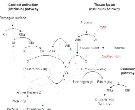
Hemophilia A (HA) is a recessive X-linked bleeding disorder that occurs in 1:5000 male new births and is due to the lack or reduced activity of coagulation factor VIII (FVIII) (1). FVIII is a non-enzymatic cofactor part of the coagulation cascade. When activated by thrombin FVIII is able to bind the factor IX (FIX) in a complex that activates factor X (FX), which converges in the common pathway of the coagulation cascade converting fibrinogen to fibrin and clot formation (Figure 1).
Figure 1. schematic representation of coagulation cascade
Based on the residual FVIII activity, there are three forms of hemophilia A: the severe form, in which the levels of FVIII are below 1%, the moderate form, between 1 and 5%, and the mild form, from 5 to 40% of FVIII activity (2). The most frequent mutation that affects approximately 45% of patients with severe hemophilia is the inversion of the first 22 exons caused by intrachromosomal recombination between the intron 22 (int22h1) with two homologous sequences that are distant 500kb from the gene (int22h3 and int22h3) (3). Other mutations causing the severe form are small deletions or insertions, missense and non-sense mutations and taken together they occur in 45% of patients. Less frequent mutations are large deletions, splice site mutations and intron 1 inversion (4). Instead, missense mutations are mainly associated with moderate and mild form (Figure 2).
7
Figure 2. FVIII mutations frequency reported by CDC Hemophilia A Mutation Project (CHAMP)Disease clinical manifestations range from spontaneous bleeding, with frequent haemarthroses in the most severe form, to secondary bleeding with rare haemarthroses in milder form (2). Diagnosis is made in relation to family history or following the first haemarthrosic episodes that occur at different ages according to the clinical form of hemophilia (0-3 years for severe, 2-7 for moderate, 5-14 for mild) (5). The main diagnostic technique used in laboratory is the evaluation of activated partial thromboplastin time (aPTT), which depends on the ability of patient's plasma to shorten the time required for clot formation in a standard FVIII-free plasma (6). However, this method can sometimes be inaccurate to diagnose the mild form and provides in 5-10% of cases false negative results. This implies the use of other more sensitive techniques such as two-stages clotting, chromogenic assay and genetic investigation (6). The current therapeutic practice consist
8
in the administration of recombinant FVIII or blood products and is carried out based on the disease severity; patients with severe hemophilia receive every other day prophylaxis, while patients with mild and moderate form receive replacement therapy on demand (7). Primary prophylaxis is defined as regular continuous treatment initiated in the absence of documented osteochondral joint disease, determined by physical examination and/or imaging studies, and started before the second clinically evident large joint bleed and age of 3 years. Continuous means the intention of treating for 52 weeks/year and at least 45 weeks/year (85%) (8). Patients receive 25-40 IU kg-1 per dose administered intravenously three times a week (8). The main complication related to this treatment is the development of neutralizing antibodies (inhibitors) to FVIII that occurs within the first 50 exposure days and is common in 20-40% of patients with the severe form. This worsens the clinical aspect because it makes ineffective the treatment and further reduces the residual activity of the endogenous FVIII (9). Among the risk factors for inhibitor formation the F8 genotype and the bleeding phenotype are highly predictive, indeed multidomain deletions are associated with nearly 90% risk to develop neutralizing antibodies (10). Another factor related to inhibitors development consist in FVIII haplotypes, indeed, for instance six protein variations were reported (H1-H6). H1 and H2 haplotypes are predominant in white population and are the most frequently associated with recombinant and plasma derived FVIII where as H3, H4 and H5 were reported only in black people (11). It was suggested that this haplotypes mismatch lead a two folds higher susceptibility to developed antibodies by African patients (11). Recent discovery showed that, in comparison with non-neutralizing antibodies, neutralizing antibodies mainly belong to the IgG4 subclass, display high affinity to FVIII and have long lifespan in circulation (12).
Cell mechanisms underlying the immune response to FVIII are not completely understood. However is likely that T helper lymphocytes (CD4+ cells) activated after FVIII presentation by antigen presenting cells (APC) plays an important role. Generally, the interaction between MHCII and T cell receptor triggers the first signal for CD4+ activation; even so, T cells become fully activated only in the presence of a costimulatory signal such as B7/CD28 or CD40/CD40L. This “two signal model” is required to generate T lymphocytes able to produce cytokines necessary to sustain B-cell differentiation, maturation and antibodies formation. An indirect evidence of CD4+-dependent inhibitory response was assessed in HA patients infected by HIV in the first years of replacement therapy. Indeed, in these patients the inhibitory titers decreased concurrently to the reduction of CD4+ cells and increased again after HIV therapy (13, 14). In addition, it was demonstrated that blocking costimulatory signal is sufficient to reduce antibody
9
titers in HA mice which have developed anti-FVIII inhibitors, proving that T cells are fundamental in the biological process ending with inhibitors formation (15, 16). Moreover, CD4+ cells isolated from patients who have developed anti-FVIII antibodies proliferated in vitro upon stimulation with exogenous FVIII (17, 18). The treatment of patients with inhibitors is particularly complicated and mainly consists in the administration of active FVII or activated prothrombin complex (bypassing agents) and in techniques to temporarily reduce the amount of immunoglobulin in the plasma (plasmapheresis and immunoadsorption) (9). However, these solutions are not as efficient as FVIII replacement to restore hemostasis and do not solved definitively the problem of inhibitors formation. For these reasons, the possibility to induce sustained FVIII tolerance represents the best alternative to overcome this issue. Immune tolerance induction (ITI) for patients who developed neutralizing antibodies consists in regular daily high dose administration of FVIII for several weeks, or even months, depending on protocol, aiming to eradicate inhibitors and to restore replacement therapy efficacy by inducing antigen-specific tolerance (19). The first ITI therapy was performed more than 30 years ago showing the ability of this treatment to eradicate antibodies in a 20 years old patient (20). In patients with high inhibitors titer (>5 Bethesda units) the success rate of ITI is between 60% and 80% with low relapsing of 15% in 15 years (21). Despite the great outcomes the high costs of ITI reduced its feasibility to few patients requiring the development of other strategies such as blockade of co-stimulation, oral tolerance, immunosuppressive treatment and antigen-specific regulatory T cell (22).
Replacement therapy
Before the advent of replacement therapy, life expectancy of patient affected from the severe form was less than 20 years, and the quality of life was generally devastating from joint bleeding complications or intracranial hemorrhage (23). Replacement therapy became feasible in the mid-1960s by discovering that precipitate left from thawing plasma was rich in FVIII (cryoprecipitate). Later, manufactories developed a methods to separate FVIII from pooled plasma, allowing to purify FVIII as lyophilized concentrate product (24). However, in the late 1970s the use of contaminating pooled plasma concentrated factors caused the widespread of serious infections such as HIV, HBV and HCV in hemophilia community (25). Subsequently, manufacturers introduced several viral inactivation treatments and the control of donors became more rigorous. Anyway great concern about the safety of plasma-derived products continued among hemophilia patients. The big advance in replacement therapy was achieved following the
10
FVIII gene cloning in 1984 which allowed the development of highly purified recombinant FVIII (rFVIII) (26). The first generation of rFVIII products were synthesized by gene-transfection in mammalian cells (Chinese Hamster Ovary cells) and were first used in clinic in the late 1980s (27). These rFVIII were produced in presence of animal or human derived protein in cell culture and contained human albumin in the final formulation as stabilizers (28). Safety was improved since 2000s replacing albumin with sucrose giving rise to the second generation of rFVIII (29). Among these new products generation was introduced in the market also a rFVIII lacking the B domain (BDD-FVIII) (ReFacto) that is dispensable for hemostasis but increase the efficacy in rFVIII production by cells (30). The third generation of rFVIII products are produced in absence of any animal or human proteins reaching the highest level of safety (31). Despite the development of blood products and recombinant FVIII has drastically improved the patients quality of life, replacement therapy do not represent yet a definitive cure and some issues are still to be solved. Among these, there are the high costs, the frequent number of administration due to the short FVIII half-life in circulation and the high probability to develop neutralizing antibodies. To extend rFVIII half-life several products were developed and most of them are now in Phase III clinical trial (24). Three general bioengineering mechanisms were proposed: (i) PEGylation to reduce receptor-mediated endocytosis, (ii) conjugation to the IgG Fc to avoid clearance by reticuloendothelial cell through the interaction with the neonatal Fc receptor (FcRn) and (iii) modification of rFVIII to enhance the binding with von Willebrand factor (vWF). (i) The majority of infused FVIII is cleared by the interaction with LDL receptor-related protein (LRP) that is abundantly expressed in the hepatocytes and KC other than in other organs (32). PEG can reduce this binding by incorporating a large number of water molecules and increasing the hydrophilic properties of FVIII (33). Progresses in FVIII bioengineering have allowed the possibility to conjugate in more specific way PEG to the target protein avoiding interference with other functional molecules, in the case of FVIII for example with vWF and tenase complex. Today three PEGylated rFVIII are currently in Phase III clinical trial, one of these is randomly conjugated (BAX 855, Baxter) and the other two are site directed PEGylated (BAY 94-9027, Bayer; NN7088, Novo Nordisk). All of them are reported to extend FVIII half-life by 1,5-1,7 fold compared with a commercial not modified rFVIII (34). (ii) Albumin and IgG share a very extent half-life of 19-22 days in comparison with the typical half-life of few minute or few days of other human plasma proteins (35). This is due to the interaction between these proteins with the FcRn. IgG entering in cells by pinocytosis are accumulated in early endosomes where bind the FcRn by a pH-dependent affinity due to acidity of this cell compartment (36). This interaction
11
avoids the catabolism of IgG and the complex is recycled to the cell membrane where the neutral pH of the bloodstream induces the dissociation of FcRn-IgG complex releasing the IgG back to the circulation (36). rFVIII-Fc developed by Biogen Idec/SOBI which has completed the Phase III clinical trial showed a 1,5-1,7 fold extension of FVIII half-life (37) (iii) Moreover, CSL Behring developed a single chain rFVIII in which the light and heavy chain are covalently linked demonstrating an higher binding affinity for vWF. For now this rFVIIISingleChain is in Phase I/II clinical trial (38).
Coagulation FVIII
FVIII gene (F8), which maps to band Zq28 at the tip of the long arm of the X chromosome was characterized firstly in 1984 (39). It measure 186 kb in length and is constituted by 26 exons encoding for a mature protein of 2332 amino acids (263 KDa) plus 19 aa signal oligopeptide at the N-terminal (40). FVIII molecule is organized in 6 domains: A1, A2, B forming the heavy chain and A3,C1,C2 that constitute the light chain. Between A1/A2, A2/B and B/A3 domains are present three acidic regions (a1, a2, a3) that contain the thrombin binding sites (Arg372, Arg740, Arg1689) and are crucial for FVIII activation (41). The overall structure of FVIII is similar to FV, in particular in the A domains they share approximately 40% amino acid identity also with the copper binding protein ceruloplasmin (42). Moreover C domain share some homology between FVIII, FV and proteins that bind negatively charged phospholipids (e.g., fat globular protein and the lipid-binding lectin discoidin I) (43, 44). Instead, FVIII B domain is unique and do not show significant similarity with FV or other proteins (45). Once synthesized, FVIII enters in the endoplasmic reticulum (ER) in which undergoes two important modifications: the elimination of the signal peptide and the introduction of oligosaccharide chains on asparagine residues predominantly arranged on the B domain. The N-glycosylation is fundamental to ensure the correct folding of the protein, to prevent the aggregation of intermediate forms and to allow the interaction of FVIII with enzymes and chaperon proteins essential to the correct intracellular processing, vesicular trafficking, exocytosis and secretion of FVIII (46). The ER-Golgi transition is mediated by the interaction between FVIII B domain with specific protein complex, in particular lectin-mannose binding 1 (LMAN1) also known as endoplasmatic reticulum-Golgi intermediate compartment 53 kDa protein (ERGIC53) and multiple coagulation factor deficiency 2 protein (MCFD2) (45). Defects in these molecules cause the combined deficiency of circulating FVIII and FV, which share in part the same intracellular processing of FVIII (47). In the Golgi, FVIII is cleaved close to the C-terminal region of B domain (after the aa 1313 and aa 1648)
12
producing an heterodimer consisting in the heavy chain (200 kDa) and the light chain (80kDa) that are not covalently linked by a divalent metal ion (mainly Cu2+) between the A1 and A3 domains. Finally, the processes that complete the intracellular maturation of FVIII are the modification of saccharide groups introduced in ER and the sulfurization of some tyrosines located in the acidic regions target of thrombin proteolytic activity (48).
In physiological conditions the FVIII concentration in plasma is 200-300 ng/ml and is associated with high affinity with the vWF, which is 50-folds in excess compared to the FVIII (49). The role of this interaction is to increase the half-life of FVIII by reducing the clearance and avoiding the inactivation by protein C. Moreover, the vWF prevents the premature association of FVIII with other coagulation factors before its activation mediated by thrombin. The regions involved in the binding between FVIII and vWF are located in correspondence of the acidic sequence a3, at the C-terminal of the B domain and in the C2 domain (50). Not surprisingly, therefore, those mutations in the gene of vWF or FVIII impairing the ability of interaction between the two proteins can produce very similar pathological phenotypes (50). In the early stages of the coagulation cascade, a little amount of activated thrombin acts on FVIII, and other coagulation factors, to amplify the hemostatic process. The FVIII amino acids involved in thrombin cleavage are Arg372, Arg740 and Arg1689, that are located in the acidic regions a1, a2, a3, respectively. After activation, FVIII dissociates from vWF and is able to bind platelets surface by hydrophobic interaction between some structural motifs of the C2 domain and the membrane phospholipids, which is further enhanced by the difference in charge between the two surfaces (51). In this conformation FVIII, is able to form a protein complex with FIXa and FX, known as tenase, which leading FX activation by proteolytic action of FIXa. The FVIIIa domains involved in the maintenance of tenase are A2 and A3 domains for the interaction with FIXa and the acidic region a1 for the binding with FX (52). Under physiological conditions, the clotting process triggered by any alteration of hemostasis must always end with the fibrin cap dissolution, to ensure a steady and regulated balance between a pro-thrombotic and anti-thrombotic phenotype. In this balancing thrombin plays a key role by activating coagulation cascade inhibition systems, including, the activation of protein C. Once activated this protease is able to inactivate several coagulation factors, among which, FVIII that is cleaved at the level of the A1 and A2 domains (52). In normal condition, FVIII half-life in circulation is relatively short (12-14h) even in the presence of vWF (53). This is due to the clearance mediated by several endocytic receptors such as LDL (low density lipoprotein) receptor-related protein (LRP), asialoglycoprotein receptor, VLDL receptor and the macrophage mannose receptor (54-57). Among these the more characterized and
13
effective in FVIII removal from circulation is the LRP which is a multiligand hepatic receptor belonging to LDL receptor superfamily. The interaction between FVIII and LRP1 was established firstly in 1999 by two separate groups (57, 58). Starting from this finding, it has been demonstrated that the blocking of LRP1 by infusion of an antagonist (RAP) results in FVIII clearance reduction with a longer half-life (57, 59). Additionally a conditioned mouse model in which LRP1 was specifically knocked out in the liver showed increase levels of FVIII (60). Several studies identified different FVIII domains involved in the binding with LRP. Initially, it was described that A2 domain amino acid region 484-509 and 558-565 displayed high affinity for LRP interaction (57, 61, 62). Furthermore, also peptide sequences in A3 and C1 domains are reported to be necessary for FVIII endocytosis (63-65). An important observation derived from the fact that alteration of such amino acids crucial for LRP binding did not affect the coagulation activity of FVIII (62, 63) providing a novel approach to improve FVIII half-life.
14
FVIII cell sources
Since orthotopic liver transplantation (OLT) corrected hemophilia A, liver has been considered the primary site of FVIII production (66-68). However, the identity of liver cells expressing FVIII was controversial and still now remains a question to be definitively clarified. Initially and for a long time hepatocytes were considered the FVIII expressing cells both at mRNA and protein level by in vivo and in vitro experiments (69-72). Although in early years the presence of FVIII was reported mainly in LSEC and macrophages rather than hepatocytes (73-75). These observation was strengthened by others (76) ending with the demonstration that LSEC but not hepatocytes secrete FVIII (77). Moreover, recent papers confirmed FVIII expression in endothelial cells (EC) by using a Cre/Lox strategy to selective knocking out FVIII expression in several cell types and concluding that FVIII is not synthetized in hepatocytes but mainly secreted by EC (78, 79). Furthermore, in addition to LSEC, several authors have reported FVIII transcript and protein also in EC of other organs such as kidneys, spleen and lungs (78, 80, 81) but not in heart and brain isolated EC (78). The importance of LSEC, both human and murine, in FVIII secretion was further confirmed by several transplantation studies in hemophilic mice (82-84). Indeed transplantation of healthy LSEC in HA mice restored therapeutic levels of FVIII activity and correct the bleeding phenotype of recipients up to 2 months (82). Conversely, the ability of hepatocytes to improve the clotting avtivity in HA mice after transplantation is not unequivocal and can depend by the site of transplantation. Ohashi and collegues demonstrated to achieve therapeutic FVIII levels up to 5 weeks by transplanting hepatocytes under the kidneys capsule of HA mice (85). Otherwise, intraperitoneal transplantation of hepatocytes do not provide detectable FVIII activity in recipients plasma in a short term experiment (1week) (84). The authors hypothesized that this result was due to the absence in the peritoneal cavity of vWF required to stabilize FVIII (84). Even if liver represent the main source of FVIII many studies suggested the presence of extrahepatic sites of FVIII production. The first evidence that other organs were able to express and secrete FVIII was reported in 1971 when Webster at al. demonstrated in dogs that transplantation of hemophilic liver in normal recipient caused only a partial reduction of circulating FVIII (86). More recently a similar result was obtained also in humans by Madeira (87). In this study a patient affected by stage 2 hepatocellular carcinoma with normal FVIII levels was transplanted with a liver coming from an hemophilic patient died for a diffuse brain injury. In the days after transplant, aPTT seconds of recipient did not increased meaning normal FVIII levels in circulation deriving from sources different from liver (87). Among the other organs, spleen transplantation showed to temporarily increase FVIII expression in HA dogs (88-90) and
15
more encouraging in humans, hemophilic patients transplanted with healthy spleen reached long term FVIII activity which ameliorate the clinical feature of the disease (91, 92). Other than spleen FVIII mRNA and protein were also detected in the glomeruli and in the tubular epithelial cells of kidneys and in mesenchymal cells isolated from different sites (93-96). Despite FVIII cDNA was firstly cloned from a T-cell line (97), the FVIII expression in hematopoietic cells was uncertain and poorly investigated. However, an early study described the presence of FVIII protein in pulmonary alveolar macrophages and in cells present in the splenic red pulp (75). A recent insight on a novel extrahepatic sources of FVIII demonstrated that BM transplantation significantly contribute to hemostasis in HA mice by rescue therapeutic levels of FVIII up to 6 months after treatment and allowing survival of mice after tail clip assay (93). This study highlights the role of hematopoietic cells in FVIII synthesis and secretion and offers a new potential therapeutic approach for hemophilia A. Despite these discoveries, the contribution of other sources of FVIII may be investigated and for instance little is known about FVIII transcriptional regulation. At this point the characterization of FVIII promoter activity could offer new insight to extend our knowledge about FVIII expression. Initially studies on F8 gene reported a 1175bp region upstream the ATG and containing the transcription starting site (-170) that probably was involved in transcriptional regulation of FVIII (39). Further investigation was later performed by Figueiredo at al. in 1995 by cloning the 1175bp proximal region in a luciferase plasmid system and testing the promoter activity in hepatic cell lines. By this strategy they identified a 200bp region containing the element for the maximal promoter activity and chromatin immune precipitation assay (ChIP) demonstrated the ability of 4 hepatic transcriptional factor (TF) such as HNF1, HNF4, C/EBPα and C/EBPβ to interact with FVIII promoter (98). However, this study did not analyzed the promoter activity in other cell types in
vitro or in vivo and for instance the characterization of this region was not more deeply
investigated.
Gene therapy
Gene therapy is a form of molecular medicine that has developed since the early nineties and is still evolving: the main purpose is to introduce into the target cells (but also in tissues or organs) DNA sequences in safe and efficient way, with the ultimate goal to obtain a therapeutic effect, or at least slow the disease progression. Fundamental point for gene therapy is to develop a system capable to achieve an efficient gene transfer in tissues without causing risks to the patient. For this reason, initial efforts have been spent to develop a system with these characteristics, in order
16
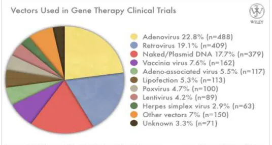
to transfer therapeutic gene in the cells directly into the body (in vivo) or in cells taken, modified, and finally reintroduced (ex vivo). Whatever the strategy used, gene transfer required the use of a vector able to overcome the biological barriers between cells and surrounding environment and capable to transfer the DNA into the cells. Vectors for gene therapy are commonly divided in viral and non-viral. Examples of non-viral gene delivery are naked/plasmid DNA, lipofection and nanoparticles however viral vectors are the major employed in gene therapy (Figure 4). These vectors derived from an extensive engineering of several classes of virus in order to satisfy the biosafety requirement and to make them able to carry the gene of interest. The most characterized and used viral vector in gene therapy application are those derived from adenovirus, adeno associated virus, retrovirus and lentivirus (Figure 4). The first successful gene therapy clinical trial in humans was achieved in the 1990 for the treatment of an inherited disease: the adenosine deaminase deficiency (ADA) who leads to a severe immunodeficiency. In this trial patients were infused with autologous T cell corrected ex vivo using a retroviral vector carrying the adenosine deaminase gene. As result, ADA was cured in one out two enrolled patients (99). Up to day, more than 1900 clinical trials were approved over the world for the treatment of several disease using different vectors. Although monogenic diseases would appear the ideal target for gene therapy, this approach was applied for many other disorder, first of all for cancer (Figure 5).
17
Figure 5. Diagram representing the diseases treated by gene therapy.As shown by the initial success, combination of gene and cell therapy when applicable, provide a great solution for several diseases. Historically, BM transplantation represented one of the most practiced example of cell therapy for many applications such as oncohematology, anemias and immunodeficiencies. The first allogenic BM transplantation was performed in 1957 on six cancer patients under chemo and radio therapy regimen by E. Donnall Thomas (100) and to date it is estimated that in the world over than 50000 patients per year have been treated with BM transplantation for various diseases (101). However, for some pathologies, for example for monogenic disease where autologous transplantation is useless, allogenic solution is the unique possible approach, which poses several concerns. Among the most relevant there is the graft versus host disease (GvHD). This reaction occurs in the absence of full histocompatibility between donor and recipient in allogeneic transplants and corresponds to the cytotoxic action mediated by the transplanted cells, which once reaching the competence, attack the recipient organism (102). Gene therapy associated with BM or hematopoietic stem cell (HSC) transplantation combines the self-renewal properties of HSC providing a sustained expression of transgene with the possibility of autologous transplantation overcoming the GvHD. This strategy was used in different clinical trial over the years, mainly for the treatment of immunodeficiency (103, 104). Very recently, an italian group combines with success gene and cell therapy using lentiviral vectors (LV) in a clinical trial for two severe and lethal diseases: Metachromatic leukodystrophy and Wiskott-Aldrich syndrome (105, 106).
18
Lentiviral vectors
Lentiviruses are a class of single-strand RNA (ssRNA) based virus belonging to the retroviridae family, associated with slow and progressive disease characterized by immune deficiency and neurological disorders; among these viruses there are those that caused acquired immunodeficiency in humans, primates and other mammals (HIV-1 and 2, SIV, FIV, BIV and CAEV). The human immunodefiency virus (HIV) that caused the acquired immunodeficiency syndrome is the most characterized and employed for gene transfer purpose. Similarly to all retroviruses, lentivirus replicate through a DNA intermediate but unlike the onco-retroviruses are able to integrate their genome even in post-mitotic cells such as neurons and macrophages (107). The virus particle contains two identical copies of 9,5Kb long ssRNA. The genome is constituted by 9 genes arranged on more than one reading frame. In addition to genes encoding for the structural and enzymatic proteins (gag, pro, pol, and env), there are other genes for accessory (vpu, vpr, nef and vif) and regulatory proteins (tat and rev) that are involved in the expression, assembly and in the pathogenicity of the virus (108). The lentiviral vectors used for gene therapy exploiting only the first stage of the viral replicative cycle to transfer and integrate the transgene within the chromatin of target cell. Once integrated, vector proviral DNA is defective for some sequences required for genome production and for proteins necessary to assemble new virion particles in transduced cells (109). In order to build safe vectors the lentivirus genome was modified over the years by segregating the cis- and trans-acting sequence in different plasmid constructs. Cis-acting sequences that regulate the incapsidation (ψ sequence), the reverse transcription (PBS, cPPT-CTS e 3’PPT and R), the nuclear translocation (cPPT-CTS) and the integration of provirus genome (the att sequence present in the U3 and U5) were maintained in the transfer construct in which is inserted the expression cassette. Another cis-sequence preserved in the plasmid design is RRE, the Rev responsive element important for RNA transport to the cytoplasm (Figure 6). To improve safety, self-inactivating transfer constructs were developed by eliminating part of U3 enhancer/promoter from parental LTR (∆U3) that was replaced by CMV enhancer/promoter retaining only a short U3 sequences required for genome integration (110). This strategy avoid the synthesis of genome RNA in transduce cells reducing the risk to produce replication-competent virus. Further, truncated LTR limit the interference with the expression cassette internal promoter and reduce the de-regulation of gene harboring the site of integration (110) (Figure 6). Other elements were then included in the transfer construct to improve transduction efficacy of LV. One of these is the central polypurine tract (cPPT) that facilitates
19
nuclear import of the lentiviral preintegration complex (PIC) before vector integration (111, 112). In addition the woodchuck hepatitis posttranscriptional regulatory element (WPRE) inserted after the transgene increased mRNA export, polyadenilation, and half-life (113) (Figure 6).
Figure 6. Schematic representation of modification made on LV transfer construct over the years
To improve vector biosafety extensive modifications were realized also on packaging construct. In the first generation of LV packaging construct part of the ENV was partially eliminated, but RRE sequence was maintained, LTRs were removed and the packaging sequence of ψ was mutated. LTRs were removed and replaced with the stronger promoter of cytomegalovirus (CMV) and polyA of chicken beta actin was inserted to substitute the parental polyA (114). Further, LV was pseudotyped by replacing HIV-1 envelope gene with the vesicular stomatitis virus glycoprotein (VSV-G) gene, increasing the cellular tropism virtually to all mammalian cells and augmenting the particles stability (114) (Figure 7). In this generation, packaging function was separated in two different construct. In the second generation of packaging construct the four accessory genes (Nef,Vip,Vpu,Vpr) were removed leaving gag and pol genes, which encoded for the structural and enzymatic components of the vector particles, and tat and rev genes with transcriptional and post-transcriptional functions (115) (Figure 7). In the third generation the
20
remaining genome was split in two plasmids: a packaging construct containing the gag and pol genes and a second plasmid containing rev gene and removal of tat gene (116) (Figure 7).
Figure 7. Schematic representation of modification made on LV packaging constructs over the years
Actually, the SIN transfer vector and the third generation packaging construct represent the best LV platform regarding biosafety and transduction efficacy. Since LV are integrating vectors the genotoxicity represent a big concern even because in an initial X-linked combined immunodeficiency (SCID-X1) ex vivo gene therapy clinical trial using a murine oncoretroviral vector carrying the interleukin 2 γ-chain some patients developed leukemia (117, 118). Further analysis showed vector integration close to LMO2 proto-oncogene promoter. Likely, retroviral LTR have induced LMO2 overexpression triggering unregulated cell proliferation and clonal expansion. This reflects the natural tendency of retrovirus to integrate their genome near the start of transcriptional unit (119). Conversely, LV integrate preferentially in active genes or in local hot spots but far from transcriptional regulation sequence (120). Indeed, under equal experimental conditions, in comparison with retroviral vectors, LV showed to promote low
21
insertional mutagenesis event and low tumors development (121, 122). Moreover, clinic outcome of several gene therapy trials using LV did not report any oncogenic occurrence (123-125). Since regulation of transgene expression in define cell types could be important for some gene therapy applications several strategies were approached. First, is possible to modify the LV tropism by replacing the most common VSV-G envelope with others viral proteins which have an intrinsic tissue specificity or display more efficacy to transduce certain cell types. For example, baboon envelope pseudotyped LV are more efficient to transduce un-stimulated or resting CD34+ cells in comparison to VSV-G (126). The H and F envelope from paramixoviridae family instead provide to LV high targeting and rate of transduction to non-activate B and T lymphocytes (127). In addition, to restrict targeting selectively to hepatocytes the baculoviridae gp64 envelope was used (128). Regarding the central nervous system, great interest get the observation that lymphocytic choriomeningitis virus (LCMV) envelope showed to target specifically astrocytes, the main cells involved in glioma formation (129). Despite the targeting issue, VSV-G pseudotyping has associates with some drawback such as in vivo dose-dependent cytotoxicity and the susceptibility to be recognize by human serum complement (130, 131). A second level of targeting is obtained by working on transcriptional regulation. Ubiquitous promoters such as CMV, spleen focus forming virus (SFFV), human polypeptide chain factor-1alpha (EF-1alpha), phosphoglycerate kinase (PGK) allowed to a strong transgene expression, even though they are affected by some limitations. Among these there are the trend to be inactivated over the time impairing a sustained transgene expression and the presence of enhancer sequence within the promoter increased risk of insertional mutagenesis (132, 133). Furthermore, the ubiquitous transgene expression may be detrimental. This is the case of adverse reaction given by proteins ectopically expressed or citotoxicity mediated by suicides genes or toxin used for anti-tumor gene therapy (134-136). Another issue is represented by the innate or adaptive immune response against the transgene once is expressed by antigen presenting cells (APC) (137, 138). At this point the use of cell specific promoter to de-targeted transgene expression in APC allowed the sustained expression of therapeutic gene in the selected cells, for example hepatocytes, by reducing immune response (139-141). For these reasons, over the years many authors have used several tissue specific promoters to drive transgene expression in cells of interest including endothelial, hepatocytes, dendritic cells, hematopoietic stem cells, megakaryocytes, B cells etc (142-147). Finally, a more recent post-transcriptional regulation based on miRNA biology provided an additional degree of cell targeting. For gene transfer purpose, the insertion of complementary sequences to a specific miRNA (miRNA target sequence, mirT) to the 3' of the
22
expression cassette, offers the possibility to reduce selectively the transgene synthesis in the cell types in which that particular miRNA is expressed. This strategy was firstly developed by Brown and colleagues to avoid transgene expression in APC showing that the presence of miRT142-3p, complementary to miRNA 142-3p which is selectively expressed in hematopoietic cells, limits immune response against GFP by preventing transgene expression in those cells (148). Since miRNA is differentially expressed also during cell differentiation miRTs can be useful to regulate transgene expression in define stages of cell maturation. This characteristic was exploited by Gentner at al. to correct globoid cell leukodistrophy using the miRT-126 to de-target GALC expression which is highly toxic in the early stages of hematopoietic differentiation and allowing its synthesis in mature cells when miRNA-126 is downregulated (149).
Gene therapy for hemophilia A
Gene therapy could constitute a powerful therapeutic approach for many diseases, in particular for monogenic diseases. Additionally, hemophilia A represent an ideal target for gene therapy since restoring FVIII levels higher than 1% is sufficient to ameliorate the bleeding phenotypes of patients with an overall increase of quality of life. Hemophilia B gene therapy has provided good results in clinical trials by using adeno associated-viral vector (AAV) to deliver FIX into the patients. In a first clinical trial a sierotype 2 AAV vector carrying the FIX cDNA under the control of the ubiquitous cytomegalovirus promoter (CMV) was intramuscularly injected in 8 enrolled patients affecting by the severe form of hemophilia B (FIX activity less than 1%) (150). By this approach no local and systemic toxicity was reported and no immune response against the vector occurred even in case of pre-existing immunity (150). Despite the evidence of gene transfer and transgene expression, only modest circulating FIX levels (<1%) were achieved in most of the patients (150). The same group has repeated the study by injecting the AAV2 into the hepatic artery and changing the ubiquitous CMV promoter with an hepatocyte specific promoter (human alpha-1-antitrypsin) to drive FIX expression selectively to hepatocytes. However, gene therapy efficacy was hampered by a strong immune response against the vector with subsequent CD8 mediated destruction of transduced hepatocytes (151). Best results were obtained in a third clinical trial by using an AAV pseudotyped with AAV8 capsid protein. It has been reported that in comparison to AAV2 the AAV8 capsid has stronger liver tropism, lower seroprevalence in human and provide less virus uptake by antigen presenting cells. Moreover is able to mediate effective transduction in animals with pre-existing immunity to AAV2 (152). Regarding the expression cassette, an artificial liver specific promoter (LSP) was used to drive the expression of
23
a codon optimized FIX cDNA (153). Finally, the AAV8.LSP.FIX was administered by peripheral vein injection in 10 patients enrolled in the study. By these strategies FIX activity was restored at level between 2-6% up to a median of 3,2 years. However 4 patients were treated with prednisolone following the increase of liver enzyme and all of them have received at least one recombinant FIX infusion (154). Nevertheless, this clinical trial showed the feasibility of gene therapy for hemophilia B, increasing the quality of life of patients and encouraged new efforts to improve this approach making it a suitable alternative to replacement therapy. Despite the relevant results obtained for hemophilia B, gene therapy for hemophilia A has seen significantly less progress into the clinic due to some factors that complicates FVIII expression in comparison with FIX. (i) The size and complexity of FVIII (9 kb) make it too large for some vector system, such as AAV. (ii) Using a comparable vector delivery, transduced cells express 100 fold less FVIII level than FIX (155) (iii) FVIII is naturally 5-6 fold more immunogenic than FIX, making the transgene mediated immune response a big concern. For instance, several approaches for hemophilia A gene therapy using different vector systems were attempted.
AAV vectors are impaired by their limited capacity to packaged genome larger than 5 kb. To circumvent this problem FVIII light and heavy chain were split in two distinct AAV and upon co-injection in mice, biologically active FVIII was detected in circulation (156, 157). However, since the interaction between the two chains occurs inside the cells, it is necessary that both vectors co-transduce the same cell to allow the production of functional FVIII. This quite reduced the efficiency of the strategy and concurrently increased the overall dose of vector to use. Another option to overcome the size limit of AAV vectors was to use a B domain deleted FVIII that reduced by one third the final size of cDNA without compromising the biological activity of the protein. Even though a minimum promoter is required to not exceed the package capacity of AAV. By this attempt, two studies have showed sustained FVIII levels in mice (158, 159). However, in both, the authors have reported in most of treated mice anti-FVIII antibodies formation that was overcome by the second group by using the sierotype AAV8 instead the AAV1 (158). AAV vectors containing the BDD-FVIII have demonstrated to be suitable to induce FVIII expression also in hemophilic dogs (160, 161). Even so the doses needed to reach therapeutic correction were significantly higher than the maximum doses of AAV-FIX administered to human in clinical trials (162) and given the dose-dependence of immune response to capsid, the use of this vector could be not feasible in humans. The reduction of doses could be obtained by increasing FVIII expression, and it was gained by using a full length promoter (163). Although this required an oversized AAV genome (5-7,5 kb) that has reported to reduced
24
packaging efficiency (164). Additionally, a recent study has demonstrated to induce remarkable therapeutic expression of FVIII in non-human primate by targeting FVIII expression in hepatocytes combined with the use of a codon optimized FVIII (165). However, most of macaques needed transient immunosuppression to reduce anti-FVIII antibodies titer (165). Among the other viral vectors lentiviral and retroviral were the more employed for the treatment of hemophilia A. The first proof of concept for in vivo gene therapy using a γ-retroviral vector was assessed in neonatal hemophilic mice. Approximately 50% of injected mice expressed physiological or even higher FVIII levels that were sustained up to 14 months. In this study, the remaining animals that showed only transient or undetectable FVIII expression developed anti-FVIII specific antibodies (166). This approach was also successful in a canine model of hemophilia A by targeting transgene expression in the liver without antibodies formation (167). However when retrovirus was used for human gene therapy only low circulating FVIII was detected in patients (168, 169). In 2000, Park and colleagues demonstrated that also LV could be used in vivo to induce human FVIII expression in wild type mice. By intraportal injection they direct FVIII expression predominantly to the liver with an ubiquitous promoter (EF1alpha) and reaching human FVIII levels of about 15%. Unfortunately hFVIII expression was only transient due to antibodies formation despite the fact that mice were not hemophilic and normally expressed murine FVIII (170). Later, similar results (5% of activity) was obtained in hemophilia A mice by intraperitoneal (IP) injection or ex vivo bone marrow transduction using a LV carrying the BDD-FVIII under the control of an ubiquitous promoter. However also in this case neutralizing antibodies were developed, with higher frequency in IP group rather than in ex vivo bone marrow transduction (171). Best outcome was achieved by improved targeting FVIII expression to hepatocytes. To do this a feline immunodeficiency virus-(FIV) based LV was pseudotyped with the baculovirus GP64 envelope that has been demonstrated to have a strong tropism for hepatocytes. Additionally, to further restrict transgene expression to these cells an hybrid hepatocyte-specific promoter (murine albumin enhancer/human alpha1-antitrypsin promoter) was included in the expression cassette. Liver targeting demonstrated to allow long-term therapeutic levels of FVIII without antibodies formation. Although slight inhibitors titer occurred by changing strain of mice, from C57BL/6 to a more immune reactive BALB/c background (128).
Up to day liver and in particular hepatocytes have represent the favorite target for hemophilia A gene therapy (165, 166, 170, 172-176) by their behavior to limiting transgene mediated immune response. Nevertheless, the anti-FVIII antibodies development is still a current drawback for the
25
feasibility of hemophilia A gene therapy. This issue point out the necessary to identify other suitable cell type to target FVIII expression in order to induce tolerance to the transgene. At this regard, encouraging results in mice were obtained by restrict FVIII expression in platelets with the use of megakaryocytic specific promoters. Doing so, FVIII was released after platelets degranulation only in the site of injury when occur, otherwise no circulating proteins are present to trigger immune response (177). Indeed, ex vivo BM transplantation transduced with a LV carrying FVIII under the control of the platelet αIIb gene promoters correct the bleeding phenotype of hemophilic mice by several coagulation assay without neutralizing antibodies detection (177, 178). More important, this strategy has proven effective also in presence of pre-existing anti FVIII antibodies (179, 180). Platelets targeting provide good results also in dog and in immunodeficient mice transplanted with genetically modified human HSC (181, 182). Another approach to overcome antibodies development is to target FVIII expression specifically in organs or cell types with immuno-tolerant properties. LSEC represent an optimal candidate at this regard. In fact it has been demonstrated their attitude to induce tolerance against antigen presented by themselves (183). Moreover, because the interaction between FVIII and vWF is crucial for the stability and activity of FVIII, endothelial cells and megakaryocytes, that are the main vWF producers, represent an interesting cell target for cell and gene therapy (50). Other than promoting FVIII production in specific cell type, immune response can be reduced by abrogates transgene expression in antigen presenting cells. This was obtained by using the miRT142-3p that provided a sustained FVIII expression in mice without antibodies formation (173). A further target examined for FVIII expression in hemophilia A gene therapy application was BM-derived stem cells, in particular HSC. At this point both LV and Retroviral vectors were employed in ex vivo gene therapy. Interestingly, this approach induced FVIII expression and seemed to be functional to induce tolerance to FVIII by reducing inhibitors even in presence of pre-existing antibodies or after rFVIII challenging (184-187). However, the best results in these experiments were achieved using a porcine FVIII that is expressed 10-14 fold better than the human (185). FVIII expression was also drive specifically to B cells using an immunoglobulin heavy chain promoters obtaining sustained long term levels of FVIII with bleeding phenotype correction (188).
FVIII codon optimization
Beyond immunological issue, HA gene therapy is hampered by the inefficient expression of FVIII. To overcome the problem many modifications were done on FVIII coding sequence in
26
order to improve the expression, the intracellular maturation and the secretion pathway of FVIII. For instance, deletion of the hFVIII B-domain, which is not required for co-factor activity, resulted in a 17-fold increase in mRNA levels over full-length wild-type FVIII (30). Nevertheless secretion of BDD-FVIII is impaired in comparison of full size due to the absence of crucial asparagin-link oligosaccharides on B domain, in fact at protein level the absence of B domain provide only 30% more secretion than the wild type. This obstacle was overcome by Pipe and colleagues maintaining the proximal 226 amino-acid portion of the B-domain (FVIIIN6) that is rich in asparagine-linked oligosaccharides (189). This retention led to a 5-10 fold FVIII secretion by promoting a better interaction of FVIII with the LMAN1/MCFD2 complex that is involved in the FVIII ER-Golgi transition. Additionally, it has been described that a 110 aa region within the FVIII A1 domain contains sequences able to bind the immunoglobulin-binding protein (BiP) causing reduction in secretion by entrapping FVIII in the ER (190). At this regard it was reported that the single amino acid F309S mutation increase FVIII secretion up to 3-fold by reducing the binding of BiP with subsequent lower FVIII retention in the ER (191). Recently, other single amino acid changes were identified to improve FVIII secretion. The combined 1899G/C1903G mutations increase 2,2 fold FVIII secretion by eliminating a dispensable disulfide group (192) and more recently, Siner et al. showed that a minimal modification in the retained B domain sequence, the mutation R1645H, is sufficient to increase FVIII secretion by 2,4 fold (193). The latest mutation was setup by comparing the amino acid sequence of hFVIII with cFVIII that is more efficiently expressed and is more stable in circulation. This is due in part by the fact that cFVIII is mainly secreted as single chain whereas the human as heterodimeric form. This difference it could be explained through the different intracellular cleavage of FVIII by the protease-paired basic amino acid cleaving enzyme (PACE)/furin. cFVIII escapes from furin cleavage because in comparison with other mammalian FVIII has a unique furin consensus sequence (HXXR instead of RXXR) that confers resistance to the protease activity (193). Finally, another two fold improvement in potency was achieved by replacing the 226 amino-acid N6 spacer with a novel Asparagine reach peptide of 17 aminoacids (V3) (165).
27
Materials and Methods
28
Bladder epithelial cells isolation and culture
To analyze the presence of FVIII from healthy and tumoral bladder, specimens were isolated from several cystectomies in collaboration with Prof. Valente and Dr. Volpe from which informal consent was obtained by the patients. Bladder fragments were snap frozen in liquid nitrogen, disaggregated and lysed for mRNA extraction. Epithelial cells were isolated through mechanical removal of the bladder epithelium from stroma. Cells were maintained in culture in serum free EpiLife Medium (Life Technologies) containing Human Keratinocyte Growth Supplement (HKGS) (Life Technologies) and presence of FVIII mRNA and protein expression were assessed by RT-PCR and immunofluorescent stainings at different cell passages.
Human and mouse cells isolation, culture and differentiation
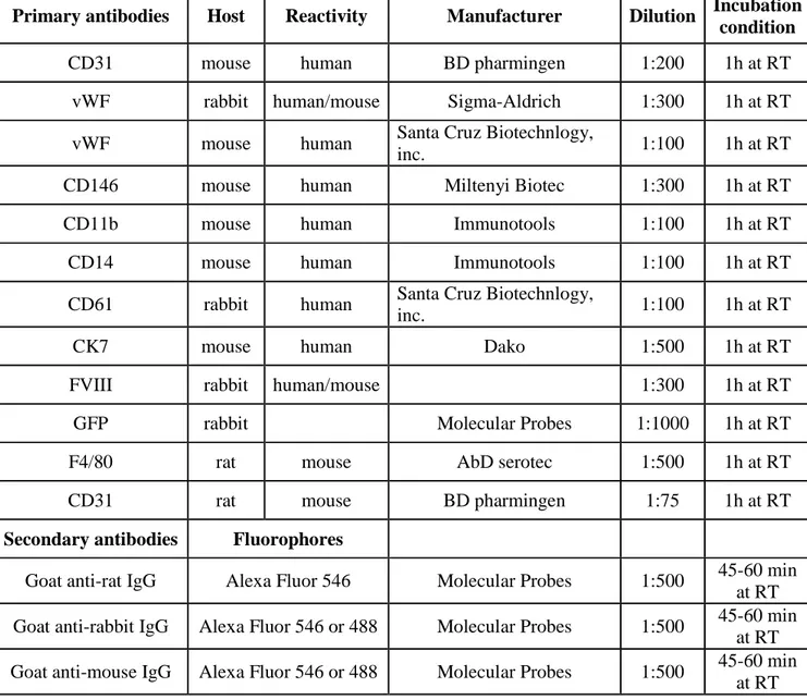
Human. Human liver samples were perfused as previously described (82). After liver dissociation with a cell scraper, cells were passed through Dacron fabric with 80-µm pores and centrifuged twice under 50 g for 5 minutes to separate hepatocytes from non-parenchymal cells (NPCs). NPCs were washed and pelleted under 350 g for 10 minutes. Human LSECs were selected from NPC fraction by immunomagnetic sorting incubating NPCs with anti-human CD31 biotin-conjugated (eBioscience) for 20 min at 4°C followed by an incubation with Streptavidin MicroBeads (MiltenyiBiotec) for 15 min at 4°C and finally isolated by MS Separation Columns (Miltenyi Biotec), according to the manufacturer’s protocol. Isolated LSECs were plated at a density of 1.5-2x10⁵ cells/cm2 and cultured on collagen-coated tissue culture dishes in EGM-2 medium (Lonza). Human Kupffer cells (KCs) were isolated by plastic adhesion from NPCs after LSEC isolation. Briefly, CD31-negative fraction of NPCs was resuspended in serum-free RPMI and plated on plastic tissue culture dishes. After 45 minutes of incubation at 37°C supernatant was removed, plates were washed twice in PBS and finally fresh RPMI containing 10% fetal bovine serum (FBS) and 10ng/ml rhM-CSF was added. KCs were cultured for up to 3 weeks under these conditions. To isolate human peripheral blood mononuclear cells (PBMCs), blood was layered on Ficoll-Paque TM (PREMIUM, GE Healthcare). PBMCs were seeded in serum free RPMI medium to promote monocytes adhesion. After 30 minutes the medium was changed with RPMI containing 5% FBS and cells cultured for 12h. To obtain macrophages, monocytes were cultured in DMEM with 10% FBS, 1mM sodium pyruvate, 1mM non-essential amino acids, 0,25mM HEPES (Lonza), 10ng/ml M-CSF.
Human HSC were isolated from cord blood obtained according to an approved protocol by the Ethical Committees of UPO, Novara, Italy. First, cord blood mononuclear cells (CBMCs) were
29
obtained by density gradient stratification of blood on Ficoll. Than CD34+ cells were isolated from CBMCs by immunomagnetic positive selection using CD34 MicroBead Kit (MiltenyiBiotec). To obtain macrophage differentiation from HSC 105 CD34+ cells/ml were plated in STEM-SPAM medium (STEMCELL Technologies Inc.) containing 20% FBS, 2mM glutamine, 50 U/ml penicillin,50 µg/ml streptomycin, 30 ng/ml interleukin 3, 30 ng/ml M-CSF, 30 ng/ml Flt-3 ligand, 25 ng/ml SCF, as previously described (194). Medium was changed every 2 days. After 14 days cells acquired macrophage morphology.
Megakaryocytes were differentiated by culturing 1.5x10⁶ CD34+ cells/ml in STEM-SPAM medium containing 2mM glutamine, 50 U/ml penicillin,50 µg/ml streptomycin, 10 ng/ml interleukin 6, 10 ng/ml interleukin 11 and 20 ng/ml thrombopoietin (TPO). The medium was changed at day 3, 7 and 10 (195). After 13 days, megakaryocytes were harvested and used for experiments.
Mouse. Murine monocytes were obtained from total peripheral blood. After removal of red blood
cells using red blood lysis buffer (RBLB) (150mM NH4Cl,10mM NaHCO3, 1 mM disodium
EDTA) total white cells were plated at the density of 1.5-2x10⁶/ml in DMEM containing 10% FBS. After 24 hours, monocytes were attached to the plate.
BM cells were flushed from tibias and femurs of 8-9 weeks-old wild type (WT) or HA mice with DMEM containing 5% FBS. After RBC lysis 0.8-1x10⁶ of total BM cells/cm2-plastic were differentiated in macrophages (BMDMs, bone marrow-derived macrophages) by culturing cells in IMDM containing 10% FBS and 5 ng/ml recombinant mM-CSF in vitro. For studies, BMDMs were released 5-7 days later by Versene (Gibco).
For mouse MK differentiation, c-Kit+ cells were isolated using the mouse CD117 MicroBeads kit (Miltenyi Biotech) from total BM cells obtained as described above. Isolated cells were cultured for 2 days in Stem Span containing 20 ng/ml SCF. Then 106 cells/ml were cultured for 3-4 days in Stem Span containing 100 ng/ml TPO, 10 ng/ml IL-6 and 10 ng/ml IL-11. All cytokines were purchased from Immunotools.
pF8 cloning in LV transfer construct
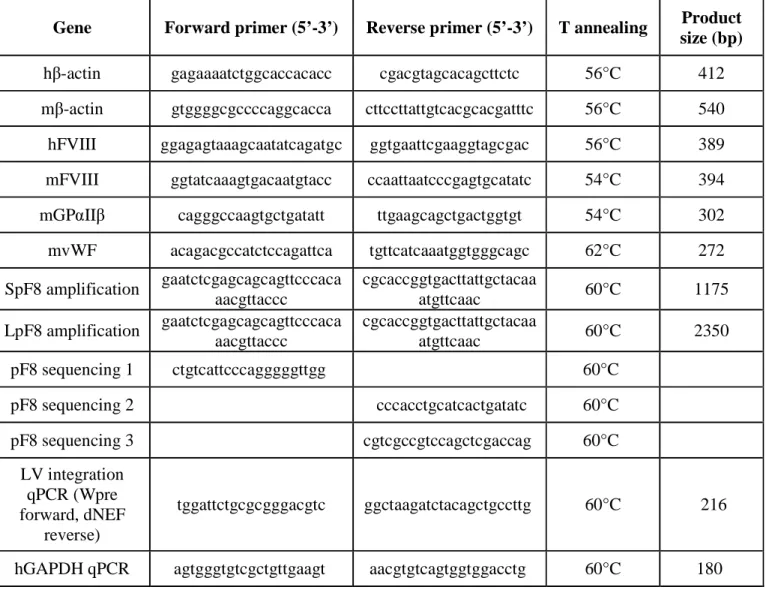
SpF8 and LpF8 were amplified by PCR from human genomic DNA by inserting at 3' and 5' ends the restriction sites for the enzymes XhoI and AgeI. These sites were used to inserted F8 promoter in place of ubiquitous PGK promoter in LV.PGK.GFP in order to obtain the LV.SpF8.GFP and LV.LpF8.GFP. To generate LV.pF8.FVIII we inserted the BDD-FVIII in place of GFP in the LV.pF8.GFP. For cloning SalI and AgeI endonucleases were used to excide
30
both GFP and FVIII from LV.SpF8.GFP and LV.PGK.FVII, respectively. Ligase product identity was assessed by restriction analysis and sequencing. Primers used for cloning and sequencing are reported in Table 1.
Lentiviral Vectors production
Third-generation LV were product using published protocoll (196). 293T cells were cotransfected with four plasmids by calcium phosphate precipitation; these vectors were the pMDLg/RRE packaging plasmid (12,5 ug); the pMD2.VSV-G envelope-coding plasmid (9 ug); pRSV-Rev (6,25 ug) and transfer vector plasmid LV.PGK.GFP, LV.SpF8.GFP and LV.LpF8.GFP (28 ug). All four plasmids were added to cells in a 15-cm dish and thirty hours following transfection the culture supernatant, containing the packaged viral particles, was collected and concentrated by ultracentrifugation. Collected viral particles were titrated on 293T using limiting dilution analysis. Briefly, one hundred thousand 293T were cultured in DMEM with progressively lower dilutions of each lentivirus (1:10, 1:100, 1:1000, and 1:10,000). For the LV expressing GFP, each dilution was quantified by FACS as percentage of GFP+ cells. Calculation from the titration analysis indicated about 1 -2 X 10⁹ transducing viral particles per milliliter. Instead, for LV not expressing GFP, genomic DNA was isolated from 293T and titer was calculated by qPCR.
Genomic DNA isolation and LV titration by qPCR
Genomic DNA was isolated from transduced 293T cells with ReliaPrepgDNA Tissue MiniPrep System (Promega). The quantitative real time PCR was carried out in a 20-ul total volume containing 1X SYBR green PCR master mix (PROMEGA), 1 uM forward and reverse primers (wpre-∆nef) and 1 µM forward and reverse primers (h GAPDH), 50 ng of genomic DNA. Quantitative PCR were performed by incubation at 95°C for 3 minutes and 40 amplification cycles of 95°C for 3 minutes and then 60°C for 30 seconds. Primers used are reported in Table 1
Animals
Animal studies were performed according to an approved protocol by the Animal Care and Use Committees of UPO, Novara, Italy. In vivo experiments were performed on 8-10 weeks old mice. For GFP expression studies, LVs were delivered in C57Bl/6 WT mice. C57Bl/6 Hemophilia A mice were used for in vivo and ex vivo gene therapy studies using LV.SpF8.FVIII. Immunocompromised NOD/SCID-γNull HA mice (NSG-HA) were generated in our laboratory by crossing NOD/SCID HA mice with NOD.Cg-PrkdcscidIl2rgtm1Wjl/SzJ (γNull) purchased by
31
Jackson lab (197). For HSC transplantation studies busulfan myeloablation was performed on recipient mice. The busulfan solution for injection was prepared as follow: 25 mg of drug were solved in 1 ml of acetone and than resuspended in 9 ml of peanut oil. Immunocompetent HA mice were lethally conditioned by intraperitonal injection of 25mg/kg of busulfan from days -4 to -1 before transplantation while NSG-HA mice received a sublethal conditioning by only one injection of 50mg/kg of busulfan the day before transplantation. NSG-HA mice were kept in autoclaved microisolator cages and fed with sterile food and water at the animal facilities of the Università del Piemonte Orientale. Moreover, all animals procedures made on NSG-HA mice were performed under sterile hood.
Mouse and human Hematopoietic Stem Cells isolation and transplantation.
To isolate murine HSC (lineage negative cells, Lin-) BM was flushing from femurs, tibiae and humerus of 6-8 weeks old donor mice. After red blood lysis, Lin- cells were obtained by immunomagnetic negative selection from total BM cells using Lineage Cell Depletion Kit (MiltenyiBiotec). After isolation cells were transduced with LVs at MOI 100 and cultured at density of 1x10⁶/ml in serum free STEM-SPAM medium without cytokines. Human HSC were isolated from cord blood obtained as decribed above and cultured at density of 1x10⁶/ml in serum free STEM-SPAM medium (Lonza) added with 50 ng/ml hTPO, 50 ng/ml hSCF, 50 ng/ml hIL-3 and 50ng/ml hFlt3-L. On the basis of experiment CD34+ cells were transduced with LV at MOI 30. For transplantation, 24h after isolation a total of 3 x10⁵ or 6x10⁵ CD34+ or 106 lin- cells were resuspended in serum free STEM-SPAM without cytokines and tail vein injected in 400µl of volume in busulfan-conditioned mice.
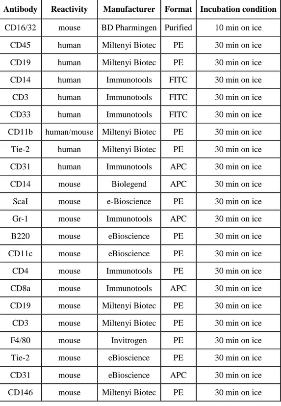
Analysis of blood and organs of treated mice.
The engraftment of transplanted mice was evaluated at several time point in the peripheral blood as percentage of GFP+ or human CD45+ cells. For each time point blood was collected by retro-orbital puncture using a glass capillar. Eritrocytes were eliminated by incubating RBLB for 10 min at 4°C. Total white cells were directly analyzed by cytofluorimetry for GFP or incubated with anti-human CD45 PE conjugated antibody to assess the engraftment after xenotransplant. Total spleen cells were obtained by spleen digestion for 30’ at 37°C in HBSS (Sigma Aldrich) containing 10% FBS and 0.2 mg/ml collagenase IV and then filtered through a 70-µm cell strainer (Falcon). BM cells were obtained by flushing tibiae and femurs. For both spleen and BM red blood cells were lysed for 8’ with RBLB. Thymus were mechanically disrupted through a