Original Articles
Irreversible inhibition of Δ16HER2 is necessary to suppress
Δ16HER2-positive breast carcinomas resistant to Lapatinib
Martina Tilio
a,1, Valentina Gambini
a,1, Junbiao Wang
a,1, Chiara Garulli
a,
Cristina Kalogris
a, Cristina Andreani
a, Caterina Bartolacci
a, Maria Elexpuru Zabaleta
a,
Lucia Pietrella
a, Albana Hysi
b, Manuela Iezzi
b, Barbara Belletti
c, Fiorenza Orlando
d,
Mauro Provinciali
d, Roberta Galeazzi
e, Cristina Marchini
a,1,*, Augusto Amici
a,1,*
aDepartment of Biosciences and Veterinary Medicine, University of Camerino, Camerino 62032, ItalybAging Research Centre, G. d’Annunzio University, Chieti 66100, Italy
cDivision of Experimental Oncology 2, Centro di Riferimento Oncologico, National Cancer Institute, Aviano 33081, Italy dAdvanced Technology Center for Aging Research, IRCCS-INRCA, Ancona 60121, Italy
eDipartimento di Scienze della Vita e dell’Ambiente, Università Politecnica delle Marche, Ancona 60128, Italy
A R T I C L E I N F O
Article history: Received 7 March 2016
Received in revised form 21 July 2016 Accepted 22 July 2016 Keywords: HER2 isoform Breast cancer Δ16HER2 mice Targeted therapies Drug resistances A B S T R A C T
HER2 tyrosine kinase receptor is a validated target in breast cancer therapy. However, increasing evi-dence points to a major role of Δ16HER2 splice variant commonly coexpressed with HER2 and identified as a clinically important HER2 molecular alteration promoting aggressive metastatic breast cancer. Con-sistently, mice transgenic for the human Δ16HER2 isoform (Δ16HER2 mice) develop invasive mammary carcinomas with early onset and 100% penetrance. The present study provides preclinical evidence that Δ16HER2 expression confers de novo resistance to standard anti-HER2-therapies such as Lapatinib and acquired resistance to the selective Src inhibitor Saracatinib in breast cancer. Of note, Dacomitinib, an irreversible small molecule pan-HER inhibitor, was able to completely suppress Δ16HER2-driven breast carcinogenesis. Thus, only Dacomitinib may offer benefit in this molecularly defined patient subset by irreversibly inhibiting Δ16HER2 activation.
© 2016 Elsevier Ireland Ltd. All rights reserved.
Introduction
Breast cancer (BC) is the most common female cancer and the second cause of cancer death in women[1]. HER2-positive (+) BC accounts for 18–20% of all BC cases and is associated with poor prog-nosis[2,3]. The advent of HER2-targeted therapies, including tyrosine kinase inhibitors (TKIs) such as Lapatinib and anti-HER2 antibod-ies such as Trastuzumab, considerably improved the overall survival and time-to-disease progression of HER2+ BC patients[4]. However, many patients do not benefit from HER2-targeted treatments because of resistance to therapy[5]. In particular, Lapatinib (GW572016, Tykerb) is a small molecule member of the 4-anilinoquinazoline class of kinase inhibitors that targets the intracellular tyrosine kinase domains of both HER1 and HER2. Thus, it is classified as a dual TKI. Lapatinib reversibly binds to the cytoplasmic ATP-binding site of the targeted kinase and blocks receptor phosphorylation and acti-vation, thereby preventing subsequent downstream signaling events
[6]. Lapatinib treatment is well tolerated and has been approved for Trastuzumab-resistant HER2+ advanced BC patients[7,8]. However, its efficacy is limited by the development of acquired re-sistance that typically occurs within twelve months after initial treatment[9]. In the past decade, different irreversible TKIs di-rected against HER family receptors have been developed for fighting drug-resistant tumors. Dacomitinib (PF-00299804) is a potent, second-generation quinazoline-based irreversible inhibitor of HER1, HER2, and HER4, which competes for ATP binding but then cova-lently binds a nucleophilic cysteine residue at the edge of the ATP binding cleft[10]. Dacomitinib is orally active and demonstrated clinical efficacy with acceptable toxicity in several solid tumors[11]
and the ability to overcome resistance to HER2-targeting agents in BC models[12]. In fact, prolonged target suppression as well as ex-cellent pharmacodynamic properties confer an improved antitumor activity to Dacomitinib compared with reversible inhibitors[13]. Recently, an HER2 splice variant lacking exon-16 (Δ16HER2) has been detected in more than 50% of HER2+ BC[14,15]. Significantly, 90% of patients expressing Δ16HER2 suffer from metastatic disease and increasing evidence points to a major role for the Δ16HER2 splice variant in resistance to Trastuzumab[15]. Nevertheless, therapies specifically targeting this highly pathogenic isoform are still missing and few studies have examined the impact of Δ16HER2 on the * Corresponding authors. Fax:+39 0737 403290.
E-mail addresses:[email protected](C. Marchini),augusto.amici@ unicam.it(A. Amici).
1 These authors contributed equally to this work.
http://dx.doi.org/10.1016/j.canlet.2016.07.028
0304-3835/© 2016 Elsevier Ireland Ltd. All rights reserved.
Contents lists available atScienceDirect
Cancer Letters
response to HER2-targeted therapies, even reporting contradicto-ry results[15–18]. A new mouse model transgenic for the human Δ16HER2 isoform (Δ16HER2 mice), developing invasive mammary carcinomas with early onset and 100% penetrance, has been re-cently generated and represents an excellent and unique preclinical model to test the efficacy of BC therapies against Δ16HER2-driven carcinogenesis[19]. Δ16HER2 harbors an in-frame deletion which promotes constitutive homodimerization of the receptor thereby coupling Δ16HER2 to unique oncogenic signaling pathways medi-ated by Src kinase[15]. Src is a non-receptor tyrosine kinase considered a critical convergent point of multiple upstream signals, conferring resistance to anti-HER2 therapies[20]. The first exper-imental evidence that Src kinase is a crucial effector of Δ16HER2 tumorigenic properties was reported by Mitra and coworkers in cells ectopically expressing Δ16HER2 protein[15]. Interestingly, they also observed that the expression of activated Src kinase is asso-ciated with Δ16HER2 expression in human invasive BCs[15]. Saracatinib (AZD0530), an orally available small molecule of the anilinoquinazoline class, is a reversible inhibitor of Src-family ty-rosine kinases (Src, Yes, Lck) and Bcr-Abl that, acting as an ATP competitor, blocks the ATP binding site of the target enzyme[21]. This particularly potent and selective Src inhibitor has shown anti-invasive and antitumor activity in several cancer cell lines and xenograft models[22,23]and is well tolerated in phase I trials[24,25], but, until now, has never been evaluated in preclinical models of Δ16HER2+ BC. Here we report that Saracatinib has anticancer effects in Δ16HER2 mice by blocking the oncogenic Δ16HER2/Src signal-ing axis. Unfortunately, initial inhibition of mammary carcinogenesis by Saracatinib is counteracted by acquired resistance mecha-nisms. Also the dual HER1/HER2 TKI Lapatinib failed to block tumorigenesis in Δ16HER2 mice. Of note, only irreversible inhibi-tion of Δ16HER2, achievable by Dacomitinib, led to suppression of Δ16HER2-driven breast carcinogenesis.
Materials and methods
Establishment of transgenic mammary carcinoma cell lines
Tumors surgically excised from 8-month-old transgenic female mice were minced and transferred into culture flasks. Cells were maintained in DMEM (Invitrogen, Carls-bad, CA) plus 20% fetal bovine serum (FBS, Invitrogen, CarlsCarls-bad, CA). They were cloned and sub-cultured to establish cell lines, according to the previously described method [26].
Cell cultures
Human BT474 and SKBR3 cells (ATCC) and the new established murine CAM3 and CAM6 cells were maintained in DMEM respectively plus 10% FBS or 20% FBS and 1% penicillin–streptomycin (P/S, Invitrogen, Carlsbad, CA). Human ZR-75-30 cells (ATCC) were cultured in Dulbecco’s MEM/F-12 (Euroclone) plus 10% FBS and 1% P/S, supplemented with 5 μg/mL insulin.
Drugs
Dacomitinib (PF-00299804), Lapatinib and SCH772984 were obtained from Chemietek while Saracatinib (AZD0530) from LC Laboratories. For in vitro experi-ments Saracatinib was dissolved in water, Dacomitinib and Lapatinib were solubilized in DMSO (Sigma-Aldrich) and directly added to the medium at the indicated con-centrations. For in vivo experiments Saracatinib, Dacomitinib and Lapatinib were daily dissolved in a vehicle consisting of 0,5 Hydroxypropyl methylcellulose, 0,1% Tween 80 (Sigma). SCH772984 was first dissolved in DMSO and further suspended in saline solution.
Immunofluorescent analysis
Refer to the Supplementary material.
Flow cytometry
Refer to the Supplementary material.
Soft agar assay
Refer to the Supplementary material. Capillary-like tubule formation assay
Refer to the Supplementary material. Transplanted tumors
Refer to the Supplementary material. Immunoblotting analysis
Cell and tumor lysates were obtained using RIPA buffer (1% NP40, 0.5% Na-deoxycholic acid and 0.1% SDS in PBS) supplemented with protease inhibitors. Lysates were separated by 4–20% gradient precast SDS-PAGE (Bio-Rad) and transferred onto polyvinylidene difluoride (PVDF) membranes (Immobilion P, Millipore). Antibod-ies to Src, pSrc, p38, pP38, Erk, pErk, HER2, pHER2, HER3, pHER3, Akt, pAkt, STAT3, pSTAT3, vinculin, β-actin were from Cell Signaling. Antibodies to EGFR and pEGFR were from Epitomics. Secondary antibodies conjugated with peroxidase were from Sigma-Aldrich. The immunoreactive bands were detected by using LiteAblot PLUS (Euroclone) reagents and images were acquired with ChemiDoc Imaging System (Bio-Rad).
Cell viability assay
Saracatinib, Dacomitinib and Lapatinib effects on cell viability were evaluated by seeding 4× 10⁴cells/well (CAM3 and CAM6 cells), 104cells/well (SKBR3 and ZR-75-30 cells) or 2,5× 10⁴cells/well (BT474 cells) in 96-well plates in DMEM plus FBS. The day after, fresh medium containing appropriate Saracatinib, Dacomitinib and Lapatinib concentrations were added. Cell viability was determined using an MTT (Sigma Aldrich, St. Louis, MO) assay.
Detection of apoptosis by annexin-V FITC staining
Apoptosis of cells was evaluated by measuring the exposure of phosphatidylserine on the cell membranes using an Apoptosis Detection Kit (BD Pharmingen) and flow cytometry according with previously described methods[27,28]. Experiments were repeated three times.
Animals
Δ16HER2 mice were housed under controlled conditions. Routine screening of Δ16HER2 transgenic mice was done as previously described[19]. Saracatinib (25 mg/ kg), Dacomitinib (6 mg/kg) and Lapatinib (50 mg/kg) were administered orally via gavage[13,29,30], while SCH772984 (15 mg/kg) by intra-peritoneal injection[31]. All mice have been treated for 10 weeks, starting drug administration at 10 weeks of age. Body weight and food intake did not significantly differ between pharmacologically-treated and control mice (data not shown), suggesting the absence of drug toxicity at the selected dose level. Tumor monitoring was performed twice a week by palpation. Progressively growing masses>1 mm mean diameters were regarded as tumors. Two perpendicular diameters (a and b) were measured on each tumor using caliper and volumes were calculated by the V= π/6[(a + b)/2]3formula. All animal experiments were carried out in accordance with the U.K. Animals (Sci-entific Procedures) Act, 1986 and associated guidelines, EU Directive 2010/63/EU for animal experiments.
Histology and immunohistochemistry
Refer to the Supplementary material. Statistical analysis
Quantitative data are presented as means± SEM from three independent ex-periments. The significance of differences was evaluated with two-tailed Students t-test, or one-way ANOVA followed by Bonferroni post-test. Statistical analysis was carried out with GraphPad Prism5 Software. P≤ 0.05 was used as the critical level of significance.
Results
CAM6 cells as in vitro counterpart of Δ16HER2-transgenic mouse model
We established several cell lines from a carcinoma spontane-ously developed in a Δ16HER2 transgenic female mouse. Among them we selected two epithelial cell lines (CAM3 and CAM6) and
one spindle cell line (P5D7) (Fig. 1A and B). In CAM3 and CAM6 cells, the expression of the human Δ16HER2 transgene correlates with a high tumorigenic potential, on the contrary P5D7 cells, not ex-pressing the transgene product, are not able to form any tumor when injected in syngeneic mice (Fig. 1B and C). CAM3 and CAM6 trans-planted tumors are morphologically and immunohistochemically similar to autochthonous tumors in Δ16HER2 females, consisting of epithelial Δ16HER2+ cells growing in solid sheets characterized by some necrotic areas and surrounded by stroma (Fig. 1D). In this work we chose CAM6 cells as representative in vitro model of the tumor of origin on the basis of their higher tumor growth rate in syngeneic mice (Fig. 1C) and because Δ16HER2 signaling is prefer-entially transduced by Src kinase as shown by western blot analysis (Fig. 1E), consistently with data previously reported in Δ16HER2 tumors[19]. Src kinase has been already identified as a key actor in Δ16HER2+ BCs conferring them a particularly aggressive behav-ior[15]. In CAM6 cells, Src kinase is recruited at the cell membrane, where it colocalizes with Δ16HER2 (Fig. 1F), and it is directly acti-vated by physical interaction with phosphorylated Δ16HER2 (Fig. S1). Interestingly, Src kinase activation is combined with that of STAT3, a downstream signal transducer in Src family kinase-mediated tu-morigenesis. Conversely, the signaling activity of wild-type (wt) HER2 in SKBR3 cells is mainly mediated by activated Akt (Fig. 1E). The epithelial identity of CAM6 cells was confirmed by immuno-cytochemical and flow cytometric analysis of selected epithelial and mesenchymal markers (Fig. S2A and B). CAM6 cells are positive for E-cadherin, cytokeratin-18 and β-catenin, whereas they are nega-tive for the mesenchymal marker vimentin. In particular, β-catenin and E-cadherin exhibited a membranous distribution. However, rather than forming well-differentiated round spheroids, CAM6 cells developed poorly organized tubular structures when cultured in a Matrigel-based three-dimensional (3D) culture system (Fig. S2C). Such aspect suggests a poor degree of differentiation that usually correlates with a poor prognosis in invasive breast carcinoma. In addition, when grown in soft agar, CAM6 cells formed large and compact foci demonstrating their capability of anchorage-independent growth, in agreement with their tumorigenic potential (Fig. S2D).
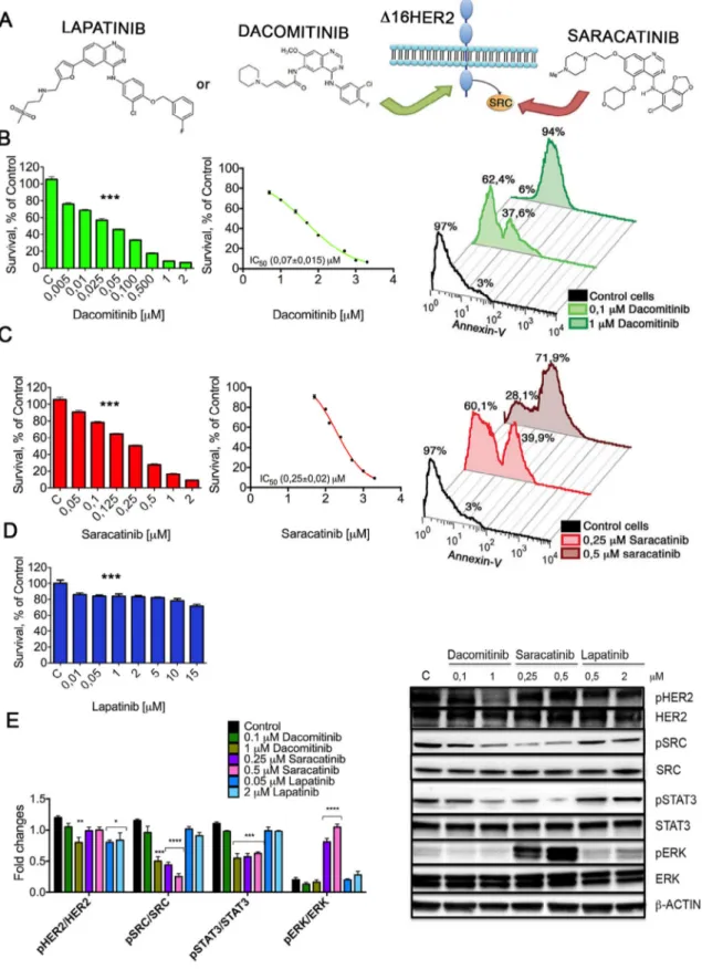
Saracatinib and Dacomitinib impair CAM6 cell viability blocking the oncogenic Δ16HER2/Src axis
Src kinase activation is a critical event in Δ16HER2-mediated mammary carcinogenesis, thus we evaluated the effect of pharma-cological interference with Δ16HER2/Src oncogenic axis on CAM6 cells viability. Targeting directly Δ16HER2 receptor by Dacomitinib or its main effector protein Src by Saracatinib (Fig. 2A), we found a significant dose-dependent decrease of CAM6 cells viability (Fig. 2B and C, left panels). In particular, the IC50value analysis indicated
that Dacomitinib is the most effective drug, probably due to its ir-reversible action (Fig. 2B). On the contrary, the reversible HER2-targeted drug Lapatinib failed to impair CAM6 cells viability (Fig. 2D). Interestingly, Saracatinib did not affect the viability of wt HER2-overexpressing SKBR3 cells, which instead were responsive to both Lapatinib and Dacomitinib, confirming that Δ16HER2 expression is coupled with a distinct signaling cascade (Fig. S3). Consistently, both murine CAM3 and human ZR-75-30 cells, an HER2+ cell line ex-pressing a relatively high level of the Δ16HER2 isoform[16], quite faithfully reproduced the CAM6 cell response to Saracatinib, Dacomitinib and Lapatinib, while BT474 cells, characterized by a modest level of the spliced variant[16], showed an intermediate behavior (Fig. S3). These data suggest an inverse correlation between the proportion of Δ16HER2 isoform with respect to the wt HER2 and the response to Lapatinib.
We thereafter investigated whether the inhibitory effect of Saracatinib and Dacomitinib on CAM6 cells viability was the result
of apoptosis. Flow cytometry analyses with annexin V staining strongly proved the ability of both drugs to induce CAM6 cells apop-tosis (Fig. 2B and C, right panels). Then, the impact of a 6 h treatment with Saracatinib, Dacomitinib and Lapatinib on Δ16HER2 signal-ing pathways was verified in CAM6 cells by western blot analysis. As shown inFig. 2E, Dacomitinib significantly reduced Δ16HER2 phosphorylation, thereby leading to a dose-dependent reduction of Src phosphorylation, while Saracatinib was able to impair Src ac-tivation without affecting Δ16HER2. Moreover, we observed a decrease in total Δ16HER2 in CAM6 cells treated with the highest concentration of Dacomitinib, indicating that this treatment may also lead to a destabilization of Δ16HER2. Of note, both Dacomitinib and Saracatinib treatments on CAM6 cells resulted in a dose-dependent inhibition of constitutively activated STAT3. Taken together, these results confirm that Src kinase is directly placed in the signaling cascade downstream Δ16HER2 and controls cell be-havior through STAT3 phosphorylation. In agreement with viability data, Src and STAT3 Δ16HER2-downstream signaling pathways re-mained unaffected upon Lapatinib treatment, despite a reduction in Δ16HER2 phosphorylation. Interestingly, although Δ16HER2 did not require a direct activation of MAPK/Erk axis, Erk phosphoryla-tion was switched on by Saracatinib. Such unexpected de novo Erk activation might be considered a treatment escape signaling pathway. We suppose that Erk upregulation caused by Saracatinib is the result of a dynamic model of crosstalk between the two major MAPK cas-cades, p38 and MEK/Erk. In this context, Src can induce the activation of p38 through the Src/Dock180/Rac1 signaling axis[32], and con-sequently downregulate Erk. The inhibition of Src by Saracatinib may release this negative regulatory feedback loop, triggering Erk activity.
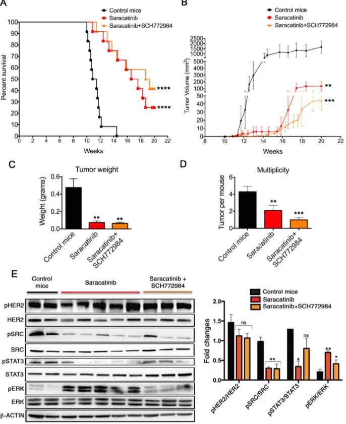
Mammary carcinogenesis inhibition by Saracatinib is counteracted by Erk-mediated escape mechanisms in Δ16HER2 mice
To evaluate the ability of Saracatinib to inhibit Δ16HER2-driven autochthonous carcinogenesis in vivo, we performed a chemopreventive study in Δ16HER2 females, treating them with Saracatinib or vehicle from 10 weeks of age, when mice were still free from macroscopic mammary lesions. In Δ16HER2 mice, the ex-pression of only five human Δ16HER2 transgene copies can drive neoplastic transformation of mammary epithelial cells with a short latency time. All transgenic females develop multiple asynchro-nous mammary tumors (4–5 tumors/mouse) with an average latency of about 15 weeks[19]. Treatment of these transgenic mice with Saracatinib resulted in significant tumor growth delay. Indeed, while the first palpable tumor appears at 11 weeks of age in the vehicle-treated group, Saracatinib-vehicle-treated mice displayed tumor development only five weeks later (Fig. S4A). Moreover, Saracatinib-treated mice developed smaller and fewer tumors compared with the control animals, as shown by the mammary tumor growth curve and the multiplicity graph (Fig. S4B and C). Accordingly, histopatho-logical analysis disclosed the presence of a significantly lower number of mitotic divisions (Fig. S4D) associated with a decrease in PCNA expression (Fig. S4E), demonstrating that Saracatinib ad-ministration inhibits tumor cell proliferation. On the other hand, although the Src inhibitor significantly slowed down tumor devel-opment, only 20% of the mice eventually remained tumor-free at 20 weeks of age (Fig. S4A). Thus, we decided to investigate the treat-ment escape signaling pathways activated in those tumors that grew despite Saracatinib administration. Western blot analysis con-firmed that autophosphorylation of Δ16HER2 was not reduced, while Src kinase activity was strongly inhibited in the Saracatinib-treated tumors. In addition, Src kinase inhibition was associated with a down-regulation of phosphorylated STAT3. Most interestingly, as already observed in vitro, we found that Erk phosphorylation was induced by Saracatinib, suggesting that the inhibition of Src might be counteracted by compensatory mechanisms involved in tumor
Fig. 1. Isolation and characterization of cell lines from a Δ16HER2 mammary tumor. (A) Schematic representation of the method used to isolate P5D7 (fibroblast-like), CAM3
and CAM6 (epithelial-like) cell lines: we established a bulk primary cell line from a Δ16HER2 tumor. After cloning by limiting dilution, three different cell lines have been isolated. (B) Upper panel: primary bulk cell culture was visualized by phase-contrast (left) and fluorescence microscopy (right). Lower panel: morphology and flow cytometry analysis of P5D7, not expressing the transgene (left panel), and CAM3 and CAM6, expressing the transgene (middle and right panel), after staining with an antibody against human HER2. Original magnification, 10×. (C) CAM3, CAM6 and P5D7 cells were transplanted in syngeneic FVB mice (n = 10) to test tumorigenicity and tumor growth rate. (D) Representative images of Hematoxylin and Eosin (H&E) staining and HER2 immunohistochemical staining of CAM3 and CAM6 transplanted tumors, grown in Δ16HER2 transgenic males or in wt FVB females. Original magnification, 20×. (E) Western blot analysis of HER2 downstream signaling pathways in SKBR3, CAM3 and CAM6 cells. Cell extracts were probed with antibodies to HER2, pHER2, HER3, pHER3, EGFR, pEGFR, p38, pp38, Erk, pErk, AKT, pAKT, STAT3, pSTAT3, Src, pSrc and vinculin (loading control); representative blot, n= 3 for each panel. (F) Δ16HER2 directly couples to Src kinase in CAM6 cells. Immunofluorescent detection of Δ16HER2 (red) and Src kinase (green) in CAM6 cells. The merged image shows colocalization (yellow). The merge includes DAPI (blue) stained nuclei. Original magnification, 40×. (For interpretation of the refer-ences to color in this figure legend, the reader is referred to the web version of this article.)
Fig. 2. Impact of Saracatinib, Dacomitinib and Lapatinib on CAM6 cell viability and signaling pathways. (A) Schematic representation of the two alternative ways of
block-ing Δ16HER2/Src oncogenic axis: targetblock-ing Δ16HER2, usblock-ing Dacomitinib or Lapatinib, or Src usblock-ing Saracatinib. (B,C,D) Left panel: CAM6 cells were incubated for 24 h in the presence of vehicle or increasing concentrations of Dacomitinib (B), Saracatinib (C) or Lapatinib (D) and cell viability was determined by MTT assay. The results are ex-pressed as percentage of living cells compared to control. Columns, mean of three separate experiments wherein each treatment was repeated in 16 wells; bars, SEM. ***P≤ 0.001, one-way ANOVA followed by Bonferroni’s multiple comparison test. Middle panel: IC50values were calculated for both drug treatments tested by fitting the concentration– effect curves data obtained in the three experiments with the sigmoid-Emax model using nonlinear regression, weighted by the reciprocal of the square of the predicted effect. Right panel: quantification of apoptosis through Annexin V assay. CAM6 cells, untreated or treated with indicated concentrations of Dacomitinib (B) or Saracatinib (C) for 24 h, were labeled with Annexin V-FITC and analyzed by flow cytometry. (E) Right panel: western blot analysis of HER2 downstream signaling pathways in CAM6 cells, untreated (control) or treated with indicated concentrations of Dacomitinib, Saracatinib or Lapatinib for 6 h. Cell extracts were probed with antibodies to HER2, pHER2, Src, pSrc, STAT3, pSTAT3, Erk, pErk and β-actin (loading control); representative blot, n= 3 for each panel. Left panel: densitometric quantification of pHER2/HER2, pSrc/Src, pSTAT3/STAT3, pErk/Erk expression from three independent experiments. *P≤ 0.05; **P ≤ 0.01; ***P ≤ 0.001; ****P ≤ 0.0001; one-way ANOVA test followed by Bonferroni’s multiple comparison test.
growth (Fig. S5A). Erk activation and nuclear translocation upon Saracatinib treatment was confirmed by immunofluorescence anal-ysis of tumor sections from control and Saracatinib-treated mice (Fig. S5B). To verify the role of Erk activation in Saracatinib resis-tance, a further in vivo experiment was carried out, including one additional experimental group of mice treated with Saracatinib in combination with a specific Erk inhibitor (SCH772984)[31]. SCH772984 is a selective and potent Erk1/2 inhibitor, which inhib-its both Erk enzymatic activity as well as inhib-its phosphorylation by MEK, and it was recently developed for treating malignancies depen-dent on dysregulated MAPK signaling[31]. This combined therapy resulted in a significant increase in the number of tumor-free mice (45% at 20 weeks of age) in comparison with Saracatinib treat-ment alone (Fig. 3A). Of note, pharmacologically treated mice developed significantly smaller tumors than untreated mice (Fig. 3C) and both tumor growth rate and multiplicity were further dimin-ished when SCH772984 was administered together with Saracatinib (Fig. 3B and D). These results correlate with a decreased level of phos-phorylated Src and Erk as shown by western blot analysis (Fig. 3E). Nevertheless, although this combined pharmacological strategy pre-vents acquired resistance to Saracatinib, it is not able to completely tackle mammary carcinogenesis and almost half of the treated animals ultimately developed cancer.
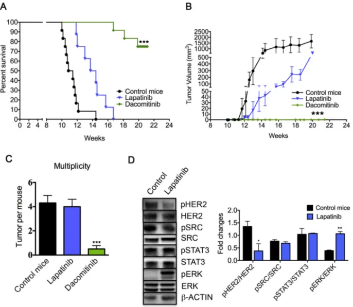
Dacomitinib suppresses Lapatinib resistant Δ16HER2-driven carcinogenesis
To evaluate the responsiveness of Δ16HER2 splice variant to HER2-targeted therapies, Δ16HER2 females were treated with Dacomitinib or Lapatinib from 10 until 20 weeks of age. Lapatinib failed to hamper Δ16HER2 mammary carcinogenesis suggesting that Δ16HER2 expression elicits intrinsic Lapatinib resistance in HER2+ BC. In particular, Lapatinib-treated animals showed a slight delay in tumor onset, but they all eventually developed tumors, charac-terized by a small reduction in tumor growth rate (Fig. 4A and B) and by comparable tumor multiplicity in comparison with control mice (Fig. 4C). To understand the molecular mechanisms beneath Lapatinib resistance, a western blot analysis was performed using tumor lysates. As expected, despite Δ16HER2 expression level did not change, its phosphorylation state was reduced upon Lapatinib treatment. However, Src and STAT3 signaling pathways down-stream Δ16HER2 remained unaffected by Lapatinib, confirming the key role of these effector proteins in promoting tumor develop-ment, while Erk was activated upon Lapatinib treatdevelop-ment, exerting a buffer effect on the already weak drug action (Fig. 4D). On the con-trary, Dacomitinib treatment completely inhibited autochthonous mammary tumors formation. Indeed 75% of treated Δ16HER2 mice remained tumor-free with only 3 animals out of 12 developing a single small sized tumor mass until the end of the experiment (20 weeks of age) (Fig. 4A–C). These data proved that the irreversible Δ16HER2 inhibition by Dacomitinib exerts relevant therapeutic effects on Δ16HER2+ BC.
Discussion
Four HER2-targeted therapies have been currently approved by FDA to treat BC: Trastuzumab and Pertuzumab, two monoclonal an-tibodies directed against the extracellular domain of HER2, Trastuzumab emtansine (T-DM1), an antibody-drug conjugate, and Lapatinib, a small molecule ATP-competitive reversible kinase in-hibitor of HER2 and EGFR. Lapatinib has been predominantly used as a second-line therapy, mainly in combination with capecitabine
[7]. Despite these therapies have shown robust clinical benefits most patients eventually relapse after treatment, indicating the need for a deeper understanding of the involved resistance pathways in order to improve patient outcomes[33]. Although Δ16HER2 isoform has
been found coexpressed with the wt HER2 form and it has been defined as the real transforming variant of the HER2 oncoprotein
[16], few studies have examined its clinical impact as well as its role in HER2-targeted therapies resistance[15,17]. Based on the exist-ing literature, the prevalence of Δ16HER2 expression in human HER2+ BCs ranges from 52 to 90%[14–17]and we sought that a suc-cessful strategy to combat HER2+ BC would have implied the suppression of Δ16HER2 aggressive splice variant. Thus, we tested both in vitro and in vivo Δ16HER2 sensitivity to TKIs in an attempt to disengage Δ16HER2 from downstream tyrosine kinase path-ways, demonstrating that Δ16HER2 expression is a strong molecular predictor for TKIs’ responsiveness and efficacy. Δ16HER2 arises from the in-frame deletion of exon-16, causing a loss of cysteine resi-dues in the extracellular domain of HER2. This molecular alteration promotes Δ16HER2 homodimerization via intermolecular disul-fide bonds, and results in enhanced oncogenic signaling strictly dependent on Src activation, providing the rationale for targeting Src in Δ16HER2+ breast cancer[15]. Src is a non-receptor tyrosine kinase found to be deregulated in numerous tumor types[34–36]
and linked to resistance to various therapies used in BC[20,37]. In particular, Src activation participates in Trastuzumab resistance mechanisms and indicates poor prognosis in patients with HER2+ BC[38]. Several small molecule inhibitors for Src kinase are under-going clinical trials after promising preclinical studies[39]. Among these drugs, Saracatinib, a dual Src/Abl kinase inhibitor, emerged as a particularly potent and selective Src inhibitor, displaying a >10-fold preference for Src over Abl kinases[22]. Given this distinct Src-targeted inhibition profile, in the present study we selected Saracatinib to block the oncogenic Δ16HER2/Src axis.
Herein reported results indicate that Saracatinib inhibits the growth and controls the survival of Δ16HER2+ breast cancer cells. By contrast, Saracatinib treatment had no effect on SKBR3 cell line with wt HER2 amplification and not expressing activated pSrc, cor-roborating previous reports describing SKBR3 as Saracatinib-resistant breast cancer cell line[40]. The in vitro antitumor action of Saracatinib on CAM6 cells was associated with a strong inhibi-tion of the activated Src kinase and with a concurrent downregulation of activated STAT3. On the other hand autophosphorylation of Δ16HER2 remained unaffected, indicating that inhibition of Src activation alone is sufficient to impair Δ16HER2-driven carcinogenesis. Most importantly, the antitumor activity of Saracatinib was confirmed in vivo, against autochthonous Δ16HER2-driven mammary oncogenesis, in a preclinical chemoprevention trial. In fact, Saracatinib administration was able to delay tumor onset and to reduce tumor multiplicity in Δ16HER2 mice. However, this encouraging anticancer effect was not durable, and Saracatinib-treated mice eventually developed tumors. Interestingly, we observed activation of MAPK/Erk signaling cascade as alternative prolifera-tion pathway developed during drug treatment, suggesting that the inhibition of Src by Saracatinib is counteracted by compensatory mechanisms. Thus, we tried a strategic combinatorial therapy aimed at resistance reversal, consisting of Saracatinib plus the Erk inhib-itor SCH772984. Although concurrent targeting of Src and Erk ensured a better protection in comparison with Saracatinib treat-ment alone, this therapeutic approach failed to completely block Δ16HER2-driven carcinogenesis. We therefore decided to target di-rectly the Δ16HER2 receptor with therapies conceived against wt HER2 protein, comparing the efficacy of HER2-targeted reversible TKI Lapatinib versus the irreversible pan-HER TKI Dacomitinib in Δ16HER2 mice. Dacomitinib, currently being tested in phase 3 trials for the treatment of advanced non-small-cell lung cancer, belongs to the second-generation TKIs, developed to circumvent first-generation HER receptor TKIs resistance[41]. Interestingly, we identified Δ16HER2 as molecular driver of Lapatinib resistance in BC. The mechanism of resistance might be based on incomplete and “leaky” inhibition of Δ16HER2 by Lapatinib, unable to block the
on-cogenic signaling mediated by Src kinase. Consistently, resistance to Lapatinib was previously reported in Src activated cell lines[37]. Resistance of Δ16HER2 to Lapatinib inhibition may also depend on protein conformational dynamics. Since Lapatinib binds
preferen-tially to an inactive kinase conformation in a reversible form[6], the constitutively active state of Δ16HER2 might interfere with Lapatinib binding and explain the different sensitivity to Dacomitinib, which conversely binds to the active site of kinases in a covalent
Fig. 3. Erk inhibition counteracts Saracatinib acquired resistance. (A) Kaplan–Meier disease-free survival plot for vehicle- (n= 12), Saracatinib-treated (n = 12) and Saracatinib+ SCH772984-treated (n = 12) Δ16HER2 mice. Mice were treated from 10 until 20 weeks of age (experimental end point). (B) Tumor growth curves in Saracatinib and Saracatinib+ SCH772984 vs control Δ16HER2 mice. (C) Tumor weight analysis in Saracatinib and Saracatinib + SCH772984 vs control Δ16HER2 mice at the end point. (D) Average tumor multiplicity in Saracatinib, Saracatinib+ SCH772984 vs control mice at the end of the experiment. The in vivo statistical significance was assessed by one-way ANOVA test followed by Bonferroni’s multiple comparison test with **P≤ 0.01; ***P ≤ 0.001; ****P ≤ 0.0001. (E) Left panel: representative western blot analysis of pHER2, HER2, pSrc, Src, STAT3, pSTAT3, pErk, Erk and β-actin (loading control) in tumor from 20-week-old mice. Right panel: relative densitometric quantification of pHER2/ HER2, pSrc/Src, pSTAT3/STAT3, pErk/Erk from three independent experiments; *P≤ 0.05, **P ≤ 0.01; one-way ANOVA test followed by Bonferroni’s multiple comparison test.
and irreversible form, leading to a persistent inhibition of the re-ceptor. In fact, we found that Dacomitinib is able to control Lapatinib-refractory Δ16HER2+ BC, displaying a relevant, long-lasting anticancer effect in vivo. These findings are concordant with previous in vitro results, where Dacomitinib inhibited the growth of HER2-amplified lines resistant to Trastuzumab and to Lapatinib[12]. However, we cannot rule out that the potent anti-cancer activities of Dacomitinib are also related to a cross-reactivity with HER3, although Dacomitinib is defined a pan-HER inhibitor targeting EGFR, HER2, and HER4. Con-sistently, treatment of CAM6 cells with Dacomitinib completely blocked HER3 phosphorylation (Fig. S6). In fact, despite the kinase domain of HER3 has long been assumed to be inactive, Shi F. et al. have recently shown that it binds ATP and promotes trans-autophosphorylation within HER receptor dimers, such as HER2/ HER3 heterodimers[42]. Moreover, the weak kinase activity of HER3 may contribute to resistance to Lapatinib, which indeed is able to inhibit HER3 transactivation in the context of HER2/HER3 dimers, but it is not able to impair HER3 autophosphorylation[42].
In conclusion, the present study identifies Δ16HER2 as a key factor in determining resistance to HER2-directed therapy and reveals that
Δ16HER2 expression defines a subgroup of highly aggressive BC, whose treatment is particularly challenging. Indeed, such BC subtype is Lapatinib-refractory and prone to develop acquired resistance to Saracatinib, proposed as alternative therapy in both Lapatinib and Trastuzumab resistant BC[12,20]. Noteworthy, we verified that Δ16HER2 isoform is sensitive to Dacomitinib, providing a ratio-nale for the treatment of Δ16HER2+ BC patients with irreversible HER inhibitors.
Acknowledgments
This study was supported by the Italian Association for Cancer Research (AIRC, IG11889). CG was a fellow of the Umberto Veronesi Foundation.
Conflict of interest
None.
Fig. 4. Dacomitinib suppresses Lapatinib resistant Δ16HER2-driven breast carcinogenesis in a preclinical chemoprevention trial. (A) Kaplan–Meier disease-free survival plot
for vehicle- (n= 12), Lapatinib-treated (n = 12) and Dacomitinib-treated (n = 12) Δ16HER2 mice. (B) Tumor growth curves in Lapatinib and Dacomitinib vs control Δ16HER2 mice. (C) Average tumor multiplicity in Lapatinib and Dacomitinib vs control Δ16HER2 mice at the end of the experiment. The in vivo statistical significance was assessed by one-way ANOVA test followed by Bonferroni’s multiple comparison test with ***P≤ 0.001. (D) Representative western blot analysis of pHER2, HER2, pSrc, Src, STAT3, pSTAT3, Erk, pErk and β-actin (loading control) and densitometric quantification of pHER2/HER2, pSrc/Src, pSTAT3/STAT3, pErk/Erk expression from three independent ex-periments; *P≤ 0.05; **P ≤ 0.01; one-way ANOVA test followed by Bonferroni’s multiple comparison test.
Appendix: Supplementary material
Supplementary data to this article can be found online at
doi:10.1016/j.canlet.2016.07.028.
References
[1] R. Siegel, C. DeSantis, K. Virgo, K. Stein, A. Mariotto, T. Smith, et al., Cancer treatment and survivorship statistics, 2012, CA Cancer J. Clin. 62 (4) (2012) 220–241.
[2] D.J. Slamon, G.M. Clark, S.G. Wong, W.J. Levin, A. Ullrich, W.L. McGuire, Human breast cancer: correlation of relapse and survival with amplification of the HER-2/neu oncogene, Science 235 (4785) (1987) 177–182.
[3] M.M. Moasser, The oncogene HER2: its signaling and transforming functions and its role in human cancer pathogenesis, Oncogene 26 (2007) 6469– 6487.
[4] D. Mendes, C. Alves, N. Alfonso, F. Cardoso, J.L. Passos-Colho, L. Costa, et al., The benefit of HER2-targeted therapies on overall survival of patients with metastatic HER2-positive breast cancer – a systematic review, Breast Cancer Res. 17 (2015) 140–153.
[5] B.N. Rexer, C.L. Arteaga, Intrinsic and acquired resistance to HER2-targeted therapies in HER2 gene-Amplified breast cancer: mechanisms and clinical implications, Crit. Rev. Oncog. 17 (1) (2012) 1–16.
[6] E.R. Wood, A.T. Truesdale, O.B. McDonald, D. Yuan, A. Hassell, S.H. Dickerson, et al., A unique structure for epidermal growth factor receptor bound to GW572016 (Lapatinib): relationships among protein conformation, inhibitor off-rate, and receptor activity in tumor cells, Cancer Res. 64 (2004) 6652– 6659.
[7] C.E. Geyer, J. Forster, D. Lindquist, S. Chan, C.G. Romieu, T. Pienkowski, et al., Lapatinib plus capecitabine for HER2-positive advanced breast cancer, N. Engl. J. Med. 355 (2006) 2733–2743.
[8] R.Y. Tsang, R.S. Finn, Beyond trastuzumab: novel therapeutic strategies in HER2-positive metastatic breast cancer, Br. J. Cancer 106 (2012) 6–13. [9] F.L. Chen, W. Xia, N.L. Spector, Acquired resistance to small molecule ErbB2
tyrosine kinase inhibitors, Clin. Cancer Res. 14 (2008) 6730–6734.
[10] J.A. Engelman, K. Zejnullahu, C.M. Gale, E. Lifshits, A.J. Gonzales, T. Shi-mamura, et al., PF00299804, an irreversible pan-ERBB inhibitor, is effective in lung cancer models with EGFR and ERBB2 mutations that are resistant to gefitinib, Cancer Res. 67 (2007) 11924–11932.
[11] X. Wang, K.M. Batty, P.J. Crowe, D. Goldstein, J.L. Yang, The potential of panHER inhibition in cancer, Front. Oncol. 5 (2015) 2.
[12] O. Kalous, D. Conklin, A.J. Desai, N.A. O’Brien, C. Ginther, L. Anderson, et al., Dacomitinib (PF-00299804), an irreversible pan-HER inhibitor, inhibits proliferation of HER2-amplified breast cancer cell lines resistant to Trastuzumab and Lapatinib, Mol. Cancer Ther. 11 (2012) 1978–1987.
[13] A.J. Gonzales, K.E. Hook, I.W. Althaus, P.A. Ellis, E. Trachet, A.M. Delaney, et al., Antitumor activity and pharmacokinetic properties of PF-00299804, a second-generation irreversible pan-erbB receptor tyrosine kinase inhibitor, Mol. Cancer Ther. 7 (2008) 1880–1889.
[14] F. Castiglioni, E. Tagliabue, M. Campiglio, S.M. Pupa, A. Balsari, S. Menard, Role of exon-16-deleted HER2 in breast carcinomas, Endocr. Relat. Cancer 13 (2006) 221–232.
[15] D. Mitra, M.J. Brumlik, S.U. Okamgba, Y. Zhu, T.T. Duplessis, J.G. Parvani, et al., An oncogenic isoform of HER2 associated with locally disseminated breast cancer and trastuzumab resistance, Mol. Cancer Ther. 8 (2009) 2152–2162. [16] A. Alajati, N. Sausgruber, N. Aceto, S. Duss, S. Sarret, H. Voshol, et al., Mammary
tumor formation and metastasis evoked by a HER2 splice variant, Cancer Res. 73 (2013) 5320–5327.
[17] F.C. Huynh, F.E. Jones, MicroRNA-7 inhibits multiple oncogenic pathways to suppress HER2D16 mediated breast tumorigenesis and reverse Trastuzumab resistance, PLoS ONE 9 (12) (2014) e114419.
[18] L. Castagnoli, M. Iezzi, G.C. Ghedini, V. Ciravolo, G. Marzano, A. Lamolinara, et al., Activated d16HER2 homodimers and SRC kinase mediate optimal efficacy for trastuzumab, Cancer Res. 74 (2014) 6248–6259.
[19] C. Marchini, F. Gabrielli, M. Iezzi, S. Zenobi, M. Montani, L. Pietrella, et al., The human splice variant Δ16HER2 induces rapid tumor onset in a reporter transgenic mouse, PLoS ONE 6 (4) (2011) e18727.
[20] S. Zhang, W.C. Huang, P. Li, H. Guo, S.B. Poh, S.W. Brady, et al., Combating trastuzumab resistance by targeting SRC, a common node downstream of multiple resistance pathways, Nat. Med. 17 (2011) 461–469.
[21] L.F. Hennequin, J. Allen, J. Breed, J. Curwen, M. Fennell, T.P. Green, et al., N-(5-Chloro-1,3-benzodioxol-4-yl)-7-[2-(4-methylpiperazin-1-yl) ethoxy]-5-(tetrahydro-2H-pyran-4-yloxy) quinazolin-4-amine, a novel, highly selective, orally available, dual-specific c-Src/Abl kinase inhibitor, J. Med. Chem. 49 (2006) 6465–6488.
[22] T.P. Green, M. Fennella, R. Whittakerb, J. Curwen, V. Jacobs, J. Allen, et al., Preclinical anticancer activity of the potent, oral Src inhibitor AZD0530, Mol. Oncol. 9 (2009) 248–261.
[23] G. Cavalloni, C. Peraldo-Neia, I. Sarotto, L. Gammaitoni, G. Migliardi, M. Soster, et al., Antitumor activity of Src inhibitor saracatinib (AZD-0530) in preclinical models of biliary tract carcinomas, Mol. Cancer Ther. 11 (2012) 1528–1538. [24] J. Baselga, A. Cervantes, E. Martinelli, I. Chirivella, K. Hoekman, H.I. Hurwitz,
et al., Phase I safety, pharmacokinetics, and inhibition of Src activity study of saracatinib in patients with solid tumors, Clin. Cancer Res. 16 (2010) 4876–4883. [25] S. Kaye, S. Aamdal, R. Jones, G. Freyer, E. Pujade-Lauraine, E.G.E. de Vries, et al., Phase I study of saracatinib (AZD0530) in combination with paclitaxel and/or carboplatin in patients with solid tumours, Br. J. Cancer 106 (2012) 1728–1734. [26] M. Galiè, C. Sorrentino, M. Montani, L. Micossi, E. Di Carlo, T. D’Antuono, et al., Mammary carcinoma provides highly tumourigenic and invasive reactive stromal cells, Carcinogenesis 26 (2005) 1868–1878.
[27] C. Kalogris, C. Garulli, L. Pietrella, V. Gambini, S. Pucciarelli, C. Lucci, et al., Sanguinarine suppresses basal-like breast cancer growth through dihydrofolate reductase inhibition, Biochem. Pharmacol. 90 (2014) 226–234. S0006-2952(14):00291-3 [pii].
[28] C. Garulli, C. Kalogris, L. Pietrella, C. Bartolacci, C. Andreani, M. Falconi, et al., Dorsomorphin reverses the mesenchymal phenotype of breast cancer initiating cells by inhibition of bone morphogenetic protein signaling, Cell. Signal. 26 (2014) 352–362.
[29] K.S. Park, M. Raffeld, Y.W. Moon, L. Xi, C. Bianco, T. Pham, et al., CRIPTO1 expression in EGFR-mutant NSCLC elicits intrinsic EGFR-inhibitor resistance, J. Clin. Invest. 124 (7) (2014) 3003–3015.
[30] B. Gril, D. Palmieri, J.L. Bronder, J.M. Herring, E. Vega-Valle, L. Feigenbaum, et al., Effect of lapatinib on the outgrowth of metastatic breast cancer cells to the brain, J. Natl. Cancer Inst. 100 (15) (2008) 1092–1103.
[31] E.J. Morris, S. Jha, C.R. Restaino, P. Dayananth, H. Zhu, A. Cooper, et al., Discovery of a novel ERK inhibitor with activity in models of acquired resistance to BRAF and MEK inhibitors, Cancer Discov. 3 (7) (2013) 742–750.
[32] T. Watanabe, M. Tsuda, S. Tanaka, Y. Ohba, H. Kawaguchi, T. Majima, et al., Adaptor protein Crk induces Src-dependent activation of p38 MAPK in regulation of synovial sarcoma cell proliferation, Mol. Cancer Res. 7 (9) (2009) 1582–1592. [33] D.M. Gagliato, D.L. Jardim, M.S. Marchesi, G.N. Hortobagyi, Mechanisms of resistance and sensitivity to anti-HER2 therapies in HER2+ breast cancer, Oncotarget (2016), doi:10.18632/oncotarget.7043.
[34] R.B. Irby, T.J. Yeatman, Role of Src expression and activation in human cancer, Oncogene 19 (2000) 5636–5642.
[35] R.S. Finn, Targeting Src in breast cancer, Ann. Oncol. 19 (2008) 1379–1386. [36] L.C. Kim, L. Song, E.B. Haura, Src kinases as therapeutic targets for cancer, Nat.
Rev. Clin. Oncol. 6 (2009) 587–595.
[37] L. Formisano, L. Nappi, R. Rosa, R. Marciano, C. D’Amato, V. D’Amato, et al., Epidermal growth factor receptor activation modulates Src-dependent resistance to lapatinib in breast cancer models, Breast Cancer Res. 16 (2014) R45. [38] G. Peiró, F. Ortiz-Martínez, A. Gallardo, A. Pérez-Balaguer, J. Sánchez-Payá, J.J.
Ponce, et al., Src, a potential target for overcoming trastuzumab resistance in HER2-positive breast carcinoma, Br. J. Cancer 111 (4) (2014) 689–695. [39] A. Aleshin, R.S. Finn, SRC: a century of science brought to the clinic, Neoplasia
12 (8) (2010) 599–607.
[40] H.J. Nam, S.A. Im, D.Y. Oh, P. Elvin, H.P. Kim, Y.K. Yoon, et al., Antitumor activity of saracatinib (AZD0530), a c-Src/Abl kinase inhibitor, alone or in combination with chemotherapeutic agents in gastric cancer, Mol. Cancer Ther. 12 (2013) 16–26.
[41] E.L. Kwak, R. Sordella, D.W. Bell, N. Godin-Heymann, R.A. Okimoto, B.W. Brannigan, et al., Irreversible inhibitors of the EGF receptor may circumvent acquired resistance to gefitinib, Proc. Natl. Acad. Sci. U.S.A. 102 (21) (2005) 7665–7670.
[42] F. Shi, S.E. Telesco, Y. Liu, R. Radhakrishnan, M.A. Lemmon, ErbB3/HER3 intracellular domain is competent to bind ATP and catalyze autophosphorylation, Proc. Natl. Acad. Sci. U.S.A. 107 (17) (2010) 7692–7697.