THE SOLID-STATE CONVERSION OF KAOLIN TO KAlSiO
4MINERALS: THE
EFFECTS OF TIME AND TEMPERATURE
DA N I E L ANO V E M B R E1A N D DO M I N G OGI M E N O2
1
Dipartimento di Ingegneria e Geologia, Universita` degli Studi ‘‘G. D’Annunzio’’ Via dei Vestini 30, 66013 Chieti, Italy
2
Departamento de Geoquimica, Petrologia i Prospeccio´ Geologica, Universitat de Barcelona, 08028 Barcelona, Spain Abstract—In recent years KAlSiO4polymorphs have become minerals of interest from an industrial point
of view; they have various applications in technological and medical fields. The costs of synthesis processes are often significant and so, in the present study, an attempt was made to develop a new synthesis protocol using a widely available and inexpensive, natural starting material. The KAlSiO4polymorphs
synthesized here were kalsilite and KAlSiO4-01 01 refers to the high-temperature polymorph of
KAlSiO4(Cook et al., 1997; Gregorkiewitz et al., 2008; Kremenovic et al., 2013). KAlSiO4polymorphs
were synthesized using kaolin; the effects of time and temperature on the synthesis process were investigated. A solid-state synthesis protocol was developed which required the mixing of the calcined kaolin with K2CO3in stoichiometric proportions at temperatures of 700 and 800ºC at atmospheric pressure.
Crystallization of kalsilite at 700ºC was demonstrated while that of KAlSiO4-01 was revealed at 800ºC.
Synthetic kaliophilite H2 was found in both of the experiments as a metastable phase. The products of synthesis were characterized by powder X-ray diffraction (XRD), scanning electron microscopy (SEM), inductively coupled plasma optical emission spectrometry (ICP-OES), infrared spectroscopy (IR), and29Si magic-angle spinning solid-state nuclear magnetic resonance spectroscopy (29Si MAS NMR). Calculation of cell parameters (through Rietveld refinement) and the density and specific surface area of the phases synthesized was also achieved. The amount of amorphous phase in the synthesis powders was estimated by means of quantitative phase analysis using the combined Rietveld and reference intensity ratio methods. In particular, the results of the spectroscopic, chemical, and morphological characterizations are in agreement with the data available for these minerals in the literature, thus confirming the effectiveness of the experimental protocol. The quantitative phase analysis (QPA) also indicated the high purity of the powders synthesized, thus allowing for industrial applications.
Key Words—ICP-OES, IR, Kaliophilite, Kalsilite, Kaolinite, SEM, 29Si MAS NMR, Solid-state Synthesis, XRD.
INTRODUCTION
A detailed review of the several polymorphic forms of KAlSiO4 that have been recognized and refined
structurally in the past was given by Okamoto (1997). One of the polymorphs is represented by low kalsilite (P63), a room-temperature variant of stuffed trydimite, the structure of which was first refined (Perrotta and Smith, 1965) with a = 5.16 and c = 8.69 A˚ ; low kalsilite was synthesized (Andou and Kawahara, 1984) under hydrothermal conditions (600ºC, 15 days) with hexago-nal symmetry (a = 5.15; c = 8.69); low kalsilite was later synthesized also by Okamoto (1997) (600ºC, 14 days), who observed its modifications at high temperature (>865ºC) in the high kalsilite polymorph which is also hexagonal (P63mc or P63/mmc). This point was also investigated by Kawahara et al. (1987) and the structural change was fixed at 865ºC.
Another high-temperature polymorph is represented by KAlSiO4-01, first synthesized by Cook et al. (1997)
and refined as having orthorhombic symmetry in the
s p a c e g r o u p P 2 1 2 1 2 a n d r e f i n e d f u r t h e r b y Gregorkiewitz et al. (2008) as being the space group P1211. More recently, the structure of single crystals of
KAlSiO4-01 in the monoclinic system was resolved by
Kremenovic et al. (2013) (P21). The transformations of
KAlSiO4-01 into the polymorph KAlSiO4-02 at
tem-peratures of >1450ºC were investigated by Cook et al. (1997).
Another polymorph known as ‘‘synthetic kaliophi-lite’’ was synthesized by Tuttle and Smith (1958) and later named ‘‘kaliophilite H2’’ by Merlino (1984). The
structure was refined (Okamoto and Kawahara, 1996; Okamoto, 1997) as having hexagonal symmetry (P63mc) with a = 5.17 and c = 8.49 A˚ .
Due to their framework structure of linked (Si,Al)O4 tetrahedra, KAlSiO4 plymorphs are, in fact, very
versatile minerals finding applications in various indus-trial fields. The application of kalsilite as a cement in restorative dentistry has been explored in the recent past (Liou et al., 1994; Zhang et al., 2007). During the preparation of aluminosilicate glass-ceramics, multi-phase products (e.g. kalsilite, leucite, and sanidine) are often produced, according to Bogdanoviciene et al. (2008). More recently, kalsilite was used as a hetero-geneous catalyst for transesterification of soybean oil * E-mail address of corresponding author:
[email protected] DOI: 10.1346/CCMN.2017.064077
with methanol to biodiesel (Wen et al., 2010). High thermal-expansion ceramics have been prepared from KAlSiO4-01 for bonding on copper or silver (Ota et al.,
1996). The material is very important in terms of high-temperature technologies (Gregorkievitz et al., 2008) as has been found in blast-furnace linings (Rigby and Richardson, 1947), magnetohydrodynamic generators (Cook et al., 1977), and hazardous-waste incinerator clinkers (Li et al., 2003).
The synthesis of KAlSiO4polymorphs was achieved
in the past by various methods: Cation exchange from nepheline (Dollase and Freeborn, 1977); the sol-gel method using TEOS (tetraethoxysilane, Si(OH)4)
(Hamilton and Henderson, 1968) or hydrated SiO2,
CH3COOH, and Al(NO3)3·9H2O (Bogdanovicieni et
al., 2007); hydrothermal methods (Smith and Tuttle, 1957; Kopp et al., 1961; Andou and Kawahara, 1984; Okamoto and Kawahara, 1996; Okamoto, 1997; Becerro et al., 2009; Becerro and Mantovani, 2009); the flux technique (Kremenovicic et al., 2013); and solid-state synthesis from zeolite, silicate compounds, and kaolinite (Dimitrijevic and Dondur, 1995; Kosanovic´ et al., 1997; Heller-Kallai and Lapides, 2003). The reactions of kaolinite at elevated temperatures have found many applications in the past; several studies have focused, in particular, on the reactivity of metakaolinite with alkali solutions in successful synthesis of zeolite (Gualtieri et al., 1997; Akolekar et al., 1997; Demortier et al., 1999; Sanhueza et al., 1999; Zhao et al., 2004; Heller-Kallai and Lapides, 2007; Rı´os et al., 2009; Novembre et al., 2005, 2011) or in the synthesis of kalsilite (Becerro and Mantovani, 2009; Novembre et al., 2017b).
As far as the solid-state method is concerned, the pioneering work was done by Gorgeu (1887) who mixed metakaolin with potassium carbonate leading to the formation of a crystalline salt such as kaliophilite. Synthesis of KAlSiO4 minerals using kaolinite as the
starting material has been explored under modern laboratory conditions by Heller-Kallai and Lapides (2003) only; the thermal reactions of kaolinite with potassium carbonate was explored by those authors at 700ºC for 1 h, yielding synthetic kaliophilite as the final product. No characterization of the products synthesized is available from those authors, nor, in fact, was any investigation of the phase transitions of the ‘‘metastable’’ phase obtained, kaliophilite (sensu Tuttle and Smith, 1958), made known.
The aim of the present study was to synthesize minerals with the formula KAlSiO4using kaolin as the
starting material and to study the effects of time and temperature on the crystallization times and stability field of the minerals obtained. In particular, the thermal reaction of kaolinite with potassium carbonate during a certain time interval was of interest in terms of establishing which are the metastable and which are the stable phases and to provide full characterization of the phases synthesized with a view to possible industrial applications.
EXPERIMENTAL Materials and reagents
The kaolin sample used in the present study was Standard Porcelain from IMERYS Minerals Ltd (Cornwall, UK). For the chemical composition of kaolin and its mineralogical characterization, see Novembre et al. (2011). Preliminary calcination of kaolin was carried out using the following procedure: aliquots of kaolin were placed in open porcelain crucibles which were heated in a Gefran Model 1200 furnace (Gefran Spa, Brescia, Italy) to the calcination temperature (650ºC) at a pressure of 1 atm. The heating rate of the sample was 1.5ºC s 1. Once the calcination temperature was reached, the crucibles were left in the furnace for 2 h and then removed and cooled at room temperature.
The K2CO3 used in the synthesis protocol was
purchased from Riedel-de Hae¨n (Honeywell Riedel-de Hae¨n, Bucharest, Romania). The purity of the reagent was 599%.
Syntheses
Two synthesis runs were performed by the mixing of calcined kaolinite (4 g) with K2CO3(4 g) at 700 or 800ºC
(Table 1). Powders, finely mixed, were placed inside porcelain crucibles in a muffle furnace and held at a fixed temperature (700 or 800ºC) and ambient pressure. During the periodic sampling, a loss of mass in the open crucible was observed (Novembre et al., 2010; Pasculli and Novembre, 2012) and attributed to CO2diffusion through
the granular material contained in the crucible. Techniques
The synthesis products were analysed by Powder X-ray diffraction (PXRD, Siemens D5000,
Siemens-Table 1. Starting-mixture compositions and mineralogical assemblages obtained from experimental runs.
Synthesis run Temperature (ºC) Starting mixture Mineralogical assemblage
1 700 4 g metakaolinite + 4 g K2CO3 2 24 h: kaliophilite H2 + kalsilite 10 days: kalsilite 2 800 4 g metakaolinite + 4 g K2CO3 1 5 h: kaliophilite H2; 5 days: KAlSiO4-01
Bruker, Billerica, Massachussetts, USA) operating with Bragg-Brentano geometry; CuKa = 1.518 A˚ , 40 kV, 40 mA, 4 45º2y scanning interval, step size of 0.020º2y. Samples collected periodically were analyzed by PXRD; samples, collected at other time intervals, were washed thoroughly with distilled water and oven dried at 40ºC for 24 h. The powder samples were prepared as smears on glass slides; identification of KAlSiO4 polymorphs and relative peak assignments
were performed with reference to the following JCPDS codes: 00-011-0579 for kalsilite; 00-018-0987 for KAlSiO4-01; and 00-011-0313 for kaliophilite H2.
Both the crystalline and amorphous phases in the synthesis powders were estimated using QPA, applying the combined Rietveld and reference intensity ratio (RIR) methods; corundum NIST-SRM676a (National Institute of Standards and Technology – Standard Reference Material, U.S. Department of Commerce, Gaithersburg, Maryland, USA) was added to each sample, making up 10% of each (according to the strategy proposed by Gualtieri, 2000), and the powder mixtures were homogenized by hand grinding in an agate mortar. Data for the QPA refinement were collected over the angular range 5 120º2y with steps of 0.02º2y and 10 s step 1, a divergence slit of 0.5º, and a receiving slit of 0.1 mm.
Data were processed using the GSAS software (Larson and Von Dreele, 1997) and the graphical interface EXPGUI (Toby, 2001). The unit-cell para-meters were determined, starting with the structural models proposed by Kremenovic et al. (2013) for KAlSiO4-01 and by Andou and Kawahara (1984) for
kalsilite. The refined parameters were background parameters, zero shift, cell parameters, and peak profiles.
IR studies of the products synthesized were con-ducted with an FTLA2000 spectrometer (ABB Analytical, Quebec, Canada), served by a KBr beam splitter and a deuterated triglycine sulfate (DTGS) detector; the source of IR radiation was a SiC (Globar) filament. The samples were treated according to the method of Robert et al. (1989) using powder pressed pellets (sample:KBr ratio of 1/100 and pellitization pressure of 15 t/cm2); spectra were acquired with 64 scans and processed with the program GRAMS-AITM (Thermo Fisher Scientific, Waltham, Massachusetts, USA).
29
Si MAS NMR was performed using a Bruker Advance-Spectrospin 300 MHz instrument (Bruker, Billerica, Massachusetts, USA) (speed of rotation at the magic angle of 4000 Hz, variable number of scans from 100 to 200 400, and pulse length (90º) p1 = 8 ms, and scanning time t1 = 5 s. The spectra were processed using the program WINNMR from Bruker. The chemical shifts were referenced to trimethylsilyl propionic (2,2,3,3)-d4 acid, sodium salt TSP (Sigma Aldrich, St.
Louis, Missouri, USA) for29Si.
The SEM analyses were carried out using a JEOL JSM-840 instrument (JEOL, Akishima, Tokyo, Japan) operating at 15 kV and with window conditions ranging from 18 to 22 mm, following the procedure outlined by Ruggieri et al. (2011).
ICP-OES analysis of synthesized powders was carried out using a Perkin Elmer Optima 3200 RL (Perkin Elmer Corporation, Waltham, Massachusetts, USA), following the procedure and analytical error range as explained by Fernandez-Turiel et al. (2003) and references therein, i.e. the analytical quality assurance was performed using replicate samples, triplicate mea-suring of each sample analyzed by ICP-MS (ICP mass spectrometry), internal calibration using 115I. Sample preparation was by means of prior alkaline fusion in and subsequent acid solubilization as explained by Aulinas et al. (2010).
The density of kalsilite and KAlSiO4-01 was
calculated by He-picnometry using an AccuPyc 1330 pycnometer (Micromeritics Instrument Corporation, Norcross, Georgia, USA).
Differential thermal analysis (DTA) and thermogra-vimetry (TG) were performed on raw kaolin using a M e t t l e r T G A / S D T A 8 5 1 e i n s t r u m e n t ( 1 0 º / m i n , 30 1100ºC, sample mass of ~10 mg, Al2O3 crucible)
(Mettler Toledo, Greifensee, Switzerland), as explained by Novembre et al. (2014).
RESULTS AND DISCUSSION Characterization of the starting material
Morphological (SEM), thermal (DTA-TG), and spec-troscopic (IR and 29Si MAS-NMR) characterizations of
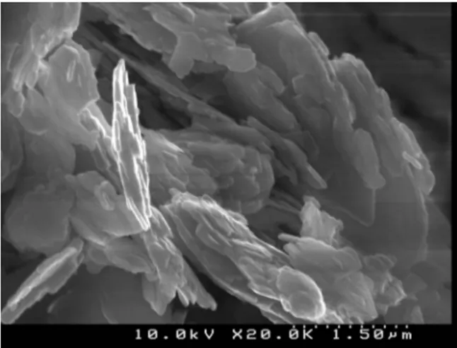
kaolin were performed. The morphological analysis showed that the kaolinite particles had an irregular shape and were up to micrometers long (Figure 1).
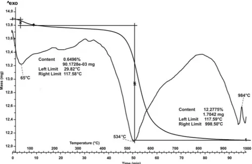
Thermal analysis (Figure 2) revealed the typical endothermic and exothermic peaks of kaolin with a continuous water loss between 29 and 998ºC. Two
endothermic peaks were observed; the first at 65ºC was related to the loss of adsorbed water and the second at 534ºC was ascribed to the dehydroxylation of kaolinite. An exothermic peak was found at ~780ºC and is related to the disruption of the kaolinitic structure. The exothermic peak located at 984ºC is due to the formation of the Al-Si spinel phase from kaolinite. Two different steps in the loss of water were observed; the first ranging between 29 and 117ºC and the second between 117 and 998ºC. The total mass loss was 1.71 mg corresponding to a starting water content of 12%.
Infrared analysis was conducted on kaolin raw material (Figure 3). The bands between 3750 and 3500 cm 1are attributed to OH stretching. Five bands, in particular, were noted at 3648, 3653, 3667, 3674, and 3686 cm 1, and related to the inner surface OH in
phase/out-of-phase stretching vibrations (Cheng et al., 2010a; Frost et al., 2001b); a band located at 3619 cm 1 is related to the inner OH stretching vibration (Cheng et al., 2010a; Frost et al., 2001b). In the lower-w a v e n u m b e r r e g i o n o f t h e I R s p e c t r a (1800 400 cm 1) a band at 1113 cm 1 was revealed Figure 2. DTA and TG analysis of raw kaolin.
and attributed to apical Si O stretching vibrations (Cheng et al., 2010b); in the same wavenumber region, two bands located at 1024 and 1002 cm 1 were noted and are related to apical Si O stretching vibrations (Cheng et al., 2010b); two other bands at 935 and 910 cm 1 are attributed to Al OH bending vibrations
(Qtaitat and Al-Trawneh, 2005); three bands at 788, 749, and 685 cm 1are related to OH (Al-OH) translational vibrations (Frost et al., 2001a).
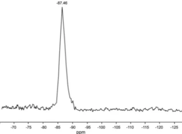
The 29Si MAS-NMR spectrum of kaolin (Figure 4) reveals a peak located at 87.46, the position of which is consistent with the data of Rı´os and Williams (2010).
Mineralogical, crystallographic, and chemical characterization of synthetic products
The results of PXRD analyses conducted on samples of the two experimental runs (Figures 5, 6; Table 1) revealed interesting phases and phase transitions. In synthesis run 1 (Figure 5) the crystallization of synthetic kaliophilite H2 as a metastable phase (sensu Tuttle and Smith, 1958) is shown together with kalsilite in the run lasting 2 24 h; after this, kaliophilite H2 became unstable and kalsilite was the only phase remaining after 10 days.
In synthesis run 2 (Figure 6), kaliophilite H2 was the first phase to occur in the synthesis run lasting 2 5 h; after this, kaliophilite H2 became unstable and KAlSiO4
-01 was the only phase remaining after 5 days.
This temporal synthesis trend can be read in two ways. (1) From an industrial point of view, it offers the possibility of an economic synthesis of both products. (2) From a strictly geological point of view, kalsilite has been reported as a magmatic product in some rare volcanic environments. The present result seems to indicate, however, that the pyrometamorphic genesis of both is viable in terms of the short time of magma-chamber residence, at least in terms of a direct relation-ship of alkaline basaltic magma (especially if K-rich magmas are involved) by digestion of tonstein-like sedimentary xenoliths (for a discussion on pyrometa-morphism associated with basaltic rocks and products, Figure 4.29Si MAS-NMR spectrum of raw kaolin.
see the study by Sabine and Young (1975) and by Novembre et al. (2017a), and references therein).
The end-products of syntheses 1 and 2, consisting of monomineralic powders of kalsilite and KAlSiO4-01,
r e s p e c t i v e l y , w e r e c o n s i d e r e d f o r f u r t h e r characterizations.
The observed and calculated (Rietveld refinement) profiles and difference plot (Figure 7) for kalsilite and KAlSiO4-01, each mixed with corundum NIST 676a,
were obtained for the end run samples of synthesis runs 1 and 2 (Figure 7a,b, respectively). (Corundum was added in order to perform the QPA analysis; this is a procedure in which a known amount of corundum NIST 674a or 676a, is added to the mixture (10 wt.%) and considered as a component itself. The refined values of the Rietveld phase fractions are converted into weight fractions and rescaled into absolute values with respect to the amount of added spike.) The cell parameters for kalsilite, having hexagonal symmetry (space group P63)
were calculated from the Rietveld refinement (Table 2) and were a0= b0= 5.1652 (0.00053) A˚ and c0= 8.6894
(0.00069) A˚ ; the cell parameters of KAlSiO4-01, refined
as having monoclinic symmetry, space group P21, were
a0= 15.67871(0.00437) A˚ , b0= 9.06004(0.00217) A˚ ,
and c0= 8.56471(0.00427) A˚ .
When comparing the refined cell parameters with bibliographic data, good agreement with values proposed for kalsilite by Andou and Kawahara (1984) and for KAlSiO4-01 by Kremenovic et al. (2013) was achieved.
The results of the QPA analysis (Table 2) indicated that the calculated amount of amorphous phase in the sample after 10 days of synthesis in run 1 was 6.6(9)%, thus resulting in a final product of 93.4(9)% kalsilite; the
calculated amorphous phase in the sample at 5 days of synthesis in run 2 was 6.5(8)%, thus resulting in a final product of 93.5(8)% KAlSiO4-01.
The results of chemical analyses of the samples of kalsilite (after 10 days of synthesis in run 1) and of KAlSiO4-01 (after 5 days of synthesis in run 2) and Si/
(Si+Al) ratios are shown in Table 3. The chemical formulae of kalsilite and KAlSiO4-01 were calculated
based on the chemical analysis results reported in Table 3: (K8.01)(Al7.98Si8.01)O32 and (K6.98)(Al8.03Si8.23)O32,
respectively.
Morphological and physical characterization
The SEM image (Figure 8a) of the sample of synthesis run 1 at 10 days provides evidence of the crystalline growth of hexagonal crystals of kalsilite, the maximum axis of which measured 4.5 mm; powder of synthesis run 2 at 5 days (Figure 8b) indicated the presence of a plate-like prismatic habit of KAlSiO4-01
with an average length of 4 mm.
Density values of 2.624 (0.05) and 2.611(0.02) g/cm3 (Table 4) were calculated for KAlSiO4-01 and kalsilite,
respectively, which are comparable to those obtained by Gregorkievitz et al. (2008), Kremenovic´ et al. (2013), and Hokamoto (1997).
IR and 29Si MAS NMR
The IR spectra of kalsilite and KAlSiO4-01 from both
of the synthesis runs (Figures 9, 10), along with the assignments for the absorption bands (Table 5), were analyzed in the context of known data for kalsilite (Becerro et al., 2009; Henderson and Taylor, 1988) and for KAlSiO4-01 (Dimitrijevic and Dondur, 1995).
For kalsilite, a single band was noted in the region of the asymmetric Si O stretch, located at 966 cm 1; a minor band associated with symmetric Si O stretch vibrations was observed at 685 cm 1.
For KAlSiO4-01, eight bands were found in the
asymmetric Si O stretch region located at 1106, 1071, 1034, 988, 973, 959, 941, and 930 cm 1, respectively. Two bands related to symmetric Si O stretching were located at 693 and 663 cm 1.
The29Si MAS-NMR spectrum of kalsilite revealed a single peak at 88.9 ppm corresponding to the Si type of environment Q4 (4Al) (Figure 11a, Table 6). The data are consistent with values reported for this mineral by Becerro et al. (2009) for a kalsilte synthesized under hydrothermal conditions using kaolinite as the starting material; good agreement was also found with the data of Dimitrijevic and Dondur (1995) for a kalsilite obtained by cationic exchange on zeolite LTA. As Figure 7. Rietveld refinement plots: observed (+) and calculated profiles (solid lines) and difference plots for: (a) kalsilite + corundum NIST 676a; and (b) KAlSiO4-01 + corundum NIST 676a. The tick marks show the positions of the Bragg peaks.
Table 2. Rietveld refinement of samples at 10 days of synthesis in run 1 and 5 days of synthesis in run 2: experimental conditions and crystallographic data for kalsilite and KAlSiO4-01 plus 10% corundum NIST 676a. The results of the QPA
analyses of kalsilite and KAlSiO4-01 in each sample are reported as percentages of the amorphous component.
Wavelength (A˚ ): CuKa of the X-ray tube.
Sample 10 days, synthesis run 1 +
10% Corundum NIST 676a
5 days, synthesis run 2 + 10% Corundum NIST 676a
Wavelength (A˚ ) 1.5418 1.5418 No. of observations 9504 7114 Rwp 0.18 0.15 Rp 0.13 0.11 CHI2 2.37 1.75 % amorphous 6.6(9) 6.5(8) % phase kalsilite 93.4(9) % phase KAlSiO4-01 93.5(8) Space-group kalsilite P63 a (A˚ ) 5.1652(0.00053) b (A˚ ) 5.1652(0.00053) c (A˚ ) 8.6894(0.00069) Space-group KAlSiO4-01 P21 a (A˚ ) 15.6787(0.00437) b (A˚ ) 9.0600(0.00217) c (A˚ ) 8.5647(0.00427)
Table 3. Chemical characterization of products of synthesis in runs 1 and 2. Values for the MgO, MnO, TiO2, and P2O5oxides
were below their respective detection limits (*) (Stebbins et al., 1986).
Temperature (ºC) K2O (%) SiO2(%) Al2O3(%) Time (h) PXRD
700 29.75 37.95 32.10 30 kalsilite
(*) 28.50 39.3 31.60
800 26.62 40.01 33.15 20 KAlSiO4-01
(*) 26.00 40.60 32.10
Figure 8. SEM images of kalsilite synthesized at 10 days of synthesis in run 1 (a) and of KAlSiO4-01 crystals synthesized at 5 days of
noted by Becerro et al. (2009), an additional peak attributed to Q3 (3Al) was reported for kalsilites by Stebbins et al. (1986) and by Hovis et al. (1992), who obtained them by cationic exchange on plutonic nephelines, and who interpreted it as being a result of silica content in excess of the ideal 1:1 Si/Al ratio. The absence of this peak in the sample synthesized in the present study is consistent with a 1:1 Si/Al ratio, thus resulting in complete Si/Al order in the kalsilite sample of synthesis run 1.
For KAlSiO4-01 the sample at 20 h of synthesis in
run 2 is characterized by a29Si MAS-NMR band located at 85.51 (Figure 11b, Table 6) and related to Q4(4Al) sites. These data are consistent with the findings of Stebbins et al. (1986) who also reported the presence of another secondary peak at 93.9 ppm for a KAlSiO4-01
grown by high-temperature sintering of oxides and two other secondary peaks for a KAlSiO4-01 obtained after
thermal treatment of kalsilite.
CONCLUSIONS
The results of the present study have proven that the reaction of kaolin with potassium carbonate leads to the
formation of kaliophilite H2 and that this is indeed a metastable phase. The evolution of this phase was followed over time and allowed the authors to identify the stable phases.
In particular, crystallization of kalsilite and of KAlSiO4-01 as isolated phases was achieved after
10 days and 5 days, at 700 and 800ºC, respectively. In other studies (e.g. Gorgeu, 1887; Heller-Kallai and Lapides, 2003), KAlSiO4 minerals were synthesized
starting from a natural kaolinitic precursor; thermal activation of kaolinite with K2CO3at 700ºC for 1 h gave
kaliophilite H2 as the final product (Heller-Kallai and Lapides, 2003). The syntheses presented here were performed over long periods compared with the experi-Table 4. Density analysis of the mineral phases.
Sample Density g/cm3 KAlSiO4-01 (5 days) 800ºC 3 (*) 2.57 (**) 3 Kalsilite (10 days) 700ºC 3 (***) 2.64
*Gregorkievitz et al. (2008); **Kremenovic et al. (2013), and ***Hokamoto (1997).
Figure 9. IR spectrum of synthesis run 1; sample at 10 days.
Figure 10. IR spectrum of synthesis run 2; sample at 5 days.
Figure 11.29Si MAS-NMR spectra: (a) sample at 10 days of
ments conducted in the other studies (e.g. Gorgeu, 1887; Heller-Kallai and Lapides, 2003) and illustrated the phase transitions of kaliophilite H2 during a specific time interval.
Detailed characterization of the products synthesized found that the chemical-physical, morphological, and spectroscopic characteristics of the experimental pro-ducts are comparable with those reported in the literature for the same minerals synthesized using different methods. In particular, the QPA analyses indicated a high purity for the minerals synthesized and this allows for industrial application.
ACKNOWLEDGMENTS
The authors acknowledge the technical staff at the University of Barcelona for their help during the devel-opment of the work. This is a contribution of the Spanish MICCIN PROJECT CGL2011-28022.
REFERENCES
Akokelar, D., Chaffee, A., and Howe, R.F. (1997) The transformation of kaolin to low-silica X zeolite. Zeolites, 19, 359 365.
Andou, Y. and Kawahara, A. (1984) The refinement of the structure of synthetic kalsilite. Mineralogical Journal, 12, 153 61. Aulinas, M., Gimeno, D., Fernandez-Turiel, J.L., Font, L.,
Perez-Torrado, F.J., Rodriguez-Gonzalez, A., and Novell,
G.M. (2010) Small-scale mantle heterogeneity on the source of the Gran Canaria (Canary Islands) Pliocene Quaternary magmas. Lithos, 119, 377 392.
Aznar, A.J. and La Iglesia, A. (1985) Obtencio´n de zeolitas a partir de arcillas aluminosas espan˜olas. Boletı´n Geologı´co y Minero, 96, 541 549.
Becerro, A.I. and Mantovani, M. (2009) Hydrothermal synth-esis of kalsilite: a simple and economical method. Journal of the American Ceramic Society, 92, 2204 2206.
Becerro, A.I., Escudero, A., and Mantovani, M. (2009) The hydrothermal conversion of kaolinite to kalsilite: influence of time, temperature, and pH. American Mineralogist, 94, 1672 1678.
Bogdanoviciene, I., Jankeviciute, A., Pinkas, J., Beganskiene, A., and Kareiva, A. (2007) Sol-gel synthesis and character-ization of kalsilite-type alumosilicates. Materials Science (Medzˇiagotyra), 13 (3), 1392 1320.
Bogdanoviciene, I., Jankeviciute, A., Pinkas, J., Beganskiene, A., and Kareiva, A. (2008) Study of alumosilicate porce-lains: sol-gel preparation, characterization and erosion evaluated by gravimetric method. Materials Research Bulletin, 43, 2998 3007.
Cheng, H., Liu, Q., Zhang, J., and Frost, R.L. (2010a) Delamination of kaolinite-potassium acetate intercalates by ball-milling. Journal of Colloid and Interface Science, 348, 355 359.
Cheng, H., Liu, Q., Zhang, J., and Frost, R.L. (2010b) A spectroscopic comparison of selected Chinese kaolinite, coal bearing kaolinite and halloysite a mid-infrared and near infrared study. Spectrochimica Acta Part A: Molecular and Biomolecular Spectroscopy, 77, 856 861. Cook, L.P., Roth, R.S., Parker, H.S, and Negas, T. (1977) The
system K2O-Al2O3-SiO2. Part 1. Phases on the KAlSiO4–
KAlO2join. American Mineralogist, 62, 1180 90.
Demortier, A., Gobeltz, N., Lelieur, J.P., and Duhayon, C. (1999) Infrared evidence for the formation of an inter-mediate compound during the synthesis of zeolite Na-A from metakaolin. International Journal of Inorganic Materials, 1, 129 134.
Dimitrijevic, R. and Dondur, V. (1995) Synthesis and characterization of KAlSiO4 polymorphs on the SiO2
-KAlO2 join. II. The end-member of ANA-type zeolite
framework. Journal of Solid State Chemistry, 115, 214 224. Dollase, W.A. and Freeborn, W.P. (1977) The structure of KAlSiO4with P63mc symmetry. American Mineralogist, 62,
336 340.
Fernandez-Turiel, J.L., Gimeno, D., Rodrı´guez, J.J., Carnicero, M., and Valero, F. (2003) Spatial and seasonal water quality in a Mediterranean catchment: the Llobregat river (NE Spain). Environmental Geochemistry and Health, 25, 253 474.
Table 5. Asymmetric stretch of inner bonds and symmetric stretch of external Si O bonds for samples of synthesis runs 1 (10 days) and 2 (5 days).
Sample Symmetric Si O Assymetric Si O
Synthesis run 700ºC 10 days 966 685 (*) Kalsilite 1037 985 684 (**) Kalsilite 1030 983 688 Synthesis run 800ºC 5 days 1106 1071 1034 988 973 959 941 930 691 663 KAlSiO4-01 (***) 1100 1070 1040 990 940 880 700 665
*Becerro et al. (2009); **Henderson and Taylor (1988); and ***Dimitrijevic and Dondur (1995).
Table 6.29Si chemical shift (ppm) for synthesis runs 1 and 2.
Sample 29Si Chemical shift (ppm)
Run synthesis 1 10 days 88.9 (*) Kalsilite 89.1 (**) Synt. kalsilite 89.63 (***) kalsilite 88.8, ( 94.0) Run synthesis 2 5 days 85.51 (***) KAlSiO4-01 85.6, 88.8, ( 92.0, 97.0)
*Becerro et al. (2009); **Dimitrijevic and Dondur (1995); and ***Stebbins et al. (1986).
Frost, R.L., Locos, O.B., Kristof, J., and Kloprogge, J.T. (2001a) Infrared spectroscopic study of potassium and c es i u m a ce t at e- i n t e rc ala t e d k a ol i n i t e s. Vibrat ional Spectroscopy, 26, 33 42.
Frost, R.L., Mako´, E´ ., Kristo´f, J., Horva´th, E´., and Kloprogge, J.T. (2001b) Modification of kaolinite surfaces by mechan-ochemical treatment. Langmuir, 17, 4731 4738.
Gorgeu, A. (1887) Action du kaolin sur plusieurs compose´s alkalins: silicates doubles d’alumine et de potasse ou de soude. Annales de Chimie et de Physique, 2e`me se´ries, X, 145 166, Masson, Paris.
Gregorkiewitz, M., Li, Y., White, T.J., Withers, R.L., and Sobrados, I. (2008) The structure of ‘‘orthorhombic’’ KAlSiO4-01: evidence for Al-Si order from MAS NMR
data combined with Rietveld refinement and electron microscopy. The Canadian Mineralogist, 46, 1511 1526. Gualtieri, A.F. (2000) Synthesis of sodium zeolites from a
natural halloysite. Physics and Chemistry of Minerals, 28, 719 728.
Gualtieri, A., Norby, P., Artioli, G., and Hanson J. (1997) Kinetics of formation of zeolite Na-A (LTA) from natural kaolinites. Physics and Chemistry of Minerals, 24,191 199. Hamilton, D.L. and Henderson, C.N.B. (1968) The preparation of silicate compositions by a gelling method. Mineralogical Magazine, 36, 832 838.
Heller-Kallai, L. and Lapides, I. (2003) Thermal reactions of kaolinite with potassium carbonate. Journal of Thermal Analysis and Calorimetry, 71, 689 698.
Heller-Kallai, L. and Lapides, I. (2007) Reactions of kaolinites and metakaolinites with NaOH comparison of different samples (Part 1). Applied Clay Science, 35, 99 107. Henderson, C.M.B and Taylor, D. (1988) The structural
behaviour of the nepheline family: (3) Thermal expansion of kalsilite. Mineralogical Magazine, 52, 708 711. Hovis, G.L., Spearing, D.R., Stebbins, J.F, Roux, J., and Clare,
A. (1992) X-ray powder diffraction and23Na,27Al and29Si
MAS-NMR investigation of nepheline kalsilite crystalline solutions. American Mineralogist, 77, 19 29.
Kawahara, A., Andou, Y., Marumo, F., and Okomo, M. (1987) The crystal structure of high temperature form of kalsilite (KAlSiO4) at 950ºC. Mineralogical Journal, 13, 260 270.
Kopp, O.C., Harris, L.A., and Clark, G.W. (1961) The hydrothermal conversion of muscovite to kalsilite and an iron-rich mica. American Mineralogist, 46, 719 727. Kosanovic´, C., Subotic, B., S´mit, I., and Cˇ izˇmek, A. (1997)
Study of structural transformations in potassium-exchanged zeolite A induced by thermal and mechanochemical treat-ments. Journal of Materials Science, 32, 73 78.
Kremenovic´, A., Lazic, B., Kru¨ger, H., Tribus, M., and Vulic´, P. (2013) Monoclinic structure and nonstoichiometry of ‘‘KAlSiO4-01’’. Acta Crystallographica, C69, 334 336.
Larson, A.C. and Von Dreele, R.B. (1997) Los Alamos National Laboratory report: Document Laur 86-748. Li, Ying, Laursen, K., White, T.J., and Gregorkievitz, M.
(2003) The crystal chemistry and microstructure of fluidised bed incinerator clinker tridymite. Journal of Material and Engineering, 14, 119 125.
Liou, C.L., Komarneni, S., and Roy, R. (1994) Seeding effect on crystallization of KAlSi3O8, RbAlSi3O8 and CsAlSi3O8
gels and glasses. Journal American Ceramic Society, 77, 3105 3112.
Merlino, S. (1984) Feldspathoids: their average and real structures. Pp. 435 470 in: Feldspars and Feldspathoids (W.L. Brown, editor). NATO ASI Series C, Vol. 137. Reidel Publishers, Dordrecht, The Netherlands.
Novembre, D., Di Sabatino, B., and Gimeno, D. (2005) Synthesis of Na-A zeolite from 10 A˚ halloysite and a new crystallization kinetic model for the transformation of Na-A into HS zeolite. Clays and Clay Minerals, 53, 28 36.
Novembre, D., Gimeno, D., Pasculli, A., and Di Sabatino, B. (2010) Synthesis and characterization of sodalite using natural kaolinite: an analytical and mathematical approach to simulate the loss in weight of chlorine during the synthesis process. Fresenius Environmental Bulletin, 19, 1109 1117.
Novembre, D., Di Sabatino, B., Gimeno, D., and Pace, C. (2011) Synthesis and characterization of Na-X, Na-A, hydroxysodalite and Na-P zeolites from metakaolinite, Clay Minerals, 46, 336 354.
Novembre, D., Pace, C., and Gimeno, D. (2014) Synthesis and characterization of K-F and W merlinoite-type zeolites using a diatomite precursor. Mineralogical Magazine, 78, 1209 1225.
Novembre, D., Pace, C., and Gimeno, D. (2017a) Synthesis and characterization of wollastonite-2M by using a diatomite precursor. Mineralogical Magazine, https://dx.doi.org/ 10.1180/minmag.2017.081.025.
Novembre, D., Gimeno, D., d’Alessandro, N. and Tonucci, L. (2017b) Hydrothermal synthesis and characterization of kalsilite by using a kaolinitic rock (Sardinia, Italy) and its application in the production of biodiesel. Mineralogical Magazine, https://doi.org/10.1180/minmag.2017.081.080. Okamoto, Y. (1997) Structural modification of KAlSiO4
minerals. Okayama University Earth Science Reports, 4, 41 72.
Okamoto, Y. and Kawahara, A. (1996) Interpretation of the crystal structure of synthetic kaliophilite from the domain structure of kalsilite. Okayama University Earth Science Reports, 3, 57 64.
Ota, T., Takebayashi, T., Takahashi, M., and Hikichi, Y. (1996) High thermal expansion KAlSiO4 ceramic. Journal
of Materials Science, 31, 1431 1433.
Pasculli, A. and Novembre, D. (2012) A phenomenological-mathematical approach in simulating the loss in weight of chlorine during sodalite synthesis. Computers and Geosciences, 42, 110 117.
Perrotta, A.J. and Smith, J.V. (1965) The crystal structure of kalsilite KAlSiO4. Mineralogical Magazine, 35, 588 95.
Qtaitat, M.A. and Al-Trawneh, I.N. (2005) Characterization of kaolinite of the Baten El-Ghoul region/south Jordan by infrared spectroscopy. Spectrochimica Acta, (A) 61, 1519 1523.
Rigby, G.R. and Richardson, H.M. (1947) The occurrence of artificial kalsilite and allied potassium aluminum silicates in blast furnace linings. Mineralogical Magazine, 28, 75 88. Rı´os, C.A., Williams, C.D., and Fullen, M.A. (2009)
Nucleation and growth history of zeolite LTA synthesized from kaolinite by two different method. Applied Clay Science, 42, 446 454.
Rı´os, C.A. and Williams, C.D. (2010) Hydrothermal transfor-mation of kaolinite in the system K2O-SiO2-Al2O3-H2O.
Dyna, 77, 55 63.
Robert, J.L., Della Ventura, G., and Thauvin, G. (1989) The infrared OH-stretching region of synthetic richterites in the system Na2O-K2O-CaO-MgO-SiO2-H2O-HF. European
Journal of Mineralogy, 1, 203 211.
Rocha, J. and Klinowski, J. (1991) Synthesis of Zeolite Na-A from metakaolinite revisited. Journal of the Chemical Society, Faraday Transactions, 87, 3091 3097.
Ruggieri, F., Fernandez-Turiel J.L., Saavedra, J., Gimeno D., Polanco, E., and Naranjo, J.A. (2011) Environmental geochemistry of recent volcanic ashes from Southern Andes. Environmental Chemistry, 8, 236 247.
Sabine, P.A. and Young, B.R. (1975) Metamorphic processes at high temperature and low pressure: the petrogenesis of the metasomatized and assimilated rocks of Carneal, Co. Antrim. Philosophical Transactions of the Royal Society of London, Series A, Mathematical and Physical Sciences,
280, 225 269.
Sanhueza, V., Kelm, U., and Cid, R. (1999) Synthesis of molecular sieves from Chilean kaolinites: I. Synthesis of Na A type zeolites. Journal of Chemical Technology and Biotechnology, 74, 358 363.
Smith, J.V. and Tuttle, O.F. (1957) The nepheline-kalsilite system: 1. X-ray data for the crystalline phases. American Journal of Science, 255, 282 305.
Stebbins, J.F., Murdoch, J.B., Carmichael I.S.E., and Pines, A. (1986) Defects and short-range order in nepheline group minerals: a Silicon-29 nuclear magnetic resonance study. Physics and Chemistry of Minerals, 13, 371 381. Toby, B.H. (2001) EXPGUI, a graphical user interface for
GSAS. Journal of Applied Crystallography, 34, 210 213. Tuttle, O.F. and Smith, J.V. (1958) The nepheline-kalsilite
system II: phase relations. American Journal of Science, 256, 571 589.
Wen, G., Yan, Z., Sinth, M., Zhang, P., and Wen, B. (2010) Kalsilite based heterogeneous catalyst for biodiesel produc-tion. Fuel, 89, 2163 2165.
Zhang, Y., Lv, M., Chen, D., and Wu, J. (2007) Leucite crystallization kinetics with kalsilite as a transition phase. Materials Letters, 61, 2978 2981.
Zhao, H., Deng, Y., Harsh, J.B., and Flury, M. (2004) Alteration of kaolinite to cancrinite and sodalite by simulated Hanford tank waste and its impact on cesium retention. Clays and Clay Minerals, 52, 1 13.
(Received 2 November 2016; revised 27 November 2017; Ms. 1147; AE: J. Brendle´-Miehe´)