PhD in
“Experimental and Regenerative Medicine”
XXIX cicle
“Study of molecular basis of Oculo-Auricolo-Vertebral-Spectrum”
Tutor: Prof. Angelo Vescovi PhD Student: Francesca Piceci Sparascio Co-Advisor:Dott.ssa Valentina Guida
________________________________________________________________
Index
Abstract ... 3
1.Introduction ... 5
1.1 Definition ... 5
1.2 The OAVS clinical phenotype ... 6
1.3 Etiology ... 10
1.3a Murine models ... 15
1.4 Branchial Arch Syndromes ... 17
1.4a Molecular mechanisms of patterning in the branchial arches ... 18
1.4b Differential diagnosis ... 21
Treacher Collins syndrome ... 22
Townes–Brocks syndrome ... 24
CHARGE syndrome ... 25
Branchio-oto-renal spectrum disorders ... 26
Mandibulofacial dysostosis, Guion–Almeida ... 27
1.5 Treatment considerations ... 30
2. Aim of the project ... 32
3. Materials and Methods ... 34
Patients ... 34
DNA extraction ... 34
2 3.1 NGS approach ... 35 a) Targeted sequencing ... 35 b) Nextera XT sequencing ... 39 c) Exome sequencing: ... 43 4. Results ... 44 5. Discussion ... 50 6. Conclusions ... 71 7. Bibliografy ... 72
3
Abstract
Oculo auriculo vertebral spectrum (OAVS; OMIM 164210) is a clinically and genetically heterogeneous disorder originating from an abnormal
development of the first and second branchial arches. Main clinical characteristics include defects of the aural, oral, mandibular, and vertebral development. Anomalies of the cardiac, pulmonary, renal, skeletal, and central nervous systems have also been described. The aetiology of OAVS remains largely obscure. Nevertheless, the identification of several families with proof of both autosomal dominant and autosomal recessive inheritance, as well as detection of several chromosomal aberrations, strongly suggest that OAVS has a genetic basis. The advent of next-generation sequencing (NGS) surmounts these issues and changes the landscape of rare-genetic-disease research, with the possibility of identifying genetic disease causative genes at an accelerating time-rate.The aim of this work is to study the OAVS molecular bases
identifying genes included in microriarrangements using SNP array technology and new candidate genes throughout targeted or exome sequencing. In a large court of 84 clinically well-characterized OAVS patients, some
microduplications and microdeletions have been identified, involving genes belonging to the EYA-SIX- and PAX pathways, previously associated with OAVS. Starting from the genes involved in these microriarrangements, a panel of 78 genes has been drawn, including genes implicated in the molecular pattern of the first and second branchial arch development and genes associated with pathologies with a clinical phenotype overlapping with OAVS. In detail, NGS analysis has identified two missense variants in GCS and PLCB4 genes in two sporadic patients belonging to two unrelated families. Animal models have shown that both of these genes are involved in the early stages of embryonic development and, specifically, in the formation of branchial arches, confirming their role in the etiology of OAVS. Two missense variants and a maternally inherited in-frame deletion were also identified in the MYT1 gene recently associated with OAVS. MYT1 gene is involved in the retinoic acid pathway as one of the teratogenic agents associated with OAVS. The identification of these three variants confirms the involvement of MYT1 gene in the etiopathogenesis of this pathology.A second nonsense mutation in the HOXA2 gene was also identified in a family only presenting isolated microtia. Retrospective studies on
4
the only other patient reported in literature, showed that both patients displayed a particular shape of the outer helix of the ear, compared to other isolated microtia forms. Finally, the analysis of the whole exome was performed in four affected subjects, belonging to a family of three generations, in which the clinical features segregated as autosomical dominant way with incomplete penetrance and variable expression. Two splicing variants and a missense variant were identified in the RNF213, SHPRH and ITGB4 genes, respectively, in all affected and non-affected members of the family.Unfortunately, none of these genes seems to suggest a possible correlation with the aetiological hypotheses brought up until now. This work, therefore, contributed to the expansion of the microdeletion and microduplication spectrum associated with OAVS, presented the first time a point mutation in the GCS gene previously associated with OAVS throughout linkage studies, and the identification of a mechanism of allelic heterogeneity linked to the presence of a mutation in PLCB4 gene, previously associated with Auricolar Condylar Syndrome, in a patient with a Goldenhar phenotype.Finally, it has been contributed to the identification of the second nonsense mutation in the HOXA2 gene in a patient with isolated microtia and a distinctive ear morphology. All these findings provide further evidence of the molecular mechanisms underlying OAVS, confirming the genetic heterogeneity of this pathology and helping to identify new genotype-phenotypic correlations. Identification of new candidate genes
better provide the basis for appropriate genetic testing, clinical management, genetic counseling, with improvements in risk assessment, prognosis and prevention, and for the development of new therapeutic approaches.
KEYWORDS: Oculo auriculo vertebral spectrum; SNP-array analysis; Next Generation Sequencing
5
1.Introduction
1.1 Definition
Oculo-auriculo-vertebral spectrum (OAVS) is a rare congenital disorder
involving the first and second branchial arch derivatives (that affects primarily aural, oral and mandibular development). The estimated incidence is 1/5,600 (Gorlin., 1990), although a recent work showed an incidence of 1/26370 live birth (Barisic et al., 2014).
This spectrum includes conditions previously known as hemifacial microsomia and Goldenhar syndrome (Goldenhar., 1952), and is clinically heterogeneous, ranging from isolated unilateral microtia to multiple visceral malformation.
Most patients present with unilateral hemifacial microsomia. In addition, epibulbar dermoids and upper eyelid colobomas are found frequently, often associated with macrostomia or facial clefts (Rollnick et al., 1987). Moreover, the involvement of the structures of the face, cardiac, renal, pulmonary and central nervous system anomalies are described.
The first patients with OAVS was described by Professor M. Goldenhar in 1952 as a combination of anomalies such as epibulbar dermoid, preauricular tags, ear malformations and abnormalities of the cervical vertebrae; this
condition was clinically defined as Goldenhar syndrome (Goldenhar., 1952). A few years later, in 1963, it was suggested the use of the term oculo-auriculo-vertebral dysplasia to describe a syndrome characterized by epibulbar dermoid and/or lipodermoid, ears and vertebral anomalies (Gorlin et al., 1963). In 1976, finally, Gorlin and Pindborg concluded that the hemifacial microsomia,
Goldenhar syndrome and oculo-auriculo-vertebral dysplasia, despite they had different clinical severity phenotype, show clinical features almost overlapping (Gorlin et al., 1976). Only later, in 1989, the research group of Cohen coined the term oculo-auriculo-vertebral spectrum (OAVS), today still used (Gorlin et al., 2010 ; Hartsfield., 2007).
Due to the variable expressivity, there is no consensus regarding the minimum diagnostic criteria for OAVS. After the initial suggestion to consider as a minimum diagnostic criterion the presence of microtia (Gorlin et al., 2010), there are different classification systems for OAVS (Cousley., 1993 ; Rollnick et al., 1987 ; Vento et al., 1991). Those described by Cousley (Cousley., 1993)
6 and Vento et al. (Vento et al., 1991) focus only on the facial anomalies and do not include other frequently associated anomalies as vertebral anomalies, congenital heart defects or limb anomalies. Rollnick et al. (Rollnick et al., 1987) do not take the laterality of involvement into consideration. More recently, Tasse et al.suggest new clinical diagnostic criteria based on the presence of facial asymmetry, in association with ear anomalies and/or preauricular appendages or fistulas. More in detail, Tasse et al. classifyied 53 OAVS patients into three groups based on the presence of the main clinical findings, consisting of microtia (group 1), microtia/preauricular tags plus hemifacial microsomia (group 2), microtia/preauricular tags plus hemifacial microsomia plus vertebral anomalies (group 3), and further differentiated each group into two subgroups: ‘u’ for unilateral and ‘b’ for bilaterally affected. (Tasse et al., 2005). This classification takes into account not only the presence of all the clinical findings but also the affected side (monolaterally or
bilaterally).
By comparing unilaterally and bilaterally affected patients, it has been observed that patients with bilateral involvement require more surgical and supportive treatment, e.g. physiotherapy and speech therapy suggesting to examine these patients more carefully to detect additional clinical findings. The statistically significant correlation between subgroup and number of additional clinical findings suggests that this new classification might be a useful predictor of the likelihood that associated malformations are present in a patient with OAVS.
Recent evidences in literature suggest to take in consideration isolated hemifacial microsomia when associated with a family history of OAVS as a diagnostic criteria.
1.2 The OAVS clinical phenotype
The main clinical features of OAVS include craniofacial abnormalities that are associated with vertebral, cardiac, pulmonary, renal and central nervous system anomalies. The OAVS patients may also have a low birth weight and postnatal growth retardation. About 60% of patients present with unilateral involvement, while 10-33% of patients show abnormalities in both sides with a more serious involvement of the right side than the left (Tasse et al., 2005). The
7 hemifacial microsomia is present in 65% of patients (Figure 1A), associated with a medical case of different gravity, which also depends on the presence of mandibular or maxillary hypoplasia. Often the asymmetry is not appreciable in the first years of life, but it starts to become clear only later, around 4 years of a child's life (Gorlin et al., 2010). About 35% of patients with agenesis of the mandibular branch also have microsomia more frequently associated with epibulbar dermoid.
The ear abnormalities may relate to the external pinna, in the form of microtia (Figure 1B), present in 90% of patients, and atresia / stenosis of external ear occurring in 25-50% of cases. The latter anomaly is related to the problem of conductive hearing loss which can be also sensorineural affecting approximately 50% of patients (Barisic et al., 2014).
There can also be present preauricular skin tags or fistula, described in approximately 40-60% of patients, which sometimes can be located at the base of the neck (Figure 1C). The skin tags are the only feature of the syndrome related to epibulbar dermoid because of, histologically they are both choristomas (Gorlin et al., 2010).
Vertebral defects (Figure 1D) are reported in 18-60% of patients with OAVS and include the presence of hemivertebrae in the lumbar, thoracic and cervical spine, or scoliosis, spina bifida and butterfly vertebrae (Barisic et al., 2014 ; Anderson et al., 2005). Radial anomalies were observed in 10% of patients, and mainly concern the hypoplasia or aplasia of the radius and/or the thumb (Gorlin et al., 2010).
They may also be present different types of ocular abnormalities. In about 10% -20% of patients can also be present epibulbar dermoid often associated with clinically more severe Goldenhar phenotype (Figure 1E). Patients with epibulbar dermoid frequently show anomalies both extraocular and palpebral of the tear drainage, microcornea, ptosis, and anophthalmia (Figure 1F). OAVS patients may also present microphthalmia (5-35%), coloboma (5-10%) and epicanthus (10%). In particular coloboma in the upper part of the eyelid is found in 20% of cases , meanwhile aniridia or absence of the lens and the retina is present with a frequency <1% (Barisic et al., 2014 ; Gorlin et al., 2010). In addition to skeletal malformations, the OAVS patients may have congenital heart disease in 5-58% of cases. Typically , in literature are described
8 arch which alone represented 65% of all cardiac abnormalities noticed (Digilio et al., 2009 ; Gorlin et al., 2010). Finally, even patients with transposition of the great arteries have been described, tubular hypoplasia of the aortic arch
associated with a mild coarctation of the aorta, cardiomegaly, PDA (Patent Ductus Arteriosus ), pulmonary stenosis, dextrocardia, double outlet right ventricle, aortic arch anomalies, hypoplasia of the external carotid arteries and situs ambiguus (Gorlin et al., 2010).
In 5% of patients was observed the presence of tracheoesophageal fistulas while lung abnormalities ranging from incomplete lobulation until hypoplasia and agenesis, unilateral and bilateral. Generally the absence of the lung appears to be in the same side where the craniofacial anomalies are present.
Urogenital defects are present in the 2.5% -50% of patients, including renal ectopia and mono or bilateral renal agenesis, cryptorchidism and multi cystic kidney (Barisic et al., 2014 ; Ritchey et al., 1994) and also, double ureter, renal artery blood flow abnormalities, hydronephrosis, hydroureter. The presence of imperforate anus are also described. Brain abnormalities in OAVS patients have been repeatedly reported and encompass the presence of tumor (e.g., teratoid rhabdoid tumor, medulloblastoma) (Lafay-Cousin et al., 2009), anterior or occipital encephalocele (Kerckoff Villanueva et al., 2008 ; Gustavson and Chen., 1985 ; Aleksic et al., 1984), obstructive hydrocephalus (Kerckoff Villanueva et al., 2008 ; Aleksic et al., 1984 ; Kumar et al., 2000), pons and brainstem abnormalities (Pane et al., 2004 ; Chong et al.,2015), cerebellar hamartoma (Arzimanoglou et al., 1999), hemispheric hypotrophy (Jena and Duggal., 2006), brain calcifications, midline lipomas (Jeanty et al., 1991 ; Beltinger et al., 1988 ; Thommen et al.,1986), and corpus callosum abnormalities (Aleksic et al., 1984).
Few cohort studies investigated systematically the presence of brain abnormalities by means of MRI (Strömland et al., 2007 ; Rosa et al., 2010 ; Tasse et al., 2005) and found them in a high proportion of OAVS patients (17 – 50%). A recent study (Brotto et al., 2017) validated previous findings, both in terms of brain abnormality high frequency (56% in our cohort) and variability. In addition, Brotto et al.,2017 confirmed a previous observation (Rosa et al., 2010) that brain abnormalities are more frequent among OAVS patients with concomitant ophthalmologic abnormalities, probably because the eye is an outpouching of the primitive brain and likely shares some of its development
9 milestones. Nonetheless, it has also been found an association with spine or cranial nerve abnormalities that became even stronger when both
ophthalmologic and spine abnormalities were present. Brain abnormalities seem to be associated with a more severe phenotype thus suggesting a greater or earlier damage during fetal life.
According to Brotto et al., findings, the detection of eye and spine
abnormalities in the context of OAVS should prompt for proper neuroimaging to investigate the presence of brain abnormalities (Brotto et al., 2017).
Figure 1. Clinical features OAVs: facial asymmetry (A), microtia (B), preauricular skin tags (C), vertebral anomalies (fusion C6 / C7, T1) (D), epibulbar dermoid (E), microphthalmia (F) ( Kelberman et al., 2001 ; Cox et al., 2014 ; Tasse et al., 2007 , Anderson et al., 2005)
10
1.3 Etiology
Although knowledge of the genetic basis of human disease and its effect on embryonic development has greatly expanded in recent years, the etiology of OAVS are still largely unknown, and the involvement of both genetic and environmental factors have been suggested (Hennekam et al., 2010 ; Heike et al., 2009). Fetal exposure in the first weeks of pregnancy to vasoactive drugs or teratogenic substances, such as pirimidone, thalidomide and retinoic acid , may cause OAVS. In this regard, one of the hypotheses widely shared by the
scientific community is that these teratogenic substances lead to an aberrant vascular development in utero. In particular, in a study of 154 pregnancies in which pregnant women were exposed to a retinoid substance (isotretinoin) prescribed for severe cystic acne, not only they were observed a high number of abortions but also the presence, in 21 infants,of cranio-facial malformations, (microtia / anotia, micrognathia, cleft palate), or cardiac malformations (conotruncal heart defects, aortic arch anomalies), and defects of the central nervous system (Lammer et al., 1985).
The OAVS phenotype has also been noticed in infants born to diabetic mothers (Grix., 1982 ; Ewart-Toland et al., 2000 ; Wang et al., 2002). The teratogenic effect of maternal diabetes was confirmed by a study of 21 infants of diabetic mothers who had hemifacial microsomia (67%), microtia (52%), sensorineural hearing loss / conductive (43%), epibulbar dermoid and vertebral fusions (24%) (Ewart-Toland et al., 2000). A plausible explanation is that the state of hyperglycemia in pregnant women alter the migration of fetal neural crest cells. This concept is further strengthened by the observations carried out on mice and chicken embryos, in which the induction of a state of
hyperglycemia clearly leads to the presence of defects in cardiac development and neurulation (Wang, et al., 2002). Embryonic hematoma formation or ectodermal non disjunction in early development, which is reminiscent of the occult spinal dysraphisms, have been also suggested to be involved in the pathogenesis of OAVS (Poswillo., 1975 ; Hartsfield., 2007).
Experiments conducted on mouse model by Poswillo., have shown that by injecting intraperitoneally the triazene (antifolate drug) and inducing the formation of bleeding stapedial artery, the murine fetuses showed hypoplasia involving one or both sides of the skull (Poswillo., 1974). Moreover the presence of discordant monozygotic twins and the high frequency of OAVS
11 children born from assisted reproduction pregnancies suggest that the base of OAVS, may be due to aberrant epigenetic mechanisms. Studies with model organisms reveal how the identity and patterning of vertebrate-specific portions of the skull are epigenetically regulated and how epigenetic dysregulation in cranial neural crest cells induces severe skull vault defects (Haberland et al., 2009). Epigenetic factors have been suggested as a possible pathogenic mechanism leading to histone acetylation-dependent imbalance of allelic expression of the BAPX1 gene, a member of the NKX family of homeobox-containing proteins, which play a role in skeletal development and patterning of the middle ear, in five patients with OAVS (Fischer et al.,2006). However there are many evidences of the presence of the genetic contribution to the etiology of OAVS, including the presence of family cases. Despite of most OAVS patients are sporadic, familial studies reported that 20-45% of individuals with OAVS has family history (Rollnick and Kaye., 1983 ; Llano-Rivas et al., 1999 ; Klockars et al., 2007) . Rollnick et al (Rollnick and Kaye., 1983) in a study of 97 cases observed that 45% patients had a family history of some features of the disorder, with first-degree relatives being most frequently affected, often with only mild phenotypic expression (for example, isolated preauricular pits or tags), thus suggesting that what is often presumed to be a sporadic event in a family may be the more severe manifestation of a familial condition. The authors have reported that first degree relatives were most often affected (35/433, 8%). Of 176 siblings cited, 11 (6%) were considered affected. Furthermore, the authors observed a broad phenotypic spectrum within families, an aspect that has also been reported by others (Rollnick.,1988 ; Rollnick et al., 1987) and have proposed that familial inheritance is more frequent than originally reported (Rollnick and Kaye., 1983). Subsequently, a genetic predisposition has been proposed based on growing evidence from the literature (Kaye et al., 1992 ; Tasse et al., 2007 ; Vendramini-Pittoli and
Kokitsu-Nakata ., 2009 ; Tsai FJ and Tsai CH.,1993 ; Goodin et al., 2009). Both autosomal dominant and recessive family were described. Kaye et al., provided evidence for an autosomal dominant model of inheritance with reduced
penetrance troughout a study of segregation analysis performed in 311 members of the families of 74 probands with OAVS ( Kaye et al., 1992 ). Tasse et al., describe an autosomal dominant family where the disease was present with rare bilateral extracranial anomalies (Tasse et al., 2007)(Figure 2). Some authors
12 have suggested that patients with autosomal dominant inheritance of OAVS are more often bilaterally affected than patients with sporadic occurrence (Tasse et al., 2007 ; Vendramini-Pittoli and Kokitsu-Nakata., 2009), and rarely present extracranial abnormalities (Vendramini-Pittoli and Kokitsu-Nakata., 2009). The evidence, so far, does not suggest that the familial OAVS phenotype is different from the sporadic OAVS cases. Later, in 2011, Farra et al. identifyied a family where the ocula-auriculo- vertebral spectrum was characterized by microtia, mandibular hypoplasia, sacral vertebral defects, heart and brain, and with an apparent autosomal recessive inheritance (Farra et al., 2011) (Figure 2).
Figura 2. Examples of autosomal dominant and autosomal recessive families with OAVS reported in literature (Tasse et al., 2007 ; Farra et al., 2011).A) VI-1_Female patient with facial asymmetry, diaphragmatic hernia, encephalomeningocele, and preauricular skin tag. VI-2_Her sibling presented with facial asymmetry and abnormal ears. B) III-1_Female patient with hemifacial microsomia, microtia, preauricular tags. III-2_ Sister of III-1 presented with preauricular skin tags on the right side, mild asymmetrical mandibular hypoplasia. II-2 showed phenotypic expression of OAVS with scars of previously removed preauricular pits on both sides, right-sided hemifacial microsomia and a highly arched palate. I-2 The maternal grandmother showed that the position of her right ear and right palpebral fissure were higher than that on the left side and a mild unilateral lower jaw hypoplasia on the right side.
A number of chromosome abnormalities have been reported to be
associated with OAVS including del(5p), monosomy (6q), del(8q), trisomy 18, ring chromosome 21, del(22q), dup(22q), trisomy 22 and 47,XXY. In addition, there are also reports of chromosomal mosaicism including trisomy 7(mosaic) and trisomy 9 (mosaic) which may account for localized features and low recurrence risk (Josifova et al., 2004). Among these, a terminal deletion of 5p
13 was reported in 8 unrelated patients, suggesting that it may harbor a gene or genes involved in OAVS (Ala-Mello et al., 2008 ; Josifova, et al., 2004 ; Tasse et al., 2007 ; Wang and Khan, 2010). Rooryck et al. performed high density oligonucleotide array-CGH on 86 OAVS patients and identified in 11 patients 12 novel genomic rearrangements (4 deletions and 8 duplications) ranging in size from 2.7 kb to 2.3 Mb, confirming genetic heterogeneity of OAVS (Rooryck et al., 2010). The best evidence for the possibility of a single gene having a major effect comes from familial cases, where the condition appears to segregate in a dominant manner (Kelberman, et al., 2001 ; Tasse, et al., 2007 ; Vendramini-Pittoli and Kokitsu-Nakata., 2009), with variable penetrance and phenotypic expression within and between families. A high lod score has been found for the 14q32 genomic region which harbors the Goosecoid gene (Gsc) (Kelberman et al., 2001). Recently, Huang et al., used bead cheep analysis on a three-generation family with autosomal dominant inheritance and reduced penetrance and found a significant load score on chromosome 15q26.2-q26.3(Huang et al.,2010). More recently, Ballesta-Martinez et al., described a family with clinical diagnosis of OAVS and autosomal dominant inheritance pattern in which they detected a 14q23.1 duplication of 1.34 Mb in size which segregates with the phenotype. This region contains OTX2, which is involved in the development of the forebrain, eyes, and ears, and appears to be a good candidate gene for OAVS (Ballesta-Martinez et al., 2013). Ou et al., had previously described a father and son with clinical features of OAVS and branchiootorenal (BOR) syndrome and the detection of an 11.79 Mb duplication of chromosome 14q22.3-q23.3 including SIX1, SIX6, or OTX2 genes. He proposed that the increased dosage of genes included in the
duplication might be responsible for the BOR and OAVS-like features in this family(Ou et al.,2008). This finding, together with previous findings supports the important role of 14q in OAVS phenotype and narrows down the candidate region to 14q23.1. Although a complex disorder, there is strong evidence to suggest a major genetic determinant in some cases of this condition. Anomalies in 22q have been frequently documented in patients with OAVS, particularly the 22qter deletion (Herman et al.,1988), 22q11.2 deletions (Xu et al., 2008 ; Digilio et al.,2009 ; Tan et al.,2011), the 22q11.1-q11.21 (Cat-eye)
region(Quintero-Rivera and Martinez-Agosto., 2013 ; Torti et al.,2013), and a partial 22 trisomy (47,XX,+der(22)t(11;22)(q23;q11)), which duplicates the
14 22q11 region (De Ravel et al., 2001). Chromosomal mosaicism for trisomy 22(De Ravel et al., 2001) has also been described, making this region a good candidate for some cases of OAVS. More recently, array-CGH analysis identified 22q11 dosage anomalies in 10 out of 22 index cases screened hypothesizing that the 22q11 locus may harbour genes that are important in aspects of the regulation of craniofacial symmetry and 1st and 2nd branchial arch development (Beleza-Meireles et al., 2015). Interestingly, a central role of Crkl, a gene located in the 22q11 syndrome region, has been demonstrated in regulating signalling events in the developing of pharyngeal arches, with potential contribute to craniofacial dysmorphism. In fact, an altered retinoic acid and endothelin signaling has been evidenced in a Crkl mutant mouse. These two signalling pathways play an important role in the migrating and differentiation of neural crest cells in the branchial arches during
embryogenesis (Miller et al., 2014).Among several published subtelomeric deletions in 20q13.33, a small heterozygous deletion including only PCMTD2 and MYT1 genes was described in a girl presenting with severe mental
retardation and mild dysmorphic features (Kroepfl et al., 2008). More recently, in a study of 2016, Lopez et al., screened a cohort of 169 OAVS patient by whole exome sequencing (WES) identifying two mutation in the myelin transcription factor 1 (MYT1). A de novo nonsense mutation was found in a patient with facial asymmetry, preauricular and jugal tags, small and dysplastic ear and moderate conductive hearing loss with stenosis of the external auditory canal, epibulbar dermoid, and vertebral malformation. Furthemore a missense mutation was found in a patient and in his father both affected by OAVS. The proband presented a more severe phenotype with craniofacial anomalies with the presence of a sacrococcygeal dimple, pyloric stenosis and lumbar
dysraphism without vertebral anomalies whereas his father show a mild phenotype characterized by a progressive bilateral sensorineural hearing loss diagnosed at the age of 25, a myopia diagnosed during adolescence and a thoracolumbar scoliosis with right convexity without vertebral malformation (Lopez et al., 2016). Functional studies by transient knockdown of myt1a, homologue of MYT1 in zebrafish, led to specific craniofacial cartilage alterations. Treatment with all-trans retinoic acid (RA), a known teratogenic agent probably causing OAVS, led to an upregulation of cellular endogenous MYT1 expression. Additionally, cellular wild-type MYT1 overexpression
15 induced a downregulation of RA receptor β (RARB), whereas mutated MYT1 did not (Figure 3 ).
Figure 3 Myelin transcription factor 1 (MYT1) is involved in retinoic acid (RA) pathway and belongs to craniofacial disorders causative genes network. (A) Schematic model depicting
MYT1 action on RA pathway. Grey and dashed arrows link RA to MYT1 overexpression and MYT1 inhibition of RA receptor ß (RARB) expression as shown in the present study. (B)
Network of protein-protein interactions is shown, produced by STRING software
(http://www.string-db.org/), including MYT1 (framed) and its closest partners SIN3B, HDAC1 and HDAC2 (underlined) along with craniofacial disorders causative proteins characterised in human and animal models. Thicker lines represent interactions of higher confidence. Proteins in bold have been reported in the literature as related to RA.
1.3a Murine models
Studies in mouse models, have also confirmed the involvement of genetic factors in the pathogenesis of OAVS.
Initially, in 1994, some transgenic mice caused by an insertional mutation on chromosome 10 produce a phenotype in hemizygous mice resembling HemiFacial Microsomia (HFM) in humans , including microtia and abnormal bite occlusion , secondary hypoplasia of the second and third branchial arch accompanied by a hemorrhagic event (Naora et al., 1994) (Figure 4).
16
Figure 4. Phenotypes of transgenic line HFM. Microtia left (A), unilateral anotia dx (B) and abnormal dentition (C) (Naora et al 1994)
Meanwhile the mouse models with Otx2 +/- genotype showed a wide spectrum of craniofacial anomalies that included micrognathia, agnazia,
anophthalmia and a narrowing of the head with lacking involvement of headset (Matsuo et al., 1995).
Additional evidence were later found by studies on mice with mutations in ZIC3, transcription factor involved in the lateralization during development. These mouse models developed multiple birth defects including: abnormalities of the skeleton/ribs, craniofacial abnormalities, left-right axis defects,
congenital heart defects, alteration of the hyoid bone and laryngeal cartilage abnormalities (Zhu et al., 2007).
Some mouse models created by Minoux and colleagues in 2013, allowed to evaluate the expression of homeobox Hoxa2 gene in mesenchymal cells derived from neural crest cells of the first branchial arch, where normally this gene is not expressed. As a result of this expression, mice showed specular earphones duplication; this observation linked the expression of Hoxa2 with the
development of the second branchial arch. Subsequently, the research group of Cox, in 2014, considering the results reached by the expression of ectopic Hoxa2 in mice, noticed that there were both duplicated and specular auricular pinnae and both some small appendixes, just as also observed in patients with a OAVS phenotype, suggesting that the altered expression of the gene Hoxa2 could be involved in these anomalies , focusing on some downstream genes such as Hoxa2 as Bmp4, Bmp5 and Twsg1 (Minoux et al., 2013). None of these genes is specific molecular interpreter of the second branchial arch
17
development, rather they are necessary to the proliferation and differentiation of the auricular cartilage and other cell types.Consistently with what has been said previously, each of these genes when silenced, in mouse models, originates simple microtia phenotypes (Di Leone et al., 1998 ; Minoux et al., 2013 ; Petryk et al., 2004).
1.4 Branchial Arch Syndromes
OAVS involves primaryly the derivatives of the first and second
pharyngeal arches, so it has been proposed that the aetiology and mechanisms of OAVS are related to the development of these structures. The first branchial arch is involved in the development of the face. During the 4th week to 8th week of gestation, the frontonasal prominence gives rise to the median facial structures.
The paired maxillary and mandibular prominence develop into the lateral facial structures. Small hillocks develop at the dorsal end of the first and second branchial arches from the 24th day of gestation. These hillocks gradually fuse to form the pinna of the external ear. The second branchial arch enlarges during the 5th week, forms the mandibular prominence and overgrows the 3rd and 4th arches. Both arches will develop into nerves, muscles, ligaments and skeletal structures, through a complex but poorly characterised signaling network (Szabo-Rogers et al., 2010 ; Chai and Maxson., 2006 ; Minoux and Rijli., 2010).
Disregulation of these signaling pathways triggered by genetic or
environmental factors constitute a potential source of facial maldevelopment. The morphogenesis of the pharyngeal arch derivatives depends on continuous and reciprocal tissue–tissue interactions. One of the key features of craniofacial development is the formation of cranial neural crest cells, which migrate ventrolaterally as they populate the craniofacial regions. Disturbances in the specification, migration, proliferation, survival and ultimate fate determination of the cranial neural crest cells have been proposed as a possible mechanism for OAVS (Szabo-Rogers et al., 2010 ; Chai and Maxson., 2006 ; Minoux and Rijli., 2010). Manifestation and severity of the defect will thus depend on how the expression and activation of genes and proteins have been shifted during facial development.
18 1.4a Molecular mechanisms of patterning in the branchial arches
Appropriate epithelial-mesenchymal signaling is essential for proper development of the pharyngeal arches (Figure 5). Genetic analyses in mice provide evidence that numerous homeobox genes, including Msx, Dlx, goosecoid (Gsc), and Prx, and other transcription factors, such as Hand2, are expressed in pharyngeal arch mesenchyme and play essential roles in
development of PA1 (Graham., 2003 ; Richman and Lee., 2003). Homeobox genes are a large group of genes that encode transcription factors responsible for regulating the expression of downstream target genes. The homeobox is a highly conserved 180 base pair sequence originally discovered in the homeotic selector genes of the fruitfly Drosophila melanogaster. Homeotic genes are a family of master regulatory homeobox genes, ultimately responsible for specifying segment identified along the anterior–posterior axis of the fly’s developing. The vertebrate homologues are the Hox genes and these genes specify the vertebrate embryonic body axis during development
(Akam.,1989). In addition, segment-specific combinatorial Hox gene expression in migrating neural crest cells is also responsible for generating diversity in the branchial arch system(Hunt and Krumlauf.,1991).However, Hox genes are not expressed in the first branchial arch and it has been suggested that this loss of Hox gene expression has been essential for the
skeletal rearrangements to occur that are necessary for the development of a jaw (Cohn., 2002). Indeed, overexpression of Hox genes in the first branchial arch neural crest leads to a failure of differentiation into cartilage and bone (Couly et al., 1998 ; Grammatopoulos et al., 2000). If Hox genes are not directly
responsible for patterning the first arch, then other mechanisms will exist to establish the positional fate of these ectomesenchymal cells.The ectoderm and endoderm that cover the first arch derivatives is characterised by distinct tempero-spatial regions of gene expression. Many of these genes encode secreted molecules and one candidate for playing an early role in patterning of the first branchial arch is the signalling peptide encoded by Fgf-8. Inactivation of mouse Fgf8 specifically in PA1 epithelium revealed that Fgf8 promotes mesenchymal cell survival and induces a developmental program required for PA1 morphogenesis (Trumpp et al., 1999). It has been proposed that this specific expression of Fgf-8 is involved in the early determination of polarity in the first branchial arch (Tucker et al., 1999). Lhx-6 and Lhx-7 are two LIM
19
homeobox domain genes that are characteristically restricted to the
ectomesenchyme within the oral half of the first arch (Tucker et al., 1999 ; Grigoriou et al., 1998). However, this restricted expression domain is not an inherent property of these cells ; ectomesenchyme derived from the second branchial arch, which does not normally express either of these genes, can be induced to express both Lhx-6 and Lhx-7 when recombined with ectoderm from the oral surface of the first branchial arch (Tucker et al., 1999). Fgf-8 is the most likely candidate as the endogenous inducer, its epithelial expression domain complements the ectomesenchymal domains of both genes and beads soaked in recombinant Fgf-8 protein can induce both 6 and
Lhx-7 expression in a concentration-dependent manner in isolated mandibular ectomesenchymal cultures(Tucker et al., 1999). Goosecoid (Gsc) is another homeobox-containing gene expressed within the ectomesenchyme of the first branchial arch, however, in contrast to Lhx-6 and Lhx-7, Gsc expression is restricted to the aboral regions (Gaunt et al.,1993). Consistent with this restricted expression, mice with targeted mutations in Gsc have skeletal abnormalities of the mandible, including hypoplasia and malformations in Meckel’s cartilage (Rivera-Perez et al., 1995 ; Yamada et al., 1995). Therefore, whilst Fgf-8 is essential for normal patterning of the majority of the axis of the first arch, distally in the future incisor regions an alternative regulatory cascade seems to be important (Trumpp et al., 1999)
Distal-less genes incorporate a six-gene family of mammalian homeobox genes (Dlx-1, -2, -3, -5, -6 and -7) that also exhibit highly nested domains of expression in the branchial arches during early development (Bulfone et al., 1993 ; Depew et al., 1999 ; Qiu et al., 1997). Within the mammalian genome, these genes are arranged in convergent pairs, with each pair having similar domains of expression (Dlx-1/-2; Dlx-3/-7; Dlx-5/-6) (Qiu et al., 1997). In particular, along the rostral-caudal axis of the branchial arches Dlx-1 and -2 are expressed more-or-less continuously, whilst the expression domains of Dlx-5/-6 and Dlx-3/-7 are found to be progressively more restricted in a caudal direction (Depew et al., 1999 ; Qiu et al., 1997). The study of mice with targeted mutations in Dlx genes has suggested that a Dlx code of expression might be important in establishing inter-arch identity within the branchial region. Certainly, mice with loss of either Dlx-1, Dlx-2 or Dlx-1/-2 function exhibit progressively more severe anomalies in structures derived from the
20
more rostral regions of the branchial arches, in particular the maxillary process of the first branchial arch (Qiu et al., 1997 ; Qiu et al., 1995). Even though they are expressed in caudal structures, the loss of Dlx-1/-2 does not seem to affect the patterning of these regions because of compensatory action by
other Dlx genes, a finding confirmed by the presence of defects in regions of the mandibular arch of Dlx-5−/− mice (Depew et al., 1999 ; Qiu et al., 1997 ; Qiu et al., 1995 ; Acampora et al., 1999). Further, in mice lacking the function of both Dlx-5 and -6, genes that are only expressed in more caudal regions of the branchial arches, a homeotic transformation is found to occur; these mice have a conversion of mandibular arch structures to maxillary (Depew et al., 2002). Thus, nested Dlx gene expression appears to play a fundamental role in establishing both the identity of different branchial arches and the identity of the maxillary and mandibular processes of the first branchial arch. Members of the Bmp family also have important roles in outgrowth of PA1 (Tucker et al.,1999). PA1 development appears very sensitive to the level of Bmp
signaling during the initial outgrowth phase, and the level of Bmp signaling is tightly regulated by various factors (Massague and Chen., 2000).
The secreted protein Shh, a vertebrate ortholog of the Drosophila segment polarity gene, Hedgehog, is essential for normal development of many organs and is implicated as a cause of HPE (Holoprosencephaly). Shh is expressed in the pharyngeal arch epithelium and targeted disruption of Shh in mouse leads to near complete absence of craniofacial skeletal elements along with multiple organ defects (Chiang et al., 1996). Recent studies using chick and mouse embryos have suggested that Shh may play a role in NCC (neural crest cells) development and pharyngeal pouch patterning (Ahlgren and Bronner-Fraser., 1999 ; Jeong et al., 2004 ; Moore-Scott and Manley., 2005).
21
Figure 5.Molecular pathways involved in the specification of frontonasal, maxillary, mandibular and hyoid structures. A section through an imaginary plane is shown, which is mid-sagittal at the level of the frontonasal process (FNP) and paramid-sagittal (slightly more lateral than the midline) at the level of the first and second pharyngeal arches (BA1 and BA2, respectively). The indicated genetic interactions are representative of data collected in mouse, chick or zebrafish. Some differences might exist in these pathways in different species. The ectodermal, endodermal and mesodermal localization of secreted signalling molecules is indicated,
including bone morphogenetic protein 4 (Bmp4; green), sonic hedgehog (Shh; blue), fibroblast growth factor (Fgf8; red), endothelin-1 (Et1; violet), Fgf8/Fgf3 and Et1 (pink). Receptor molecules are expressed in the neural crest mesenchyme (yellow), though not exclusively, and they induce the activation and/or maintenance of the indicated transcription factors. In turn, the activity of transcription factors specifies the morphogenetic identity of structures both along the anteroposterior (inter-pharyngeal) and the dorsoventral (intra-pharyngeal) axes.
1.4b Differential diagnosis
The list of syndromes involving structures derived from the first and second pharyngeal arches is extensive (Gorlin., 2001) , but they are often associated with malformations derived from other embryological origins than these arches. First and second branchial arch syndromes manifest as combined tissue
22
and the mandibular arches and are the second most common craniofacial malformation after cleft lip and palate. Bilateral anomalies are present in 30% of these patients. Therefore, the presence of a familial history and a detailed clinical examination with biological evaluation may help to identify a known syndrome. Nowadays there is a tendency to group the clinical cases here described in a group of malformations called “LATERAL FACIAL
MICROSOMIA” or “OTOMANDIBULAR DYSPLASIAS” (see Table 1).
The wide spectrum of otomandibular dysplasias makes them difficult to classify, but these deformities can be broadly considered as involving skeletal, auricular and soft tissue. Some patients with OAVS have clinical findings that overlap with other syndromes involving structures derived from the first and second pharyngeal arches. Usually, there is a tendency to consider as overlapping with OAVS fenothype, a lot of syndromes with known causative genes: Treacher Collins syndrome (Katsanis and Jabs., 2012) , Townes–Brocks syndrome (Kohlhase., 2007) , CHARGE syndrome (Lalani et al., 2006) , Branchio-oto-renal spectrum disorders (Smith., 2013) and the phenotypic spectrum associated with mutations in EFTUD2 (Gordon et al., 2012 ; Voigt et al., 2013). The crux of the matter is that these conditions are distinctive and recognisable phenotypic entities. The presence of anal anomalies, for instance, points strongly towards Townes–Brock syndrome; the shape of the ear and semicircular canal abnormality in CHARGE syndrome are very characteristic, and individuals with EFTUD2 mutations have microcephaly and may also have associated with esophageal atresia.
Treacher Collins syndrome
Treacher Collins syndrome (TCS, MIM 154500; TCS2, MIM 613717; TCS3, MIM 248390) is a rare congenital disorder of craniofacial development. TCS is characterized by hypoplasia of the facial bones, particularly the
mandible and zygomatic complex, together with cleft palate, downward slanting of the palpebral fissures, and anomalies of the external and middle ear (Figure 6). Interestingly, there is a considerable degree of phenotypic variability in the severity and combination of these characteristic anomalies both between and within families (Dixon et al., 2004 ; Trainor et al., 2009). TCS occurs with an estimated incidence of 1:50000 live births and is primarily associated with autosomal dominant mutations in TCOF1 (Wise et al., 1999). However, despite
23
extensive searches, the causative mutation in a subset of patients exhibiting classic features of TCS remained unidentified. The use of genome-wide copy number analysis in a child with TCS who was negative for a TCOF1 mutation, led to the identification of a de novo 156-kb deletion within human
chromosome 13q12.2 that resulted in deletion of the entire POLR1D gene (Dauwerse et al., 2011). POLR1D encodes a subunit of RNA polymerase I and III (Dauwerse et al., 2011). Subsequently, a further 242 individuals with classic features of TCS, but who were negative for TCOF1 mutations, were sequenced, leading to the identification of additional POLR1D mutations (Dauwerse et al., 2011). In addition to 10 heterozygous nonsense mutations, seven heterozygous missense mutations located in exon 3 of POLR1D were discovered. Without exception, the missense mutations affected evolutionary-conserved amino acids in the RNA polymerase dimerization domain of POLR1D.Given the strong interaction between POLR1D (RPAC2) and POLR1C (RPAC1) in yeast (Yao et al., 1996) , POLR1C, which also encodes a subunit of RNA polymerase I and III,was sequenced leading to the identification of mutations in both POLR1C alleles in three affected individuals. In all cases, one mutant allele was inherited from each phenotypically unaffected parent, confirming autosomal recessive inheritance in a very small subset of TCS patients (OMIM248390) (Dauwerse et al., 2011 ; Vincent et al., 2016).
24
Figure 6.Characteristic findings of Treacher Collins syndrome include downward slanting palpebral fissures, lower eyelid colobomas, midface and zygomatic hypoplasia, microtia, and mandibular microretrognathia. Patients can be fitted with a removable bone-assisted hearing aid to help speech and language development prior to definitive implantation after ear
reconstruction.
Townes–Brocks syndrome
Townes-Brocks syndrome (TBS; MIM 107480) is characterized by three main clinical signs: imperforate anus in '82% of patients, in 88% of patients were observed ear abnormalities (dysplasia of the upper helix and preauricular pits ) associated in 65% of cases with sensorineural or conductive hearing loss, and malformations of the thumb.
Minor clinical signs showed renal abnormalities occurred in 27% of cases, cardiac abnormalities in 25% of patients and in 56% of cases, deformities of the feet (flat feet or overlapping fingers). Finally also genitourinary malformations (36% of cases have been reported) and intellectual disability in about 10% of cases. The gene responsible for the disease is SALL1 (MIM 602218), located in the chromosomal region 16q12.1. Point mutations or deletions / duplications of the gene are responsible for 70% of cases of Townes-Brocks. SALL1 encodes a transcription factor, which performs its function as a repressor during the early stages of embryogenesis. Functional studies have also shown that the promoter region binds directly to SIX1 gene promoting its expression through a
dose-25 dependent effect and making this also a candidate gene to be tested in patients with an atypical clinical phenotype of BOR syndrome (Chai et al., 2006). CHARGE syndrome
CHARGE syndrome is a pleiotropic disorder whose name is derived from the acronym summarizing its six clinical principal features: ocular coloboma, heart defects, choanal atresia, retardation of growth/development, genital anomalies, and ear anomalies/deafness (Figure 7). CHD7 is the only gene mutation of which is known to cause CHARGE syndrome, and mutations are identified in approximately two-thirds of patients with a clinical diagnosis of CHARGE syndrome (Yasuda et al., 2016)
Various chromosomal rearrangements have been reported in rare patients with a CHARGE-like phenotype. These include balanced translocation between chromosomes 6 and 8(Hurst et al., 1991), unbalanced translocations involving chromosomes 2 and 18, 3 and 22(Clementi et al., 1991), partial trisomy of 19q with partial monosomy 21q (De Krijger et al., 1991), inverted duplication of chromosome 14 (14q22R q24.3) (North et al., 1995) and partial trisomy of 2q (Lev et al., 2000). Based in part on the inconsistent chromosomal aberrations in rare patients with CHARGE association, it is most likely that this condition is genetically heterogeneous. Within the group of children with CHARGE association, there is clearly a subgroup with distinctive clinical characteristics that appears to have a recognisable syndrome (Graham., 2001) Previously, a systematic scan for loss of heterozygosity using microsatellite markers in 10 such patients failed to identify a discernible submicroscopic deletion (Lalani et al., 2003). Although several candidate genes such as PITX28 and PAX29 have been investigated, no mutations have been identified in patients with CHARGE syndrome. Recent evidences demonstrate a de novo mutation in SEMA3E in an affected patient, identified upon mapping the translocation breakpoints in an unrelated individual with a de novo balanced translocation involving
26
Figure 7:Dysmorphic features and cardiovascular malformations in Case 1. (a) “CHARGE ear.” (b) Hockey-stick palmar crease (arrow). (c) Cleft foot. (d) 3D-CT angiography showed truncus arteriosus type A4, interrupted aortic arch type B, and aberrant origin of the right subclavian artery. AAo, ascending aorta; DAo, descending aorta; LCCA, left common carotid artery; LPA, left pulmonary artery; LSCA, left subclavian artery; PDA, patent ductus arteriosus; RCCA, right common carotid artery; RPA, right pulmonary artery; RSCA, right subclavian artery.
Branchio-oto-renal spectrum disorders
Branchiootorenal spectrum disorders comprise branchiootorenal (BOR) syndrome and branchiootic syndrome (BOS).
▪ BOR is characterized by malformations of the outer, middle, and inner
ear associated with conductive, sensorineural, or mixed hearing impairment, branchial fistulae and cysts, and renal malformations ranging from mild renal hypoplasia to bilateral renal agenesis. Some individuals progress to end-stage renal disease (ESRD) later in life.
▪ BOS has the same features as BOR syndrome but without renal involvement.
27 Extreme variability can be observed in the presence, severity, and type of branchial arch, otologic, audiologic, and renal abnormality from right side to left side in an affected individual and also among individuals in the same family. BOR syndrome and BOS can be seen in the same family.The diagnosis of branchiootorenal spectrum disorders is based on clinical criteria. Molecular genetic testing of EYA1 (BOR1, BOS1) detects pathogenic variants in
approximately 40% of individuals with the clinical diagnosis of BOR/BOS. Pathogenic variants can be detected in an additional 5% and 4% of individuals with the clinical diagnosis of BOR/BOS by molecular genetic testing of SIX5 (BOR2) and SIX1 (BOR3, BOS3), respectively. BOR syndrome and BOS are inherited in an autosomal dominant manner. The offspring of an affected individual are at a 50% risk of inheriting the pathogenic variant.
Prenatal testing for pregnancies at risk is possible if the pathogenic variant has been identified in a family member (Smith RJH et al,.1999 updated 2015 Oct 22).
Mandibulofacial dysostosis, Guion–Almeida
Mandibulofacial Dysostosis, Guion-Almeida Type (MFDGA,
OMIM#610536, also called mandibulofacial dysostosis with microcephaly) is a multiple malformation syndrome characterized by progressive microcephaly, choanal atresia, cleft palate, mandibular hypoplasia, microtia, preauricular tags, conductive deafness, congenital heart and/ or thumb anomalies and
developmental delay (Wieczorek et al., 2009 ; Lines et., 2012). Phenotypes of mandibulofacial dysostosis also overlap with other syndromes such as Pierre-Robin, Miller, Treacher Collins, and Nager syndromes, as well as
oculoauriculovertebral (OAVS) spectrum disorders such as VATER
association, CHARGE, Goldenhar and Feingold syndromes (Lines et al., 2012 ; Gordon et al., 2012 ; Need et al., 2012). MFDGA is inherited in an autosomal dominant manner and is caused by mutations in the EFTUD2 gene. EFTUD2 (OMIM#603892, elongation factor Tu GTP binding domain containing 2, also called U5 snRNP-specific protein, 116-KD) coded by EFTUD2, is a small nuclear ribo- nucleoprotein and is a component of the spliceosome complex which plays an important role in pre-mRNA splicing process (Fabrizio et al., 1997; Lines et al., 2012). Actually, 29 causative mutations have been identified in patients with MFDGA. These mutations are: missense (7/29), nonsense
28 (6/29), splicing (4/29), small deletion/insertions (8/29), and large deletion (4/29). The majority of the reported cases have a de novo heterozygous loss of function mutation (Lines et al., 2012 ; Gordon et al., 2012 ; Need et al., 2012).
Table 1 Differential diagnoses of OAVS
Diagnosis Main clinical features Gene Inheritance Treacher
Collins syndrome OMIM 154500, 613717, 248390
Hypoplasia of the zygomatic bones and mandible
External ear abnormalities frequently associated with hearing impairment
Coloboma of the lower eyelid
Absence of the lower eyelashes
Preauricular hair displacement onto the cheeks
Craniofacial involvement is generally symmetrical TCOF1 POLR1D POLR1C Sporadic (60%) Autosomal Dominant(40%) Townes–Brocks syndrome OMIM 107480 Imperforate anus
Dysplastic ears (overfolded superior helices and preauricular tags) frequently associated with sensorineural and/or conductive hearing impairment
Thumb malformations (triphalangeal thumbs, duplication of the thumb, preaxial polydactyly or hypoplasia of the thumbs)
Renal impairment with or without structural abnormalities
SALL1 Autosomal Dominant
CHARGE syndrome
Coloboma of the iris, retina-choroid, and/or disc
CHD7 Autosomal
29 OMIM 214800 Unilateral or bilateral choanal atresia
or stenosis
Ear abnormalities (external ear malformation, ossicular
malformations, Mondini defect of the cochlea and/or absent/hypoplastic semicircular canals)
Cryptorchidism in males and
hypogonadotrophic hypogonadism in both males and females
Cardiovascular malformations
Orofacial clefts
Tracheoesophageal fistula
Cranial nerve dysfunction Branchio-oto-renal spectrum disorders (branchio-oto-renal and branchio-otic syndromes) OMIM 113650, 610896, 602588
Malformations of the outer, middle and inner ear
Conductive, sensorineural, or mixed hearing impairment
Branchial fistulae and cysts,
Renal malformations ranging from mild renal hypoplasia to bilateral renal agenesis.
Branchio-otic syndrome has the same features as branchio-oto-renal
syndrome but without renal involvement. EYA1, SIX5 and SIX1 Autosomal Dominant Mandibulofacial dysostosis, Guion– Almeida—type OMIM 610536
Oto-facial abnormalities (acrofacial dysostosis)
Oesophageal atresia
EFTUD2 Autosomal
30 Thumb anomalies Intellectual disability Zygomatic anomalies Microcephaly
1.5 Treatment considerations
The treatment strategy for patients should prioritize procedures that result in functional improvement while delaying those with cosmetic goals until growth maturity. The following functional categories may necessitate early intervention: airway, feeding, hearing, speech, vision, and socialization.
a) Airway
Maxillary and mandibular hypoplasia can contribute to a compromised airway. Choanal atresia or stenosis can compound this prob- lem,which may require immediate intervention ranging from observation with pulse oximetry and positioning to mandibular advancement with distraction osteogenesis, or tracheotomy
b) Feeding
An incompetent lip seal or cleft lip and or palate may compromise adequate nutrition. Considerations to cleft treatment algorithms or G-tube placement may be necessary.
c) Hearing
Pediatric otolaryngology consultation and formal audiology testing is required early in order to establish successful hearing.
d) Vision
Pediatric ophthalmologist should be consulted to evaluate for any extra ocular muscle (EOM) dysfunction or deficits in visual acuity. When the lateral and inferior orbital supporting structures are hypoplastic or missing the corneas are unprotected,which may necessitate earlier reconstruction of the orbital and zygomatic structures.
e) Speech
Correction of a cleft palate should follow the standard cleft treatment recommendations.
31 Secondary treatment strategy should take place at the end of skeletal
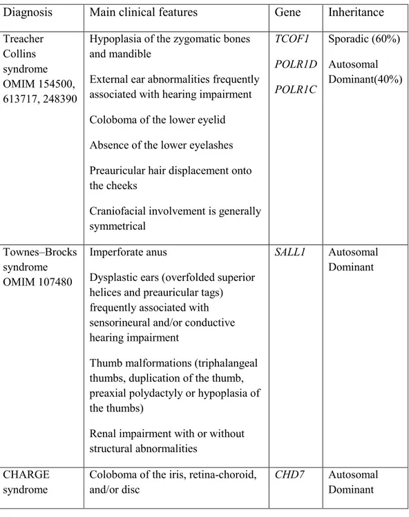
maturity and include orthognathic surgery and zygomatic-orbital reconstruction. Malar and orbital reconstruction uses a full-thickness calvarium bone graft through a coronal incision and exposure. It is recommended that this procedure be performed after the age of 6 years, for skeletal maturity of the midface, as well as the ability to reconstruct the donor calvarium site with a local split-thickness calvarium graft. Maxillomandibular reconstruction is best approached with traditional orthognathic surgery at 13 to 15 years of age, at the time of early skeletal maturity(Figure 8). Le Fort I and rami osteotomies with sliding genioplasty are usually indicated. When the deformity results in absent ramus, condyle, or glenoid fossa, then reconstruction may be performed at the time of malar and orbital reconstruction. The use of costochondral bone graft to reconstruct the ramus-condyle is advocated. A second reconstruction with conventional osteotomies is almost certain to be warranted after skeletal maturity. As with conventional orthognathic surgery, nasal reconstruction should be performed as the final reconstructive procedure when indicated (Alfi et al., 2014).
Figure 8. Schematic drawings of the application of two distracters on the maxilla and mandible in HFM show application of the maxillary and mandibular distractors after performing the Le Fort I osteotomy and the horizontal osteotomy of the mandibular ramus.
32
2. Aim of the project
OAVS is a complex and heterogeneous disorder that involves first and second branchial arch derivatives including maxillo-facial complex, kidneys, skeletal system and heart. It is a rare disease with a prevalence of about 0.3% in the general European population. Although the etiology of this disease is still unknown, a connection between OAVS and maternal diabetes or teratogens was mentioned. Embryonic hematoma formation or ectodermal non-disjunction early in development, was also suggested to be causative for OAVS. However, the presence of familial cases following autosomal dominant or autosomal recessive inheritance, as well as detection of several chromosomal aberrations, and the hemifacial microsomia mouse model, indicate that a significant number of OAVS cases may have a genetic basis. Genetic heterogeneity has been hypothesized and the identification in OAVS patients of many distinct
microdeletions and microduplications throughout cytogenetic and array-based studies, confirm this hypothesis (Rooryck et al., 2010 ; Beleza-Meireles et al., 2015). With the advent of next generation sequencing (NGS) technologies, allowing investigation of multiple genes through one single reaction, the genetic basis of several Mendelian diseases characterized by genetic
heterogeneity have been solved. Recently, NGS has been applied to the analysis of the whole exome of five OAVS patients. The authors identified two
mutations in MYT1, a candidate gene involved in the retinoic acid pathway (Lopez et al., 2016). In our laboratory we have collected a large OAVS cohort.
This clinically-well characterized cohort has been already studied for the presence of microdeletion and microduplication by SNP array, showing that part of the patients carry de novo microrearrangements. Starting from this data, my research project is focused on the use of high-throughput targeted and exome sequencing approaches to search for novel gene(s) causing OAVS. Moreover, this kind of approach allows further insight into the pathways underlying craniofacial development with implications for other similar disorders as isolated microtia or syndromes of the first and second pharyngeal arches. For this purpose I have: 1) used target sequencing [Truseq Custom Amplicon (TSCA)] technology to analyze a panel of candidate genes in a group of selected OAVS individuals (n=65); 2) used Nextera XT technology in order to study the contribute of MYT1 gene’s mutations in our cohort of OAVS
33 patients (n=73); 3) scanned for mutations HOXA2 gene in a family with isolated microtia; 4) analyzed the entire exome of a family with four affected
individuals, where OAVS segregated as Mendelian autosomal dominant trait in two different branches[Ion Proton PCR based enrichment kit (Life
Technologies)]. Identification of the underlying causes of OAVS will provide the basis for appropriate genetic testing, clinical management, genetic
counseling, with improvements in risk assessment, prognosis and prevention, and for the development of new therapeutic approaches.
34
3. Materials and Methods
Patients
Over these three-year’s project, I have collected peripheral blood’s samples of 55 individuals (23 patients and 22 non affected family members) expanding the cohort of previously available samples up to 232 individuals including 118 patients (51 females and 67 males) with oculo-auriculo-vertebral spectrum (OAVS) and 114 non affected family members for a total of 113 indipendent family. Among the 118 patients with OAVS, 105 were sporadic cases and 13 were familiar forms belonging to eight independent families, including a family with a pair of monozygotic twins with discordant phenotype. All participants in this study were selected based on the minimum diagnostic criteria described by Tasse et al., 2005, which include the presence of facial asymmetry associated with ear malformations and / or preauricular pits or fistulas. The OAVS cohort include a total of 20 patients with Goldenhar syndrome, the most severe form of OAVS, where in addition to craniofacial abnormalities might be present ocular abnormalities (such as coloboma or epibulbar dermoid), vertebral anomalies, cardiac and genitourinary malformations.
DNA extraction
Genomic DNA was extracted from peripheral blood using manual kit (Macherey-Nagel) according to the manufacturers instructions. DNA concentration was assessed throughout Qubit™ fluorometer(Invitrogen) and purity parameters (260/280=1.8/2 ; 230/280=1.8/2 ) evaluated using the NanoDrop1000 Spectrophotometer (Thermo Scientific, Waltham, USA).
SNP-array approach
In the first place, given a lot of evidences of genomic rearrangements identified in OAVS patients, samples have been evaluated, using SNP-array approach in order to determine the prevalence and the types of genomic micro-rearrangements and check for recurrent abnormalities associated with specific phenotypes. 64 OAVS samples previously collected, were already analyzed
35
with SNP Chip 6.0 (Affymetrix). Subsequently, I have collected other 23 OAVS samples enlarging the cohort of patients analyzing them with Cytoscan HD chip (Affymetrix).
Data analysis was conducted considering all the duplications and deletions (CNV, copy-number-variants) with a size greater than 75 Kb, and including at least 25 probes. The identification of CNVs, candidate genes and genomic regions associated with OAVS was conducted using different databases of
genomic variants including "Genomic Variants"
(http://projects.tcag.ca/variation/), "UCSC Genome browser "(http://www.genome.ucsc.edu/), and" DECIPHER "
(http://www.sanger.ac.uk/PostGenomics/decipher). All CNVs containing genes coding for proteins and/or microRNA and all CNVs lacking of genes, but flanking genes (far no more than 1Mb from the rearrangement) as well as CNVs that were not present in our laboratory’s database of internal genomic variants and/or absent in control populations listed in the previous reported database of genomic variants, were validated by quantitative real-time PCR assays, using a protocol based on detection with SYBR-Green molecule (Applied Biosystems) using real-time PCR ABI 7900 (Applied Biosystems).
3.1 NGS approach
a) Targeted sequencing
Starting from the genes involved in the rearrangements identified by SNP array analysis, genes belonging to protein functional networks involved in craniofacial development, genes related to disorders with clinical features overlapping with OAVS, I have used bioinformatics prediction software (BIOGRID, GeneMania, String, IPA) to built a network of 78 genes used in order to draw a targeted panels based on an amplicons’ approach (TruSeq Custom Amplicon (TSCA), Illumina San Diego CA) (Figure 9).
36
Figure 9. Genes network built considering those genes involved in the genomic rearrangements detected troughout SNPs array approach, genes belonging to protein functional networks involved in craniofacial development; genes related to disorders with clinical features overlapping
The design of the probes was performed using the "Design Studio software" (Illumina), available online. The probes were designed in the exonic regions and in the UTR regions of selected genes considering several parameters including the GC content, the specificity for a particular gene, the interaction between the probes and the "coverage". The first panel had a target region of 242.944 kbp, divided in 1003 amplicons and included 98% of the target sequence. The second panel covering a target region of 206.055 kbp, was divided into 889 amplicons and included 97% of the target sequences (Figure 10).
37
Figure10. Two targeted genes panels designed troughout "Design Studio software" (Illumina), available online. The first panel had a target region of 242.944 kbp, divided in 1003 amplicons and included 98% of the target sequence. The second panel covering a target region of 206.055 kbp, was divided into 889 amplicons and included 97% of the target sequences.
65 OAVS samples have been analyzed using 250 ng of DNA as input and the enrichment kit "TruSeq Custom Amplicon v1.5" (Illumina, San Diego, California), following the Illumina’s user guide (Figure 11).The produced libraries were sequenced using the "MiSeq Reagent Kit-600 v3”, with a" theoretical throughput " of about 15 Gb for run. The sequencing analysis was carried out paired-end throughout a second generation sequencer "MiSeq Desktop Sequencer" (Illumina). Paired-end sequencing allows users to sequence both ends of a fragment and generate high-quality, alignable sequence data. Paired-end DNA sequencing also detects rearrangements such as small insertions and deletions.
38
The data obtained were analyzed with the software "MiSeq Reporter". The "reads" are aligned considering the reference sequence Homo Sapiens GRCh37/hg19 using two software (BWA and BOWTIE2). SNPs, insertions/deletions and annotations were identified using the Genome Analysis Toolkit software (GATK3.6). Bioinformatic analysis identified a total of about 400 variants for each analyzed sample. Prioritization was performed by selecting the exonic and splicing variants, excluding the synonymous ones. All the variants that had a frequency greater than 1% in certain population’s
database available online such as dbSNP143
(https://www.ncbi.nlm.nih.gov/projects/SNP/) Go-Exp (http://evs.gs.washington.edu/EVS/) and Exac (http://exac.broadinstitute.org/) were eliminated. Troughout this filtering criteria, it has been selected an average of four variants for each sample whose pathogenicity was assessed using 13 pathogenicity predictors including SIFT, Polyphen, Mutation Assessor and CADD. All variants identified, have been confirmed subsequently by Sanger sequencing. Where parents of proband were available, it has been studied the segregation of variants identified also in unaffected members.
39 Figure 11: Adaptation of Illumina TruSeq Custom Amplicon Kit to allow for single molecule tagging. (A) Schematic of method showing amplification of target DNA using custom probes and flanking primers. The P5-SMT primer is the same as the standard P5 index primer, but contains a degenerate 12 N-mer sequence in place of the index. The incorporation of an Ampure Bead size selection step after two rounds of PCR removes unused P5-SMT, and the P5 primer is added to facilitate downstream amplification.
b) Nextera XT sequencing
Following the identification of two OAVS patients with point mutations in the MYT1 gene involved in the retionoic acid pathway (Lopez et al., 2016), I have screened 73 OAVS samples selected from our cohort by Nextera XT DNA