Department of Molecular Medicine
PhD program in “Immunological, haematological
and rheumatological sciences” - XXXII Cycle
Curriculum: Immunology and Immunopathology
Mast cell-derived nanovesicles potentiate allergic
inflammation
PhD candidate: Dott. Mario Lecce
Supervisor
Prof. Rossella Paolini
PhD Coordinator
Prof. Angela Santoni
Contents
Summary ... 3
Chapter 1: Introduction ... 4
1.1 Biology of Mast cells ... 4
1.2 IgE and its high affinity receptor FceRI ... 11
1.3 FceRI structure ... 12
1.4 Regulation of FceRI expression ... 13
1.5 FceRI signal transduction ... 14
1.6 Negative regulation of FceRI-mediated signaling ... 18
1.7 Exosomes: molecular composition and biogenesis ... 20
1.9 Functions of exosomes ... 27
1.10 Mast cell-derived exosomes ... 30
Aim of the study ... 32
Chapter 2: Results ... 35
2.1 MC-derived nanovesicle characterization ... 35
2.2 Antigen-mediated MC activation induces increased level of vesicle release and promotes FceRI/IgE/Ag sorting into exosome-like vesicles ... 36
2.3 Nanovesicle-delivered antigen promotes MC exosome re-uptake by endocytosis ... 39
2.4 Nanovesicles carrying IgE and antigen induce MC functions ... 42
2.5 Vesicles exposing FceRI-IgE complexes are present in sera of atopic patients ... 46
Chapter 3: Discussion ... 51
Chapter 4: Materials and methods ... 55
4.1 Cell maintenance and stimulation ... 55
4.2 Human samples ... 55
4.2 Exosome purification ... 56
4.3 Western blot analysis ... 57
4.4 Ultrastructural analysis and immunoelectron microscopy ... 58
4.5 Dynamic light scattering (DLS) ... 58
4.6 Flow cytometry analysis ... 60
4.7 Small interfering RNA (siRNA) ... 60
4.9 Degranulation assay ... 61
4.10 MagPix IL-6, IL-13 and TNF-a detection assay ... 61
4.11 IgE evaluation ... 62
4.12 Statistical analysis ... 62
Summary
Mast cells (MCs) play a major role in allergic diseases: After activation by the high affinity receptor for IgE (FceRI), they release a high number of pro-inflammatory factors. Moreover, MCs can also produce exosomes, nanosized vesicles that might act as vehicles for intercellular communication. However, evidence of a role for MC-derived exosomes during allergic inflammation is limited.
The aim of my PhD project was to characterize exosome-like vesicles released by unstimulated and IgE-stimulated MCs.
Secreted nanovesicles were purified from the supernatant of MCs and from human sera by a series of centrifugations and ultracentrifugations and, in order to assess their purity, they were characterized in terms of morphology, size and marker expression. The presence of FceRI/IgE complexes was evaluated by western blotting combined with FACS analysis and ELISA. MC activation was measured by degranulation assay and multiplex immune-assay, and exosome uptake was analysed by confocal microscopy and flow cytometry.
We found that FceRI engagement increases the amount of exosome release compared to the constitutive production. Moreover, only exosomes secreted upon antigen stimulation display both surface IgE and antigen. We have also demonstrated that vesicles exposing IgE and antigen are efficiently internalized by sensitized MCs and are able to induce their degranulation as well as pro-inflammatory cytokine production. Finally, we found that exosomes purified from serum of atopic patients are also endowed with FceRI/IgE complexes.
All together, these findings reveal a potential novel mechanism of allergic reaction amplification.
Chapter 1: Introduction
1.1 Biology of Mast cells
Mast cells (MCs) are granulated tissue resident cells (Figure 1), which are best known for their role in type I hypersensitivity, anaphylaxis and protective responses against parasites. However, thanks to the expression of many different activating receptors, they are also involved in several functions both in physiology and pathology including wound healing, angiogenesis, coagulation, immune modulation, tolerance, autoimmunity and cancer (Bulfone-Paus et al., 2017; Frossi et al., 2017).
Figure 1. Electron micrograph of mast cells. Mast cells (MCs) are granulated cells of the
immune system. They are characterized by a central nucleus and numerous granules containing a large panel of biologically active mediators including histamine and proteases. Almost the entire cytoplasm of MCs is occupied by electron-dense granules.
MCs come from CD34+ / CD117+ / CD13+ pluripotent stem cells which reside in the bone
marrow and undergo maturation mainly under the influence of stem cell factor (SCF) and interleukin-3 (IL-3). After precursors egress the bone marrow and circulate in the bloodstream, they enter in several peripheral tissues where they terminally differentiate in mature MCs under the influence of cytokines and growth factors present in the microenvironment (Arock, 2016; Kalesnikoff and Galli, 2008; Moon et al., 2010). Mast cell precursor (MCp) recruitment hasn’t been completely elucidated yet, but it seems constitutive, even though inflammation and tissue
adhesion molecules and chemokines drive the homing of precursors in tissues, such as integrin a4b7 which is involved in MCp recruitment in the gut (Kalesnikoff and Galli, 2008; Krystel-Whittemore et al., 2016). MCs have been found in almost all vascularized tissue and are mainly located in skin, airways, genito-urinary and gut mucosa that represent districts exposed to external environment (Tsai and Galli, 2012). Given their strategic location at the organ interfaces, MCs can be considered the first line of defense against pathogens and external insults.
As already mentioned, MCs express a great array of surface receptors enabling them to sense and recognize a high number of stimuli consistent with their tissue sentinel role (Migalovich-Sheikhet et al., 2012; Olivera et al., 2018; Redegeld et al., 2018). The high affinity receptor for IgE, namely FceRI, is the most studied activating receptor expressed by MCs and plays a pivotal role during the allergic responses (Conner and Saini, 2005; Kraft and Kinet, 2007; Olivera et al., 2018; Reber et al., 2017). Moreover, some MC populations in human and mice are positive for anaphilatoxin binding receptors C3aR and C5aR which promote degranulation in vivo (Migalovich-Sheikhet et al., 2012). Several Toll-like receptors (TLRs) are also expressed, including TLR-1, 2, and 4–6 on the cell surface and TLR-3, and 7–9 intracellularly whose activation generally results in cytokine production upon pathogen recognition (Sandig and Bulfone-Paus, 2012). Beyond the receptors previously listed, MCs are positive also for surface IgG receptors (FcgRs), interleukin receptors including IL-1R and IL-33R, cytoplasmic sensors RIG-I, MDA-5, NOD1, NOD2, NLRP-3 and many others (Migalovich-Sheikhet et al., 2012). It is an understatement to affirm that MCs can be found only in an active or inactive state. It is globally accepted that MCs represent a “tunable” cell population, sampling continuously the microenvironment and reacting accordingly, thus it’s reasonable to affirm that the division in resting and activated state don’t represent exhaustively MC functions (Frossi et al., 2017; Galli et al., 2005).
Two main MC subsets have been described in both human and rodent. In human, MCs are grouped depending on tissue location and proteases content as tryptase-only MC (MCT), found
mainly in mucosal tissues such as airways and lungs, and MCs positive for tryptase and chymase (MCTC), located particularly in serosal tissues such as skin, the gastrointestinal tract,
and conjunctiva. Two major subtypes have been described in mice as well, and they’re defined according to their localization, specific proteases expression and T-cell dependence. Mucosal MCs (MMCs) are positive for MC protease (MMCP)-1 and -2, have an intraepithelial
localization and importantly, their presence depends on T-cell-induced inflammatory response. Connective tissue MCs (CTMCs) express MMCP-4, -5, -6 and carboxypeptidase A, they reside close to blood vessels and nerves, and are defined as constitutive population whose number is independent from T-cells presence. CTMCs characteristics resemble human MCTCs, while
MMCs are generally compared to human MCTs (Figure 2) (Galli et al., 2011).
Figure 2. Mast-cell subsets in mice and human. MCs can be grouped according to anatomical
location and molecular content. Murine connective tissue-MC are characterized by high concentrations of heparin and histamine accompanied by the expression of tryptases (mMCP-6 and mMCP-7), chymases (mMCP-4 and mMCP-5) and carboxypeptidase A. They are mainly located in skin and serous membranes. Mucosal MCs don’t contain heparin, possess low level of histamine and express mMCP-1 and mMCP2 chymases.
Similarly, in humans MCs can be divided in MCT, which are positive for heparin and tryptases,
and MCTC which express heparine, tryptases, chymases and MC-CPA.
However, differently from rodents, human MC subset distribution doesn’t respect completely this division. Indeed, in some tissues both MC types can be found and, interestingly, it has been
This established MCs subtype division results in a functional diversification. For example, MCs in human alveolar parenchyma are almost negative for FceRI receptor while a high expression was detected in MCs present in bronchial connective tissue (Andersson et al., 2009). Related to this, a recent report demonstrated that murine MCs from different tissues are characterized by a similar transcriptional profile of about 100 genes, but they showed clear tissue-based specific transcriptomes. Interestingly, they clustered independently from basophils, generally considered the circulating counterpart of MCs (Dwyer et al., 2016).
One of the main feature of MCs is the large number of electron-dense granules contained in their cytoplasm where several biologically active mediators are stored such as proteases, cytokines, chemokines, amines (e.g. histamine and serotonine) and enzymes including β-hexosaminidase (Wernersson and Pejler, 2014). Almost all the roles exerted by MCs critically depend on these compounds, for this reason they are deeply involved in the regulation of several immune processes (Jain et al., 2019).
MC granules are also known as secretory lysosomes due to their similar features with canonical lysosomes in terms of acidic pH and protease presence. Several evidences showed that granule formation is a tightly regulated multi-step process which starts from the Golgi membrane where small progranules are generated. Subsequently, they undergo maturation upon progressive fusion process with other granules which results in fully-maturated granules which populate almost the entire MC cytoplasm. Upon MC stimulation, these organelles undergo exocytosis with subsequent mediator release in the extracellular space (Blott and Griffiths, 2002; Wernersson and Pejler, 2014). Several stimuli have been described which are able to induce MC degranulation such as toxins, proteases, endogenous mediators, and allergen-induced FceRI activation (Gaudenzio et al., 2016; Migalovich-Sheikhet et al., 2012).
Given the expression of a large panel of receptors as well as of immunologically active mediators, MCs are able to influence and regulate effector functions of several components of both myeloid and lymphoid cells. They may impact on several aspects including cell recruitment, phenotype, and survival depending on which products are released. For instance cytokine or chemokine MC production, will either enhance or suppress immunological responses (Jain et al., 2019).
Evidences showed that MCs are able to internalize, process and present antigens on MHC class I and II molecules in vitro (Galli and Gaudenzio, 2018; Gong et al., 2010), however this process hasn’t been described in vivo so far. Moreover, antigens internalized together with IgE and
FceRI can be captured, upon MCs apoptosis, by other APCs which in turn present them to specific lymphocytes (Kambayashi et al., 2008). The clear immunomodulatory role is further confirmed by the expression of co-stimulatory molecules such as B7-1, B7-2 and CD28, molecules included in TNF family, CD40 and CD40L allowing MCs to interact with numerous cell type such as DCs, B and T cells (Galli et al., 2008a). MC-derived products influence DC activation and mobilization, moreover, they are able to skew them into a Th2-polarizing phenotype (Kitawaki et al., 2006). Furthermore, MC-derived TNF-a mediates DC recruitment into the site of bacterial infection, contributing to pathogen elimination (Shelburne et al., 2009; Suto et al., 2006).
Efficient antigen presentation to CD4+ T cells and subsequent Th2-polarized activation requires
OX40L whose expression is induced by TLR and FceRI stimulation (Bulfone-Paus and Bahri, 2015).
Besides having sentinel effector functions against bacteria, several evidences suggest that MCs may be able also to limit viral infections, indeed they express viral-sensitive receptors such as TLR-3 which trigger production of anti-viral cytokine including IFN-a and IFN-b (Orinska et al., 2005). Besides interferons, upon recognition of viral products, MCs release CD8+ T-cells
recruiter CCL5 thus enhancing infected cell elimination (Orinska et al., 2005).
MCs play a major role in allergic inflammation which represents a very common disease in the western countries; There are many pathologies depending on tissue localizations such as allergic rhinitis (upper airways), asthma (lower airways), atopic dermatitis (skin), and food allergies (gastrointestinal tract) (Galli et al., 2008b; Kubo, 2018).
Two phases have been generally recognized during the allergic inflammation. An early-phase characterized by a fast release of diverse classes of mediators only few seconds upon allergen challenge, and a late-phase which occurs within hours (Reber et al., 2017).
Generally, allergic response starts with the production of IgE from B cells against innocuous antigens called allergens that induce type 2 immune response elicited mainly by T helper 2 lymphocytes. These allergen-specific antibodies are immediately bound by the high affinity receptor for IgE, the FceRI (Figure 3 upper panel). When a second exposure to the same at least bivalent antigen occurs, a cross-linking of IgE receptors on the MCs surface is induced resulting in the activation of a complex biochemical pathway depending on phosphorylation
Figure 3. IgE/Antigen-mediated Mast cell activation. First exposure to allergen in
predisposed individuals, promotes the production of IgE which can be immediately bound to mast cell through FceRI resulting in MC sensitization (upper panel). FceRI crosslinking induced by a second exposure to the same multivalent antigen results in MC degranulation and subsequent release of proinflammatory mediators such as histamine, prostaglandins, cytokines and chemokines (bottom panel).
The first mediators to be released within minutes are granule-associated preformed molecules such as histamine, prostaglandin D2 (PGD2) and Tumor necrosis factor a (TNF-a). These all have a large number of effects including increased vascular permeability, mucus production
and smooth muscle contraction (Wernersson and Pejler, 2014). The second class comprises the newly synthesized molecules derived from the metabolism of arachidonic acid such as eicosanoids consisting in leukotriene C4 (LTC4) and prostaglandin D2 (PGD2). These compounds aren’t stored in the granules but upon an appropriate stimulus, they are released after synthesis and act in an autocrine and paracrine manner. Together, these two classes of mediators deeply affect vascular endothelium in term of permeability and differential expression of adhesion molecules provoking a higher recruitment of proinflammatory cells in the interested tissue. After few hours, a large panel of cytokines, chemokines and growth factors are released by MCs such as IL-3, IL-5, IL-13 which contribute, together with recruited leukocytes and activated resident cells, to the late phase of the response which results in a prolonged inflammation and subsequent tissue remodeling and fibrosis (Galli, 2016; Galli et al., 2008b). In addition, sneezing, itching and coughing are induced thanks to the effects exerted by MC mediators on nociceptors localized in various tissues (Reber et al., 2017),
Of note, MCs could respond to more than one antigen simultaneously because only a small part of FceRI receptors need to be engaged in order to trigger degranulation (Kawakami and Galli, 2002). Once the response is over, MCs are not subjected to apoptosis as happens for the majority of immune cells, but soon they’re available to be activated again (Tsai and Galli, 2012). The inflammatory response triggered by allergens is not always the same, but it can vary depending on the existing affinity with their specific IgE. MCs activated by low-affinity antigens release chemokines, but they don’t undergo degranulation. FceRI cluster stability and the signaling adaptor recruited are highly affected by the strength of IgE-Ag interaction that in turn influences the composition of mediators released (Frossi et al., 2017; Gaudenzio et al., 2016).
Both genetic and environmental factors are involved in susceptibility to allergies. Type of allergen, its concentration and the route by which contacts the immune system deeply affects the strength of the response in predisposed individuals (Madore and Laprise, 2010). At the same time, several polymorphisms are connected to a certain probability to counteract allergic diseases. It has been clearly demonstrated a hereditary transmission of the pathologies and, thanks to the advances in gene identification methodologies, many loci have been linked to allergies contributing to elucidate also their molecular mechanisms. Certain polymorphisms of IL-33, ST2, HLA-DQ genes have been associated with asthma (Vercelli, 2008). Moreover,
represent a major cause of asthma. In this context, certain polymorphisms of CDHR3-encoding gene contribute to dampen epithelial cell interaction which results in asthma exacerbation (Vercelli, 2008).
Given a clear proinflammatory role of MCs during innate and adaptive immunological responses, an increasing number of reports demonstrate an opposite function in several contexts. Related to tolerance, MCs exert an important role in allograft tolerance in mice thanks to an IL-9-based crosstalk with activated CD4+CD25+Foxp3+ regulatory T cells (Lu et al.,
2006). In addition, using diverse mouse models of skin inflammation, it has been shown that MC-derived IL-10 significantly reduce leukocyte recruitment, inflammation and tissue damage (Grimbaldeston et al., 2007).
1.2 IgE and its high affinity receptor FceRI
IgE plays a major role in protection against helminthic infection and type I hypersensitivity reactions (Conner and Saini, 2005; Gould and Sutton, 2008). It is the isotype with the lowest quantity in vivo, its serum concentration may vary from 50 up to 200 ng/ml in healthy individual. In patients suffering of allergic disease, total serum IgE concentration could greatly increase over 10 times respect to physiological values, while the level of antibody specific for a certain allergen could raise even 1000 times (Galli, 2016).
Beside their participation in allergic inflammation and host defense against parasites, IgE is also involved in the elimination of external insults such as toxins, xenobiotics and venoms (Luker et al., 2019). Recently, increasing evidences have been uncovered an additional role for IgE in autoimmune diseases. For instance, clinical studies showed that a certain percentage of patients affected by systemic lupus erythematosus (SLE) produce antinuclear-IgE antibodies. Moreover, these authors showed that in the included cohort, 74% of enrolled patients with enhanced disease showed the presence of specific IgE for self-antigens. For this reason it could represent an effective biomarker of disease condition (Dema et al., 2014).
Structurally, IgE is composed of e heavy-chains containing one variable chain and four constant domains which are bound by diverse receptors (Gould and Sutton, 2008).
Two IgE receptor have been found so far, a low affinity receptor namely FceRII/CD23 which is a member of C-type lectin superfamily and the most studied high affinity receptor FceRI which binds the Fc portion of IgE with an affinity characterized by a dissociation costant of
1010M-1 (Sutton and Davies, 2015). The majority of IgE is captured by this receptor in
peripheral tissues, a process that increase its half-life which normally is of about 3 days in the human serum (Gould and Sutton, 2008).
1.3 FceRI structure
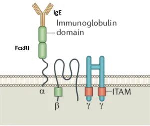
As previously mentioned, MCs play major roles in IgE-associated immune responses thanks to the constitutively expression of FceRI. It’s a cell surface multimeric receptor, composed of 4 subunits characterized by specific functions; The immunoglobulin superfamily member a-chain contains two extracellular immunoglobulin-like domains which bind the Fc portion of a single molecule of IgE with high affinity (Figure 4). It includes a transmembrane region and a N-terminal domain harboring glycosylation sites important for its stability (Kraft and Kinet, 2007).
Figure 4. Structure of high-affinity receptor for IgE (FceRI). FceRI recepotor is composed by one a-chain containing two immunoglobulin-like domains capable of binding one single molecule of IgE, one transmembrane b-chain with 4 hydrophobic domains, a dimer of disulfide-bond g chains. Both b and g chains play a crucial role in signal transduction since they possess cytoplasmic motifs termed immunoreceptor tyrosine-based activation motifs (ITAMs).
binding but contains four hydrophobic transmembrane domains able to amplify and stabilize the FceRI surface expression (Donnadieu et al., 2000). The g-chains comprise two identical disulfide-bound transmembrane proteins. Both b- and g-chains contain in their cytoplasmatic side immunoreceptor tyrosine-based activation motives (ITAMs) which are crucial in the initiation of the signaling cascade (Figure 4) (Kraft and Kinet, 2007).
Two different structural compositions have been found in human: A trimeric receptor composed of an a chain and g-g chains, and the full tetrameric structure which contain also the b chain. MCs and basophils mainly express the tetrameric form, while eosinophils and monocytes express the trimeric one and, under certain circumstances, this structure has been detected in DCs and skin-located Langerhans cells, as well as airway smooth-muscle, bronchial and intestinal mucosa (Campbell et al., 1998; Maurer et al., 1994; Novak et al., 2003; Untersmayr et al., 2010).
In rodents, only the tetrameric FceRI complex exists, whose expression is exclusively limited to MCs and basophils (Kraft and Kinet, 2007).
It’s well known that FceRI activation on the surface of MCs and basophils leads to degranulation and release of diverse proinflammatory mediators resulting in type I hypersensitivity, while in trimeric complex-harboring cells such as monocytes and DCs, this receptor could enhance antigen internalization and subsequent presentation to specific CD4+ T
cells, thus acting as main initiator of type 2 immune responses (Maurer et al., 1994; Novak et al., 2003).
1.4 Regulation of FceRI expression
FceRI subunit assembly in the endoplasmic reticulum (ER) represents a crucial step for its surface expression and therefore, it’s tightly regulated. In particular, the a-subunit possesses aminoacidic sequences responsible for its retaining in the ER, and only the non-covalently interaction with the g-g chains masks this ER retention site. Moreover, glycosylation of the a-chain is critically important for a proper folding, and upon terminal glycosylation processes in Golgi apparatus, the trimeric or tetrameric complexes could finally reach the cell surface. In mice, b-chain production is critical for FceRI surface translocation. This observation suggests
the presence of multiple ER-retention sequences in a-chain, absent in human, which need to be hidden through b-subunit association (Kraft and Kinet, 2007).
Th2-polarizing cytokine IL-4 is the main inducer of FceRI-a subunit expression in human MCs, DCs and monocytes, while no effect has been observed in the murine counterpart (Kraft and Kinet, 2007).
Another important regulator of FceRI surface expression in MCs and basophils is the antibody IgE. Related to this, the first evidence came from the observation of a positive correlation existing between total seric IgE levels and receptor expression (Malveaux et al., 1978). Later, it has been shown that surface receptor stabilization occurs as a result of IgE binding which completely prevent FceRI constitutive internalization and degradation. Furthermore, FceRI basal synthesis is not interrupted resulting in an increase of complexes on cell membrane of both MCs and basophils while subunit gene transcription is not affected by IgE (Conner and Saini, 2005; Kraft and Kinet, 2007).
1.5 FceRI signal transduction
Upon IgE and antigen-induced FceRI crosslinking, ITAM domains included in b and g-g chains undergo tyrosine phosphorylation by the kinase Lyn triggering a complex biochemical pathway which ultimately leads to degranulation and proinflammatory mediators release (Turner and Kinet, 1999). Lyn together with Fyn is a member of Src protein kinases, which play a crucial role in the proximal signaling cascade. Lyn associates with b-subunit in a constitutive way, moreover, thanks to its palmitoyl and myristoyl residues, it is particularly abundant in plasma membrane domains with high content of cholesterol and sphingolipids known as lipid rafts. Lipid rafts are extremely important in controlling the efficacy of the signal transduction, indeed, upon FceRI activation, these receptors converge in large structures resulting from the fusion of diverse rafts where they can interact properly with the transducer machinery in order to achieve a sustained phosphorylation (Alvarez-Errico et al., 2009). Moreover, this mechanism likely provides protection against phosphatases which may turn off the signaling. Related to this, it has been demonstrated that substitution of multivalent antigen with monovalent hapten results in a dislocation of engaged receptors from lipid rafts provoking a rapid dephosphorylation of
mechanisms which readily acts stopping the signal as soon as the aggregating multivalent antigen is removed.
Lyn activity is tightly regulated by intra-domains interactions and its phosphorylation status. Inactive Lyn possesses a phosphorylated residue in its C-terminus which is bound to its SH2 domain resulting in a close conformation. Transition to active form is mediated by FceRI aggregation which induce Lyn dephosphorylation by CD45 leading to an open structure allowing the kinase catalytic site to be completely operative (Alvarez-Errico et al., 2009). The phosphorylation exerted by Lyn on b and g-g ITAMs allows the formation of binding site for the tyrosine kinase Syk which recognizes the modified tyrosines through Src homology (SH2) domains. Syk molecules are activated by Lyn-induced phosphorylation and the signal is further amplified by activation of other Syk proteins. Lyn is also able to phosphorylate ITAM domains in other additionally recruited complexes. (Alvarez-Errico et al., 2009).
Syk is crucial for downstream signal propagation and its depletion results in absence of proinflammatory mediators release in both MCs and basophils (Siraganian et al., 2010; Zhang et al., 1996). For this reason, it has been considered an important therapeutic target for MC and basophil-driven allergic diseases.
LAT1 and LAT2 (NTAL), adaptor membrane proteins phosphorylated by Syk, are main players in coordinate and amplify the biochemical pathway initiated by the kinases Lyn and Syk. They don’t possess enzymatic activity but act as scaffold concentrating many signaling molecules including Gads, Grb2, SLP-76, Sos and Vav1, phospholipase Cg1 (PLCg1) and PLCg2. Once activated, PLCg converts phosphatidylinositol-4,5-bisphosphate [PI(4, 5)P2] into inositol-1, 4,
5-trisphosphate (IP3) and 1,2-diacylglycerol (DAG). They are second messengers that induce an increase of cytoplasmatic calcium (Ca2+) level and protein kinase C (PKC) activation,
respectively. IP3 exerts its function by binding ER receptor STIM1 responsible for Ca2+ egress
(Alvarez-Errico et al., 2009). Then, activation of store-operated calcium channels (SOCCs) is required for a second influx of Ca2+ leading to an adequate sustained signal.
Ca2+ increase triggers the fusion of the granules with the plasma membrane resulting in the
release of their cargo. Moreover, calcium, together with activated PKC, is responsible for calcineurin activation which dephosphorylates the nuclear factor for T cell activation (NFAT) that in turn enters in the nucleus and positively regulates the transcription of several cytokine genes.
Guanidine exchange factors Sos and Vav1 are activated upon FceRI engagement and induce GTP loading on small GTPases such as Ras, Rac and Rho resulting in an increase of their functions. These molecules regulate complex biochemical pathways which have as final targets mitogen-activated protein (MAP) kinases. Generally, they are involved in the initiation of a cascade signaling in which activated kinase phosphorylates and subsequently activate the following kinase involved in the pathway which has as end point transcription factor activation. For instance, Ras induce Raf-1/MEK/ERK pathway that culminate in the activation of Elk-1 while, together with Rac, triggers the activation of AP-1. Moreover, Ras is involved in eicosanoid production by regulating phospholipase A2 (PLA2). NFAT, AP-1, NfkB which is
induced by PKC, are the main transcription factors involved in the late phase of allergic response, upregulating a great number of genes mainly of cytokines such as TNF-a and IL-6 (Alvarez-Errico et al., 2009; Kraft and Kinet, 2007).
A proximal pathway parallel to Lyn has been reported which involve the kinase Fyn. Upon IgE/antigen-induced FceRI activation, Fyn phosphorylates GAB2 which in turn activates PI3K which generates an important second messenger, phosphatidylinositol-3,4,5- trisphosphate (PtdIns(3,4,5)P3 or PIP3), leading to PDK1 and PKCd induced degranulation (Alvarez-Errico et al., 2009).
Lyn and Fyn don’t act independently. Indeed Lyn depletion results in Fyn increased activity thus suggesting a negative modulation exerted by Lyn (Xiao et al., 2005). Fyn knock out in murine MCs results in poorly altered Ca2+ signal but dampened MAPK activity with reduced
degranulation accompanied by decrease in lipid mediator generation and cytokine production (Parravicini et al., 2002). Based on these observations, Fyn is able to potentiate and amplify Lyn-dependent pathway. All these pathways induced upon FceRI engagement are depicted in
Figure 5. Representation of IgE/Antigen induced FceRI signaling. FceRI activation exerted by IgE and antigen promotes activation of Lyn, which induces tyrosine phosphorylation of ITAM domains placed in b and g-g chains. Syk is then activated recognizing ITAM phosphorylated motifs through SH2 domain. Several adaptors molecules, including LAT and NTAL, are phosphorylated by activated Lyn and Syk with subsequent recruitment of Gab2, GRB2, SOS, VAV and Phospholipase C-g (PLC-g). Grb2 and SOS are responsible for the activation of RAS-ERK-MAPK cascade which ultimately leads to lipid mediator synthesis and transcription factors activation. PLC-g is responsible for the production of diacylglycerol (DAG) and inositol-1,4,5-trisphosphate (IP3) which induce protein kinases C (PKCs) activation and Ca2+ release from ER store respectively. Raise of intracytoplasmic Ca2+ leads to
STIM1-dependent store-operated channel (SOC) opening resulting in a further increase of Ca2+ levels.
PKCs together with Ca2+ mobilization promote transfer and fusion of cytoplasmic granules with
plasma membrane resulting in mediators release.
Along with Lyn, the src kinase Fyn contributes to signal propagation by mediating GAB2 phosphorylation which in turn activates PI3K which produce phosphatidylinositol-3,4,5- trisphosphate (PtdIns(3,4,5)P3 or PIP3), an important lipidic second messenger responsible for PDK1 and phospholipase D activation.
FceRI signaling may also be induced by the solely interaction of some IgE clones, in the absence of the multivalent antigen. For this reason, the clones able to induce the signal transduction have been named cytokinergic, indeed they may promote degranulation, cytokine production and enhanced survival. However, the molecular mechanism underling this process hasn’t been completely clarified yet. It has been proposed that the majority of this IgE clones could bind autoantigens that most likely are able promote FceRI crosslinking (Kraft and Kinet, 2007).
1.6 Negative regulation of FceRI-mediated signaling
Of note, IgE/Antigen-induced FceRI engagement not only generates phosphorylation-based positive signals, but also negative signals as control system of the immune response.
Related to this, murine Lyn -/- MCs are more active in term of degranulation with respect to the wild type counterpart and show a prolonged FceRI signaling (Xiao et al., 2005). The balance between this opposite dual role of Lyn likely depends on how strength is the stimuli triggering IgE-bound FceRI. Indeed, it has been reported that monomeric IgE or IgE bound to small amount of antigen induces degranulation and cytokine production, while higher level of antigen results in Lyn-mediated shutdown of FceRI signaling (Xiao et al., 2005).
Other important regulator includes SH2-domain-containing inositol polyphosphate 5′ phosphatase (SHIP) and phosphatase and tensin homologue deleted on chromosome 10 (PTEN) which acts on PIP3 provoking its dephosphorylation resulting in signal termination and subsequent repression of MC degranulation and cytokine release by inactivation of Ca2+ influx
and of several MAPKs (Sibilano et al., 2014).
Post-translational modifications are also involved in negative regulation of FceRI-mediated signaling. Cbl proteins represent main players during this process. They contain several domains involved in protein-protein interaction, a N-terminal consisting of a SH2 domain which is followed by a RING domain that activates the ubiquitin pathway (Paolini et al., 2002). Ubiquitination process consists in addition of a small peptide, ubiquitin (Ub), to specific proteins directing them to multiple different fates. For instance, proteasomal degradation occurs when a chain of at least 4 molecules of ubiquitins is added to the target protein, while
monoubiquitination promotes receptor internalization from the plasma membrane and sorting towards the endocytic pathway.
Ubiquitin peptide is added thanks to 3 enzymes which act in sequence. E1 enzyme which activates Ub; E2 that binds the activated Ub; E3 that transfers activated ubiquitin from E2 to a specific protein target. Cbl are members of the latter group of proteins which represent crucial mediators in this process since they specifically recognize their target. Three isoforms have been identified in mammalians, c-Cbl, Cbl-b and Cbl-c. In murine MCs, upon FceRI aggregation, both c-Cbl and Cbl-b are phosphorylated and subsequently recruited into lipid rafts where they induce ubiquitination and endocytosis of receptor subunits and ubiquitination and proteasome-mediated degradation of Syk (Paolini et al., 2002).
Moreover, receptor endocytosis is potentiated thanks to the interaction of Cbl with Cbl-interacting protein of 85 kDa (CIN85). CIN85 is an adaptor protein which possesses multiple domains involved in protein-protein interaction such as SH3 and prolin-rich regions that participate in the formation of large multimolecular complexes with diverse functions such as signal transduction and endocytic trafficking. It has been demonstrated by our group that upon FceRI activation, in rat MCs c-Cbl interacts with CIN85 which in turn associates endophilins, a protein member of clathrin-coated pits, promoting internalization of engaged receptors and their sorting through the endocytic pathway in lysosomes for degradation (Molfetta et al., 2005). For this reason, FceRI availability is greatly reduced on the MC membrane thus dampening IgE-associated immune response.
An additional regulatory mechanism is represented by inhibitory receptors. Typically, they contain a cytoplasmic immunoreceptor tyrosine-based inhibitory motif (ITIM) which promotes SHIP or SHP-mediated dephosphorylation. These receptors could be divided in two main groups, based on their N-terminal extracellular domain: They can be members of immunoglobulin or C-type lectin superfamily that include the inhibitory low- affinity IgG receptor FcgRIIB and KLRG1, respectively.
Coaggregation of FceRI and FcgRIIB leads to ITIM phosphorylation by Lyn resulting in the recruitment of inositol phosphatase SHIP-1 into the plasma membrane portion where both receptors are triggered. SHIP-1 lowers the cellular levels of PIP3 which represents a critical molecule in FceRI signaling provoking its termination (Sibilano et al., 2014).
Moreover, MC activation could be also reversed by soluble mediators such as IL-10 and TGFb (Caslin et al., 2018). An additional mechanism of MC function modulation is provided
by regulatory CD25+, Foxp3+ CD4+ T-cell, which are able to reduce FceRI expression dampening leukotriene production during the early-phase response (Kashyap et al., 2008).
1.7 Exosomes: molecular composition and biogenesis
Member of the large group of extracellular vesicles (EVs), exosomes are nanovesicles of about 100 nm in diameter characterized by an endocytic origin that are released by a vast majority of cell types including MCs. All biological fluids examined such as plasma, bronchoalveolar lavage (BAL) fluid, breast milk and urine contain these vesicles. For this reason, they represent an important mediator of intercellular communication either locally or distantly (Raposo and Stoorvogel, 2013).
They are encapsulated by a phospholipid bilayer which comprises several lipidic molecules conferring great stability to the numerous bioactive mediators contained by them (Hegmans et al., 2008).
Exosomes harbor a large panel of proteins, lipids and nucleic acids which derive from the producing cells (Colombo et al., 2014) (Figure 6).
Figure 6. Molecular composition of exosome. Exosomes are characterized by a lipidic bilayer
containing transmembrane and intraluminal protein, DNA and RNA molecules and several lipids which are sorted by means of several mechanisms.
Moreover, given their origin, it is reasonable that their molecular composition resembles the endosomal one. The presence of lumen and transmembrane biomolecules into exosomes is a result of specific sorting mechanisms which haven’t been completely elucidated yet.
Specific array of proteins, closely related to the producing cellular population, is contained in these nanovesicles, while a second group of proteins is common in almost all exosomes regardless the cellular type. This latter group includes ubiquitous proteins such as actin, tubulin, heat shock proteins Hsc70 and 90, the membrane trafficking regulators Rab2 and Rab7 and proteins involved in the formation of the multivesicular bodies (MVBs) such as Alix and Tsg101. Several tetraspanins such as CD9, CD63 and CD81 are commonly exposed by exosomes, for this reason, they are generally considered specific molecular markers of these vesicles. On the other hand, determined class of proteins has been detected specifically in one cell type such as transferrin receptors which is carried only by reticulocytes-derived exosomes (Hegmans et al., 2008). Moreover, the status in which the cell resides is critically important, for example a greater amount of MHC class II molecules has been detected in exosomes produced by phenotypically maturated DCs respect to nanovesicles produced in resting status. While ER-derived proteins haven’t been detected as well as lysosomal ones (Van Niel et al., 2018).
As stated above, tetraspanins are particularly enriched in exosomes. Their role isn’t completely clear, they don’t possess an enzymatic activity but enhance the stability of multiproteic complexes (Van Niel et al., 2018).
Exosomes may also carry receptor tyrosine kinases (RTKs) on their surface. For instance, depending on the nanovesicle source, epidermal growth factor receptor (EGFR) and c-kit have been detected. Additionally, this list includes several classes of receptors such as cytokine and chemokine receptors, T-cell receptor, and frizzled and many others (Hegmans et al., 2008). These evidences showed that exosomes are able to trigger signal transduction distantly through carried receptors, moreover, they may render cells sensitive for certain signal they weren’t able to detect before.
Exosomal lipid composition is peculiar: they are particular rich of cholesterol, sphingomyelin and ceramide which together with phosphatidylethanolamine and phosphatidic acid confers a high membrane curvature and stability. Exosomes showed a great resistance to detergents treatment, a feature that could be addressed to a molecular similarity with lipid rafts. Numerous studies have pointed out that the overall exosomal lipidic composition is quite different from plasma membrane of the producing cell, thus suggesting a specific transfer (Hegmans et al., 2008).
A major finding was the evidence that both mRNAs and miRNAs were contained in exosomes. These molecules could be specifically transferred to target cells where they exerted their biological functions modulating their phenotype and roles (Van Niel et al., 2018). Additional recent studies showed that exosomes also contained tRNA, vault RNA and Y RNA. Interestingly, DNA has been also detected in exosomes. A recent report provided evidence that nanovesicles produced by tumor cell lines contained double-stranded DNA (dsDNA) mirroring the mutational profile of the releasing cells, thus representing an useful and no-invasive pathological biomarker (Balaj et al., 2011). Genetic transfer by means of exosomes represent an additional way of cell communication, moreover, given their good biocompatibility and no immunogenicity, they can be considered an ideal candidate as carrier of therapeutical genes. As mentioned before, exosomes originate from an inward budding of late endosome membrane which results in the formation of multivesicular bodies (MVBs) containing intraluminal vesicles (ILVs). MVBs either fuse with lysosome promoting cargo degradation, or fuse with plasma membrane with subsequent release of exosomes in the extracellular space (Hessvik and Llorente, 2018) (Figure 7).
Figure 7. Schematic view of exosomes biogenesis. Exosomes are generated through
invaginations driven by endosomal sorting complex required for transport (ESCRT) complexes of endosome membrane generating multivesicular bodies (MVBs) containing intraluminal vesicles (ILVs) endowed with cargo molecules. MVBs can either fuse with lysosome promoting its cargo degradation or with plasma membrane resulting in the release of ILVs in the extracellular space as exosomes.
Diverse MVB subgroups have been identified whose distinction is based on their morphology, size and the structure of their luminal ILVs. Several works found cholesterol and lyso bis-phosphatidic acid (LBPA) as main molecules which allow to distinguish at least two kind of MVBs. Cholesterol-positive MVBs are prone to fuse with the plasma membrane thus releasing the ILVs outside the cell, while, LBPA-enriched MVBs mostly fuse with lysosome (Hessvik and Llorente, 2018). Recently, it has been further clarified the molecular mechanism underlying MVB fate. These authors observed that enhanced ISGylation, an ubiquitin-like posttranslational modification, resulted in impaired exosome secretion demonstrating that ISGylation was able to shift the balance towards fusion with lysosomes (Villarroya-Beltri et al., 2016).
The most studied process for MVB generation and subsequent exosomes production has as major player the endosomal sorting complex required for transport (ESCRT), a multiproteic
Early endosome ESCRTs ILV MVB Lysosome Exosomes mRNA, miRNA and proteins
complex containing about thirty proteins which compose ESCRT-0, -I, -II and III that act sequentially in a multi-step process. Hrs, the main component of ESCRT-0, binds specifically ubiquitinated transmembrane proteins recruiting them in the endosomal membrane. Hrs then interacts with TSG101, a member of ESCRT-I, which in turn recruits ESCRT-II promoting membrane invagination. Finally, ESCRT-III is responsible for vesicle scission resulting in the generation of ILVs (Colombo et al., 2013).
It has been showed that ESCRT proteins have a crucial role in exosome biogenesis (Figure 7). A screening based on multiple RNA interference gene knock down revealed that Hrs, TSG101 and the member of ESCRT-I STAM1, are involved in exosomes formation since their depletion resulted in a significant decrease of vesicle production (Colombo et al., 2013). Additionally, using different experimental approach and system, several reports identified Hrs as a key molecule in exosome generation; these authors demonstrated that Hrs depletion strongly interferes with this process (Hoshino et al., 2013; Tamai et al., 2010).
However, multiple mechanisms have been found, and importantly it has been highlighted that exosomes generation could be ESCRT-independent.
Tetraspanins are particularly involved in exosomes biogenesis. For example, CD63 gene deletion in HEK293 cells by (CRISPR)/Cas9 technology, impaired small vesicle secretion as detected by Nanoparticle tracking analysis (Hurwitz et al., 2016).
Nanovesicle formation could be also driven by lipids. Related to this, it has been demonstrated that ceramide-producing neutral sphingomyelinase 2 (nSMase2) inhibition resulted in essential reduction of exosome production (Trajkovic et al., 2008). Although the mechanism isn’t completely clear yet, it seems that ceramide formation in endosomal membrane promotes membrane deformation and invagination due to its peculiar cone-shaped structure.
All the processes involved in exosomes biogenesis described so far might not be mutually exclusive, indeed they might act together giving rise to different nanovesicle subgroups according to the molecular platform adopted, which in turn could depend on the cellular population and/or status.
The release of exosomes in the extracellular space occurs upon fusion of ILV-containing MVBs with the plasma membrane (Figure 7). This process is regulated by a complex protein network where actin and microtubule together with Rab GTPases play an important role (Hessvik and Llorente, 2018). Indeed, this family of proteins drives vesicle transport thanks to the ability to
deeply involved since their shRNA-mediated knockdown prevents the transport of MVB close to plasma membrane, subsequently dampening nanovesicle secretion (Ostrowski et al., 2010). Fusion of MVB with the plasma membrane is required for the release of exosomes. However, its molecular mechanism isn’t completely understood. This process is driven by multiple interactions involving several proteins and lipids where a major role is played by soluble N-ethylmalemide-sensitive factor attachment protein receptors (SNAREs) which generally promote the docking and subsequent fusion of vesicle with a specific membrane target. Every vesicle, including MVB, bears a particular v-SNARE protein which binds to its specific cognate ligand t-SNARE which is exposed by target membrane. The resulting “trans-SNARE complex” induces the fusion between MVB and the plasma membrane (Colombo et al., 2014).
As stated above, the molecular composition of exosomes is a result of still unclear specific sorting mechanisms. It has been demonstrated that chaperone Hsc70 is an intraluminal exosomal component and that it is involved in sorting of transferrin receptor in reticulocyte-derived exosomes (Géminard et al., 2004).
Although tetraspanins are commonly recognized as nanovesicle markers, a number of reports demonstrated that they also have a role in cargo selection. Perez-Hernandez et al. showed that intracellular tetraspanin-enriched microdomain (TEM) binds to a specific group of proteins, including Rac GTPase, mediating their transfer into exosomes. Moreover, in CD81 KO mice, nanovesicles were almost deprived of this protein (Perez-Hernandez et al., 2013).
ESCRT proteins are able to bind ubiquitinated proteins and route them into ILVs from where they are subsequently degraded. However, whether these complexes could also promote sorting into ILVs later secreted as exosomes isn’t completely clear. Lower production of CD63+,
CD81+ and MHC-II+ exosomes was observed upon RNAinterference (RNAi)-mediated
knockdown of diverse ESCRT proteins, including Hrs and tsg101 in HeLa cells compared to control group (Colombo et al., 2013).
Recently, Ageta et al. reported that the presence of a large group of proteins into exosomes importantly depends on the modification exerted by ubiquitin-like 3 (UBL3)/membrane-anchored Ub-fold protein (MUB) to target proteins. Interestingly, exosomes isolated from serum of Ubl3-deprived mice showed a strong reduction of total protein contents (Ageta et al., 2018).
How nucleic acids are transferred to exosomes is still far from being resolved as well. It has been identified a particular nucleotide sequence in a group of miRNAs which is recognized by
heterogeneous nuclear ribonucleoprotein A2B1 (hnRNPA2B1) that in turn mediates their loading (Villarroya-Beltri et al., 2013). In addition, an in silico study identified nucleotide combinations found mostly in mRNAs contained by exosomes, suggesting a cis regulatory function. These sequence seemed to be associated to a short half-life of nanovesicle-encapsulated mRNAs (Batagov et al., 2011).
Once released in the extracellular space, exosomes need to interact with a target cell in order to affect its phenotype and function. If nanovesicles carry a particular ligand on their surface, the only contact with cells expressing its specific receptor is able to impact target cell functions by triggering signal cascade (Gross et al., 2012). Differently, concerning molecules contained in the exosomal lumen, the vesicle needs to enter into the target cell.
An initial step may involve several adhesion molecules which confer specificity to this process, such as LFA-I expressed by DCs which recognize ICAM-I exposed by nanovesicles thus establishing a contact (Morelli et al., 2004). Afterward exosomes may undergo to different fates: they may release the molecular content by fusing with recipient cell plasma membrane, or they may be taken up through different mechanisms (Raposo and Stoorvogel, 2013). Fusion is a dynamic process where the macromolecules are “injected” into the cytosol of the target cell (Raposo and Stoorvogel, 2013). Many different internalization processes have been observed depending on the cell type. For instance, macrophages are able to phagocyte exosomes by means of actin polymerization promoted by PI3 kinase (Barrès et al., 2010). Internalization may occur also through receptor-mediated endocytosis which can be clathrin, lipid-raft or caveolae-driven (Raposo and Stoorvogel, 2013). Once internalized by recipient cell, exosomes may fuse with the membrane of endocytic compartments or with lysosome releasing their content (Colombo et al., 2014) (Figure 8).
Figure 8. Exosome internalization by recipient target cell. Exosome docking to plasma
membrane [1] represents the first step of internalization by target cell. Once established this contact, nanovesicle could fuse with the plasma membrane [2] or may be internalized through several mechanisms [3]. Exosomes may then fuse with endocytic compartment or lysosome in order to release their molecular content into the cytosol [4].
1.9 Functions of exosomes
In 1984 exosomes were described as collector of unwanted molecules, thus representing an alternative route to lysosomal degradation or autophagy by which cellular components can be eliminated (Harding et al., 1984). As previously stated, exosomes have been now generally recognized as vehicles of communication between cells either locally or at a distance and are involved in a large panel of biological functions both in physiology and pathology, as depicted in Figure 9. They participate in angiogenesis, apoptosis, coagulation, cellular signaling and inflammation. Exosomes take part also in numerous diseases such as cancer, cardiovascular disease, autoimmunity and Alzheimer (Raposo and Stoorvogel, 2013).
Figure 9. Exosome functions. Exosome are involved in a wide range of biological functions
including intercellular communication, regulation of apoptosis, angiogenesis, inflammation, coagulation and antigen presentation. Moreover, numerous findings showed that they are implicated in the pathogenesis and exacerbation of several pathologies such as Alzheimer, autoimmune diseases and cancer.
Great efforts have been placed in studying the role of these small vesicles in the context of the immunological processes. The first evidence was in 1996 when Raposo et al. demonstrated that infected B cells release exosomes endowed with MHC class II bound to antigen which are able to directly promote TCR-specific T cell activation and proliferation in vitro thus providing a first evidence of antigen presentation by exosomes (Raposo et al., 1996). This ability was further confirmed in vivo when it has been found that mice treatment intravenously with exosomes produced by tumoral antigen-pulsed DCs activates CD8+ T cells which in turn
promoted tumor regression (Zitvogel et al., 1998). Moreover, exosomes represent source of antigens which can be internalized by DCs. For example, tumor cells, bacterial or virus-infected cells are able to release nanovesicles carrying antigens which indirectly activate T cells responses (Robbins and Morelli, 2014). Furthermore, it has been shown that peptide-MHC complexes carried by DC-derived exosomes can be transferred to other DCs conferring the ability to present the antigen which they haven’t seen before, thus amplifying the immune
Exosomes are also able to regulate immunological responses through multiple mechanisms which do not depend on the presence of antigens. For example, it has been clearly demonstrated that, besides immunostimulatory properties of tumor-derived exosomes, they promote immunosuppression establishing an environment permissive for tumor growth and proliferation (Robbins and Morelli, 2014). The proapoptotic molecule CD95L was identified on the surface of exosomes released by tumor cell lines and primary patient cells which were able to promote T cell apoptosis (Andreola et al., 2002). Moreover immune suppressive regulatory T cell (T reg) functions are enhanced by nanovesicles produced by tumors (Clayton et al., 2007). Natural killer (NK) cells and cytotoxic CD8+ lymphocytes play an essential role in cancer immunosurveillance. Exosomes produced by tumors are able to impair IFN-g production and cytolytic function by significantly downregulating surface expression of NKG2D, an important NK cell activating receptor (Clayton et al., 2008). Recently, one of the major immunosuppressive molecule, PD-L1, has been found on the surface of tumor exosomes which are able to promote T cell exhaustion in vivo (Poggio et al., 2019).
Exosomes exert important functions also in several pathological inflammatory conditions such as allergic diseases. First of all, it was reported the presence of exosomes in human bronchoalveolar lavage fluid (BALF) which express MHC class I and II and several costimulatory molecules such as CD86. This evidence suggested a role for exosomes in airway immunoregulation (Admyre et al., 2003). Interestingly, it was detected a high amount of exosomal marker such as CD63 and CD81 in nanovesicles purified from BALF of asthmatic patients that bear a different molecular composition respect to nanovesicles purified from healthy donors. These vesicles when incubated with bronchial epithelial cells promote the synthesis and release of several leukotrienes and cytokines which are major mediators in asthma pathogenesis (Torregrosa Paredes et al., 2012).
Exosomes produced by eosinophils, which represent one of the main effector cells in asthma inflammation, have the ability to enhance eosinophil functions by increasing nitric oxide and ROS production (Cañas et al., 2017).
The contribution of exosomes to airway inflammation was further confirmed in vivo by using different mouse models. In OVA-sensitized mice, respect to healthy controls, an increased quantity of exosomes produced by epithelial cells was found in BALF. These vesicles are able to recruit macrophage enhancing tissue inflammation, moreover, mice treated with GW4869,
an inhibitor of exosome production, showed a better phenotype in terms of asthmatic features (Kulshreshtha et al., 2013).
Similarly, in a mouse model of house-dust mite airway inflammation an increased amount of nanovesicles was found in BALF. These exosomes promote inflammation increasing Th2 cytokines and eosinophil count (Gon et al., 2017).
Despite the clear proinflammatory role exerted by exosomes in these settings, it has been also reported that exosomes could contribute to tissue homeostasis preventing adverse reactions. For example, upon gastrointestinal antigen OVA exposure, murine intestinal epithelial cells produce nanovesicles with tolerogenic features, for this reason they’ve been named “tolerosomes” (Karlsson et al., 2001). Moreover, when injected to intranasally OVA sensitized mice, lower number of eosinophils accompanied by lower level of total and OVA-specific IgE was observed upon antigen challenge respect to control mice (Almqvist et al., 2008).
Nanovesicles with similar properties were also found in BALF of mice sensitized to respiratory antigen Ole e 1 (Prado et al., 2008). For this reason, tolerizing exosomes might represent a useful vaccination-based therapy in the prevention of allergic inflammation.
1.10 Mast cell-derived exosomes
As already discussed, MCs have a prominent role in regulating both innate and adaptive immune response thanks to the ability to release a great panel of biologically active molecules and the capacity to establish many cell contacts. Moreover, they release exosomes both constitutively and upon stimulation, providing an additional way of interaction with surrounding cells (Carroll-Portillo, 2012).
Three different endosomal compartments have been described in MCs: Type I, which is accessible for molecules arriving from external environment and also contains vesicles resembling exosomes; Type II compartment which presents vesicles and it is positive for serotonin; Type III which is characterized by an electron dense core. Both type I and type II contain MHC class II molecules and nanovesicles. However, their molecular composition is different and this most likely raises the possibility that MCs harbor different subset of exosomes which can be differently released according to the stimuli received or the status in which the cell resides (Raposo et al., 1997). Accordingly, it has been observed that these nanovesicles are
activation (Groot Kormelink et al., 2016). For this reason, endocytic and secretory compartment couldn’t be considered independent entities. Importantly, in the same work, the authors showed that MC proteases are encapsulated into exosomes suggesting that these enzymes could be delivered distantly where they may regulate several biological processes such as activation of angiotensin II or cytokine and chemokine processing (Groot Kormelink et al., 2016).
Several MC lines such as MC/9, P815, murine bone marrow-derived MC (BMMC) and peritoneal MC release constitutively exosomes bearing MHC class II molecule, CD86, LFA-1, and ICAM-I which are able to induce B and T cell proliferation and cytokine production both
in vitro and in vivo (Skokos et al., 2001a). This ability suggests that MC-derived exosomes
participate in the amplification of inflammatory response. Moreover, Ag-incubated MCs release nanovesicles capable of inducing phenotypical and functional maturation of DCs which become competent to activate a strong specific T cell response. The presence of the heat shock proteins hsp60 and hsp70 confers high immunogenicity to MC-derived exosomes allowing a proper DC activation (Skokos et al., 2003).
Exosomes deriving from MCs express also OX40L which can bind OX40 on the surface of naïve CD4+ T cells promoting their proliferation and polarization towards Th2 (Li et al., 2016).
These nanovesicles possess the capacity to regulate also B-cell function. In this context it has been shown that through CD40L, they contribute to the proliferation of a B-cell immunosuppressive subset able to produce IL-10. This B-cell subgroup is particularly important in maintaining the homeostasis in the gastrointestinal tract and its absence has been observed in pathological conditions such as chronic inflammation and tumor (Mion et al., 2014).
MCs are also able to deliver bioactive molecules by means of exosomes to other cells, including MCs themselves, in order to modify their phenotype and functions. For example, exosomes produced by RBL-2H3, a mast cell line, are internalized and routed toward endosomal compartment by other MCs transferring biologically active prostaglandins leading to PPARg pathway activation (Subra et al., 2010).
Interestingly, it has been shown that MCs exposed to oxidative stress release exosomes that enhance the viability of surrounding cells subjected to the same stress (Eldh et al., 2010). Noteworthy, MC-derived exosomes contain also mRNA. It was shown that nanovesicles purified by human and murine MC line such as MC/9, HMC-1 and BMMCs contain about 1300 genes, which have been selectively sorted since many of them weren’t detected in the
cytoplasm. Furthermore, exosomal RNAs could be transferred to other MCs where it can be translated in functional proteins thus unveiling a new way of genetic information delivery (Valadi et al., 2007). Interestingly, these molecules could be internalized also by CD34+
hematopoietic stem cells, suggesting a role in differentiation and maturation of progenitor cells (Ekström et al., 2012).
MC-derived exosomes carry also c-kit and FceRI receptors, globally considered as MC markers (Carroll-Portillo, 2012; Xiao et al., 2014; Xie et al., 2018).
A recent work has demonstrated that FceRI-positive nanovesicles purified from BMMC culture supernatant exert an allergic-suppressive role when injected intravenously in a mouse model of asthma. This effect was due to the ability of exosomes to bind free IgE through its high affinity receptor preventing sensitization (Xie et al., 2018).
However, the mechanism underlying FceRI delivery in exosomes hasn’t been elucidated yet. Moreover, whether MC, generally accepted as major player during allergic diseases, are able to release exosomes with a positive role in triggering allergic inflammation hasn’t been investigated so far.
Aim of the study
Mast cells are innate tissue sentinel cells which populate all vascularized tissues and their presence is prominent in compartments closely connected with the external environment (Tsai and Galli, 2012). Thanks to the expression of a great number of receptors, MCs are able to sense many different stimuli and microenvironments. For this reason, they participate in several physiological and pathological processes including angiogenesis, tissue homeostasis maintenance, wound healing, immune modulation and cancer (Krystel-Whittemore et al., 2016). Furthermore, they have a central role in responses against parasitic infections and in allergic disorders thanks to the expression of FceRI receptor. Upon IgE and antigen-mediated FceRI activation, MCs release a plethora of compounds which contribute in the establishment of inflammation (Galli, 2016). Besides these mediators, MCs release also biologically active exosomes, small vesicles of 100 nm diameter which originate from the endocytic
Exosomes represent an additional tool used by MCs for intercellular communication extending their immunomodulation properties. MC-derived nanovesicles could promote proliferation and cytokine production of B and T cell subsets and phenotypical and functional maturation of DCs (Skokos et al., 2001b, 2003). MC-derived exosomes could be also influence functions of other MCs by delivering acid nucleic and lipid mediators (Ekström et al., 2012; Subra et al., 2010). Given the existence of multiple vesicles subsets in MC endocytic compartments is conceivable that, depending on the status and on the stimulus, MCs could release exosomes endowed with a different molecular and functional pattern switching from an immunosuppressive to an immunostimulatory role.
Of note, nanovesicles produced constitutively by MCs are endowed with FceRI receptor subunits and are able to dampen IgE-associated response in a mouse model of allergic asthma (Carroll-Portillo, 2012; Xie et al., 2018).
To gain insight into this issue, the first aim of my PhD project was to characterize exosomes produced by MCs in two different conditions: vesicles released spontaneously or upon IgE and antigen-mediated FceRI activation.
We have initially purified exosomes from the supernatant of RBL-2H3, BMMC and from serum of healthy donors and atopic patients by a series of centrifugations and ultracentrifugations. Then, we assessed their purity by a combined approach of electron microscopy, Dynamic light scattering, western blotting and flow cytometry analysis. The contribution of endosomal adaptors in exosome production was analysed by siRNA-mediated specific knock-down, while the presence of FceRI/IgE/antigen immunocomplexes was evaluated by western blotting, ELISA and flow cytometry on beads-conjugated exosomes.
We found an increased level of exosomes in MC supernatant following IgE-antigen FceRI activation, respect to the amount released spontaneously. Moreover, nanovesicles released upon stimulation bear FceRI bound to the IgE and the antigen.
The second aim was to determine the biological functions of vesicles released upon FceRI cross-linking. First of all, we evaluated if FceRI-IgE-antigen immunocomplexes were able to drive exosome uptake and internalization by other MCs. To this aim, purified vesicles were first labelled with the red florescent dye PKH-26 and then incubated with MCs. Exosomal uptake was observed by flow cytometry and fluorescence confocal microscopy imaging. Then we investigated whether nanovesicles were able to trigger MC functions. MCs were incubated
with different amounts of exosomes produced by unstimulated or stimulated MCs and the activation was evaluated by b-hexosaminidase release assay and cytokine multiplex analysis. We demonstrated that only exosomes released upon stimulation are efficiently uptaken by IgE-loaded MCs and are able to trigger their functions in terms of degranulation and cytokine production.
Finally, we investigated the presence of FceRI subunit and IgE on nanovesicles purified from sera of atopic individuals with different IgE levels. We found that only vesicles purified from sera of atopic patients carry both IgE and its high affinity receptor FceRI.
Chapter 2: Results
2.1 MC-derived nanovesicle characterization
Exosomes are nanovesicles (80-150 nm in diameter) which originate from an inward budding of late endosome membrane and accumulate into MVBs as intraluminal vesicles (ILV). Then, the fusion of MVB with the plasma membrane induces the release of ILVs into the extracellular space as exosomes (Hessvik and Llorente, 2018).
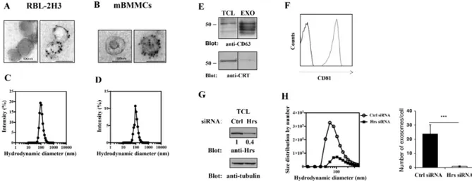
RBL-2H3 cells and BMMC cell culture supernatants were subjected to a series of centrifugations and ultracentrifugations for selective exosome isolation, as described in material and methods, and analyzed by transmission electron microscopy (TEM) and Dynamic light scattering (DLS). In accord to previously reported data (Hegmans et al., 2008), we found that isolated vesicles from both cell types showed a cup-shaped morphology with an uniform diameter of around 100 nm and are positive for the exosomal marker tetraspannin CD63, as demonstrated by immunogold staining (Figure 10A and B). Similar results were obtained by DLS analysis which shows a well-defined peak centered around 100 nm (Figure 10C and D). CD63 expression was further confirmed by western blotting in RBL-2H3-derived vesicles, that appear to be negative for calreticulin, a specific ER marker (Figure 10E). Additionally, FACS analysis of bead-bound nanovesicles showed that they were positive also for CD81 which together with CD63 is considered a common marker of these vesicles (Figure 10F).
The endosomal adapator, Hrs, protein member of ESCRT-0, is an important mediator of exosome biogenesis (Tamai et al., 2010). We assessed vesicle endocytic origin by siRNA-mediated Hrs knockdown in RBL-2H3 after having assessed by western blot the effective protein reduction (Figure 10G). We found a dramatic decrease in exosome production by Hrs-siRNA-transfected cells respect to Ctr siRNA treated (Figure 10H), as revealed by Dynamic light scattering analysis (DLS) that allows an accurate estimation of nanovesicle concentration as described in material and method section. Furthermore, the hydrodynamic size distribution showed a well-defined peak, excluding the presence of contaminants, with a mean around to 80-100 nm matching again the reported size of exosomes (Figure 10H).
Taken together, these data showed the effectiveness of the protocol adopted for purification of exosome-like vesicles which mainly derive from endocytic compartment.