U
NIVERSITÀ DEGLIS
TUDI DIM
ESSINADIPARTIMENTO DI MEDICINA CLINICA E SPERIMENTALE
DOTTORATO XXXI CICLO IN SCIENZE BIOMEDICHE, CLINICHE E SPERIMENTALI Coordinatore Dottorato: Ch.mo Prof. E. Spina
S
URGERY OF MENINGIOMAS LOCATED IN THER
OLANDIC AREA:
THE ROLE OF NAVIGATED TRANSCRANIAL MAGNETICSTIMULATION FOR PREOPERATIVE PLANNING
,
TUMOR RESECTION,
AND OUTCOME PREDICTIONTesi di Dottorato del
Dott. Giovanni Raffa
Relatore/Tutor:
Ch.mo Prof. Antonino Germanò
SSD MED/27
Introduction
Surgery of convexity meningiomas is usually considered a simple procedure, with a low
risk of postoperative neurological deficits1, 2. Surgery is more challenging in cases of
parasagittal meningiomas, since the possible involvement of the superior sagittal sinus
(SSS)3-7. In the literature the incidence of poor outcome has been reported to account up to
15%, depending on several factors including age, performance status, and the site of the
lesion8-12. Among convexity/parasagittal meningiomas the risk of postoperative motor
deficits is considered higher for lesions located in the proximity of the central region, since
the possible involvement of the motor cortex (M1)6, 9, 13-16. For this reason, it has been
suggested to consider a different specific category of “rolandic” meningiomas from other convexity/parasagittal lesions, including also falx meningiomas involving the mesial
portion of the rolandic area9. Nevertheless, such as distinction has been poorly reported in
the literature. Moreover, in this specific category of meningiomas, the risk of new permanent postoperative motor deficits seems to be higher in all that cases in which a clear cleavage plane with the M1 and the underlying corticospinal tract (CST) is not identifiable,
and the lesion infiltrates the surrounding brain17-19. For this reason, also during surgery of
“rolandic” meningiomas, as well as for others intra-axial lesions such as “rolandic” gliomas, all the available pre- and intra-operative surgical technologies and techniques should be employed by neurosurgeons with the aim to reduce the risk of postoperative motor deficits. Surprisingly, despite the intraoperative neurophysiological mapping and monitoring (IONM) is considered extremely useful to reduce postoperative motor deficits during surgical resection of “rolandic” intrinsic tumors or meningiomas located in other
been advocated only by one paper for surgery of meningiomas involving the central area9. IONM allows for a prompt identification of the functional M1 surrounding the meningioma, and could theoretically help to preserve it and the upper portion of the CST during the tumor resection, especially when a clear arachnoidal cleavage plane between the lesion and the motor pathway is not identifiable. Nevertheless, the identification of M1 merely based
on anatomical landmarks is not always straightforward 23, 24 and difficulties in locating M1
during cortical mapping could increase the operative time, reducing the efficacy of the IONM in this type of surgery. Therefore, the usefulness of IONM for surgery of supratentorial meningiomas is often neglected, and surgeons prefer to remove tumors without using IONM because the surgical procedure is usually considered simple and safe
enough by itself2.
Nevertheless, the limitation in the use of IONM related to the reliable identification of the M1 could be overpassed by using advanced preoperative techniques for the non-invasive mapping of the M1 and eventually of the upper portion of the CST.
In the last decade, a growing interest focused on the use of preoperative navigated transcranial magnetic stimulation (nTMS) for the planning of brain tumor surgery has been
recorded25-34. It has been demonstrated that nTMS is able to preoperatively disclose, in a
non-invasive way, the real motor eloquence of intrinsic brain tumors, with an accuracy that
is similar to that of IONM25, 32, 35. Its use in “rolandic” glioma surgery is associated to an
increased extent of resection and a better postoperative motor outcome29, 30. This is
particularly true when nTMS is also used to compute the nTMS-based DTI fiber tracking
(DTI-FT) of the CST 35-39. The intraoperative visualization of the nTMS-based
and is associated to a better motor outcome as compared to simple nTMS cortical
mapping25.
Nevertheless, the use of such a technique for surgery of meningiomas has been reported only in few cases among larger series mainly composed by intrinsic brain tumors located in
the rolandic region31, 32, 40. Therefore, it could be hypothesized that the nTMS-based
preoperative reconstruction of the motor pathway could be useful even for surgery of “rolandic” meningiomas.
The objective of the present study is to describe the combined experience of two European Neurosurgical Centers (the Division of Neurosurgery at the University of Messina, Italy, and the Department of Neurosurgery at the Charité Universitätsmedizin Berlin, Germany) in the use of the nTMS-based planning for surgery of convexity/parasagittal/falx meningiomas located in the rolandic area, thereby suspected to involve the motor pathway and to be at higher risk of postoperative motor deficits. In particular, we analyzed the usefulness of the use of preoperative nTMS cortical mapping of M1 and nTMS-based DTI-FT of the CST to plan the best surgical strategy, to guide IONM and tumor resection, and to preserve as much as possible the motor pathway. Moreover, we also analyzed individual parameters that could predict the occurrence of postoperative motor deficits, with the specific aim to assess the possible role of the nTMS-based planning and others preoperative factors as predictors of outcome.
Materials and methods
Patients:
We retrospectively collected clinical and neuroradiological data of patients operated for convexity/parasagittal/falx meningiomas located in the rolandic area in the period between 2012 and 2018 at the Division of Neurosurgery of the University of Messina, Italy, and at the Department of Neurosurgery of the Charité Universitätsmedizin Berlin, Germany. Inclusion criteria were age ≥ 18 years, the presence of a convexity/parasagittal/falx meningioma located in the rolandic region and suspected to involve the motor pathway (M1 and/or CST) for which an nTMS-based planning was preoperatively performed. We excluded patients aged <18 years or in treatment with antiepileptic drugs (because of the possibility to influence the results of the nTMS mapping). The involvement of the rolandic area was defined as the suspected direct contact of the meningioma with the pre-central
and/or post-central gyrus according to anatomical landmarks on the MRI scan9, 41
The real involvement of the motor pathway was confirmed or denied by the nTMS mapping of M1 in all patients. When possible, patients underwent also nTMS-based DTI fiber tracking of the CST before surgery, in order to identify the spatial relationship between the lesion and the upper portion of the CST. The study was conducted in accordance with the Declaration of Helsinki and its later amendments. All patients signed an informed consent for the collection and scientific use of their data.
MRI scan, nTMS-based planning and preoperative considerations about meningiomas
All patients underwent a specific protocol for the preoperative planning shared between the two Institutions. A volumetric T1-weighted contrast-enhanced sequence (TR/repetition time=8.1, TE/echo time=3.7, slice thickness 1 mm), a T2-weighted sequence (FS=1.5, TR= 8000, TE=331.5/7), and a DWI sequence (32 directions or gradients; TR=2383.9, TE=51.9) for DTI computation were acquired for each patient through a 1.5 T MRI scan (Philips, Ingenia, The Netherlands).
All patients underwent nTMS cortical mapping of M1 by using the NBS system 4.3 (Nexstim Oy, Elimäenkatu 9B, Helsinki, Finland). Volumetric T1-weighted gadolinium-enhanced sequences were imported in the nTMS system. Using a figure-of-eight navigated coil, the resting motor threshold (RMT) was calculated for the first dorsal interosseus (FDI)
muscle, as previously reported26, 35. Then, single pulse stimulation at 110% of the RMT was
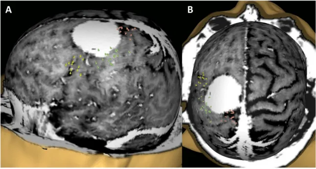
applied. Motor evoked potentials were recorded via electromyography electrodes (Neuroline 720; Ambu, Ballerup, Denmark) from two or three channels, with one muscle selected for each body segment (face, arm, leg), according to the preoperative evaluation of the spatial relationship between the meningioma and the M1, and accordingly to the preoperative motor status. The muscles typically assessed included the: FDI or abductor pollicis brevis or biceps for the arm; the tibialis anterior or rectus femoris for the leg; the mentalis or orbicularis oris for the face (Figure 1).
Figure 1: Example of the nTMS cortical mapping of the M1 in lateral (A) and cranio-caudal views (B) in a patient with a “rolandic” meningioma. The nTMS spots correspond to the cortical cortical representations of the leg (red), arm (green), and face (yellow) muscles
The final nTMS map of the M1 was exported in a DICOM format, together with the DWI sequences, into the neuronavigation system for the DTI computation. The workflow for the
nTMS-based DTI-FT of the CST was performed as described elsewhere25 using the
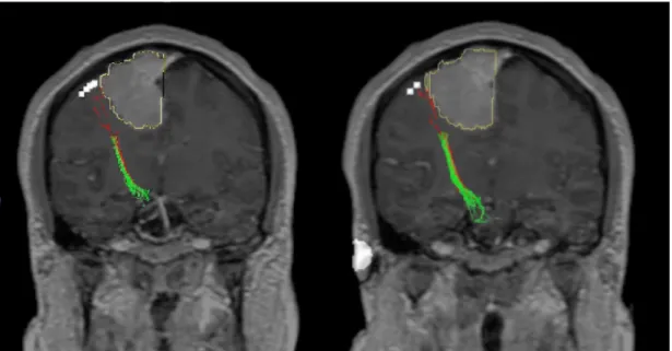
StealthViz software (Medtronic Navigation, Coal Creek Circle Louisville, CO, USA). In summary, after the co-registration of MRI sequences, the tensor was computed and the directionally encoded color (DEC) map was obtained. The nTMS map of the motor cortical representation of muscles of each body segment (arm and/or leg and/or face) was used as first seeding Region of Interest (ROI) for the computation of the corresponding CST fibers, using also a second ROI placed at ipsilateral cerebral peduncle. This process was repeated for each body segment, thus obtaining a somatotopic representation of the CST, distinguishing between leg, arm, and face fibers of the CST (Figure 2).
Figure 2: Example of the nTMS-based DTI-FT of the CST in coronal view in a patient with a parasagittal Rolandic meningioma. Please note the somatotopic organization of the CST: the red fibers are connected to the cortical representation of the arm muscles, while the green fibers are connected with that of the leg muscles. Arm fibers (red) are very close to the lesions, therefore confirming the motor-eloquence of the meningioma, and suggesting a high risk of postoperative motor deficits.
The definitive nTMS cortical map of the M1 and, when available, the reconstruction of the whole motor pathway (M1 + CST) were used to identify their spatial relationship with the meningioma.
Collectively, the preoperative patients’ evaluation included the use of: 1) T1-weighted contrast-enhanced sequences to define the pattern of enhancement (homogenous vs.
non-homogenous) after administration of gadolinium (Figure 3A)18, 42, 43; 2) T2-weighted scans
to identify the perilesional edema (present vs. absent, Figure 3B) and the suspected presence of an arachonoidal cleavage plane between the lesion and brain parenchyma
42-44; the nTMS-based reconstruction of the motor pathway to plan the best surgical strategy
to approach the lesion preserving the M1 and the upper portion of the CST25, 26, 29, 35, 38, 45, 46.
Figure 3: Example of the neuroimaging parameters evaluated to predict postoperative motor outcome and the presence/absence of an intraoperative arachnoidal cleavage plane: homogeneity vs. non-homogeneity of the enhancement after Gadolinium administration (A); perilesional edema (B); presence of the so-called “T2 cleft sign”(red arrow).
Surgical strategy, intraoperative neurophysiological monitoring and tumor resection
nTMS-based planning was used by the neurosurgeon to plan a customized surgical strategy to approach the lesion, with the aim to preserve the motor pathway. In particular, the
surgical strategy was chosen case by case, with the aim to plan to start tumor dissection and resection as far as possible from the motor pathway.
At the beginning of surgery, the nTMS-based data were used with the aid of neuronavigation to identify the M1. When considered useful, IONM was used to confirm
the navigated identification of the M1 as previously reported25. It consisted on the
identification of M1 through the use of a quadripolar electrode (strip) placed over the primary motor and the sensory cortex (S1) using the phase-reversal technique. Then the electrode was left over the M1 for continuous motor evoked potential (MEP) monitoring. In cases the phase reversal technique was not feasible (bigger lesions that displaced the rolandic region thereby not allowing to place the quadripolar electrode simultaneously over the M1 and S1, or when the S1 was not exposed through the craniotomy) the M1 was
Figure 4: Example of the use of IONM guided by the nTMS-based planning at the beginning of surgery. The position of the monopolar stimulator is verified by the navigation probe (blue stylet). This allowed for identifying the motor cortex surrounding the meningioma.
IONM was not used in cases in which the M1 was displaced by the meningioma and was located below the lesion, thereby being not accessible to the cortical strip or monopolar stimulation. In these cases the M1 and the upper portion of the CST were verified by neuronavigation only, according to the preoperative nTMS-based mapping.
Once the M1 was correctly identified by IONM and/or neuronavigation, surgery was started on the opposite side of the lesion, as far as possible from the motor pathway. A standard microsurgical technique was used. Briefly, the arachnoidal cleavage plane, when present, was identified and used to start the dissection of the meningioma from the surrounding normal brain parenchima. Then, intra-capsular debulking of the lesion began, trying to shrink the lesion and make the dissection from the surrounding brain parenchyma easier. The portion of the meningioma adjacent to the M1 was approached at the latest stages of resection. At this stage, the intracapsular resection and the dissection of the lesion through the cleavage plane was alternated to monopolar stimulation of the motor cortex to verify M1 functionality and avoid surgical damage.
In cases of the absence of an arachnoidal cleavage plane and the presence of an evident infiltration of the brain parenchyma, monopolar stimulation was used to map M1 but also the underlying upper portion of the CST. In all cases the monopolar stimulation was guided by the visual feedback provided by the nTMS-based information through the neuronavigation system. Simultaneously, when available, continuous MEP monitoring was performed to identify any suffering of the motor pathway during the resection and
dissection stages. In case of a MEP reduction was detected through continuous monitoring or monopolar stimulation, the meningioma resection and dissection were stopped for at least 5 minutes, waiting for the return of a normal MEP. The lesion resection and dissection during this stage were performed very gently and slowly, until a complete resection of the meningioma was achieved and the motor responses recorded through the continuous MEP monitoring or monopolar stimulation were still present. During the resection, cortical and bridging veins were protected and respected. In cases of suspected damage to the drainage veins, their patency was verified through intraoperative indocianin green (ICG)
video-angiography47 (Figure 5).
Figure 5: Example of preservation of the bridging veins adjacent to a parasagittal meningioma before and after complete resection. ICG was performed to verify the veins patency at the end of resection
In cases of parasagittal meningiomas with invasion of the SSS, a small fragment of the lesion invading the sinus was left in situ, and postoperative radiosurgical treatment of the
residue was planned4. The dura mater of the convexity infiltrated by the lesion was
Postoperative evaluation and follow-up
Patients underwent postoperative MRI scan with T1-weighted contrast-enhanced sequences within 48 hours from surgery. The extent of resection was evaluated on the postoperative
MRI using the Simpson grading48. As well as in the preoperative period, the postoperative
clinical evaluation of the motor performance was performed using the Medical Research
Council (MRC) scale for muscle strength49 at discharge. A new clinical evaluation and MRI
scan was performed after three months from surgery. At that time, when patients agreed, a new nTMS cortical mapping of the M1 was performed to verify its rearrangement and eventual modifications of the RMT.
Data collection, methodology of data analysis, and statistics
Clinical, neuroradiological and neurophysiological data were retrieved from the Institutional databases at two Institutions involved in the study (clinical charts, PACS archive, and outpatients clinic reports) and collected in a joint database.
Analysis of the accuracy of the nTMS-based mapping
We analyzed the accuracy of the preoperative nTMS-based mapping (nTMS risk/benefit analysis) compared to the intraoperative identification of the motor pathway trough the IONM. The preoperative involvement of the motor pathway by the meningioma was defined as the direct contact or a distance ≤ 10 mm of the lesion from the M1 and/or the
Figure 6: Example of the nTMS-based DTI-FT of the CST in a case of rolandic meningioma. The lesion was considered motor-eloquent because of a close relationship (distance ≤ 10 mm) with the CST, as it is demonstrated in the lateral (A) and oblique (B) projection.
The accuracy of the nTMS-based mapping was evaluated using the Fisher test applied to a 2X2 contingency table, analyzing sensitivity (ST), specificity (SP), positive predictive value (PPV) and negative predictive value (NPV).
Analysis of the usefulness of the nTMS-based planning for tumor resection
After surgery, the surgeon was asked to report the usefulness of the preoperative nTMS-based planning for surgical strategy, having the possibility to chose three different answers: 1) Not useful at all; 2) Useful but without inducing modification of the planned surgical strategy; 3) useful to induce a change of the planned surgical strategy (i.e. surgical approach, application of the IONM, tumor resection).
Analysis of predicting factors of motor outcome and intraoperative cleavage plane
Finally, a multivariate logistic regression analysis was used to identify independent factors that could predict postoperative outcome expressed as the MRC score after 3 months
In particular, we evaluated the possible role of several parameters as predictors of the motor outcome after three months from surgey consisting of: 1) the preoperative MRC score; 2) the preoperative RMT value obtained through the nTMS cortical mapping; 3) the presence/absence of preoperative peritumoral edema, T2 cleft sign, intraoperative cleavage plane; 4) the appearance of the meningioma in the T1-wheighted MR sequence after gadolinium administration (homogenous/non-homogenous).
Considering that the lack of an intraoperative arachnoidal cleavage plane can make surgery of “rolandic meningiomas” more complex and at a higher risk of postoperative motor deficits, we also analyzed the possible role of different preoperative variables as predictors of the presence/absence of such a cleavage plane. In particular, we analyzed: 1) the preoperative RMT value; 2) the presence/absence of preoperative peritumoral edema or T2 cleft sign; 3) the appearance of the meningioma at the T1-wheighted MR sequence after gadolinium administration (homogenous/non-homogenous).
A ROC analysis was performed to identify the cut-off values with the higher accuracy in predicting the postoperative motor outcome, the occurrence of new postoperative motor deficits, and the presence/lack of an intraoperative cleavage plane.
Analysis of postoperative nTMS cortical mapping and reorganization of the M1
All patients were asked to undergo new nTMS cortical mapping after three months from surgery. The same methodology of the preoperative mapping was employed. The pre- and postoperative RMT values were compared using the paired Student-T test. A qualitative evaluation and comparison of the size, density, and localization of the pre- and postoperative nTMS cortical map was also performed.
Statistical significance was defined as a p value <.05. Data analysis was performed using GraphPad Prism version 6.00 for MacOS, GraphPad Software, La Jolla, California, USA,
www.graphpad.com and StatCalc version 8.2.2 for MacOs, AcaStat Software, Winter Garden, Florida, USA, http://www.acastat.com.
Results
Participants and descriptive data
In the present study, 47 patients (18 males, 29 females, mean age 61.9 ± 13.3) affected by convexity/parasagittal/falx meningiomas located in the rolandic region and suspected to involve the motor pathway were included. Thirty-one patients were treated at the University of Messina, while the remaining 16 were treated at the Charité Universitätsmedizin Berlin (Figure 7). Table 1 reports all the patients’ clinical characteristics.
Figure 7: Example of ten rolandic meningiomas included in the present series suspected to involve the motor cortex.
Twenty-two patients were affected by pure convexity meningiomas, 15 by parasagittal meningiomas, and the remaining 10 by meningiomas of the falx. Twenty patients showed a preoperative motor deficit. Perilesional edema was observed in 27 cases, the lesions had a homogenous enhancement after gadolinium administration in 28 cases, and a clear cleavage
plane between the meningioma and the brain parenchyma was suspected due to the presence of the T2 cleft sign in 30 cases. The mean preoperative RMT for the FDI muscle was 34.1 ± 8.1. Postoperative histological diagnosis was WHO grade I meningioma in 26, and Grade II in the remaining 21. A Simpson I resection was achieved in 31 patients, Simpson II in 10 cases, Simpson III in 3, and Simpson IV in the remaining 3 cases.
The nTMS cortical mapping of M1 was performed in all patients, while the nTMS-based DTI-FT was feasible in 34 cases.
Main results and outcome data
Accuracy of the nTMS-based planning
A total of 39 out of 47 cases (82.9%) were defined as motor-eloquent because of the involvement of the motor pathway documented by the preoperative nTMS-based mapping of the M1 and, when available, of the CST (Figure 8).
Figure8: Case example of a right parasagittal “rolandic” meningioma (A). The nTMS cortical mapping of the M1 (B) and the nTMS-based DTI-FT of the CST (C) showed the motor pathway was located posteriorly and laterally to the meningioma, having a close spatial relationship with it. Therefore, the lesion was considered motor-eloquent and resection started from the anterior portion of the meningioma (red arrow).
Nevertheless, we were able to use the IONM to achieve a confirmation of the reliability and accuracy of the preoperative nTMS-based mapping cases in 35 cases only. Among these, the IONM confirmed the involvement of the motor pathway in 29 cases (82.8%), while it documented a safe distance from the rolandic area in the remaining 6 cases (17.2%). The accuracy of the nTMS-based planning as compared to IONM findings was 94.2%, with a ST of 93.5%, a SP of 100%, a PPV of 100% and a NPV of 66.6% (p=0.0003). Indeed, in only two cases the nTMS-based preoperative mapping was discordant with the IONM findings (5.7%) (Figure 9).
Figure 9: Accuracy of the nTMS cortical mapping as compared to IONM. nTMS findings were confirmed by IONM in 94.2% of patients, with only two false negative cases (5.7%) .
Usefulness of the nTMS-based planning
The nTMS-based planning were considered useful for surgical strategy in 89.3% of cases (42 of 47 patients), being able to induce a change of the preoperative planned strategy based on MRI in 42.5% of cases (20 of 47 patients). The latter resulted in a change of the surgical approach or in an easier/faster/more confident IONM mapping and tumor resection. In the remaining 10.7 % of cases (5 of 47 patients) it was considered not useful at all.
A tailored surgical access pathway was chosen in all cases the nTMS-based planning showed the involvement of the motor pathway, starting the tumor resection as far as possible form the M1/CST, especially when a clear cleavage plane with the brain parenchyma was not identified (Figure 10A and B). In particular, the nTMS-based reconstruction as visualized through the navigation system was considered helpful to guide the identification of the motor cortex by IONM (when available) at the beginning of surgery, making this step easier and faster (Figure 10C and B).
During resection, in cases in which a clear cleavage plane with the brain parenchyma was not identified, the nTMS-based planning was considered useful to guide IONM for mapping of the M1 and the upper portion of the CST. This made surgeons more confident with the removal of those portions of the meningiomas that have already infiltrated the surrounding brain parenchyma, influencing the surgical strategy, IONM mapping and resection. In all that cases IONM was not available, the nTMS-based reconstruction of the motor pathway was used to guide surgical strategy through its simple visualization on the neuronavigation display.
The extent of resection was not influenced in any way by the nTMS-based motor mapping and planning. The only determinant factor was the infiltration of the SSS in cases of parasagittal meningiomas (Figure 11)
Figure 10: Two example cases of “rolandic” meningiomas with a different spataial relationship with the M1: the first lesion was located anteriorly to the M1 (A) and resection started from the anterior portion of the meningioma, that is as far as possible from the motor pathway (red arrow); conversely, the second case (B) is a meningioma located posteriorly to the M1 and, therefore, the resection started from the posterior portion of the lesion (red arrow). In the bottom line, the example of a third case in which surgery started with identification of the M1 (white spots) and the upper portion of the CST (green fibers from the leg) through navigation and IONM: the motor pathway was located posteriorly to the lesion(C). Lesion debulking started from its anterior portion, as far as possible from the motor pathway.
Figure 11: Two example cases of “rolandic” meningiomas with a different Simpson grade of resection. In cases of pure convexity meningiomas, the lesion was completely resected (A); conversely, in cases of parasagittal meningiomas invading the SSS, a small remnant was left in situ (red arrow) and treated through stereotactic radiosurgery at a later stage (B).
Functional outcome and predicting factors
At discharge, 13 of 47 patients were still affected by a new postoperative motor deficit (30.2%). Nevertheless, deficits resolved in 9 of these 13 patients (69.2%) after three month from surgery, being the rate of new permanent motor deficit 8.5% (4 of 47 patients). In one of these cases, the deficit was probably related to the postoperative infarction of the surgical cave.
Multivariate logistic regression analysis showed that, in a model with six variables (preoperative MRC score, RMT value, perilesional edema, pattern of the enhancement of lesions, T2 cleft sign, and intraoperative cleavage plane) the only independent predictors of the motor outcome at three months were the intraoperative cleavage plane (p<0.02, OR 0.01), and the RMT value computed during the nTMS cortical mapping (p<0.04, OR 1.1). Moreover, we observed that, among the remaining parameters, only the T2 cleft sign was close to the statistical significance as predictor of the motor outcome (p=0.06) (Table 2). The predicting role of the intraoperative cleavage plane was probably due to the higher technical challenge for preserving the motor pathway in cases of lesions without a clear
arachnoidal plane and infiltrating the brain parenchyma and the M150. The presence of such
a plane had a strong protective effect (OR = 0.01) on the occurrence of a motor status worsening. Conversely, this is the first time that a higher preoperative RMT is disclosed to be associated to a slightly higher risk (OR 1.1) of motor performance worsening.
Concerning the prediction of the presence/absence of an arachnoidal cleavage plane, in a model with 4 variables including the preoperative RMT value, perilesional edema, pattern of the enhancement of lesions, and T2 cleft sign, only the RMT was discovered to be an independent predicting factor (p<0.008, OR 0.8). The presence of perilesional edema and the T2 cleft were close but did not reach the statistical significance (p=0.05 and p=0.06,
respectively) (Table 3). A lower preoperative RMT could therefore predict the presence of an intraoperative cleavage plane (slight protective effect against the lack of the plane) that could make surgical resection easier, thereby reducing the occurrence of new postoperative motor deficits.
In particular, an RMT cut-off value of 29.5% was discovered to be significant, with an accuracy of 65.9%, a ST of 90%, a SP of 48.1%, a PPV of 56.2% and a NPV of 86.6% (p=0.001). The accuracy was increased up to 70.2% using a cut-off = 31.5%: in such a case, the ST was 75%, the SP 66.6%, the PPV 62.5% and the NPV 78.2%.
Finally, the ability of the nTMS-computed RMT value to predict the motor outcome was further investigated. After dichotomization of the MRC score (good outcome = MRC score of 4 or 5; bad outcome = MRC score of 1 or 2 or 3), the cut-off value of 31.5% was disclosed to be able to predict the outcome with an accuracy of 65.9%, a ST of 100%, a SP 58.9%, a PPV of 33.3% and a NPV of 100% (p=0.001). Nonetheless, the accuracy increased up to 80.8% when using 38.5 as cut-off value, with a ST of 75%, a SP 82%, a PPV of 46.1% and a NPV of 94.1%.
Moreover, the preoperative RMT was also associated to the occurrence of new postoperative permanent (after three month from surgery) motor deficits. The cut-off of 30.5% was able to predict the occurrence of a new motor deficit with a ST of 100%, a SP 41.8%, a PPV of 13.7%, a NPV of 100%, but the final accuracy of 46.8%.
Postoperative nTMS cortical mapping and reorganization of the M1
At three months follow-up, only 10 patients agreed to perform a new nTMS cortical mapping. Nine of these showed a good postoperative outcome. In only one case the outcome was bad (MRC score = 3), but it was exactly the same on the admission.
In all these patients, the RMT was significantly reduced as compared to the admission (29.40±2.21 vs. 33.10 ± 5.85, p=0.01). From a pure qualitative point of view, the reduced RMT was witnessed by a greater representation of the nTMS map of the M1, and by an increase of the amplitude of the motor potentials evoked by the stimulation (figure 12).
Figure 12: Case example of the nTMS-documented reorganization of the M1 after resection of a “rolandic” right parasagittal meningioma. The cortical representation of the leg, arm, and face muscles was increased in size after surgery as documented by postoperative nTMS mapping (red arrow). This is probably due to the preoperative suffering of the M1 caused by the presence of the meningioma that resulted in a preoperative poor representation of the motor area. After surgery, the M1 came back to the normal representation.
Discussion
Key results
In the present study, we observed the nTMS-based preoperative reconstruction of the motor pathway (M1 and eventually CST) resulted useful for surgery of convexity/parasagittal/falx meningiomas located in the rolandic region. During the preoperative stages, the nTMS-based planning was able to identify the involvement of the motor pathway in a reliable manner, comparable to findings provided by IONM during surgery (accuracy = 94.2%). This aspect was considered useful by surgeons to plan a tailored surgical strategy to avoid damage to the motor pathway in more than 89% of cases.
During surgery, the nTMS-based planning was also helpful to guide IONM and to verify the spatial relationship of the meningioma with the motor pathway. Moreover, the usefulness of the nTM-based mapping was evident especially in cases without a clear arachnoidal cleavage plane between the meningioma and the brain parenchyma. In such cases, the real-time visualization of the nTMS-based reconstruction of the M1 and the upper portion of CST through navigation was helpful to guide IONM (when available), increased the confidence of the surgeon during the dissection of the meningioma from the infiltrated parenchyma, and helped the resection of the lesion preserving the motor-eloquent brain.
Apart from qualitative considerations, the most important finding was the high correlation between preoperative nTMS and intraoperative IONM findings, thus suggesting the
reliability of nTMS, as already demonstrated for intrinsic tumors 32, 46
Another important finding was the role of the nTMS preoperative findings in predicting the absence/presence of an arachnoidal cleavage plane, and the postoperative motor outcome.
The absence of an intraoperative cleavage plane is one of the most important technical aspect that could make this surgery more challenging due to the infiltration of the M1 and eventually the upper portion of CST: in the present study we observed that the lack of such a cleavage could be predicted by a higher RMT at the nTMS preoperative mapping. This could thereby be extremely useful for the preoperative stratification of patients according to the surgical risk of postoperative motor deficits. Other parameters such as the peritumoral edema, the T2 cleft sign or the non-homogenous contrast-enhancement of the meningioma were not significant independent predictors.
Moreover, the RMT was found to be an independent predictor also of outcome together with the presence/absence of an intraoperative cleavage plane between the lesion and the motor-eloquent brain parenchyma. Again, this could help the preoperative stratifications of patients according to the risk of the occurrence of postoperative permanent deficits.
Finally, in the present study we also documented that in an observed population of patients without new permanent motor deficits, the nTMS was able to disclose 1) a postoperative reorganization of the M1 that appeared larger in size that in the preoperative period, and 2) the progressive normalization of the M1 motor threshold documented by a significant reduction of the RMT as compared to the preoperative period. This could be considered as the physiological postoperative rearrangement of the motor pathway after resection of “rolandic” meningiomas that resulted in a preserved postoperative motor outcome. Such an observation has never been documented in the nTMS literature, so far.
Interpretation and generalizability
Preoperative nTMS motor mapping enables a safer surgical treatment of
motor-eloquent lesions30, 51. Several papers documented that the use of preoperative nTMS
mapping of the M1, eventually combined with the nTMS-based reconstruction of the motor pathway, results in a better outcome after surgical treatment of brain tumors suspected to be
motor-eloquent 25, 29, 52. Nevertheless, these findings have been reported mainly for surgery
of intrinsic tumors. Only few reports have included in their nTMS series few cases of
convexity/parasagittal/falx meningiomas suspected to involve the motor pathway31, 32, 40.
Indeed, also extra-axial lesions like meningiomas can involve the motor cortex, even infiltrating the motor-eloquent brain parenchyma and therefore also the upper portion of the CST. This makes surgery at risk of inducing postoperative motor deficits that sometimes
can be permanent9-11, 16. Surprisingly, in the literature meningiomas involving the rolandic
areas are not considered a nosological entity separated from other convexity/parasagittal meningiomas. Only few reports specifically investigated the risk of developing
postoperative motor deficits after this type of surgery6, 9, 13-16. The reported incidence of
postoperative permanent motor deficits varies from 7.1% to 11.7%9, 13, 16. Moreover, only
one report specifically highlighted the usefulness of using IONM during this type of surgery and suggested to consider meningiomas in the rolandic area as a different
nosological entity9. This is someway surprising, since IONM is currently used and
considered an extremely important support for surgery of meningiomas in other locations,
such as the posterior cranial fossa 20 and the spine 21, 22, 53-55, as well as for intrinsic
supratentorial brain tumors56-60.
Starting from the evidence of the usefulness of IONM and nTMS reconstruction of
the motor pathway in surgery of motor-eloquent intrinsic tumors25, we tried to assess the
first observed advantage was that the nTMS-based preoperative mapping was able di disclose the real involvement of the motor pathway as compared to IONM findings with an accuracy of 94.2%, with only two false negative cases out of 35 (5.7%). Ostry et al, in their series reported 4 of 42 cases (9.5%) as false positive according to anatomical landmarks on the MRI scan. Nevertheless, it is well known that anatomical landmarks are not always
reliable for the identification of eloquent areas, including the motor cortex23, 61. Other
preoperative techniques, such as fMRI, could be used to overcome this limitation. Nonetheless, it has been demonstrated that nTMS is more reliable than fMRI in mapping
the motor area, especially in brain tumor patients62, 63. Therefore our findings highlight the
usefulness of the nTMS planning in disclosing the real motor-eloquence of lesions even in cases of extra-axial tumors, as well as it has been widely reported for intrinsic brain
tumors25, 64. Such information could be considered useful to the preoperative stratification
of patients, thus helping in the identification of true-positive patients that are at risk of postoperative motor deficits.
In our series, in both the participating Institutions, the nTMS-based mapping of the motor pathway impacted the surgical strategy, allowing to plan a tailored strategy consisting in beginning the resection as far as possible from the M1, leaving the resection of the motor-eloquent portion of the lesion at the latest stages of surgery, eventually under the direct guidance of IONM, especially for infiltrating lesions. Even the nTMS-based visualization of the upper portion of the CST and its relationship with the lesion impacted the planning of the surgical strategy, especially in cases without a clear arachnoidal cleavage plane, helping the neurosurgeon to chose the best surgical approach to avoid damage to the M1 and the upper portion of the CST laying below it.
The usefulness of the DTI-FT has already been reported for preoperative planning
before surgery of rolandic meningiomas 6, 9, 65, but it has been poorly considered by the
international literature. To our knowledge this is the first report of the use of nTMS-based DTI-FT for rolandic meningiomas surgery. This should considered of note since it has been widely demonstrated that the reconstruction of the CST based on the nTMS mapping is
more reliable than the traditional tractography based only on anatomical landmarks 35, 36, 38,
39, 45, 66, 67
Anyway, the qualitative subjective assessment of the usefulness of the nTMS-based planning resulted in considering this technique helpful in almost 90% of cases, and induced a change of the hypothesized surgical strategy in more than 42% of cases. No other studies analyzing the usefulness of the nTMS-based planning for surgery of rolandic meningiomas have been reported, so far. Our results are not so different to that ones reported by Picht et
al. for intrinsic brain tumors64. In their series, the authors reported an overall benefit in the
78.1% of cases, and an impact of the nTMS mapping resulting in a modification of the
planned strategy in 54.8% of cases64.
In our series the nTMS-based mapping was useful also during surgery: the visualization of the nTMS-based reconstruction of the motor pathway guided the IONM, thereby making the identification of the M1 easier and faster. This aspect has been well
demonstrated for intrinsic tumor25, 29, 30 and only speculated in few series reporting few
cases of meningiomas operated using the nTMS-based planning31, 32, 40. In our series, apart
from the initial surgical stages during the identification of the M1, the usefulness of the nTMS-based reconstruction of the whole motor pathway was evident especially in cases without a clear cleavage plane between the lesion and the meningioma. This is typically the situation in which a surgical damage to the M1 and the upper portion of the CST can easily
occur50. In these cases IONM could help in identifying a new cleavage plane and to preserve the motor pathway. This allowed us to record a rate of new permanent motor deficits of 8.5% in our series, which is in the lower part of the range 7.1 - 11.7% that is
reported in the literature9, 13, 16.
No role at all was documented for the nTMS-based mapping on the extent of resection of the lesions. The only determinant was the infiltration of the SSS. In these cases
only a small remnant of the lesion was left in situ and scheduled for radiosurgery4. This is
different from what has been reported by Ostry et al. who were used to left a small remnant
over the motor cortex in cases of a clear infiltration9. In our series, the nTMS-mapping, the
tailored strategy, the constant IONM verification of the functionality of the motor pathway guided by the navigation, and a slow and gentle dissection/resection technique allowed us to achieve the complete resection of the meningiomas at the interface with the motor pathway in all cases without an increase of the postoperative motor morbidity.
Functional outcome and predicting factors
In our series we observed that only the presence/absence of an intraoperative cleavage plane, as well as the preoperative RMT value computed during the nTMS mapping, were independent predictors of the motor outcome after three months from surgery.
As previously described the lack of cleavage plane could make resection more tricky and could be responsible of a damage to the M1 and the upper portion of the CST,
resulting in a permanent motor deficit50. This has been widely reported in the literature 17-19,
The novel aspect of our findings is that the RMT value as measured during the nTMS mapping was able to predict the motor outcome. The RMT is defined as the lowest stimulation intensity capable of eliciting a response in a given relaxed muscle in 5 out of 10
stimulations and is a measure of corticospinal excitability68. It is a measure of the activation
threshold of the motor pathway. Several neurological diseases, including stroke69 and TBI70,
71, are characterized by an increased RMT as the expression of reduced corticospinal
excitability due to brain and neuronal damage. Theoretically, even the presence of an extra-axial tumor, such as a meningioma, could induce modifications of the corticospinal excitability by compressing the brain parenchyma, thereby causing brain and neuronal damage that could be responsible of an increase of the motor threshold. In our series higher preoperative nTMS-measured RMT values for the FDI muscle were associated to an increased risk of having a bad outcome (MRC score 1-2-3) or developing a new permanent motor deficit. The identified cut-off values of 31.5% (for the postoperative MRC score) and 30.5% (for new deficits) could be considered low values for RMT. Nevertheless, extra-axial lesions located over the rolandic region, including meningiomas, could be responsible of a progressive irritation of the M1, that could result in a lowering of the cortical motor
threshold and in the occurrence of seizures72, 73, 74, 75. This mechanism of increased
corticospinal excitability expressed through a reduced RMT has been already proposed for
epileptic patients with a seizure focus on the motor cortex 75. We could speculate that the
effect of the meningioma over the motor cortex could be explained with a bi-phasic action. During the initial stages of the meningioma’s growth over the M1, the focal irritative effect of the lesion over the M1 could result in a progressive lowering of the RMT. This could explain the low RMT values measured through the nTMS mapping and that exposes
the M1 and eventually of the upper portion of the CST could result in a progressive damage of the motor pathway that is witnessed by an increased RMT as measured during the nTMS mapping. This could result in a preoperative motor deficit, and could explain the role of a higher RMT value in predicting a bad motor outcome or the occurrence of a new permanent deficit. To our knowledge, this is the first study reporting the objective prognostic usefulness of the nTMS by measuring the RMT for surgery of “rolandic” meningiomas.
Moreover, the RMT value was also able to predict the presence/absence of an intraoperative cleavage plane between the meningioma and the motor-eloquent brain parenchyma. The preoperative motor status, the evaluation of the cleavage on the T2-wheighted MRI scans (T2 cleft sign), the perilesional edema, and the pattern of contrast-enhancement of the lesions at the MRI were not independent predictors. As discussed above, we could speculate that the lack of a cleavage plane reflects the infiltration of the M1, and thereby the suffering of the whole motor pathway. This could result in a higher RMT value that could predict the lack of the arachnoidal plane, and in cases of severe impairment of the motor pathway, could also predict a bad postoperative motor outcome.
In the literature, other parameters have been suggested to predict the presence of the intraoperative cleavage plane, such as the so called “T2 cleft sign” in the T2-weighted MRI
sequences18, 44, 50. Nevertheless, the accuracy of this factor has been considered low 44, 50. In
our series, the T2 cleft sign did not result to be an independent predictor factor for the absence/presence of a cleavage plane. Thenier-Villa et al reported that the cleft sign on T2-weighted MRI sequences has a ST of 88% , a SP of 28.57%, a PPV of 69%, and a NPV of 57% in predicting the presence/absence of an intraoperative cleavage plane. In our series, an RMT cut-off value of 29.5% was more accurate as predictor of a cleavage plane, with a ST of 90%, a SP of 48.1%, a PPV of 56.2% and a NPV of 86.6% .
Even the perilesional edema has been suggested as predictor of the lack of the
cleavage plane6, 18, 42, 44. It could be related to the infiltration of the brain parenchyma,
thereby resulting in a vasogenic edema6, 18. Nevertheless, the exact mechanism responsible
of perilesional edema in meningiomas is not well understood, and different causative
factors has been reported, including venous obstruction of cortical/bridging veins 43. This
could explain the lack of a role for perilesional edema in predicting the absence of an intraoperative cleavage plane in our series. Nevertheless, the predictive role was close to the statistical significance (p= 0.05). Probably, in a certain number of cases in our series, the edema was related also to the compression of cortical/bridging veins or other factors (i.e.
histology, the production of vascular endothelial growth factor, the vascular supply, etc.. 43)
and not only to the infiltration of the brain parenchyma.
Postoperative nTMS cortical mapping and reorganization of the M1:
Finally, another important aspect was that postoperative nTMS mapping in a small subgroup of patients (10 of 47) with no postoperative worsening of the motor performance showed an objective significant reduction of the RMT after three months from surgery and a qualitatively improved response (increased representation and higher MEPs) during the mapping. This probably reflects the beneficial effect of surgery that caused a recovery of the motor pathway from the initial preoperative impairment, and a normalization of the motor threshold. These finding are similar to that reported by Takakura et al. and by Barz et
al. after surgery of intrinsic brain tumors located in the motor area61, 76.
The main limitation of the study is its retrospective design involving two different Institutions that could result in potential selection and expertise biases reducing the strength of our findings. Moreover, as well as for all the nTMS studies, the mapping could be
hampered or impossible in patients with severe motor deficits77 or in whom the lesion
grows exactly over the motor cortex. In the latter case, the nTMS stimulation of the cortical boundaries surrounding the meningioma could not provide any motor evoked potential due to the interposition of the lesion between the nTMS coil and the M1.
Another limitation is the accuracy of the DTI technique that could be reduced in cases of
excessive perilesional edema 78, 79. Nevertheless, this is an intrinsic limitation of the DTI
technique that could not be eliminated.
Finally, brain shift is another unavoidable limitation of image-guided surgery, unless
intraoperative imaging is employed80-82. Nevertheless, the visual feedback provided by
nTMS-based mapping through navigation should be intended as a mere visual support to further guide the IONM during the lesion resection. Moreover, the use of tailored approaches and the continuous verification of established superficial anatomical landmarks
Conclusions
The nTMS-based mapping of the motor pathway could be considered a new, adjunctive useful strategy for surgery of convexity/parasagittal/falx meningiomas located in the rolandic area. It could provide useful information for a tailored surgical approach and for guiding IONM and surgical resection. Moreover the nTMS-based evaluation of the RMT could predict the presence/absence of an intraoperative cleavage plane between the lesions and the motor pathway, and the occurrence of postoperative motor deficits, thus representing a new interesting tool to achieve a preoperative prognostic stratification of patients affected by “rolandic” meningiomas. Therefore, as well as for intrinsic tumors, the nTMS-based preoperative mapping could represent a new tool for neurosurgeons to better face the challenge of surgery of tumor located in the rolandic region. Further prospective studies on the topic are warranted.
Table 1: Clinical characteristics of patients included in the study.
Abbreviations: DTI-FT = Diffusion Tensor Imaging Fiber Tracking; CST = Corticospinal Tract; Gd = Gadolinium; IONM= Intraoperative Neurophysiological Monitoring; nTMS = navigated Transcranial Magnetic Stimulation; RMT = Resting Motor Threshold.
Characteristics Values
Age 61.9 ± 13.3 years old
Sex 18 m, 29 f
Number of patients 47
Pure Convexity 22
Parasagittal 15
Falx 10
Perilesional edema 27
T2 cleft 30
Homogeneous enhancement after
Gd administration 28 nTMS mapping of M1 47
nTMS-‐based DTI-‐FT of the CST 34
RMT 34.1 ± 8.1
IONM Cases 35
Confirmed as motor-‐eloquent 39 of 47 (29 of 35 for IONM cases)
Simpson Grading
Grade I 31
Grade II 10
Grade III 3
Grade IV 3
Preoperative motor deficits 20
Postoperative motor status (after 3 months) Improved/Stable 43 Worsened 4 Histology Grade I 26 Grade II 21 Grade III 0
Table 2: Statistical model to identify independent predictors of motor outcome after three months from surgery
Abbreviations: Gd = Gadolinium; MRC = Medical Research Council Scale for Muscle Strength; n/a = not available; OR = Odds Ratio; RMT = Resting Motor Threshold.
Table 3: Statistical model to identify preoperative independent predictors of the presence/absence of an intraoperative arachnoidal cleavage plane
Abbreviations: Gd = Gadolinium; n/a = not available; OR = Odds Ratio; RMT = Resting Motor Threshold.
Parameters p Value OR
Statistical model at 6 variables 0.003 n/a
Preoperative MRC score 0.9 n/a
RMT value 0.04 1.1
Perilesional edema 0.4 n/a
Homogeneous enhancement after
Gd administration 0.1 n/a
T2 cleft sign 0.06 n/a
Intraoperative cleavage plane 0.02 0.01
Parameters p Value OR
Statistical model at 4 variables 0.0001 n/a
RMT value 0.008 0.8
Perilesional edema 0.05 n/a
Homogeneous enhancement after
Gd administration 0.5 n/a
References
1. Hasseleid BF, Meling TR, Ronning P, Scheie D, Helseth E. Surgery for convexity
meningioma: Simpson Grade I resection as the goal: clinical article. Journal of
neurosurgery. Dec 2012;117(6):999-1006.
2. Sanai N, Sughrue ME, Shangari G, Chung K, Berger MS, McDermott MW. Risk
profile associated with convexity meningioma resection in the modern neurosurgical era. Journal of neurosurgery. May 2010;112(5):913-919.
3. Sughrue ME, Rutkowski MJ, Shangari G, Parsa AT, Berger MS, McDermott MW.
Results with judicious modern neurosurgical management of parasagittal and falcine meningiomas. Clinical article. Journal of neurosurgery. Mar 2011;114(3):731-737.
4. Tomasello F, Conti A, Cardali S, Angileri FF. Venous preservation-guided resection:
a changing paradigm in parasagittal meningioma surgery. Journal of neurosurgery. Jul 2013;119(1):74-81.
5. Nowak A, Dziedzic T, Czernicki T, Kunert P, Marchel A. Surgical treatment of
parasagittal and falcine meningiomas invading the superior sagittal sinus. Neurol
Neurochir Pol. 2014;48(3):174-180.
6. Magill ST, Theodosopoulos PV, McDermott MW. Resection of falx and
parasagittal meningioma: complication avoidance. Journal of neuro-oncology. Nov 2016;130(2):253-262.
7. Pettersson-Segerlind J, Orrego A, Lonn S, Mathiesen T. Long-Term 25-Year
Follow-up of Surgically Treated Parasagittal Meningiomas. World neurosurgery. Dec 2011;76(6):564-571.
8. Sade B. LJH. Factors Influencing Outcome in Meningioma Surgery. In: J.H. L, ed.
Meningiomas. London: Springer; 2009.
9. Ostry S, Netuka D, Benes V. Rolandic area meningioma resection controlled and
guided by intraoperative cortical mapping. Acta neurochirurgica. May 2012;154(5):843-853.
10. Black PM, Morokoff AP, Zauberman J. Surgery for extra-axial tumors of the
cerebral convexity and midline. Neurosurgery. Jun 2008;62(6 Suppl 3):1115-1121; discussion 1121-1113.
11. Chan RC, Thompson GB. Morbidity, mortality, and quality of life following
surgery for intracranial meningiomas. A retrospective study in 257 cases. Journal of
neurosurgery. Jan 1984;60(1):52-60.
12. Rockhill J, Mrugala M, Chamberlain MC. Intracranial meningiomas: an overview
of diagnosis and treatment. Neurosurgical focus. 2007;23(4):E1.
13. Elzarief AA, Ibrahim MF. Long-term follow-up of motor function deterioration
following microsurgical resection of middle third parasagittal and falx meningioma.
Egypt J Neurol Psychiatr Neurosurg. 2018;54(1):9.
14. Skudas G, Tamasauskas A. [Prognosis of the surgical treatment of parasagittal
meningioma]. Medicina (Kaunas). 2002;38(11):1089-1096.
15. Deng WS, Zhou XY, Li ZJ, Xie HW, Fan MC, Sun P. Microsurgical treatment for
central gyrus region meningioma with epilepsy as primary symptom. J Craniofac
Surg. Sep 2014;25(5):1773-1775.
16. Bi N, Xu RX, Liu RY, et al. Microsurgical treatment for parasagittal meningioma in
17. Sindou MP, Alaywan M. Most intracranial meningiomas are not cleavable tumors: Anatomic-surgical evidence and angiographic predictability. Neurosurgery. Mar 1998;42(3):476-480.
18. Ildan F, Tuna A, Gocer AI, et al. Correlation of the relationships of brain-tumor
interfaces, magnetic resonance imaging, and angiographic findings to predict cleavage of meningiomas. Journal of neurosurgery. Sep 1999;91(3):384-390.
19. M.G. Y. Brain-tumor interface: adherence and adhesiveness. In: Yasargil MG AT,
Cravens GF, Johnson RJ, Reeves JD, Teddy PJ, Valavanis A, WichmannW, Wild AM, Young PH ed. Microneurosurgery. Vol IVA. Stuttgart: Tieme; 1994:147-148.
20. Tomasello F, Angileri FF, Conti A, et al. Petrosal Meningiomas: Factors Affecting
Outcome and the Role of Intraoperative Multimodal Assistance to Microsurgery.
Neurosurgery. May 22 2018.
21. Scibilia A, Raffa G, Rizzo V, et al. Intraoperative Neurophysiological Monitoring in
Spine Surgery: A Significant Tool for Neuronal Protection and Functional Restoration. Acta neurochirurgica. Supplement. 2017;124:263-270.
22. Scibilia A, Terranova C, Rizzo V, et al. Intraoperative neurophysiological mapping
and monitoring in spinal tumor surgery: sirens or indispensable tools?
Neurosurgical focus. Aug 2016;41(2):E18.
23. Ahdab R, Ayache SS, Brugieres P, Farhat WH, Lefaucheur JP. The Hand Motor
Hotspot is not Always Located in the Hand Knob: A Neuronavigated Transcranial Magnetic Stimulation Study. Brain Topogr. Jul 2016;29(4):590-597.
24. Sahin N, Mohan S, Maralani PJ, et al. Assignment Confidence in Localization of
the Hand Motor Cortex: Comparison of Structural Imaging With Functional MRI.
25. Raffa G, Conti A, Scibilia A, et al. The Impact of Diffusion Tensor Imaging Fiber Tracking of the Corticospinal Tract Based on Navigated Transcranial Magnetic Stimulation on Surgery of Motor-Eloquent Brain Lesions. Neurosurgery. Nov 29 2017.
26. Rizzo V, Terranova C, Conti A, et al. Preoperative functional mapping for rolandic
brain tumor surgery. Neuroscience letters. Nov 7 2014;583:136-141.
27. Raffa G, Quattropani MC, Scibilia A, et al. Surgery of language-eloquent tumors in
patients not eligible for awake surgery: the impact of a protocol based on navigated transcranial magnetic stimulation on presurgical planning and language outcome, with evidence of tumor-induced intra-hemispheric plasticity. Clinical neurology and
neurosurgery. Mar 11 2018;168:127-139.
28. Raffa G, Bahrend I, Schneider H, et al. A Novel Technique for Region and
Linguistic Specific nTMS-based DTI Fiber Tracking of Language Pathways in Brain Tumor Patients. Front Neurosci. 2016;10:552.
29. Frey D, Schilt S, Strack V, et al. Navigated transcranial magnetic stimulation
improves the treatment outcome in patients with brain tumors in motor eloquent locations. Neuro-oncology. Oct 2014;16(10):1365-1372.
30. Krieg SM, Sabih J, Bulubasova L, et al. Preoperative motor mapping by navigated
transcranial magnetic brain stimulation improves outcome for motor eloquent lesions. Neuro-oncology. Sep 2014;16(9):1274-1282.
31. Picht T, Mularski S, Kuehn B, Vajkoczy P, Kombos T, Suess O. Navigated
transcranial magnetic stimulation for preoperative functional diagnostics in brain tumor surgery. Neurosurgery. Dec 2009;65(6 Suppl):93-98; discussion 98-99.
32. Picht T, Schmidt S, Brandt S, et al. Preoperative functional mapping for rolandic brain tumor surgery: comparison of navigated transcranial magnetic stimulation to direct cortical stimulation. Neurosurgery. Sep 2011;69(3):581-588; discussion 588.
33. Picht T, Krieg SM, Sollmann N, et al. A comparison of language mapping by
preoperative navigated transcranial magnetic stimulation and direct cortical stimulation during awake surgery. Neurosurgery. May 2013;72(5):808-819.
34. Rosenstock T, Grittner U, Acker G, et al. Risk stratification in motor area-related
glioma surgery based on navigated transcranial magnetic stimulation data. Journal
of neurosurgery. Apr 2017;126(4):1227-1237.
35. Conti A, Raffa G, Granata F, Rizzo V, Germano A, Tomasello F. Navigated
transcranial magnetic stimulation for "somatotopic" tractography of the corticospinal tract. Neurosurgery. Dec 2014;10 Suppl 4:542-554; discussion 554.
36. Frey D, Strack V, Wiener E, Jussen D, Vajkoczy P, Picht T. A new approach for
corticospinal tract reconstruction based on navigated transcranial stimulation and standardized fractional anisotropy values. NeuroImage. Sep 2012;62(3):1600-1609.
37. Krieg SM, Buchmann NH, Gempt J, Shiban E, Meyer B, Ringel F. Diffusion tensor
imaging fiber tracking using navigated brain stimulation--a feasibility study. Acta
neurochirurgica. Mar 2012;154(3):555-563.
38. Raffa G, Conti A, Scibilia A, et al. Functional Reconstruction of Motor and
Language Pathways Based on Navigated Transcranial Magnetic Stimulation and DTI Fiber Tracking for the Preoperative Planning of Low Grade Glioma Surgery: A New Tool for Preservation and Restoration of Eloquent Networks. Acta
39. Raffa G, Scibilia A, Germanò A, Conti A. nTMS-Based DTI Fiber Tracking of Motor Pathways. In: M. Krieg S, ed. Navigated Transcranial Magnetic Stimulation
in Neurosurgery. Cham: Springer International Publishing; 2017:97-114.
40. Hendrix P, Senger S, Griessenauer CJ, Simgen A, Schwerdtfeger K, Oertel J.
Preoperative navigated transcranial magnetic stimulation in patients with motor eloquent lesions with emphasis on metastasis. Clin Anat. Oct 2016;29(7):925-931.
41. Ebeling U, Steinmetz H, Huang YX, Kahn T. Topography and Identification of the
Inferior Precentral Sulcus in Mr Imaging. Am J Roentgenol. Nov 1989;153(5):1051-1056.
42. Thenier-Villa JL, Alejandro Galarraga Campoverde R, Ramon De La Lama
Zaragoza A, Conde Alonso C. Predictors of Morbidity and Cleavage Plane in Surgical Resection of Pure Convexity Meningiomas Using Cerebrospinal Fluid Sensitive Image Subtraction Magnetic Resonance Imaging. Neurologia
medico-chirurgica. 2017;57(1):35-43.
43. Osawa T, Tosaka M, Nagaishi M, Yoshimoto Y. Factors affecting peritumoral brain
edema in meningioma: special histological subtypes with prominently extensive edema. Journal of neuro-oncology. 2013;111(1):49-57.
44. Takeguchi T, Miki H, Shimizu T, et al. Prediction of tumor-brain adhesion in
intracranial meningiomas by MR imaging and DSA. Magn Reson Med Sci. Dec 31 2003;2(4):171-179.
45. Rosenstock T, Grittner U, Acker G, et al. Risk stratification in motor area-related
glioma surgery based on navigated transcranial magnetic stimulation data. Journal
46. Krieg SM, Shiban E, Buchmann N, et al. Utility of presurgical navigated transcranial magnetic brain stimulation for the resection of tumors in eloquent motor areas. Journal of neurosurgery. May 2012;116(5):994-1001.
47. Della Puppa A, Rustemi O, Gioffre G, et al. Application of indocyanine green video
angiography in parasagittal meningioma surgery. Neurosurgical focus. Feb 2014;36(2):E13.
48. Simpson D. The recurrence of intracranial meningiomas after surgical treatment.
Journal of neurology, neurosurgery, and psychiatry. Feb 1957;20(1):22-39.
49. Compston A. Aids to the investigation of peripheral nerve injuries. Medical
Research Council: Nerve Injuries Research Committee. His Majesty's Stationery Office: 1942; pp. 48 (iii) and 74 figures and 7 diagrams; with aids to the examination of the peripheral nervous system. By Michael O'Brien for the Guarantors of Brain. Saunders Elsevier: 2010; pp. [8] 64 and 94 Figures. Brain : a
journal of neurology. Oct 2010;133(10):2838-2844.
50. Alvernia JE, Sindou MP. Preoperative neuroimaging findings as a predictor of the
surgical plane of cleavage: prospective study of 100 consecutive cases of intracranial meningioma. Journal of neurosurgery. 2004;100(3):422-430.
51. Frey D, Schilt S, Strack V, et al. Navigated transcranial magnetic stimulation
improves the treatment outcome in patients with brain tumors in motor eloquent locations. Neuro Oncol. Jun 12 2014.
52. Krieg SM, Sollmann N, Obermueller T, et al. Changing the clinical course of
glioma patients by preoperative motor mapping with navigated transcranial magnetic brain stimulation. BMC cancer. 2015;15:231.