Feline lungworms unlock a novel
mode of parasite transmission
Vito Colella1, Alessio Giannelli1, Emanuele Brianti2, Rafael Antonio Nascimento Ramos1, Cinzia Cantacessi3, Filipe Dantas-Torres1,4 & Domenico Otranto1
Snail-borne lungworms exert an enormous toll on the health and welfare of animals and humans. Of these parasites, Aelurostrongylus abstrusus and Troglostrongylus brevior affect the respiratory tract of felids. These lungworms share both the ecological niche and the species of snail (Helix aspersa) acting as intermediate host. Recently, the ability of H. aspersa to shed infective third-stage larvae (L3s) of A. abstrusus and T. brevior in the environment has been demonstrated, matching previous knowledge of mode of transmission of zoonotic lungworms. Here, we evaluated, for the first time, the ability of A. abstrusus and T. brevior L3s to infect new, susceptible snail hosts following their release from experimentally infected molluscs, and refer to this novel route of parasite transmission as intermediesis. The implications of snail-to-snail transmission in the epidemiology of snail-borne diseases are also discussed.
Gastropod-borne diseases exert an enormous socio-economic impact on human and animal popu-lations1. Based on recent estimates, > 300 million people are affected by a wide range of snail-borne
infections (e.g., schistosomiasis and opisthorchiasis), generally considered under the “neglected tropical diseases” umbrella (NTDs)2. Driven by global efforts to develop novel and effective strategies to control
human schistosomiasis, snail-borne diseases have attracted the interest of the scientific community on a global scale1–3. Amongst snail-borne helminths with zoonotic potential, Angiostrongylus cantonensis
(Strongylida, Angiostrongylidae) has been increasingly reported in humans4–6 as a frequent cause of
eosinophilic meningitis6–10. The life cycle of A. cantonensis involves rats as definitive hosts, snails and
slugs as intermediate hosts and various paratenic hosts (e.g., crabs, freshwater shrimps)11,12. Humans
become infected through the consumption of molluscs, paratenic hosts or food contaminated by snail secretions containing the infective third-stage larvae (L3s)13,14, with the latter transmission route
gen-erally considered less likely to cause disease14. The importation of infected snails (e.g., Achatina fulica)
in areas previously considered infection-free facilitates the spread of the parasite and thus plays an important role in the epidemiology of the disease15. In addition, A. cantonensis has been detected in
lemurs, opossums, tamarins, falcons and non-human primates, in which it causes neurological disorders, and that led to speculations that this parasite may endanger wildlife species in areas where the infec-tion is uncontrolled16–19. However, information on the fundamental biology and epidemiology of these
snail-borne nematodes is scant, mainly because of intrinsic difficulties in collecting human data and the risks associated with working with pathogenic organisms. Studies of non-zoonotic nematodes, biolog-ically and phylogenetbiolog-ically closely related to A. cantonensis, may allow us to overcome these obstacles and assist research aimed at shedding light on the effective role/s of snail intermediate hosts in the epi-demiology of these diseases. For example, snail-borne helminths parasitizing the respiratory tract of cats (= feline lungworms) are well known in veterinary medicine because of the disease they cause in infected animals20. In particular, Aelurostrongylus abstrusus (Strongylida, Angiostrongylidae) is found in the
bron-chioles and alveolar ducts of the feline definitive host, whereas Troglostrongylus brevior (Strongylida, Crenosomatidae) is usually detected in the bronchi and bronchioles21. The adult nematodes live in the
1Department of Veterinary Medicine, University of Bari, 70010 Valenzano, Bari, Italy. 2Department of Veterinary
Sciences, University of Messina, Messina 98122, Italy. 3Department of Veterinary Medicine, University of Cambridge,
Cambridge CB3 0ES, United Kingdom. 4Department of Immunology, Aggeu Magalhães Research Centre, Oswaldo
Cruz Foundation, 50670-420 Recife, Pernambuco, Brazil. Correspondence and requests for materials should be addressed to D.O. (email: [email protected])
Received: 13 March 2015 Accepted: 13 July 2015 Published: 14 August 2015
respiratory tract where they release first-stage larvae (L1s), which are excreted with the host faeces and penetrate suitable snail intermediate hosts, where they develop to L3s22. These nematodes share both
the ecological niche and the gastropod species acting as intermediate host (e.g., Helix aspersa)23; very
recently, our group has demonstrated the ability of infected H. aspersa to excrete infective L3s of both lungworms in the environment via the mucus or following death of the snail24. However, despite this
evi-dence, very little is known of the biology of these parasites in their snail intermediate hosts. Elucidating fundamental aspects of snail-parasite interactions will not only lead to a better understanding of the epidemiology and transmission patterns of these and other (phylogenetically related) species, but will also assist the identification and development of novel control strategies focussed on the snail interme-diate hosts. In particular, given that A. cantonensis, A. abstrusus and T. brevior share many taxonomic, systematic and biological features, comparative investigations of the epidemiology, ecology, biology and snail-parasite relationships of human and feline lungworms may provide useful clues towards the devel-opment of novel and effective treatment and control strategies against this group of parasites, in line with the principles of the One Health Initiative (http://www.onehealthinitiative.com). Therefore, in this study we (i) assess the potential of nematode transmission from infected to naïve susceptible snails; and (ii) evaluate the survival time of A. abstrusus and T. brevior L3s in the environment.
Results
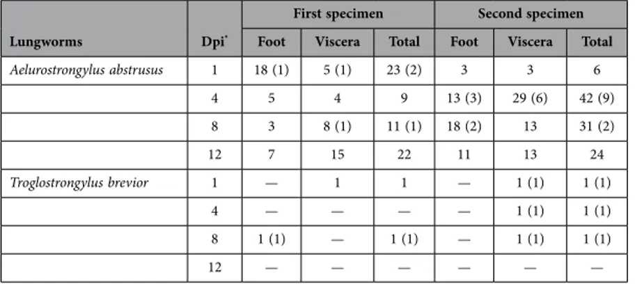
In order to assess the potential of L3s to infect naïve intermediate hosts, 24 pathogen-free H. aspersa snails were each experimentally infected with 50 L3s of either A. abstrusus or T. brevior. Larvae of A. abstrusus were 540 μ m long and 27 μ m wide, whereas larvae of T. brevior measured 435 μ m in length and 21 μ m in width, on average. L3s were recovered without the external sheaths, they were motile and identified as infective L3s based on a previous description25. The numbers of A. abstrusus and T. brevior
larvae subsequently detected in the foot and viscera of H. aspersa are reported in Table 1. Out of 168 A. abstrusus larvae retrieved (mean 21 ± 12.1), 78 (46.4%) were localized in the foot and 90 (53.6%) in the viscera. Given the initial infection dose, from 12 to 66% L3s were retrieved, with a number of live larvae significantly larger than that of the dead (p ≤ 0.05). Only five larvae of T. brevior were recovered from the viscera (n = 4) and the foot (n = 1) and, of these, only one was alive (Table 1).
Histologically, L3s of A. abstrusus were detected at all timepoints in the eight experimentally infected snails. Conversely, no larvae of T. brevior were observed at the histological examination of gastropods, which was in accordance with the small number of larvae detected at the artificial digestion (n = 1 per snail). L3s of A. abstrusus were detected in the fibro-muscular tissue of the foot (Fig. 1a,b) and in the intestine, kidney parenchyma, and locomotion-associated glands (Fig. 1c,d).
Then, in order to evaluate the occurrence of snail-to-snail transmission of L3s, molluscs infected by either A. abstrusus or T. brevior were co-housed with uninfected snails (study 1). Live L3s of A. abstrusus (n = 5) were detected in previously uninfected snails at 8 and 12 days post contact (dpc) and only one of T. brevior at 1 dpc. Subsequently, one dead snail infected by either lungworm species was co-housed with susceptible uninfected snails (study 2). Live L3s of A. abstrusus (n = 3) were then detected in unin-fected snails at 1, 8 and 12 dpc, whereas those of T. brevior were dead and detected at 1 (n = 3) and 12 dpc (n = 1), respectively.
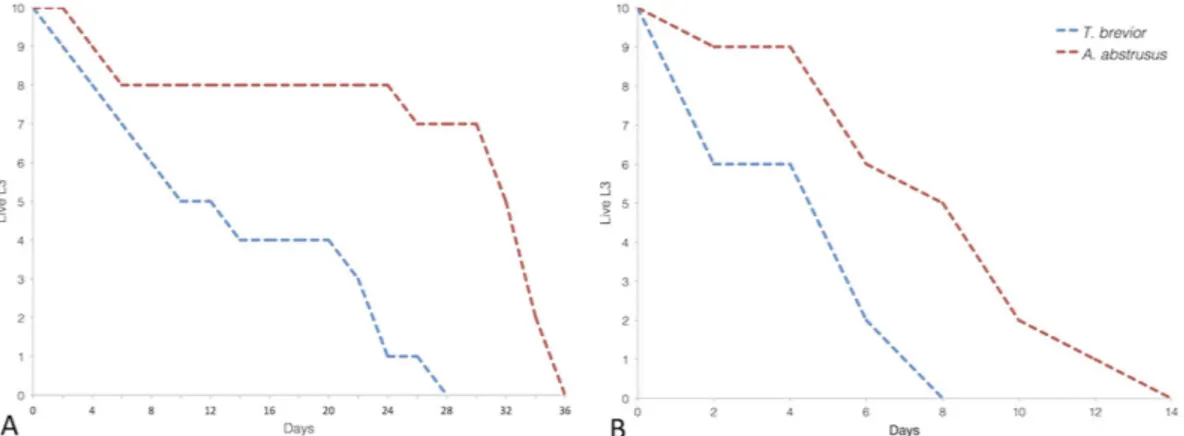
Survival of third-stage larvae. Finally, we evaluated the off-host survival time of A. abstrusus and T. brevior L3s at different temperatures via observation at 2 day-intervals. The maximum survival time of L3s at 4 and 26 °C was 36 and 14 days for A. abstrusus and 28 and 8 days for T. brevior (Fig. 2).
Lungworms Dpi*
First specimen Second specimen
Foot Viscera Total Foot Viscera Total
Aelurostrongylus abstrusus 1 18 (1) 5 (1) 23 (2) 3 3 6 4 5 4 9 13 (3) 29 (6) 42 (9) 8 3 8 (1) 11 (1) 18 (2) 13 31 (2) 12 7 15 22 11 13 24 Troglostrongylus brevior 1 — 1 1 — 1 (1) 1 (1) 4 — — — — 1 (1) 1 (1) 8 1 (1) — 1 (1) — 1 (1) 1 (1) 12 — — — — — —
Table 1. Number of third-stage larvae of Aelurostrongylus abstrusus and Troglostrongylus brevior detected in the foot and viscera of Helix aspersa snails infected with 50 L3s each.Two specimens were analysed at each time point (i.e., 1, 4, 8, 12 *Days post infection, Dpi). Number of dead larvae in brackets.
For both parasites, an inverse association (p > 0.0001) between the timepoint and the number of live larvae was registered at both temperatures. At 4 °C, the frequency of T. brevior live larvae was higher (p ≤ 0.05) than that of dead larvae until the eighth day; at 26 °C, the same trend was observed, but only on the fourth day. For A. abstrusus, a larger number of live larvae (p ≤ 0.05) was recorded until the thirty-second and sixth day at 4 and 26 °C, respectively (Fig. 2).
Molecular characterisation of larvae recovered from the experimental studies, based on amplifica-tion and sequencing of the internal transcribed spacer 2 of the ribosomal DNA (ITS-2) confirmed the morphological identification, with all sequences displaying 100% identity to those of A. abstrusus and T. brevior in GenBankTM (accession numbers KF751655 and KF751656, respectively).
Figure 1. Histopathology of snail tissue. Third-stage larvae of Aelurostrongylus abstrusus from experimentally infected gastropods were detected in the fibro-muscular tissue of the foot (1A,B), and in glands (1C,D). Scale bar 50 μ m.
Figure 2. Number of live third-stage larvae of Aelurostrongylus abstrusus and Troglostrongylus brevior at 4 °C (A) and 26 °C (B).
Discussion
In this article we report, for the first time, snail-to-snail transmission of A. abstrusus L3s from infected to naïve H. aspersa. While the transmission of infective larvae through a series of paratenic hosts, also known as paratenesis, is a well-documented mechanism occurring in selected parasites (e.g., Gnathostoma spinigerum)26, the transmission of L3s between intermediate hosts is, to the best of our knowledge, a
completely novel finding. More in general, this is the first report of an intermediate host being infected by an infective L3. Such an occurrence may have important implications for the epidemiology of a number of snail-borne parasitic diseases (e.g., A. cantonensis, Protostrongylus spp.), whose L3s may be excreted by snails13,27,28. However, the impact of this transmission route on the overall patterns of
par-asite epidemiology and ecology (i.e. compared to the ingestion of intermediate or paratenic hosts)29 is
currently unknown (given that the infection of a receptive host with L3s was not possible for ethical rea-sons) and deserves further consideration. Indeed, although the risk of human infection by A. cantonensis L3s excreted via snail mucus or contaminated water has been considered less likely to cause disease14,
snail-to-snail transmission might increase the chance for the definitive host to come across the parasite. At the same time, this phenomenon may significantly increase the survival time of infective larvae in the environment since, once released by an intermediate host, they can promptly infect a new, susceptible snail should a suitable definitive host be unavailable. This hypothesis is further corroborated by the high recovery rate of A. abstrusus L3s (i.e., up to 66%) in experimentally infected H. aspersa and by the larval transmission from infected to uninfected snails (study 1). It is known that the mucus of snail trails is a source of energy intake when ingested by congeners30,31 and it is pivotal for aggregation and mating31.
Snail clustering and the elimination of A. abstrusus L3s via the mucus may promote the circulation of the parasite among intermediate hosts, as supported also by the detection of infective larvae in the viscera of snails. Whether these metastrongyloids activate (directly or indirectly) a cascade of biochemical signals into the snail mucus, similarly to the sea snail Littorina littorea when infected by trematode parasites32,
is yet to be confirmed.
Previous studies have shown that L3s of A. abstrusus, T. brevior and Muellerius capillaris leave the infected snails soon after the death of their intermediate hosts24,33. This knowledge supports our finding
of L3s in previously uninfected snails following direct contact with a dead infected specimen (study 2). Compared to A. abstrusus, L3s of T. brevior displayed a decreased capacity to infect H. aspersa, a possible consequence of a limited adaptation of the latter species to this snail intermediate host. These data are supported by previous evidence of a higher moulting rate for A. abstrusus than T. brevior in the same snail species24.
The passage of infective larval stages between intermediate hosts is a novel pattern of parasite trans-mission. Therefore, we propose for this mode of transmission the term intermediesis.
One of the primary effects of the circulation of infective larvae among snails is the broadening of the number of intermediate hosts available to the definitive hosts, which may ultimately lead to the spread of the infection in a suitable environment. This mechanism allows the enlargement of refugia of infective larvae whose survival time is a limiting factor in hostile environments (i.e., high temperatures, low rainfall or other abiotic factors), thus persisting from season to season across time and space. For example, the effect of human activities (e.g., irrigation or water storage) has been hypothesized to play a major role in the spread of snail-borne diseases. However, the elucidation of snail-parasite relationships and of new transmission pathways is central to the development of integrated control strategies2, that
aim at overcoming concerns related to drug resistance34–36. If this transmission pattern is confirmed to
occur in nature, mathematical models will need to integrate the phenomenon of “intermediesis”, besides paratenesis, in studies of transmission dynamics of parasites with a complex life cycle37.
Methods
Experimental infection of gastropods by L3s. Infective L3s of A. abstrusus or T. brevior were obtained from infected H. aspersa snails maintained in the Parasitology Laboratory of the University of Bari (Puglia, Italy) as follows. Each snail was digested in 100 ml HCl solution (pH 2.2) and 3 mg/ml of pepsin (Sigma-Aldrich, St. Louis, Missouri, United States). The suspension was heated on a magnetic stirrer at 37 °C for 75 min, sift through a 250 μ m sieve to remove undigested material, transferred to 50 ml plastic tubes and centrifuged at 600 g for 5 min. The suspension was microscopically examined and larvae were morphologically identified, according to previous descriptions23,25, with the aid of a
computer software (Leica LAS
®
AF 4.1). Single infective doses of 50 L3s were collected under a light microscope (Leica®
, DM LB2) and kept in plastic tubes (1.5 ml), until being utilized for snail infection. Nematode-free snails, 20 months old, were purchased from a commercial provider in Barletta (Puglia, Italy), placed in plastic boxes (= vivaria) and fed every 2 days with lettuce and water. The vivaria were kept in a temperature-controlled room (23 ± 1 °C) and monitored via a thermo-hygrometer on an hourly basis. The absence of natural infections by any nematode larvae was assessed by microscopically exam-ining eight snails (10%) the day prior to the infection. On the day of the infection, two groups of 12 (nematode-free) snails each were infected with 50 live L3s of either A. abstrusus or T. brevior. Briefly, snails were individually placed in a plastic infection chamber containing a potato slice (0.5 cm thick) and the infective dose of either A. abstrusus or T. brevior. Specimens were left in the infection chamber for 48 h and subsequently returned to the vivaria. Successful infection with either metastrongyloid wasassessed at different time points (i.e., 1, 4, 8, 12 days post-infection), using the protocol described above on two sections of each snail, i.e. muscular foot and viscera.
The presence of larvae from experimentally infected gastropods was detected by histological exami-nation. At each time point, one snail infected by L3s of A. abstrusus or T. brevior was examined. Snails were anesthetized with menthol steam, deprived of their shells and fixed in a 50 mL vial with 10% neutral buffered formalin solution. Sections of 5 μ m across the body of the snail were stained with haematoxylin and eosin (H&E) and routinely processed.
Snail to snail transmission. Two studies were designed to determine the potential of snail-to-snail transmission of infective L3s from infected to naïve, susceptible gastropods. In study 1, infected snails (n = 3) marked with permanent ink were co-housed with uninfected specimens (n = 3) in a 1 L container. The experiment was repeated twice (i.e., G1a and G1b) for each nematode species.
In study 2, for each species, one dead infected mollusc was co-housed soon after its natural death with six uninfected snails in a 1 L container for 24 h. All naïve snails from both studies were artificially digested at three time points (i.e., 1, 8, 12 days post-contact; dpc) for the detection of L3s and examined at each time point (i.e., one specimen per group in study 1 and two snails for study 2). At the end of the study, infected snails from study 1 and the dead specimen from study 2 were digested to confirm the success of the experimental infection.
Survival of third-stage larvae. The survival time of A. abstrusus and T. brevior L3s was recorded at 4 and 26 °C, the minimum and maximum temperature at which snails are active. Briefly, following artificial digestion of H. aspersa, the solution was centrifuged at 600 g for 5 min, and the larvae-containing sedi-ments were collected and washed, twice. Following the last centrifugation, the sedisedi-ments were transferred to 1.5 ml tubes (one for each nematode) and maintained at 4 and 26 °C, respectively.
The motility of the nematode larvae (10 larvae per time-point) was assessed at 2-day intervals until the death of the last specimen (maximum survival time). Larvae were considered dead when no move-ments were observed under a light microscope (Leica
®
, DL MB2) for up to 10 s, or when degeneration of larval internal organs occurred.Data were analysed by Fisher’s exact test using BioEstat software (version 5.0; Mamirauá/CNPq, Belém, PA, Brazil).
References
1. World Health Organization. Accelerating work to overcome the global impact of neglected tropical diseases–a roadmap for
implementation. Geneva, Department of Control of Neglected Tropical Diseases (2012).
2. Adema, C. M. et al. Will all scientists working on snails and the diseases they transmit please stand up? PLoS Negl. Trop. Dis. 6, e1835 (2012).
3. Hotez, P. J. et al. Control of neglected tropical diseases. N. Engl. J. Med. 357, 1018–1027 (2007).
4. Tsai, T. H. et al. An outbreak of meningitis caused by Angiostrongylus cantonensis in Kaohsiung. J. Microbiol. Immunol. Infect.
34, 50–56 (2001).
5. Wang, Q. P., Lai, D. H., Zhu, X. Q., Chen, X. G. & Lun, Z. R. Human angiostrongyliasis. Lancet Infect. Dis. 8, 621–630 (2008). 6. Sawanyawisuth, K. et al. Clinical manifestations of eosinophilic meningitis due to infection with Angiostrongylus cantonensis in
children. Korean J. Parasitol. 51, 735–738 (2013).
7. Rosen, L., Chappell, R., Laqueur, G. L., Wallace, G. D. & Weinstein, P. P. Eosinophilic meningoencephalitis caused by a metastrongylid lung-worm of rats. JAMA. 179, 620–624 (1962).
8. Alicata, J. E. Biology and distribution of the rat lungworm, Angiostrongylus cantonensis, and its relationship to eosinophilic meningoencephalitis and other neurological disorders of man and animals. Adv. Parasitol. 3, 223–248 (1965).
9. Wang, X. et al. A clinical study of eosinophilic meningoencephalitis caused by angiostrongyliasis. Chin. Med. J. 115, 1312–1315 (2002).
10. Evans-Gilbert, T., Lindo, J. F., Henry, S., Brown, P. & Christie, C. D. Severe eosinophilic meningitis owing to Angiostrongylus
cantonensis in young Jamaican children: case report and literature review. Paediatr. Int. Child Health. 34, 148–152 (2014).
11. Chen, H. T. Un nouveau nematode pulmonaire: Pulmonema cantonensis ng., n. sp. des rats de Canton. Ann. Parasitol. Hum.
Comp. 13, 312–317 (1935).
12. Mackerras, M. J. & Sandars, D. F. The life history of the rat lungworm, Angiostrongylus cantonensis (Chen) (Nematoda: Metastrongylidae). Aust. J. Zool. 3, 1–21 (1955).
13. Heyneman, D. & Lim, B. L. Angiostrongylus cantonensis: proof of direct transmission with its epidemiological implications.
Science 158, 1057–1058 (1967).
14. Cowie, R. H. Pathways for transmission of angiostrongyliasis and the risk of disease associated with them. Hawaii J. Med. Public
Health 72, 70–74 (2013).
15. Macpherson, C. N. Human behaviour and the epidemiology of parasitic zoonoses. Int. J. Parasitol. 35, 1319–1331 (2005). 16. Kim, D. Y., Stewart, T. B., Bauer, R. W. & Mitchell, M. Parastrongylus (Angiostrongylus) cantonensis now endemic in Louisiana
wildlife. J. Parasitol. 88, 1024–1026 (2002).
17. Duffy, M. S., Millar, C. L., Kinsella, J. M. & De Lahunta, A. Parastrongylus cantonensis in a nonhuman primate. Florida. Emerg.
Infect. Dis. 10, 2207–2210 (2004).
18. Burns, R. E. et al. Cerebral Angiostrongylus cantonensis infection in a captive African pygmy falcon (Polihierax semitorquatus) in southern California. J. Vet. Diagn. Invest. 26, 695–698 (2004).
19. Kottwitz, J. J., Perry, K. K., Rose, H. H. & Hendrix, C. M. Angiostrongylus cantonensis infection in captive Geoffroy’s tamarins (Saguinus geoffroyi). J. Am. Vet. Med. Assoc. 245, 821–827 (2014).
20. Conboy, G. A. Helminth parasites of the canine and feline respiratory tract. Vet. Clin. North Am. Small Anim. Pract. 39, 1109–1126 (2009).
21. Brianti, E., Giannetto, S., Dantas-Torres, F. & Otranto, D. Lungworms of the genus Troglostrongylus (Strongylida: Crenosomatidae): neglected parasites for domestic cats. Vet. Parasitol. 202, 104–112 (2014).
22. Anderson, R. C. in Nematode parasites of vertebrates their development and transmission 2nd edn, Ch. 3, 129–164 (CABI Publishing, 2000).
23. Giannelli, A. et al. Development of the feline lungworms Aelurostrongylus abstrusus and Troglostrongylus brevior in Helix aspersa snails. Parasitology 141, 563–569 (2014).
24. Giannelli, A. et al. Release of lungworm larvae from snails in the environment: potential for alternative transmission pathways.
PLoS Negl. Trop. Dis. 9, e0003722 (2015).
25. Gerichter, C. B. Studies on the nematodes parasitic in the lungs of Felidae in Palestine. Parasitology 39, 251–262 (1949). 26. Beaver, P. C. The nature of visceral larva migrans. J. Parasitol. 55, 3–12 (1969).
27. Boev, S. N. in Protostrongylids. Fundamentals of nematology, Vol. 25 (Helminthological laboratory, academy of sciences of the USSR, Moscow). 1–337 (U.S. Department of Agriculture, Washington, D.C., and Amerind Publishing Co., New Delhi, India, 1984).
28. Ubelaker, J. E., Bullick, G. R. & Caruso, J. Emergence of third-stage larvae of Angiostrongylus costaricensis Morera and Cespedes 1971 from Biomphalaria glabrata (Say). J. Parasitol. 66, 856–857 (1980).
29. Kralka, R. A. & Samuel W. M. Emergence of larval Protostrongylus boughtoni (Nematoda: Metastrongyloidea) from a snail intermediate host, and subsequent infection in the domestic rabbit (Oryctolagus cuniculus). J. Parasitol. 70, 457–458 (1984). 30. Bailey, S. E. R. Daily cycles of feeding and locomotion in Helix aspersa. Haliotis 19, 23–31 (1989).
31. Ng, T. P. et al. Snails and their trails: the multiple functions of trail-following in gastropods. Biol. Rev. Camb. Philos. Soc. 88, 683–700 (2013).
32. Erlandsson, J. & Kostylev, V. Trail following, speed and fractal dimension of movement in a marine prosobranch, Littorina
littorea, during a mating and a non-mating season. Marine Biol. 122, 87–94 (1995).
33. Zmoray, I., Svarc, R. & Lestan, P. Localization of larvae of Müllerius capillaris in the tissues of the intemediary host Cepaea
vindobonensis (Fér). Biologia 24, 113–128 (1969).
34. Doenhoff, M. J., Kusel, J. R., Coles, G. C. & Cioli, D. Resistance of Schistosoma mansoni to praziquantel: is there a problem?
Trans. R. Soc. Trop. Med. Hyg. 96, 465–469 (2002).
35. Gray, D. J. et al. Schistosomiasis elimination: lessons from the past guide the future. Lancet Infect. Dis. 10, 733–736 (2010). 36. Knox, M. R. et al. Novel approaches for the control of helminth parasites of livestock VI: Summary of discussions and conclusions.
Vet. Parasitol. 186, 143–149 (2012).
37. Poulin, R. in Evolutionary ecology of parasites 2nd edn, (Princeton university, 2007).
Acknowledgements
Authors thanks Francesca Abramo (University of Pisa) for technical assistance and Merial for covering publication fee and partially supporting our studies on this topic.
Author Contributions
V.C. and D.O. conceived the study. V.C., D.O. and A.G. designed the project. V.C., A.G. and R.A.N.R. analysed the data and performed most of the experiments. E.B., C.C. and F.D.T. critically reviewed the manuscript. All authors read and approved the final manuscript.
Additional Information
Competing financial interests: The authors declare no competing financial interests. How to cite this article: Colella, V. et al. Feline lungworms unlock a novel mode of parasite
transmission. Sci. Rep. 5, 13105; doi: 10.1038/srep13105 (2015).
This work is licensed under a Creative Commons Attribution 4.0 International License. The images or other third party material in this article are included in the article’s Creative Com-mons license, unless indicated otherwise in the credit line; if the material is not included under the Creative Commons license, users will need to obtain permission from the license holder to reproduce the material. To view a copy of this license, visit http://creativecommons.org/licenses/by/4.0/