Polytechnic University of Marche
RATIONAL DESIGN OF FUNCTIONALIZED LIPIDS WITH ANTIOXIDANT AND SCAVENGING ACTIVITY AS COMPONENTS OF INNOVATIVE ARTIFICIAL TEARS
PhD course “Biomolecular Sciences” XV cycle
Academic tutor:
Dott. Roberta Galeazzi
Aziendal tutor:
Dott. Dario Rusciano
PhD Eureka course in collaboration
with SOOFT Italia company
PhD Laudadio Emiliano
INDEX
1. OBJECTIVE OF RESEARCH
2. INTRODUCTION AND STATE OF ART 2.1 TEAR FLUID LIPID LAYER
2.2 LIPOSOMES
2.3 THERAPEUTIC APPLICATIONS OF LIPOSOMIAL FORMULATIONS IN TOPICAL OCULAR DRUG DELIVERY
3. MATHERIALS AND METHODS
3.1 ENERGY AND SIMULATION OFLIPID BYLAYERS
3.1.1 CLASSICAL MOLECULAR MECHANICS APPROACH 3.1.2 FORCE FIELDS FOR LIPID BILAYER SIMULATIONS
3.2. ATOMISTIC MOLECULAR DYNAMICS SIMULATIONS OF BILAYER SYSTEMS
3.2.1. THE NEWTONS LAWS 3.2.2. ENSAMBLES
3.2.3. PERIODIC BOUNDARY CONDITIONS
3.2.4. OPTIMIZATION OF THE STARTING STRUCTURES
3.2.5. KINETIC ENERGY AND TEMPERATURE CONTROL DURING MD 3.2.6. INTEGRATOR ALGHORITMS
3.2.7. TEMPRATURE COUPLING 3.2.8. PRESSURE CONTROL 3.3. SOLVATION MODELS
4. IN SILICO DESIGN OF THE LIPOSOMIAL NANOVECTOR 4.1 CELL MEMBRANE COMPOSITION
4.2 LIPOSOMIAL NANOVECTOR: STRUCTURE AND COMPOSITION 4.3 SETTING AND TUNING THE MOLECULAR DYNAMICS MEMBRANE
SIMULATIONS PROTOCOL FOR MIXED COMPOSITION BILAYERS 4.3.1 FUNCTIONALIZED COMPOUNDS FOR GENE DELIVERY
4.3.1.1 CHOLP MOLECULES 4.3.1.2 CROWN ETHER LIPIDS
4.3.2 MD PROTOCOL VALIDATION: ANTIOXIDANT SYNTHETIC NITROXIDES (NOXs)
5. DESIGN AND DEVELOPMENT OF LIPOSOMAL NANOVECTORS WITH ANTIOXIDANT ACTIVITY
5.1 ANTIOXIDANT COMPOUND: EDARAVONE DERIVATIVE 5.2 MODELING OF THE MIXED MEMBRANE SYSTEMS 5.3 MD SIMULATION ANALYSIS
5.4 DISCUSSIONS AND CONCLUSIONS
6. OTHER ANTIOXIDANT NANOVECTORS: PHYTOSOMES 6.1 INTERACTIONS OF EGCG WITH BILAYER
6.1.1 MODELS WITH KCl 6.1.2 MODELS WITH NaCl 6.1.3 MODELS WITH MgCl2
6.1.4 MODELS WITH CaCl2
6.2 INFLUENCE OF LIPID MATRIX ON EGCG ENCAPSULATION
7. CONCLUSIONS 8. REFERENCES
1 1. OBJECTIVE OF RESEARCH
The main aim of my PhD research work is the in silico design and the development of new drugs able to efficiently deal with free radicals at the target tissue or organ, more in particular for the eye. Such objective is of great interest in the pharmaceutical field because free radicals are responsible for the processes of aging and cell death of the animal tissues, and therefore also of at the ocular level. Our aim is to create an innovative product that could be used as drug delivery nanovector in ocular drops. In fact, it is known that the formulations based on nanoparticles such as micelles, microemulsions and monolaminar liposomes exploit the fact that a high surface/volume ratio leads to a significant increase in pharmacological activity. The nanoparticle can carry the drug to the specific site, releasing it in a controlled manner and at the same time protect it from premature degradation. However, the innovation of our project involves the design of the liposome binding the new antioxidant molecule directly to a lipid molecule that thus becomes itself a constituent of the liposomal nanovector and not only incorporated inside. This strategy is aimed at improving the functionality of transport and of resistance to degradation. In fact, the new molecule synthesized will remain longer in the tear film, and will be active for a greater period. This aspect is very important because the eye in contact with light uses a large amount of ATP, and this generates the free radicals. They are molecules that possess an unpaired electron on electric orbital and this makes them highly unstable and highly reactive. Free radicals react easily with anyone molecule is in their proximity damaging and often compromising its function. Also, by reacting with other molecules, have the ability to disseminate themselves transforming their targets in free radicals and thereby causing a chain reaction that can cause extensive damage in the cell.
More in detail, during my PhD period, we explored two different strategies: (i) functionalization of known antioxidant molecules in order to insert them deeply into the bilayers; (ii) improving encapsulation of known antioxidant molecules. The final object will produce liposomal antioxidant systems for ophthalmic use that will be stable and efficient. Moreover, the antioxidant molecules will be retained on lacrimal film, and thus they will use their antioxidant activity against the free radicals generated outside the eye. For this reason, the functionalization has to improve lipophilicity on antioxidant molecules but it must not influence the proper antioxidant activity of the chosen molecules. Only in this way, we can obtain an efficient ocular drug delivery system. To reach all these goals efficiently and fast, we settled out an in silico molecular modeling protocol based on atomistic molecular dynamics simulation of lipid bilayer systems as liposomal models. Indeed, thanks to the improvement of both Molecular Modeling Software Packages and hardware resources, it is nowadays possible to get new insights on complex molecular aggregates. Extensive structural
2 analysis and accurate inter-molecular interactions previsions via computational methods arise today as a key part of laboratories protocols in many scientific fields such as chemistry, drug design, biology and materials science. To highlight the latest results attainable with computational chemistry tools, we will describe how we successfully recreated a realistic model for inhomogeneous systems such as lipid membranes. We describe our extensive analysis on particular membrane bilayers containing functionalized lipids with antioxidant activity. Molecular Dynamics (MD) simulations were implemented on the basis of our tested laboratory protocol, to better understand the behaviors of antioxidant molecules in their lipophilic carriers.
In this work, I discuss the innovation perspectives offered by an in silico analysis, both in terms of “better understanding” and “suggesting”: the first as a complementation of experimental data, the latter as a novel way to plan the experiment itself.
2. INTRODUCTION AND STATE OF ART 2.1 TEAR FLUID LIPID LAYER
Human tear film comprising lipids, proteins, metabolites, and water protects the eye from dryness and injury. The tear film have a unique structure that enables it to perform many functions, composed by lipid, aqueous, and mucous components. There is also a mucin containing glycocalyx interface that extends from the apical membranes of the corneal and conjunctival epithelia which acts as an integral part of the tear film. The seven major functions of the tear film are:
Maintaining a smooth surface for light refraction. The tears form the first refractive surface encountered by light on its path to the retina. For clear vision, it is critical to maintain the transparency of the second refractive surface that rays of light encounter, i.e. the cornea. Lubricating the eyelids.
Lubricating the conjunctiva and the cornea by providing a smooth and reflective surface, thus avoiding ocular surface mechanical damage from the surprisingly high pressures generated by each blink.
Supplying the cornea with nutrients by transporting oxygen and a limited number of other nutrients to the avascular cornea, regulating the electrolyte composition and pH.
Providing white blood cells with access to the cornea and conjunctiva.
Removing foreign materials from the cornea and conjunctiva. The tear film protects the ocular surface from the external environment by responding dynamically to a wide range of external conditions and potentially damaging situations. These external stresses include desiccation,
3 bright light, cold, mechanical stimulation, physical injury, noxious chemicals, and bacterial, viral, and parasitic infection.
Defending the ocular surface from the pathogens via specific and nonspecific antibacterial
substances [1].
In humans, tear film is composed by three distinct parts: the innermost mucin-rich layer, an aqueous layer in the middle, and the outermost tear fluid lipid layer (TFLL). The latter is a lipid layer located at the water-air interface. The function of tear film is to protect the epithelium of the eye. Tear fluid fulfils this purpose in many ways. When blinking, tear fluid forms a lubricating film between the lid and ocular surface. It possesses antibacterial properties and flushes contaminants from the ocular surface. Tear fluid also acts as a nutrient for the corneal epithelium and improves the optical properties of the eye. Tear film has an ill-defined trilaminar and concentration gradient-dependent structure [2].
Figure 1A and 1B: Structure of lacrimal film
TFLL coats the aqueous phase and may also protect the ocular surface from evaporation, though the latter property has been questioned very recently. The lipid layer prevents evaporation of tears, the aqueous layer allows spread of tears over the ocular surface and mucin layer adheres the tear film to ocular surface. In an unstimulated human eye at a normal blink rate of 15–20 blinks/min, the tear volume on the ocular surface is about 6–8 ml and the basal tear turnover rate is approximately 16%/min of the total tear volume. The ability to retain water is due to the Van der Waals interactions among hydrophobic carbon chains. Furthermore, these incompressible lipid layers have very low free volumes, preventing diffusion of small molecules through the layers. An increase in surface pressure increases the retardation of evaporation by decreasing the chain tilt and, therefore, increasing the thickness of the layer. To a certain extent, evaporation-retardant lipids can be mixed with non-retardant lipids and still hold their ability to retard evaporation [3].
Very minute changes in molecular composition may disturb the delicate balance between healthy and unhealthy ocular surfaces, highlighting the importance of understanding the role of the TFLL (with
4 regard to its dynamics, composition, structure, and mechanical properties) in maintaining the sensitive balance of the ocular surface. Nonetheless, the current understanding of TFLL properties is quite limited. Perhaps the best known aspect is its lipid composition, as recent studies have revealed the most abundant lipids present in human TFLL to be phospholipids (PLs), free fatty acids (FFAs), cholesteryl esters (CEs), triglycerides (Tgs), and wax esters (Fig. 1). Further, the relatively high ratio of PLs in the mixture (more than 50%) supports the idea of a single lipid monolayer at the air-water interface, though one cannot rule out a possibility of a more complex structure either. Meanwhile, the organization and role of neutral lipids (e.g., CEs, TGs, and fatty acids) in the lipid layer is more intriguing [4].
The ocular tissue absorption of topically administered drugs is estimated to be less than 5%. Majority of drug loss is attributed to overspill. The capacity of the conjunctival sac when lower lid is pulled away is approximately 25 ml and reduces to approximately 10 ml when eyelid returns to its normal position. The pathological conditions affecting conjunctiva may further limit the holding capacity of conjunctival sac. Hence, instillation of eye drops in a volume larger than 25 ml results in drug loss due to overspill. The amount of drug retained in the conjunctival sac mixes with the precorneal tear film before it comes in direct contact with ocular surface. It has been observed that even if the drug loss due to drainage is compensated by sustained drug delivery through a solid delivery system, the ocular bioavailability reaches only up to 10% indicating the importance of barriers on the ocular surface. Not only the barrier functions of ocular surface tissue i.e. cornea, conjunctiva and sclera are now extensively studied; investigations have also revealed the importance of precorneal tear film as a significant barrier to drug absorption [5].
Figure 2: Structures of Corneal, Tear film and scleral barriers
5
An intact tear film is essential for a healthy ocular surface. However, as described above, for the penetration of drugs applied topically to ocular surface, tear film is a significant barrier. This tear turnover rate may increase significantly due to reflex tearing which may occur due to drug instillation resulting in accelerated washout of the drug. Tears are drained through the nasolacrimal duct into the nasal cavity. Blinking creates a pumping mechanism to facilitate flow of tears into the nasolacrimal duct. Drainage of the instilled drug into the nasolacrimal system along with tears is the main factor contributing to drug loss from pre-corneal tear film and reduction in the rate of tear drainage significantly increases the drugs’ ocular bioavailability.
Considering the challenges in ocular drug delivery, ideal topical drug delivery system musts to have these characteristics:
Should be able to resist pre-corneal clearance and provide prolonged corneal contact time. Should be delivered in a dosage form that provides adequate trans-corneal absorption
(paracellular or transcellular).
Should be of suitable viscosity that provides good corneal contact time but avoids reflex blinking, tearing and blurred vision.
Should have a suitable pH that favors the absorbable form of drug molecule (non-ionized) but is non-irritant to ocular surface.
Should cause minimal adverse effects. Should be easy to administer.
Should require sufficiently low frequency of administration to ensure patient compliance.
2.2 LIPOSOMES
Liposomes are artificial vesicles consisting of outer covering of lipids that encloses the inner core. In the early twentieth century, liposomes were considered as artificial cells due to their lipid bilayer covering; after this, liposomes were recognized as important drug delivery systems and their potential uses in cancer chemotherapy were investigated. Since then, liposomes have undergone extensive investigation to develop them for targeted and sustained drug delivery.
The liposomal vesicles vary in size from 10 nm to 1 mm or greater. Structurally liposomes are classified into unilamellar vesicles (ULVs) and multilamellar vesicles (MLVs). Based on the size of vesicles, ULVs are further classified into small unilamellar vesicles (SUVs), giant unilamellar vesicles (GUVs), and large unilamellar vesicles (LUVs) (Figure 2). In ULVs, single lipid bilayer consisting of lecithin or phosphatidylglycerol encloses the aqueous core. MLVs consist of more than
6
one lipid bilayer, each separated by an aqueous compartment. Structure of liposomal vesicle allows them to serve as carrier for hydrophilic drugs that can be encapsulated into the aqueous core as well as hydrophobic and amphiphilic drugs that can be embedded in the lipid bilayer. Since the liposomes with multiple compartments have greater aqueous space, their capability to entrap hydrophilic drugs is higher than those with single compartments are. Lipids commonly used for the preparation of liposomes are phospholipids, which can be synthetic or naturally occurring (Fig. 3). The phospholipids that are commonly used include egg phosphatidylcholine, brain and synthetic phosphatidylserine, sphingomyelin, synthetic dipalmitoyl-DL-a-phosphatidylcholine, phosphatidylinositol andovolecithin. A nonionic or zwitterionic lipid is generally used as the basic lipid and to introduce surface charge other lipids are included such as stearylamine for positive charge and diacetylphosphate, phosphatidyl glycerol or phosphatidyl serine for negative charge. Incorporation of charged lipids leads to a greater overall volume for aqueous entrapment and the likelihood of aggregation reduces. However, stearylamine containing cationic liposomes have been shown to cause toxicity in rabbit [102]. Incorporation of cholesterol increases the stability, enhances
the fluidity or microviscosity of the lipid bilayer and reduces the leakage of water-soluble molecules.
The mechanisms of interaction of liposomes with cell membranes that result into intracellular drug delivery have been studied extensively but are poorly understood. Due to highly complex nature of this interaction, the interpretation of experimental data is often difficult. The initial liposome-cell membrane interaction is the key process that leads to intracellular drug delivery. This liposome-cell membrane interaction may involve different receptors on different cell types or more than one receptor on a particular cell and is greatly affected by the lipid composition of liposomes [103]. Largely,
Figure 3: Structures of different liposomes
7
four mechanisms of intracellular drug delivery by liposomes are widely accepted and are as follows (Fig 4).
Figure 4: mechanisms of intracellular drug delivery using liposomes
Adsorption: Adsorption of liposomes to cell membrane is one of the important mechanisms of
intracellular drug delivery. The adsorbed liposomes, in the presence of cell surface proteins, become leaky and release their contents in proximity of cell membrane. This results in a higher concentration of drug close to cell membrane and facilitates cellular uptake of drug by passive diffusion or transport
[104].
Endocytosis: Adsorption of liposomes on the surface of cell membrane is followed by their
engulfment and internalization into endosomes. Endosomes transport liposomes to lysosomes. Subsequently, lysosomal enzymes degrade the lipids and release the entrapped drug into the cytoplasm [105].
Fusion: Fusion of lipid bilayer of liposomes with cell membrane by intermixing and lateral diffusion
of lipids results in direct delivery of liposomal contents into the cytoplasm.
Lipid exchange: Due to the similarity of liposomal membrane lipids with the cell membrane
phospholipids, lipid transfer proteins in the cell membrane recognize liposomes and consequently cause lipid exchange. This results in the destabilization of liposomal membranes and intracellular release of drug molecules [106]. An understanding of the mechanisms of intracellular drug delivery by
liposomes provides the basis for bringing about manipulations in the characteristics of liposomes to enhance their favorable interaction with cell membranes and hence the drug delivery.
Liposomes have the advantage of delivering both the lipophilic drugs that they can entrap in their lipid covering and hydrophilic drugs that are incorporated into their aqueous core. Studies have shown that liposomes are also efficient carriers of amphiphilic drugs. Liposomes enhance the corneal
8
permeability of lipophilic, hydrophilic as well as amphiphilic drugs due to their ability to come in close contact with cornea and conjunctiva and increase the extent of corneal uptake by prolonging the corneal contact time [107]. Additionally, liposomes are completely biodegradable and relatively
nontoxic. Despite these advantages, some limitations of the liposome-based formulations have restricted their therapeutic uses in the past. Conventional liposomes do not differentiate the target cells from others and manipulation of the structure of liposomes in order to achieve targeted drug delivery is another important issue. Additionally, the requirement of prolonged sustained drug delivery also warrants manipulations of the liposome characteristics. Efficacy of liposomes as a drug carrier depends upon various factors such as charge, size, and lipid composition of the liposomal membrane, stability and corneal residence time. To circumvent the limitations of liposomes summarized above and to achieve the characteristics of an ideal drug carrier, several modifications particularly to the surface characteristics and lipid composition of the liposomes have been explored.
Modifications:
Charge: In general, charged liposomes resist aggregation and fusion better compared to uncharged
liposomes and positively charged liposomes provide greater duration of action and higher drug delivery compared to negatively charged liposomes [107]. This is because positively charged liposomes
intimately interact with negatively charged cornea leading to prolonged residence time [108]. It has
also been suggested that cationic vehicle slows down the drug drainage with lacrimal fluid by increasing the viscosity and interaction with negative charges of the mucus [109]. The effect of surface
charge of liposomes on ocular irritation has also been evaluated. Positively charged liposomes significantly increase the rabbit eye blinking rate compared to neutral liposomes, however, the mean total score on Draize test remains below ‘‘practically non-irritating level’’ and no corneal histological changes appeared.
Size: The size of the liposomes also affects their efficiency of drug delivery. MLV have prolonged
retention compared to SUV of same lipid composition and the interaction of liposomes with cornea decreases in the order of MLV+ > SUV+ > MLV- > SUV- > MLV. However, in another study it was observed that inulin-loaded neutral MLV, despite lesser affinity for cornea, provide 100-times greater and sustained inulin concentration in the anterior segment of eye compared to positively charged MLV [110]. This effect was attributed to a two-fold faster disappearance rate of positively charged
MLV from the tear pool. Other study also showed that MLV demonstrate prolonged drug delivery
[111]. The increase in vesicle size restricts drainage from the inner canthal region, hence providing
9
Lipid composition: Lipid composition of the liposomal membrane determines its capacity to resist
leakage of the entrapped drug. Tear-induced leakage of entrapped drug can be reduced by incorporating increasing amounts of cholesterol in the vesicle bilayers. The lipid composition of the liposomes needs to be optimized depending on the drug to be loaded. Similarly, drug to phospholipid ratio also affects the entrapment efficiency of the liposomes. The type of lipid used also determines the surface charge of liposomes [5].
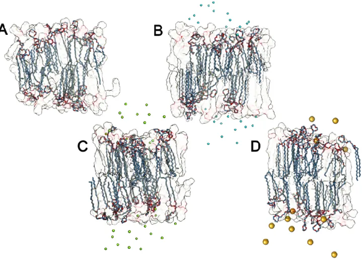
Type and concentration of salt: Considering the biological relevance of salt, the most important ones include as anions Cl- and as cations Na+, K+, Ca++, and Mg++. The binding of cations has been found to have a significant impact on the structural and dynamical properties of PC membranes: It leads to a drop in the area per lipid accompanied by an enhanced ordering of lipid acyl chains and the slowing down of lateral diffusion in the membrane plane. The effect of monovalent salt strongly depends on the type of lipids and cations. We can identify some computation studies results [7, 8], that developed a methology to include these ions into molecular dynamics simulations of bilayers (Table 1-2-3).
TABLE 1: Lennard-Jones Parameters of Ion
TABLE 2: Summary of MD Simulations of Phospholipid Membranes with Monovalent Salt
10 For a salt-free POPC bilayer at 310 K the area per lipid was found to be 0.652 +/- 0.002 nm2. On the experimental side, values of 0.66 nm2 (T = 310 K), 0.65 nm2 (T = 298 K), 0.64 nm2 (T = 298 K), and 0.63 nm2 (T = 297 K) have been reported for the area per lipid for POPC bilayers. Therefore, calculated value for the area per lipid is in good agreement with available experimental data (as well as with previous MD studies), validating thereby the molecular model.
POPC under the Influence of NaCl. Addition of NaCl drastically changes the structural properties of
a POPC lipid bilayer through the binding of cations to the lipid-water interface. Previous MD studies clearly demonstrated that ion binding is a rather slow process, which emphasizes the fact that system equilibration is one of the central issues in simulations of lipid bilayers with salt. The change of membrane parameters can modify functionality of the liposome; this is due to different solvation free energy and different size of every type of ion, so some ions can break the solvation shell and move itself near the polar heads of liposome, creating ionic interactions with the negative components phospholipids. The tight binding of Na+ ions to PC lipid bilayers leads to a notable decrease in the area per lipid accompanied by a more vertical orientation of PC head groups with respect to the membrane surface, by a considerable slowing down of the lateral lipid mobility, and by an increase in the potential difference across a monolayer.
As for potassium ions, their binding to PC membranes and, therefore, the overall salt induced effects are found to be much weaker compared to Na+. This is mostly due to the larger size of a K+ ion, which implies a smaller ionic surface charge and a less ordered first hydration shell [6].
The interaction between metal divalent cations and negative lipids plays an essential role in the structure and function of biological membranes. Aggregation and fusion processes seem to be intimately related to the ability of these cations to bind to phospholipid headgroups and form dehydrated intermembrane complexes. In this sense, the effectiveness of Ca++ and Mg++ in inducing membrane aggregation and fusion has been correlated to their respective binding constants. Recent studies showed that the different effect of Ca++ and Mg++ in membranes is more profound than a simple numerical difference in binding constants. Aggregates formed by Ca++ were branched structures, whereas dense structures were observed for PS_ clusters induced by Mg++. These reported differences also suggest the possibility of different binding modes or binding sites for Ca++ and Mg++ with negative lipids. In a system with negative lipids, Ca++ seems to interact with PO4- group of a
phopspholipid and COO- group of another phospholipid, while Mg++ seems to interact with two PO4
-groups or two COO- groups of phospholipids, suggesting different modifications on membrane structure [7].
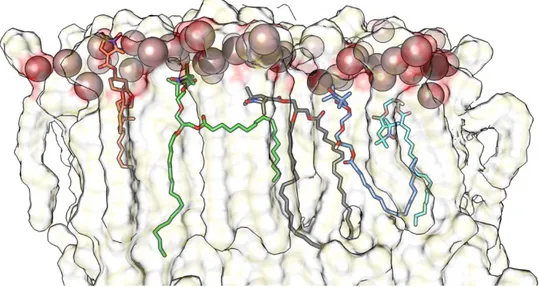
11 Figure 5: Different binding modes of Ca++ and Mg++ as observed in MD simulations. The snapshots were made with the
use of the VMD program. Cations are shown as spheres and lipid molecules are shown in the bond representation of the VMD program
2.3 THERAPEUTIC APPLICATIONS OF LIPOSOMAL FORMULATIONS IN TOPICAL OCULAR DRUG DELIVERY
Antiviral drugs
Herpes Simplex virus (HSV) keratitis is a common cause of cornea related blindness worldwide. Global incidence of HSV keratitis is roughly 1.5 million, including 40 000 new cases of severe monocular visual impairment or blindness each year. Several antiviral drugs have been used in the treatment of HSV keratitis. Acyclovir, a nucleoside analog, has been shown to be effective against HSV. However, because of its limited water solubility, poor lipophilicity and low bioavailability following topical application, it has not shown effectiveness in the treatment of HSV keratitis. It has been compared the bioavailability of topically administered acyclovir with that of subconjunctival administration [113]. The corneal levels of acyclovir were 2.5 times lower and aqueous levels were 5
times lower with topical administration compared to subconjunctival. Systemic acyclovir has been shown to be more effective than topically applied ointment in terms of recurrence rate, functional improvement and graft survival in patients after keratoplasty. Prodrug strategy using more stable amino acid esters of acyclovir has been shown to have enhanced bioavailability. Use of liposomal formulations to enhance bioavailability of topically applied acyclovir has also been evaluated. In one of the studies, in vitro corneal penetration and in vivo corneal absorption of acyclovir containing liposomes was evaluated using rabbits. Liposomes were prepared by drug-lipid hydration method. Phosphatidylcholine and cholesterol were used as lipids, stearylamine as cationic charge inducing agent and dicetylphosphate as anionic charge inducing agent. The loading concentration of acyclovir in the liposome dispersion was 1.24 mg/ml. In vitro studies showed that the acyclovir in solution had fastest permeation through cornea while the corneal permeability of negatively charged liposomes was two times lower and that of positively charged 3.6 times lower than solution. Because of the slower permeation, negatively charged liposomes provided longer permeation time and the same was
12
even longer with positively charged liposomes. In vivo studies also showed that positively charged liposomes penetrate cornea slower than negatively charged liposomes and solution, however, due to prolonged corneal contact time, the extent of corneal penetration is highest with positively charged liposomes [108]. It has been studied acyclovir delivery by topical application using liposome [114]. 1,
2-Dipalmitoyl-sn-glycerol-3-phosphocholine (DPPC) and cholesterol were used as the lipids. Negative charge was introduced by adding 1, 2-dipalmitoyl-sn-glycerol-3-phosphatidic acid (DPPA) and positive charge by stearylamine or dimethyl-dioctadecyl glycerole bromide (DDAB). Liposomes were prepared following four different techniques; thin-layer evaporation, reverse phase evaporation, frozen and thawed, and dehydrated- rehydrated. Positively charged oligolamellar liposomes were shown to have most suitable bioadhesivity towards corneal epithelium. In vivo studies exhibited 42.3-folds increase in the acyclovir concentration in aqueous humor by acyclovir-loaded liposomes compared to free drug.
Antibacterial drugs
Several antibacterial agents have been formulated in liposomal forms to enhance the ocular bioavailability of these agents in the treatment of bacterial ocular infections. Ciprofloxacin, a fluoroquinolone antibiotic, is widely used in the treatment of ophthalmic infections; however, the efficacy is often limited due to poor ocular bioavailability of aqueous solutions. In other works, it has been prepared multilamellar liposomes containing ciprofloxacin by thin film hydration method using lecithin and a-L-dipalmithoylphosphatidylcholine (DPPC) [115]. Release profile of the liposomes was
then studied in vitro. DPPC liposomes were found to significantly prolong the release half-time especially when 0.1% polymethacrylic acid was incorporated in the formulation as vehicle. Ciprofloxacin-loaded liposomes coated on soft contact lenses have also investigated for efficient drug delivery. The contact lenses with ciprofloxacin in liposomes inhibited both the Staphylococcus aureus and Pseudomonas aeruginosa on an agar plate and showed an enhanced antibacterial effect as determined by minimal inhibitory concentrations. The system was found to be nontoxic in in vitro experiment [116]. It has been studied in vitro and in vivo distribution of ciprofloxacin hydrochloride
using liposomes coated with chitosan complex [117]. Liposomes were prepared by thin film hydration
method. It was observed that positively charged liposomes entrapped high level of ciprofloxacin. However, when chitosan complex coating was added, even higher entrapment was seen with negatively charged liposomes. In vivo studies showed that negatively-charged chitosan-coated liposomes provide significantly higher level of ciprofloxacin in external eye tissues with prolonged duration of up to 8 h compared to non-coated positively-charged liposomes (4 h) and commercial eye
13
drop (2 h). In a recent study, it was reported that ciprofloxacin-loaded liposomes provide higher ocular bioavailability compared to commercially available drug [118].
Antifungal agents
Ocular fungal infections are often difficult to treat. Although, several antifungal agents are available for systemic use their ocular bioavailability after topical or systemic administration is poor and intraocular administration is associated with toxic effects. Amphotericin B is an important antifungal drug for the treatment of mycotic infections in eye, however, topical use of the drug causes irritation and ocular permeation is poor. Therefore, use of liposome encapsulated amphotericin B has been investigated. Application of one drop of amphotericin containing unilamellar liposomes to rabbit eyes provided very stable corneal amphotericin B level and had the benefit of lowered ocular toxicity. In a study involving 11 patients with culture positive Candida keratitis, the treatment was given as topical liposomal fluconazole (2 mg/ml) 3 times daily over a period of 30 d. Among all, eight patients showed complete improvement, one patient showed partial improvement while two patients had no improvement. Thus the fluconazole in liposomal form was found to be highly effective in treating Candida keratitis. Although topical fluconazole in liposomal form was found to be superior to solution form in the rabbit model of Candida keratitis, in Candida endophthalmitis model the intravitreal liposomal fluconazole showed lower efficacy than fluconazole in solution [119].
Anti-inflammatory and immunomodulatory agents
Several anti-inflammatory and immunomodulatory agents are widely used in the treatment of ocular inflammatory and immunological diseases. To enhance the ocular bioavailability and reduce the toxic effects following topical or intravitreal adminsitration, liposomal forms of many of these drugs have been evaluated. Cyclosporin is an anti-inflammatory and immunomodulatory drug and is used for the treatment of ophthalmic conditions like dry eye syndrome and in patients undergoing corneal transplant. It has been investigated the efficacy of cyclosporine encapsulated in liposomes in preventing the corneal graft rejection in rats that received allogenic graft [120]. When compared with
cyclosporine in olive oil or empty liposomes, the group treated with cyclosporine-loaded liposomes showed increased mean survival time. The graft survival rate was 77% in liposome treated group compared to 45% in cyclosporine in olive oil or 36% in empty liposomes treated group. In another study cyclosporine loaded liposomes or olive oil containing equivalent amount of cyclosporine was applied topically to rabbit eyes at 15-min intervals within the first hour and then one hourly over a 6-h period. Additionally, collagen s6-hields soaked for 30 min in t6-he liposome preparation of cyclosporine were also tested in vitro and in vivo. It was observed that cyclosporine in liposomes applied topically
14
or in collagen shields delivered significantly higher drug levels to the cornea and sclera at 1 and 3 h compared to cyclosporine in olive oil. Similarly, the levels of cyclosporine in aqueous and vitreous humor were significantly higher in animals treated with liposomes topically or in collagen shields compared to cyclosporine in olive oil. The aqueous humor concentration of cyclosporine was also found to be higher after subconjunctival injection of cyclosporine containing liposomes in rabbits compared to subconjunctival injection of the same quantity of free cyclosporine. One of the recent studies in rabbits has demonstrated that topically applied fusogenic liposomes are safe and in some eyes may produce mild conjunctival injection.
Antiglaucoma agents
Several antiglaucoma agents are used topically to lower intraocular pressure for the treatment of glaucoma. However, topical use is associated with adverse effects and the reduction in intraocular pressure is often suboptimal. Use of these drugs in liposome form may enhance their efficacy by increasing the ocular bioavailability and on the other hand may reduce the adverse effect by slow drug release over prolonged period. Latanoprost, a prostaglandin analog, is one of the most effective drugs in lowering IOP. It is a lipophilic substance and is available as oil/water emulsion. Studies have been done to encapsulate latanoprost in liposomes and achieve long-term delivery for better patient compliance. In one of the studies, latanoprost-loaded large unilamellar liposomes were given subconjunctivally in rabbits as a single injection and reduction in intraocular pressure was monitored. A comparison was made with daily topical application of latanoprost in solution. It was observed that the liposomal formulation provided a greater sustained IOP lowering effect compared with daily administration of topical latanoprost beyond 90 d and no signs of inflammation were evident in the eyes. Liposome entrapped brimonidine tartrate and diltiazem hydrochloride also exhibit greater IOP lowering effects compared to corresponding free drug solutions.
Other potential uses
Use of liposomes for gene therapy is another potential application that has recently been investigated, and this study showed that N-(alpha-trimethylammonioacetyl)-didodecyl-D-glutamate (TMAG) liposomes can effectively transfer plasmid DNA to retinal ganglionic cells.
Antioxidants are finding application in several disease processes. Edaravone, a radical scavenger, was used in liposomal formulation to study its effect against light-induced retinal damage. The liposomal formulation of edaravone was shown to reduce the photic damage as well as apoptotic cell death of the retinal ganglion cells much more effectively compared to free drug formulation.
15
Many experimental studies suggested that liposome formulations are effective for transcorneal drug delivery of anticataract agents such as disulfiram. Other studies demonstrated that liposomal CoQ10 is a promising candidate for the topical application of CoQ10. In our experimental studies we demonstrated that liposomal formulations of magnesium taurate and tocotrienol delay the onset and progression of galactose-induced cataract in rats [5].
3. MATHERIALS AND METHODS
3.1 ENERGY AND SIMULATION OF LIPID BYLAYERS
Thanks to the improvement of both Molecular Modeling software and hardware resources, it is nowadays possible to get new insights on complex molecular aggregates. Extensive structural analysis and accurate inter-molecular interactions previsions via computational methods arise today as a key part of laboratories protocols in many scientific fields such as chemistry, drug design, biology and materials science. Among these tecniques, Molecular Dynamics (MD) allows a more accurate analysis of the whole system’s kinetics and it is particularly suitable to describe dynamical behaviors.. In our study, the liposomial nanovectors were described throught MD simulations, carried out using a tested protocol developed in our laboratory, in order to get a realistic model of lipid components, and to point out the main differences arising as type of interactions of different antioxidant molecules tested. Indeed, computer simulations of lipid bilayers research has become prominent for the last couple of decades. As computational resources became more available to the scientific community, simulations play an increasingly important role in understanding the processes that take place in and across cell membranes. The scientific interest is strictly related to the biological importance of the Biomembranes, which act as barriers separating cell’s internal environment from the external one. The membranes have a heterogeneous complex composition and they include many different lipids together with proteins, steroids, carbohydrates and other membrane-associated molecules. Each of these compounds are involved in a great number of cellular processes and thus, membranes exist as dynamic structures. In the last decades, molecular dynamics simulations have become one of the most useful tool in the in silico investigations of molecular structures; in fact, such computations provide structural dynamical information, which is essential and hardly obtained by experimental methods; furthermore, it furnishes a system real-time imaging at atomistic-level resolution.
Indeed, the spectacular growth of membrane simulations in the last 10-15 years had led to a better overall picture of lipid bilayers at atomic resolution, when the only employment of experimental methods is often insufficient. Thus, the usage of high-performance computing (HPC) allows
16 investigation of complex membrane molecular systems using powerful molecular modeling techniques such as molecular dynamics (MD). In particular, full atoms MD simulations reproduce the motions of each atom in the simulation, by using an empirical potential energy function and it provides molecular atomistic interactions and energetic details that are generally hard to obtain from experiments. Thus, it represents a critical additive information for the full comprehension of membrane macroscopic behavior. The timeline for the first attempts of computer simulations of model bilayers withatomistic resolution might begin in the 1980’s with the early simulation of a solvent free decanoate lipid bilayers and with water between rigid lipid headgroups which was followed in the 1990’s by simulations of fully hydrated bilayers formed by lipids generally found in biological membranes, with both phospholipids and water represented with atomic details. Later on, the development of HPC power has made feasible simulations of more huge complex systems, with increased size. At present, simulation of hundred hydrated lipids for a time length of 50-100 ns, is considered routinely and can be easily extended for a much longer simulation time (1 μs-1ms). Scientists mede many efforts to develop new safe and efficient supramolecular vectors for transporting a pharmaceutical compound in the body to achieve the desired therapeutic effect (Drug Delivery). In fact, if a drug is not able to reach its site of action, it is essentially useless; moreover, its delivery is affected by its physico-chemical properties, and also by the interplay of these factors with binding, transport, and metabolism of the drug in the body. Thus, the choice of the delivery methods to efficiently transport hard-to-deliver compounds to the appropriate target sites, is of great importance.
The mixed lipid systems have a composition in line with the cell lipid composition and thus they represent a good choice to design safe and efficient nanovectors for Drug or Gene Delivery. In this point of view, the use of in silico models allows the lipid to target drug design allowing the saving of time and money, features that make the in vitro experiments very tiring. Computational models also allow a more detailed study of the interactions, which allows the development of vehicles for drugs even better directed towards their biological target.
17 3.1.1. CLASSICAL MOLECULAR MECHANICS APPROACH
With the aim of pursuing the full comprehension at atomistic level of biological and chemical systems, Molecular Modeling was developed as a computational theoretic technique, which allows scientists to mimic the behavior of real molecules in a virtual environment. This description at the atomistic level performed in silico, includes two possible treatment: the classical Molecular Mechanics (Classical mechanics/Newtonian mechanics) approach, or the Quantum Mechanics ones. As the Molecular Mechanics is the main approach suitable to study large biological systems, we will focus on a brief description of this method, to give a better comprehension of Classical Mechanics Model benefits and limits.
Molecular Mechanics calculations, also known as Force Field calculations, can be of considerable use in the qualitative/quantitative descriptions in these cases where we concentrate on the structural aspect and not on the electronic and/or spectroscopic properties. In essence, we describe the potential energy surface without invoking any quantum mechanical calculations or descriptions. The Born-Oppenheimer approximation, fundamental to gain a molecular description, states that the Schrödinger Equation for a molecule can be separated into a part describing the motions of the electrons and a part describing the motions of the nuclei and that these two motions can be studied independently. With Molecular Mechanics we focus on the motions of the nuclei while the electrons are not explicitly examined at all, but are assumed to find an optimal distribution about the nuclei. The Born-Oppenheimer PES (Potential Electrostatic surface) is then a multidimensional surface describing the energy of the molecule in terms of nuclear positions.
The common chemical representation of molecules as mechanical assemblies is made up of simple elements like balls (atoms), rods or sticks (bonds), and flexible joints (bond angles and torsion angles). The method then treats a molecule as a collection of particles held together by simple harmonic forces. These various types of forces are described in terms of individual potential functions that in sum constitute the overall molecular potential energy or steric energy of the molecule. In its simplest representation, the molecular mechanics equation is:
𝑬 = 𝑬𝒃𝒐𝒏𝒅+ 𝑬𝒂𝒏𝒈𝒍𝒆+ 𝑬𝒅𝒊𝒉𝒆𝒅𝒓𝒂𝒍+ 𝑬𝒆𝒍𝒆𝒄𝒕𝒓𝒐𝒔𝒕𝒂𝒕𝒊𝒄+ 𝑬𝒗𝒂𝒏 𝒅𝒆𝒓 𝑾𝒂𝒂𝒍𝒔
E is the molecular system's total potential energy in a given conformation and it is treated as a sum of individual energy terms [6, 7].
All these potential terms, together with the equilibrium bond, angle, and dihedral values, partial charge values, atomic masses and radii, and energy function definitions, are collectively known as a Force Field. Parameterization is typically done through agreement with experimental values and theoretical calculations results. The atoms of the calculated molecular system are classified as “atom
18 types” which really go further the element type definition and classification. In addition to atomic number (C,N,O, ecc), connectivity and nature of bonded atoms or group are considered.
Thus, the “atom types” could therefore be seen as identification codes that correlate every virtual atom we find in our model, with a set of specifics parameters for the real atom and its physico-chemical properties. Every set of parameters must be used for the proper force field. The main points to distinguish the chemical environment of an atom and assign the correct atom type, are as follows:
1. Hybridization 2. Atom formal charge
3. Vicinal atoms which is linked to
As an example, AMBER force field defines the following set for oxygen: O carbonyl oxygen
OH hydroxyl oxygen OS eter or ester oxygen
Each force field is parameterized to be internally consistent, but the parameters are generally not transferable from one force field to another. For all these reasons, a particular attention must be put on the use of a correct parametrization for the molecular systems that must be studied.
3.1.2 FORCE FIELD FOR LIPID BILAYER SIMULATIONS
A critical component for this aspect has been the development of a reliable force field (FF) potential energy function. A simulation cannot proceed without it, and the availability of the appropriate FF is among the first issues that a researcher must confront when considering a new system. Furthermore, proper parameterizations in the force field developed is necessary. This is a technical aspect, which needs continuous attention in molecular simulations since a force field is good if it provides agreement with all available experimental data, taking into account for the simulation and experimental uncertainty. It is true that experimental techniques are improving and thus, even if a force field have always provided satisfactory agreement with experimental data, it may later begin to show discrepancies. The only way to solve this problem is further and continuous improvements of the force field, which can lead to a better description of the molecular interactions and a better agreement with the experimental data. The three main force fields, which have been tested and used in recent years in lipid bilayers simulations, are GROMOS, AMBER and, in our case, CHARMM.
19 CHARMM force field describes all hydrogens explicitly and within this FF, a more detailed description of intramolecular interactions is provided. Thus, the Urey-Bradley term for covalent angles and a richer variety of parameters for dihedral angles are included. Concerning the force field (FF) parametrization, CHARMM parameters for lipids were introduced first by Feller et al. (Charmm22 parameter set or referred as C22) and then were updated in Charmm27 (c27) parameter set and in its extension C27r. The CHARMM force field was firstly born in the original CHARMM software, and it is originally present in NAMD simulation package. It is also implemented in a number of other simulation packages such as GROMACS. Successively, the CHARMM36 lipid force-field have been developed which have led to a quantitative accuracy for many membrane-protein properties predictions. The limit is the membrane dipole potential that is incorrectly predicted unless some form of polarization is included within the force-field. Indeed, non-polarizable FFs attempt to reproduce many-body polarization effects in an averaged way, using partial atomic charges that are invariant to their electrostatic environment. To solve this problem, recently there has been some efforts to introduce polarization for the common lipids found in cell membranes. However, there still remains only non-polarizable FFs for the treatment of other membrane molecules as sphingomyelin (SM) and Cholesterol (Chol).
AMBER Force Field
By the 1980s, enough experience had accumulated with earlier parameterizations for several groups to develop a new generation of force fields. At the first time, the tendency was not include all hydrogen atoms as explicit force centers. The importance of hydrogen bonding, however, led many investigators to adopt a compromise whereby polar hydrogens were represented but hydrogens bonded to carbon were combined into united atoms. A widely used force field at this level was developed in 1984 in the Kollman group [11]; this FF was incorporated into the Amber molecular mechanics package [12]. Charges were derived from quantum chemistry calculations at the Hartree-Fock STO-3G level. The van der Waals terms were adapted from fits to amide crystal data by Lifson’s group [13] and from liquid-state simulations pioneered by Jorgensen [14]. Force constants and idealized bond lengths and angles were taken from crystal structures, and adapted to match normal mode frequencies. Finally, torsion force constants were adjusted to match torsional barriers extracted from experiment or from quantum chemistry calculations. But were born three problems with this ‘‘polar hydrogen only’’ approach, and for this reason, there were improvements in the speed of available computers, led many groups to move to an all-atom approach:
20 the forces that affect the pseudo-rotation between conformations of five-member aliphatic rings [13] are difficult to describe when only the heavy atoms are available as force centers; it is difficult with united atom models to make comparisons between computed and vibrational
frequencies.
In 1986 was published an extension of the 1984 force field to an all-atom model. The continued increase in the speed of computers led the Kollman’s group to conclude in 1990 that a new round of force-field development was warranted; this labeled the ‘‘Cornell et al.’’ or ff94 force field [16]. In addition to improvements in the parameters, a more serious attempt was made to explicitly describe the algorithm by which the parameters were derived, so that consistent extensions could be made to molecules other than proteins. The use of fitted charges at the HF/6-31G* level appeared to offer a general procedure for quickly developing charges for all 20 amino acids in a way that would be roughly consistent with the water models that were expected to be used. The actual implementation of this scheme for developing charges had to deal with two complications:
effective charges of the more buried atoms are often underdetermined, and second the resulting charges depend on molecular conformation, often in significant ways. A method to overwhelm this problem called RESP (for restrained electrostatic potential fit) and weakly favors solutions with smaller charges for buried atoms, yielding fairly consistent charge sets with little degradation in the quality of the fit to the electrostatic potential outside the molecule.
The resulting charges depend on molecular conformation, often in significant ways. This is a manifestation of electronic polarizability, which can only be described in anaveraged way if fixed atomic charges are to be used.
Once the charges and the ‘‘stiff’’ internal parameters for bonds and angles were available, reference to densities and heats of vaporization in liquid-state simulations. Only a small number of sets of 6-12 parameters were necessary to achieve reasonable agreement with experiment. A key expansion from earlier work was the notion that parameters for hydrogens should depend in an important way on the electronegativity of the atoms they are bonded to. The question of how best to partition torsional barriers into ‘‘bonded’’ versus ‘‘nonbonded’’ interactions is a thorny one, and many developers of force fields have adopted a strictly empirical approach, fitting kφ, n, and δ so that the total profile (including the nonbonded terms) matches some target extracted from quantum mechanics or from experiment. Some account of the longer-range effects was provided in subsequent parameterizations, referred to as ff96 [17] and ff99 [18], in which the potentials were fit to tetrapeptide as well as dipeptide quantum mechanical conformational energies. In recent years, it has become computationally feasible
21 to test protein potentials (and especially their backbone torsion angle behavior) by carrying out converged or nearly converged simulations on short peptides and comparing the resulting conformational populations to those derived from experiment [19]. The experimental estimates, obtained mainly from circular dichroism or from NMR, are often only qualitative, but this can be enough to identify obvious errors in computed ensembles [20].
Many groups have noticed that ff99 (and ff94 as well) do not provide a good energy balance between helical and extended regions of peptide and protein backbones. Another problem is that ff94 variants had incorrect treatment of glycine backbone parameters; in fact, it was one of the latest attempt to improve this behavior, and was developed in the Simmerling group. It presents a careful re-parametrization of the backbone torsion terms in ff99 and achieves much better balance of four basic secondary structure elements. Dihedral term parameters were obtained through fitting the energies of multiple conformations of glycine and alanine tetrapeptides to high-level ab initio QM calculations. This force field provides much improved proportions of helical versus extended structures. In addition, it corrected the glycine sampling and should also perform well for β-turn structures, two things which were especially problematic with most previous Amber force field variants [21]. AMBER now has implements a wider range of parameters: ff10 consists of the ff99SB amino acid parameters, the BSC0 DNA parameters, the Cheatham et al. updated the ion parameters, and modifications to RNA. There is also a new fixed-charge protein force field, ff12SB, provided with AMBER v12 along with enhanced support for polarizable potentials as well as the CHARMM force fields via an auxiliary program called Chamber [22].
AMBER has been implemented to treat at best lipid systems only in recent years. The release of Amber 14 [23] includes Lipid14 [22], a lipid force field suitable for the dynamics of phospholipids. Lipid14 derives directly from Lipid 11 which was originally developed to be fully compatible with the other pairwise additive AMBER-based force fields. Currently, it includes parameters from the General Amber Force Field (GAFF), a novel charge derivation, as well as ongoing refinements of parameters for phospholipids. Furthermore, in this implementation within the AMBER MD package, it allows tensionless simulation of a wide number of lipid types. In addition, the modular nature of this force field provides a large number of combinations of head and tail groups to create different lipid types, permitting in this way the easy insertion of new lipid species. It is completely compatible with the standard AMBER force fields built for protein, nucleic acid, carbohydrate, thus it enables simulation of hybrid molecular systems
22
CHARMM Force Field
Empirical force fields (FF) have become integral tools in the study of complex phenomena in numerous fields such as materials science and biomolecular simulations. The first protein molecular dynamics (MD) simulation was performed in 1970 on a system of 458 atoms over a length of 9.2 ps on an IBM 370, a top-of-the-line computer at the time, using precursor versions of the program CHARMM and CHARMM force field [25]. From its initial conception in the late 1970s to the present time, CHARMM and the associated force field have evolved to allow MD simulation studies to be performed on a host of systems of different sizes, functions, and complexity. The current version of the CHARMM program, which has been extensively reviewed by Brooks et al [26], is compatible with and has been optimized for multiple types of computer platforms capable of running either in serial or in parallel ranging from single processors PCs to clusters of multi-core processor machines to shared-memory supercomputer installations. The molecular mechanics approach was originally pioneered by Alder and Wainwright in the late 1950’s [27]. It consists of using a “balls-on-springs” scheme to represent molecules where each ball corresponds to an atom and each spring a covalent bond. This representation allows to model molecules through Newtonian mechanics. Calculation of the forces and integration of the Newton’s equation of motion allows the time evolution of a given system to be calculated, thus giving birth to the field of MD simulations. In molecular mechanics (MM) the potential energy of a system of particles is determined using a defined functional form and associated parameter set, the combination of which is called a force field. In MM force fields the properties of a system of particles are defined by the elasticity of the covalent bonds, the suppleness of two or three adjacent bonds (valence and dihedral angles), as well as the interactions that arise between non-bonded atoms (van der Waals “vdW” and electrostatic interactions). These elements represent the individual contributions to the energy of the system, which are ultimately combined allowing for the energy and forces of a system to be calculated as a function of geometry.
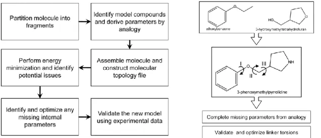
In addition, parametrization of the CHARMM force field consists of obtaining appropriate intramolecular (bond, angle, dihedral, Urey-Bradley and improper, or “out-of-plane”, terms), vdW, and electrostatic parameters that adequately reproduce the selected target data. Determination of the electrostatic parameters differs between the additive and the Drude polarizable force fields in that the polarizabilities and Thole factors need to be optimized in addition to the point charges in the additive force field. Here, we describe the strategies behind the various steps depicted in figure 6 that are involved in parameter optimization for the additive and Drude polarizable force fields.
23 Figure 6: simplified parametrization flow chart.
As was suggested earlier, parametrization is a tedious process that directly affects the outcome of subsequent usage. It is therefore a requirement that this task be performed in a careful and consistent fashion. The development of an accurate electrostatic model is one of the more tedious steps in the parametrization workflow, laying the foundation for the model to reproduce the proper electronic response as a function of environment. In the additive CHARMM force field optimization of point charges is based on a supramolecular approach where the charges are adjusted to reproduce QM HF/6-31G(d) interaction energies and geometries of the model compound with, typically, individual water molecules as illustrated in figure 7
Placement of water molecules at different orientations around the molecule has the distinct advantage of implicitly accounting for local electronic polarization, a desirable feature for accurate reproduction
Figure 7 Orientation of test water molecules around the polarizable atom of interest exemplified through ethanethiol. Carbon is in cyan, sulfur in yellow and LP in blue. “120” is probing for interactions along the S-LP axis. “180” is oriented towards the C–S bond. “BIS” water points at the bisection of the C-S-H valence angle. “PR” is pointed directly at the sulfur proton and along the S–H bond.
24 of condensed phase properties. In addition, interactions with water are often supplemented with dimers of the model compounds and with dipole moments of the models, with the dipole moments overestimated to account for condensed phase effects [28] [29].
The CHARMM27r [30] lipid force field has been used for the study of biological systems involving the lipid bilayer. Recent publications include, for example, elucidation of the free energy profile and barriers for cholesterol flip-flops in three different types of lipid environments [31], simulation of a peptide in lipid bilayers [32] ,to better understand the dynamics of peptides as a function of membrane type and thickness, and exploration of the mechanism behind which a serine protease is anchored to a bilayer [33]. Success of these studies is associated, in part, with revisions of the original CHARMM27 force field. In the revision parameters for the aliphatic tails were optimized using high level QM calculations as target data to yield better agreements with NMR order parameters for the aliphatic tails. However, one major flaw exists in the CHARMM27r version where positive surface tension of the bilayer results in shrinking of the membrane to a gel-phase state well above the transition temperature. This flaw has been recently addressed by Klauda et al. [34] where headgroup torsions parameters and LJ and partial atomic charges of the ester linkage were re-optimized to yield the CHARMM36 lipid force field. This version reproduces experimental surface areas for saturated and unsaturated lipid bilayers, which prevents unwarranted shrinking of the lipid bilayer. One issue that remains to be addressed is that of the treatment of long-range LJ interactions, which, as discussed by the authors of the CHARMM36 article, is different for monolayers with respect to bilayers, the latter being correctly treated with standard truncation methods common to all simulation programs. Accordingly, the CHARMM36 version of the lipid force field is suitable for the majority of the current research interests involving lipids including heterogeneous systems involving bilayers. An important utility of empirical force fields is to aid in the design and development of new therapeutic agents. Because of the extent of structural and chemical diversity in drug molecules, parameters geared towards transferability across a wide range of compounds are required from the computational scientific communities. Fulfilling this purpose are specialized force field parameters including MMFF [35], AMBER GAFF [36], CVFF [37], COMPASS [38], and the commercial version of CHARMm [39]. These force fields are appropriate, for example, for methods that identify biologically active or “lead” compounds from a large database of small molecules as well as computer-based de novo molecular design of drug-like molecules. However, it should be emphasized that reliable calculations such as those required for target-based structure optimization of lead compounds require the use of a well-balanced force field that properly treats both the drug and target (e.g. protein) molecules. In addition, to accurately capture the subtle differences across a series of test compounds it is generally necessary that at least some level of parameter optimization of the
25 molecules of interest be performed, due to the inherent limited transferability of parameters within a force field. With sufficient optimization, a force field can provide superior accuracy in conformational sampling and energy evaluations, leading to improved results from a variety of techniques such as 4D-QSAR [40], conformationally sampled pharmacophore (CSP) [41] and related methods, as well as free energy perturbation calculations [42]. The increasing need for computational analysis of drug-like molecules was the driving force behind the development of the CHARMM General Force field (CGenFF). Over 100 common and medicinally useful molecular scaffolds and linkers have been formally added to the existing collection of model compounds in the CHARMM force field. From this pool of model compounds, researchers can intuitively assemble compounds of interest, followed by evaluation of the quality of the resulting model and optimization of selected parameters to improve the accuracy of the treatment of the molecule of interest. Typically, one would only need to optimize the linker regions between ring systems. A summary of the steps involved in creating a new molecule is shown in Figure 8.
Figure 8: Flowchart for the determination of parameters for drug-like molecules as described in reference 16. 3-phenoxymethylpyrrolidine is assembled from two of its constituents, ethoxybenzene and 3-hydroxymethyltetrahydrofuran, available from the CGenFF. Parameters were identified for the pyrrolidine group by analogy. Optimization of the dihedrals I, II, and III is required to produce an accurate computational model for this molecule.
CGenFF was conceived using a methodology consistent with the parametrization philosophy behind the biological CHARMM additive force field. As a result, CGenFF is fully compatible with the biological portions of the force field, which makes possible simulations of heterogeneous systems such as protein-ligand interactions. This concept is similar to the philosophy used in OPLS [43] [44]. For complex systems or cases where a large number of molecules need to be studied, scripts that generate initial guesses of the atom type and parameters are available on the MacKerell group web
26 page (http://mackerell.umaryland.edu/) and efforts are ongoing with respect to the development of a web server that automatically assigns atom types, charges and parameters (https://www.paramchem.org/). The ultimate goal of that server will be the ability to validate and optimize selected parameters, a capability that is anticipated to facilitate the expansion of CGenFF to cover a larger range of chemical space. The availability of CGenFF will likely spur additional interest in computer-aided drug design using the CHARMM force field.
3.2 ATOMISTIC MOLECULAR DYNAMICS SIMULATION OF BILAYER SYSTEMS
Over the last few decades, among the other computational techniques that have emerged in Science, the atomistic Molecular Dynamics (MD) simulation have been extensively implemented and used from scientists all over the globe to obtain insight with atomic detail of steady and dynamic properties of lipid bilayers based supramolecular systems. In this regard, a critical aspect that must be identified in all the MD simulations is related to the time and length scale of process observed, the ensamble used, the pressure and temperature control. However, the amount of works on simulations of lipid membranes has achieved higher accuracy and prediction capabilities; the scale of systems that can be studied continues to increase with the increasing of the computer power and with the improvement of the MD simulation software. At present it can be identified a general limit on simulations which can be up to 5000 lipids in full atoms MD simulations and up to 50000 in coarse grained models. Concerning simulation time length, it must be pointed out that the equilibrium motions in lipid bilayers range from picosecond time scales (rattling of individual lipid hydrocarbon tails) to over many microseconds (collective motion of all lipids, m size dimension). Those motions can be investigated experimentally by means of spectroscopy methods (the former) and microscopy (the latter). Actually, hundreds of ns have become routinely for full atoms MD in bilayers, but if a further extension of the simulation time lengths is required, it must be necessary to refer to the coarse grained approach (up to ms-s time length). Usually, periodic boundary conditions (PBC) are able to avoid strong artifacts from presence of boundary planes, and this way a stack of bilayers with infinite dimension is simulated. Finally, a particular attention in simulations must be taken to identify the equilibration time to reach a steady state. To this purpose, Porasso et al. studied lipid bilayers with different composition in their liquid crystalline phase in order to establish a general criterion to identify the reaching of such a state. In particular they studies the dynamical temporal evolution of some lipids properties (i.e. area per lipid, the deuterium order parameter, the lipid hydration and lipid salt coordination) and they observed that the time required depends strictly on the single property studied. It has been found out the following order from faster equilibration property to slower: