DOLOMITE FORMATION BY NANOCRYSTAL AGGREGATION IN THE DOLOMIA PRINCIPALE OF THE BRENTA DOLOMITES (NORTHERN ITALY)
PATRICK MEISTER1 & SILVIA FRISIA2
1Department of Geodynamics and Sedimentology, Althanstr. 14, 1090 Vienna, Austria; email: [email protected] 2School of Environmental and Life Sciences, The University of Newcastle, Callaghan, New South Wales, 2308, Australia
To cite this article: Meister P. & Frisia S. (2019) - Dolomite formation by nanocrystal aggregation in the Dolomia Principale of the Brenta Dolomites (Northern Italy). Riv. It. Paleontol. Strat., 125(1): 183-196.
Abstract. Upper intra- to supratidal laminites in the Norian Dolomia Principale of the Brenta Dolomites
(Northern Italy) commonly consist of aphanitic and partially peloidal dolomite previously interpreted as very early dia-genetic or penecontemporaneous. Re-examination of a sample prepared by focused ion beam milling through high-re-solution transmission electron microscopy (TEM) revealed that dolomicrite crystals may consist of ca. 5-nm-sized na-nocrystals characterized by imperfectly aligned orientation. A similar type of dolomite was found in clay-rich Carnian laminites of the Travenanzes Formation (Venetian Alps, Northern Italy) and was interpreted as indication of primary precipitation. The observation of nanocrystals in both the Dolomia Principale and the Travenanzes Formation allows hypothesizing that dolomite mud formed directly from solution via a non-classical pathway involving nucleation and aggregation of nano-particles. Domains of nanocrystalline dolomite in the Dolomia Principale are embedded within, or cemented by, calcian dolomite showing coherent lattice at the micrometre scale and a modulated structure under the TEM. This new finding provides the first evidence that one of the largest dolomite bodies occurring in the geological record, the Dolomia Principale, commenced with deposition of sediment, consisting of dolomite mud formed from solution via non-classical crystallization, and was partially affected by later pervasive diagenetic dolomitization. Based on these new observations we propose that further nano-scale studies are necessary to substantiate the hypothesis that the formation of large dolomite bodies characterized by abundant dolomicrite may have commenced by non-classical nucleation and growth processes. This approach may provide insight on non-actualistic conditions in ancient envi-ronments that may have differed, in their boundary conditions, from their modern analogues.
Received: December 18, 2018; accepted: February 22, 2019
Keywords: Carbonate factory; Dolomia Principale; dolomite; mudstone; nanocrystals; non-classical pathway of nucleation.
I
ntroductIonThe origin of large bodies of dolomite ob-served in the geological record, such as the Norian Dolomia Principale (DP) of the Southern Alps of Italy, to date remains controversial. The so-called “dolomite problem” is both a matter of anecdo-tal “truisms” and geochemistry. First, the idea that dolomite abundance increases relative to limestone with increasing Phanerozoic age (Chilingar 1956), was challenged by Given and Wilkinson (1987), and, what appears to be true is that dolomite abun-dance increases in periods of high sea level (Morse
& Mackenzie 1990), suggesting that dolomite did not become more abundant with “age”. Second, because dolomite does not precipitate from seawa-ter due to a kinetic barrier, metastable phases, arag-onite, Mg-calcite and calcite, are deposited, but it would require 650 m3 of seawater to supply enough Mg to replace 1 m3 of metastable Ca-carbonate by dolomite during burial diagenesis (Machel 2004). Large-scale pervasive dolomitization by convective seawater flow was proposed for modern platforms, such as the Bahamas (Davans & Swart 1988; Budd 1997) and assumed, along with reflux dolomitiza-tion (Adams & Rhodes 1960), to be responsible for the alteration of ancient platform calcium car-bonate minerals into dolomite (e.g. Machel 2004).
The Norian of the Tethys, including the Dolomia Principale, stands out in relation to many other pervasively dolomitized platforms because of its wide geographic extent, which spans from Spain to Hungary, Greece and all the way to Kar-akorum (Marcoux et al. 1993; Frisia 1994), which were, then, part of the Tethys, while outside the Te-thys realm dolomite was not abundant (cf. Given & Wilkinson 1987). The Dolomia Principale consist of completely dolomitized shallow platform facies ranging in thickness from 300 to over 1000 metres thick dissected by up to 2000 m thick partially to completely dolomitized intraplatform basin facies (Jadoul 1985). Dolomitization during early to burial
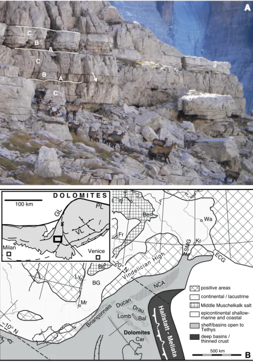
diagenesis was inferred from investigations of the Dolomite mountains, Apennines and Transdanubi-an case-studies (Frisia 1994; ITransdanubi-annace & Frisia 1994; Haas & Demeny 2002; Mastandrea et al. 2006; Meister et al. 2013; Haas et al. 2015; etc.). The first investigation of a pervasively dolomitized carbonate platform from outcrop- to micrometre-scale was carried out for the Dolomia Principale succession outcropping in the Brenta Dolomites of NE Italy (Frisia & Wenk 1993). This approach allowed dis-tinction of 6 different types of dolomite suggesting that dolomitization involved progressive changes in crystal size, stoichiometry and order. Indication that the dolomitization of the DP was mostly diagenetic Fig. 1 - A) Outcrop photograph of the Dolomia Principale lo-wer peritidal member near Rifugio Alimonta, along the track leading to Rifugio Brentei showing predomi-nant well-bedded dolomites. The thin-bedded intervals pertain to the inter- to su-pratidal facies containing the Loferites (horizons A and B); the thick-bedded inter-vals represent the subtidal facies (horizon C). A teepee structure is indicated by the arrow. B) Palaeogeographic map of the Southern Alpi-ne to Germanic domains during the middle Triassic, reproduced from Brack et al. (1999; modified). Bal: Ba-laton; BG: Burgundy Gate; Car: Carnian Alps; ECG: eastern Carpathian Gate; Lomb: Lombardy; NCA: Northern Calcareous Alps; SMG: Silesian Moravian Gate. Inset: Tectonic map of the Southern Alps (Brack et al., 1996, modified) showing the location of the Brenta Dolomites. GL: Giudicarie Line; PL: Pustertal Line; VL: Val Sugana Line.
was the observed replacement of bioclasts, such as dasycladacean algae or megalodont shells by dolo-mite, which suggested a large-scale overprint (Frisia 1994). In contrast, laminated structures, which were interpreted as microbial laminites (or Loferites; Fis-cher 1964) of the upper intertidal to supratidal fa-cies, representing the shallowest part the peritidal cycles, show aphanitic dolomite (type 1 in Frisia & Wenk 1993) of possible early, or even penecontem-poraneous, origin (i.e. dolomite that formed during or shortly after sedimentation; Frisia & Wenk 1993). Type 1 aphanitic dolomite was observed in laminae with syneresis or desiccation cracks alternating with cement, filling laminar, fenestral pores. Frisia (1994) reinforced the concept of penecontemporaneous replacement for type 1 aphanitic dolomite on the basis of its low density of growth defects and ox-ygen isotope values indicative of precipitation at surface temperatures from seawater that was only slightly modified by evaporation (Iannace & Frisia 1994). Therefore, it seemed reasonable to infer that aphanitic dolomite formation may have been tied to specific upper Triassic palaeo-environmental condi-tions.
Wenk et al. (1993), Frisia and Wenk (1993) and Frisia (1994) were the first to combine scanning electron microscopy (SEM), transmission electron microscopy (TEM) and high-resolution TEM with field-scale and microfacies analysis, whereas pre-vious microstructural investigations of dolomite were carried out on a purely crystallographic basis, focused on understanding how cation ordering is achieved and on the significance of microstructural defects (Reeder 1981; Wenk et al. 1983). This com-bined approach allowed them to interpret ancient laminated aphanitic dolomites by referring to mod-ern analogues. Frisia (1994) could, thus, conclude that dolomicrite of type 1 in the DP, which is char-acterized by dislocations, defect-free regions and incipient, fine-modulated structures, was a mimetic replacement of a precursor Mg-rich carbonate or aragonite phase. Grain-pervasive modulated struc-tures at the micrometre scale is typically observed in diagenetic Ca-dolomites replacing precursor arago-nite or Mg-calcite. The modulated microstructure is a heterogeneous structural defect with a wavelength in the range of 20 to 30 nm (Fig. 4 in Frisia & Wenk 1993) that was first observed by Reeder et al. (1978) using a TEM. It still remains an enigmatic struc-ture that appears as regular “waves” of darker and
lighter lamellae with parallel orientation, which are commonly parallel to the cleavage rhombohedron. It is, therefore, understandable why Frisia and Wenk (1993) and Frisia (1994) ascribed dolomicrite and dolomicrosparite in the DP to a diagenetic over-print.
Frisia and Wenk’s (1993) conclusion that all dolomite in the Triassic DP was diagenetic in or-igin was put into question by the work of Preto et al. (2015), who reported nanocrystal dolomite aggregates as seen by TEM in aphanitic dolomite laminae, interlayered with fine clay in the Carnian Travenanzes Formation (Fm.) of the Dolomites of Veneto (NE Italy). The Travenanzes Fm. immedi-ately pre-dates the Dolomia Principale (it is known as Raibl in the Brenta Dolomites and outcrops just below the DP) and documents a more continen-tal, proximal sabkha/alluvial plain environment (Breda & Preto 2011) compared to that of the DP (Frisia 1994). The nanometre-scale dolomite crys-tals of the Travenanzes Fm. were identified as the same defect-ridden crystals, referred to as mottled structures in recent dolomites from Deep Springs Lake (California) by Reeder (1981) and in Holo-cene Abu Dhabi sabkha dolomites by Wenk et al. (1993), and thus opened the possibility for the in-terpretation that, in the Triassic, some dolomite may have formed as a primary phase, where “primary” indicated that precipitation occurred directly from solution, without a precursor phase (Preto et al. 2015). Furthermore, the presence of nanocrystals suggested that in the Travenanzes Fm., some dolo-mite may have formed via a non-classical pathway by nano-particle attachment (Preto et al. 2015). This interpretation accounted for experimental studies demonstrating that dolomite forms inorganically via non-classical nucleation pathways (Rodriguez-Blan-co et al. 2015). Non-classical processes deviate from the classical nucleation and growth theory, e.g. given in Sunagawa (2005), as thermodynamic and kinetic barriers are fundamentally different if particles are nanometre-scale. Crucially, similar conclusions were reached by observing authigenic Ca-Mg-carbonates at the nanoscale, including dolomite that formed in deep-sea methane seeps (Lu et al. 2018). It became, then, evident that the dolomitization of the DP of the Brenta Dolomites needed to be re-evaluated in the light of non-classical crystallization pathways. It is noteworthy that non-classical nucleation and growth pathways, regardless to the intervention of
bio-mediation (Rodriguez-Blanco et al. 2015), fun-damentally change our understanding of mineral formation processes in modern and ancient envi-ronments.
On the basis of the discovery of nanocrystal aggregates in the Travenanzes Fm., we here revisit the aphanitic laminae in Loferites from the peritid-al cycles of the Dolomia Principperitid-ale of the Brenta Dolomites (NE Italy), which were investigated by Frisia and Wenk (1993) and Frisia (1994). We use high-resolution transmission electron microscopy (HR-TEM) in combination with sample preparation by focused ion beam (FIB) milling on targeted DP laminites. The Dolomia Principale has complex faci-es associations, and it was so huge that one facifaci-es is obviously not representative for dolomitization of the entire platform across space and time. Yet, the sample analysed here is representative of aphanitic dolomite in peritidal laminated facies, with overar-ching consequences for our understanding of early, or “primary” dolomite formation in an environ-ment where the current preferred hypothesis is a an early diagenetic origin, perhaps influenced through bio-mediation (Mastandrea et al. 2006; Bontognali et al. 2010). With respect to the Travenanzes Fm., the lower member of the DP in both the Brenta Dolomites and Venetian region documents a tran-sition from continental proximal clay-dominated (Carnian Raibl Group or Travenanzes Fm.) to more distal shallow-water carbonate platform environ-ments, free of siliciclastic input (Frisia 1994). Thus, the present study had the ambitious aim to provide evidence that the earliest stages of dolomite crys-tallization are also preserved in the DP, where per-vasive recrystallization could not have been inhib-ited by large amounts of clay. By considering that Frisia et al. (2018) indicated that organic molecules in all settings, both continental and marine, may facilitate the preservation of unstable phases, we revise the interpretation of the DP multi-step dol-omitization (Frisia & Wenk 1993), with the goal of finding evidence of the primary dolomite phase in the sequence of stages leading to the final massive dolostone.
Methods
The new observations have been carried out in a sample collected in 2017 from the ca. 300-m-thick lower member of the Dolomia Principale in the Brenta Dolomites near Rifugio Alimonta
(Fig. 1). In this area, the Dolomia Principale is ca. 1000 m thick and consists of a lower peritidal member, overlain by a predominantly subtidal member and capped by a third, peritidal member (Frisia 1994). The sample was taken from a ca. 30-cm-thick bed referred to as “Loferites” (interval B in Fisher 1964; see also Bosellini 1967) and labelled LC1-facies in Frisia and Wenk (1993) and Frisia (1994), within ~1-m-thick peritidal cycles (Fig. 1). The laminated portion of the sample, anectdotically ascribed to stromatolites, was cut to obtain from the same block, both a thin section and a double polished wafer from which a sample characterized by electron transparency could be cut by focused ion beam (FIB) milling. The thin section and the wafer were microstratigraphically correlated using images obtained by a high-resolution scanner.
Electron transparent foils were prepared from a specific area of microsparitic and aphanitic dolomite, in the LC1 facies, using FIB. The foils were milled perpendicular to the polished specimen, on both sides of a 2 micrometre-long section in the area of interest. FIB milling was carried out at the Advanced Microscopy Facility of Bio21 (University of Melbourne) using a FEI Technai F30 at 30 kV. The total thickness of the milled slide was about 500 nanometres. Subsequent polishing was carried out at 5 kV, to minimize potential damage to the sample. The final thickness attained by FIB was slight-ly less than 100 nanometres. FIB has the advantage that it uses Ga rather than Ar, which penetrates less into the specimen.
The foils were analysed with a JEOL 2100 LaB6 TEM op-erating at 200 kV at the Electron Microscopy and X-Ray Unit of the University of Newcastle (Australia). The JEOL 2100 LaB6 TEM allows for lattice imaging. It is equipped with an EDS-system that en-abled us to obtain chemical microanalysis of the area of interest after having taken the images (image mode), and thus matching the crystal structural imperfections at lattice resolution with the chemical data. Care was taken not to induce artefacts in the samples during TEM observations. To minimize artefacts from radiation damage (see for details Frisia et al. 2018) we used a small aperture, and kept the resi-dence time of the beam on the analysed spot to a minimum, checking the selected area diffraction pattern (SAD) every few seconds: if the SAD pattern changed from a typical pattern of a single crystal (dots) to one of a polycrystal (dots arranged in rings) after a few minutes, we knew that radiation damage occurred. If the pattern showed a series of dots arranged along rings (polycrystals) to start with, then this was clearly a primary feature and not due to radiation damage. In all cases, we noticed that radiation damage started only after 5 to 10 minutes. Furthermore, it is known that FIB may induce amorphous layers at the surface of the foil. Amorphous phases would show in the SAD as rings, with no diffraction dots. We did not notice any such feature, and, thus, we are confident that the beam penetrated through the amorphous layer and what we documented are real crys-tal structural imperfections of the dolomite sample.
The first step in our observations was to image the foil at 20,000x magnification, which was the most common magnifica-tion used by Frisia (1994). Then we increased the magnificamagnifica-tion to 600,000x for both dislocation-ridden regions and those described by Frisia and Wenk (1993) and Frisia (1994) as characterized by modu-lated structure. At 600.000x magnification we also performed lattice imaging.
r
esultsIn the outcrop, macroscopic observations re-veal that laminae range from 1 to 2 mm in thickness and seem to be relatively flat towards the subtidal
portion of the cycle, whereas towards the top they become undulated, bending upward. Where the laminae form these small folds (3-4 mm in height), elongated fenestral cavities filled by cement are more common. In this specific LC1-facies, where the sample was collected, we observed syneresis and desiccation cracks.
Under the petrographic microscope, a lami-nation is visible at the cm-scale, consisting primarily of lighter and darker dolomicrite (Fig. 2). Some of the light, thin laminae near the top may also repre-sent cemented crusts. One layer (arrow in the fig-ure) shows an erosion surface characterized by rip-up clasts of darker material embedded in a lighter matrix of dolomicrosparite in the layer above. The reworked clasts are round and elongated. Peloids can be recognized in some of the laminae in this section. These peloids are near to spherical and their size varies from layer to layer. They consist of homogeneous micrite and do not show any radial
or concentric crystal orientation. In most cases, the peloids are embedded in a micritic matrix, but they also may be densely packed to an extent that the grains are almost entirely amalgamated, whereby the interstices, or what is left of them, may still be visi-ble as infill of sparitic cement. Some laminae show a coarser, microsparitic structure, but it remains un-clear whether the microsparite represents a primary cement or a diagenetic overgrowth. Elongate fenes-trae in the thick layer in the middle show an S-shape deformation (left of “Fe” in Fig. 2). Larger scale pores showing isopachous cement rims occur in the lower half of the slide (labelled with (3); Fig. 2).
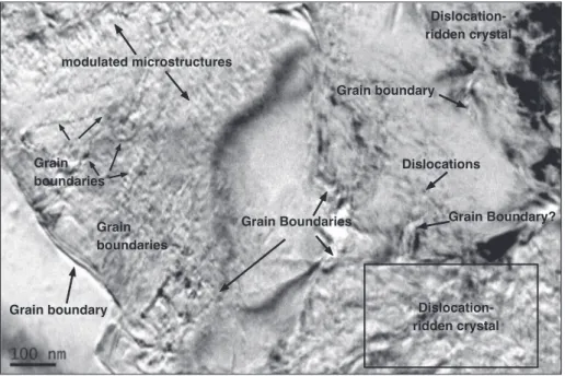
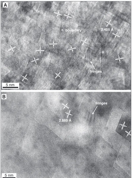
TEM observations of the targeted aphanit-ic dolomite sample, extracted from within the area outlined in Fig. 2 by a yellow circle within black pen marking, show both dislocation-ridden regions and grains with modulated structure in the low-resolu-tion overview image (magnificalow-resolu-tion 50,000x; Fig. 3). The dislocation-ridden crystal (area outlined by the rectangular frame in Fig. 3) shows heterogeneous mottled contrast, which has been previously inter-preted as the result of high density of growth de-fects (Wenk et al. 1980; Frisia & Wenk 1993). The presence of fringes in the 50,000x image highlight the presence of very small grains whose orientation is slightly different than that of the adjacent grain. At 600,000x magnification (Fig. 4A) the same re-gion is shown to consist of an aggregate of nano-crystals. The dominant d-spacing is 2.405 Å, that would coincide with the dolomite {1120} direction (Reeder & Wenk 1983). Figure 4A shows that there is some mismatch in the orientation of the grains as determined by the directions of {1120}, which is evident by fringes (highlighted by an arrow in Fig. 4A) that can be interpreted as a difference in thick-ness of the slide at the margin between adjacent grains, or a rotation of one grain with respect to the next. No sulphur or carbon enrichment was detect-ed at boundaries between nanocrystals that could bridge nanocrystals with diverse lattice orientations. In addition, we observed rod-like features, but only in the region showing mottled contrast, which was confirmed to arise from an aggregate of nanocrys-tals. Critically, the lattice spacing and direction does not change across the “rods” (asterisk in Fig. 4A).
When we moved our observations to a differ-ent spot within the investigated area, a modulated structure was observed (Fig. 3). At 600,000x (Fig. 4B) the lattice is coherent, meaning that it does not Fig. 2 - Photomicrograph of the same facies as in Fig. 1 (LC1) in a
sample taken in 2017 near Rifugio Alimonta in the Brenta Dolomites. One can see (1) aphanitic dolomite (which has peloids), (2) microsparite with peloids, (3) void filling. Ar-row indicates an erosion surface and rip up clasts (peloids) of darker material embedded in a lighter matrix in the layer above. Elongate fenestrae are indicated with Fe. The pen mark indicates the location of analysis. The width of the thin section is 3 cm.
show mismatch across the whole imaged grain. The observed dominant d-spacing in this specific grain is 2.88 Å, which coincides with the {1014} lattice spacing. Critically, all previous literature reports that the modulated structure in dolomite is parallel to {1014} or nearly so (see Wenk et al. 1983). Our TEM observations show the presence of fringes, which suggest possible subtleties in thickness due to orientation, but these may also be due to uneven milling.
d
IscussIonNanocrystal Structures of Primary and Replacement Dolomite
New HR-TEM investigation of L1C-faci-es aphanitic dolomite revealed two different typL1C-faci-es of nanometre-scale defects: nanocrystal aggregates that are observed by TEM as mottled contrast and modulated micro-structure in dolomite with a co-herent lattice. Wenk et al. (1993) and Frisia (1994) documented crystals with mottled contrast in mod-ern dolomites from the Abu Dhabi sabkha, using a PIP system to obtain electron transparency (ion beam thinning of a detached thin section portion mounted on a copper grid 3 mm in diameter). Now, by using FIB and carrying out observations with HR-TEM we demonstrate that the mottled contrast observed in aphanitic dolomite of the L1C-facies of the DP is due to nanocrystal aggregates. Cru-cially, having observed the same mottled contrast
in specimens prepared with different techniques in rock and in unlithified sediment, we are confident that the crystal defects we report here for the DP are not due to deformation or to technical artefacts. Preto et al. (2015) and Lu et al. (2018) suggested that the presence of nanocrystal aggregates in do-lomite grains is indicative of their primary origin. Even if we cannot unequivocally prove that dolo-mite nanocrystals were the first phase that formed directly from the solution, we can nevertheless con-clude that finding a nanocrystalline structure in the DP is either a primary feature or the product of a rapid transformation of an amorphous or poor-ly ordered precursor into dolomite that occurred pre-attachment (cf. Rodriguez-Blanco et al. 2015).
A nanocrystal structure is expected to be thermodynamically metastable, due to the small grain size, and would be inevitably subject to a grain coarsening upon replacement (Ostwald ripening). As an example, in the Quaternary Abu Dhabi sab-kha sediments, Frisia (1994) documented micro-metre-sized Ca-rich rhombohedra with modulated structures enclosing sub-micrometre (hundreds of nanometre) rhombohedra, with ideal composition and characterized by dislocations. Frisia (1994) in-terpreted the Ca-rich domains as indicative of a secondary phase replacing either aragonite and/or the dislocation-ridden dolomite. The observation that dolomite showing a modulated structure in the DP has a coherent lattice at high magnification sup-ports the interpretation that it represents a second-ary phase and, most likely, a product of Ostwald
Fig. 3 - TEM image of FIB foil obtained from dolomicrite peloid from DP LC1 facies at 50,000x magnification (base of photograph is circa 0.5 micrometres), showing two types of crystal struc-ture. A dislocation-ridden structure (frame) is similar to the mottled structure obser-ved by Wenk et al. (1993) in Abu Dhabi sabkha dolomi-te. A structure with uniform orientation and modulated nano-structure is observed on the upper left. Grain boundaries can be recogni-zed by fringes due to interfe-rences that form if the grain boundary is not perpendicu-lar to the slide plain.
ripening. The close association of dolomite with modulated microstructure and ideal dolomite with “mottled” contrast in the DP may indicate that the former may be a very early product of diagenesis. Hence, we have documented the preservation of nanocrystal aggregates as a possible precursor of dolomite with modulated structure in the DP, a rock that is known to have been subjected to multi-step diagenesis.
A Pathway of nearly oriented Attachment of Nano-Particles
The observation of nanocrystals in dolo-mites from different sites (Preto et al. 2015; Lu et al. 2018; and the results presented here) supports
the hypothesis that a primary dolomite phase may form under Earth surface conditions and that na-nocrystal aggregation is one of the crystallization pathways. These new findings also support the no-tion that mottled contrast in Mg-calcite and dolo-mite observed in Holocene sediments results from high density of boundaries bordering nanocrystals with a slight misorientation. This is different from what was proposed by Wenk et al. (1983) who inter-preted mottled contrast as reflecting heterogeneous microstructures “possibly including dislocations, faults and subgrain boundaries” related to compo-sitional domains (for example more Ca in one re-gion and more Mg in another; Reeder 1981; Wenk et al. 1983). Since we did not find any difference in Fig. 4 - A) Close up of framed area in
Fig. 3 at 600,000x magnifica-tion. The light and dark con-trasts are nanocrystals with diverse orientations (as indi-cated). The mismatch of the nanocrystal lattices seems to be accommodated by an amorphous phase, for which we have no data. In addition to lattice mismatch, there are some features that cannot be easily explained, such as a rod-like feature [*] and lat-tice fringes. B) TEM image of microsparite with modu-lated nano-structure similar to the one shown in Fig. 3 at 600,000x magnification. The crystals show almost perfectly aligned crystal orientation. Fringes suggest that there are still some dif-ferences in thickness, but these could be due to sample preparation.
chemical composition at the nanoscale, we cannot support the notion that variations in lattice orien-tations are in any way related to compositional var-iations, such as in the Mg-content. The interpreta-tion of Wenk et al (1983) was that recent dolomites with mottled contrast were single grains character-ized by areas with accumulation of dislocations due to slight lattice misorientations in some parts that resulted in the formation of sub-grains. Subgrain boundaries should develop through deformation and accumulation of stress/strain in certain regions of the grain and are commonly observed to develop in deep ice cores where ice grains underwent stress (Weikusat et al. 2009). However, this would be hard-ly the case for recent dolomites that were not deephard-ly buried. Here, we demonstrated that dolomite with mottled contrast, rather than being a single micro-metre-sized grain with subgrain boundaries, may consist of nanocrystals, whose lattice is mismatched relative to adjacent nanocrystals.
Based on the orientation distribution in the nanocrystals, aggregation occurred through a near-ly oriented attachment pathway, which is one pos-sible pathway among several ones suggested by De Yoreo et al. (2015; Fig. 1 therein). The reason why a nano-particulate (non-classical) pathway is preferred is readily understandable from a kinetic point of view. Thermodynamic and kinetic barriers strongly depend on the distribution of nano-struc-tures (also referred to as the “energy landscape”; De Yoreo et al. 2015) and generally are much lower for small particles, which is not accounted for in the classical nucleation theory. Since dolomite is noto-rious for its high kinetic barrier (e.g. Land 1998), a nucleation via a nano-particulate (metastable) phase may provide a preferred route to its primary for-mation. This behaviour is not surprising and simply adheres to Ostwald’s step rule, according to which a metastable phase with the lower kinetic barrier forms first.
Nanocrystals showing imperfectly oriented attachment with considerable mismatch are well known from experimental studies: they still may ex-hibit a long-distance ordering, giving rise to crystal-lographically ordered mesocrystals, as demonstrated by Cölfen and Antonietti (2005, 2008). Imperfectly attached crystals may then spontaneously rearrange over time and fuse together to form a coherent lat-tice. This would be consistent with the observations of non-evenly distributed Mg-Ca compositional
domains by Wenk et al. (1983) and Lu et al. (2018) and with the passage from oriented attachment of the particles to ordered crystals. In the sample an-alysed from the DP, we observed both nanocrystal aggregates and dolomite with coherent lattice and modulated structure, which suggests progressive ordering. In any case, the finding of nearly oriented attachment in the DP provides a new basis for the interpretation of similar dolomite bodies in the ge-ologic record.
Drivers of Nanocrystal Formation and Aggregation in a Triassic Platform Environment
The sequence in which a nanocrystalline do-lomite phase forms first, either directly or via a pre-cursor amorphous or poorly ordered Mg/Ca-car-bonate phase, then aggregates to form mesocrystals and subsequently fuses to a coherent lattice, is as observed in the experiment of Rodriguez-Blanco (2015) but also complies with Ostwald’s step rule, which predicts the formation of metastable phases at high supersaturation and ripening to the stable phase at lower supersaturation. A scenario involv-ing such conditions has been suggested by Deel-man (1999; 2001), who reported the successful precipitation of dolomite in the laboratory under pH fluctuating between 5 and 9. Deelman’s experi-ments essentially substantiated Ostwald’s step rule, as at high pH metastable carbonate should form due to high supersaturation, while at low pH the metastable phase is undersaturated and ripens to ordered dolomite. While Deelman’s experiments help understanding dolomite formation, we suggest a slightly modified interpretation, which considers a non-classical multistage crystallization pathway, such as the one documented here. Nano-particles differ in their energy landscape from macroscopic crystals, often favouring aggregation-based crys-tallization as opposed to a layer-by-layer growth of single crystals proposed in the classical theory (De Yoreo et al. 2015). As a result of lower energy barriers, subtle fluctuations, e.g. in pH, ionic com-position, and supersaturation in the parent solution may cause spontaneous nucleation of dolomite na-nocrystals and their ripening. While the range of pH variations in Deelman’s experiments may be questioned as not realistic for shallow marine or pe-ritidal environments of deposition, smaller fluctua-tions due to tidal or seasonal rainfall variafluctua-tions may have been sufficient to influence pathways of
do-lomite formation at a nano-particle level. Meteoric water today has a pH of around 7, while seawater is around 8.1, but it could have reached even high-er values in the Tethys due to enhanced continen-tal weathering under a high pCO2 atmosphere (see discussion below). Thus, supersaturation may not have been high enough to cause the precipitation of amorphous calcium carbonate (as in the experi-ments of Rodriguez-Blanco 2015, and Purgstaller et al. 2015), but high enough to precipitate a nanocrys-talline dolomite phase prior to attachment.
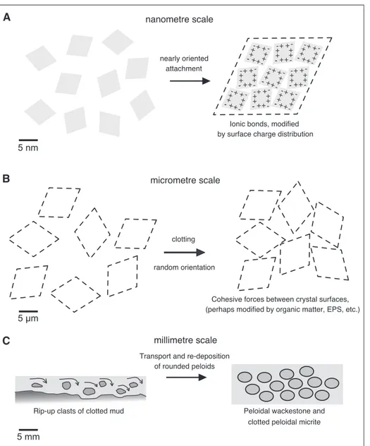
If nanocrystals could nucleate at high pH and high supersaturation, a lowering of the pH would not only lead to a ripening of the nano-particles (in case they formed from a precursor phase), but may have also affected aggregation of the nanocrystals. Particles have a surface charge (zeta-potential), and Ca/Mg-carbonate particles are known to remain in suspension at high pH due to the repulsive forces of the negatively charged surfaces. This has been observed, for example, in Lake Neusiedl (Austria) and adjacent alkaline ponds, where modern formation of fine mud of high-Mg calcite and protodolomite occurs (Krachler et al. 2012; Neuhuber et al. 2015). Typically, lake waters show high turbidity at high pH, while ponds with lower pH tend to be clear. Apparently, nano-particle attachment occurs upon pH decrease, as a result of reduced zeta-potential. Likewise, pH-changes on the Norian platform may have been sufficient not only to induce precipitation dolomite at a nano-particle level and induce subsequent aggregation of the nano-particles to form mesocrystals. According to the theory of colloidal attraction and repulsion (Derjaguin - Landau - Verey - Overbeek; DVLO theory; Derjaguin & Landau 1941) two types of forces play a role, the attracting Van der Waals (dipole-dipole) forces and the repulsive electrostatic (Coulomb) forces. Thereby, charge distribution may not be homogeneous along the nano-particle surfaces, as exemplified by negative and positive charges indicated in Fig. 5A. The nearly oriented attachment observed in the lattice-scale image then could be explained by the difference in surface charge between edges and centres of the crystal faces, which would not favour an entirely oriented attachment.
Particle attachment may be modified (facili-tated or inhibited) by additional organic or inorgan-ic constituents of the aqueous solution. Colloidal
particles, such as clay minerals, could interfere with the surfaces of carbonate nano-particles, and this scenario has been suggested for the clay-rich Trav-enanzes Fm. (Frisia et al. 2018). However, the DP LC1 facies studied here, and the DP in the Brenta Dolomites in general, is depleted in clay minerals with respect to the Travenanzes, thus, aggregation by adsorption on to clay minerals may not provide a suitable model for dolomite formation in the Brenta Dolomites DP.
It is also often discussed that polymeric substances secreted by bacteria (so-called extracellular polymeric substances; EPS) are involved in the formation of dolomite (Dupraz et al. 2008; Bontognali et al. 2013). EPS may be positively or negatively charged, which could both support or inhibit the attachment of nano-particles. One problem of an EPS matrix, as it is observed to occur in microbial mats and suggested to have formed the laminites in the supratidal intervals of the Lofer cycles, would be its permeability for single molecules but not for particles. In other words, an EPS matrix would prevent the aggregation of particles for permeability reasons. In contrast, organic matter in solution could adsorb on mineral surfaces and support the attachment of nano-particles. Therefore, charged amino acids or certain carbohydrates could potentially alter the surface conditions and energy landscape and favour an aggregation of dolomite nanocrystals. Research on biomimetic minerals demonstrated that non-classical crystallization processes explain the formation of high Mg-calcite and protodolomite via attachment of nanoparticles precursors and that the presence of organics may facilitate the process (Rodríguez-Navarro et al. 2016). However, EDX analysis did not reveal any carbon enrichment at boundaries between nanocrystals. Moreover, despite a potential role of organic molecules as modifiers, imperfectly oriented attachment is also observed in pure abiotic experiments, e.g. with barium sulphate (Judat & Kind 2004; Cölfen & Antionetti 2005).
It remains to be explained as to why a ripen-ing to dolomite with coherent lattice and modulated structure occurs in one area, in the other one not. A passage from disordered Mg-calcite with mottled contrast to dolomite with coherent lattice has been attributed to the catalytic action of high levels of sulphide and extrapolymeric substances (Lu et al. 2018). Although, a sulphate reduction zone
com-monly occurs at shallow depth in modern sabkhas, especially in areas where microbial mats produce EPS (e.g. Bontognali et al. 2010), carbon isotope compositions usually do not indicate a significant effect of sulphate reduction on dolomite formation (see e.g. McKenzie et al. 1980). Likewise, in the DP of the Brenta Dolomites, carbon isotope data do not support the formation of dolomite mud (aphanitic dolomite) and its subsequent recrystallization under sulphate-reducing conditions (Frisia 1994). And as already mentioned, we did not detect any sulphur and carbon enrichments in the interstices between the nanocrystals. However, we may speculate that partial recrystallization also pertains to Ostwald’s step rule. While in the Travenanzes Fm., aphanitic dolomite is largely shielded from ambient fluids by clay minerals, microporosity in some of the coarser laminae may partially allow fluid circulation while permeability in the dense aphanitic dolomite e.g. in
the peloids is very low. Recrystallization then occurs in the more permeable domains, at times of low pH, upon lowering the ion activity product of the fluid below saturation of the metastable phase.
Origin and Microfacies of Aphanitic Dolomite
While a pathway of nanocrystal aggregation is suggested by TEM analysis and supported by independent findings in abiotic laboratory experi-ments, it remains to be discussed whether it is con-sistent with the environmental conditions and dep-ositional facies on the Upper Triassic DP platform in the Brenta Dolomites. The upper intertidal to su-pratidal L1C facies reveals fine micrite and peloidal wackestone that would be consistent with a primary formation. The peloids, one of which shows the nanocrystal structure, occur in several laminae and could have formed from already existing mud or by
A B C Figure 6 + ++ + ++ + + --- -+ -+-++ ++ ++ --- -+ -+-++ ++ ++ --- -+ -++ + ++ + + --- -+ -++ + ++ + + --- -+ -+-++ ++ ++ --- -+ -++ + ++ + + --- -+ -+-++ ++ ++ --- -+ -++ + ++ + + --- -nearly oriented attachment
Ionic bonds, modified by surface charge distribution nanometre scale
micrometre scale
clotting random orientation
millimetre scale
Rip-up clasts of clotted mud Peloidal wackestone and clotted peloidal micrite Cohesive forces between crystal surfaces, (perhaps modified by organic matter, EPS, etc.)
Transport and re-deposition of rounded peloids 5 nm
5 μm
5 mm
Fig. 5 - Conceptual model for the attachment pathway at three different size scales. A) At the nano-metre scale, chan-ges in the energy landscape may dominate the way how crystals attach. Surface char-ge effects at the edchar-ges may favour a nearly oriented attachment over an orien-ted attachment. B) At the meso-scale, a few micron scale crystals attach by sur-face charge (not ionic bon-ds). The attachment leads to “clotting” as observed in cheese or clotted cream. C) At the mm-scale, the clotted material may be reworked, and depending on the flow regime different degrees of rounding and sorting may occur. The result is what is commonly described as “clotted peloidal micrite”.
primary precipitation within an EPS matrix. Peloids may become amalgamated to form a structure also referred to as clotted peloidal micrite, a microfaci-es that is commonly associated with microbialitmicrofaci-es (Kazmierczak et al. 1996; Riding 2000). However, precipitation within an EPS matrix is not consistent with the observations of nanocrystal aggregates, for the reasons already discussed above, and it re-mains difficult to find hard criteria in the Brenta Dolomites DP that would allow for unambiguous-ly assigning an in-situ precipitation as opposed to a sedimentary deposition.
Instead, the clotting could be a result of pH decrease, reducing the negative surface charge of the phaenocrystals (or mesocrystals). In this case, micrite mud would be primary in origin and, hence, consistent with our proposed model of mud-grains consisting of nanocrystal aggregates. Further ag-gregation of phaenocrystals (or mesocrystals) at the micrometre-scale, after deposition, could occur as a result of cohesive forces as indicated in Fig. 5B. Such hierarchical aggregation is indeed evidenced by the observations at different scales (TEM vs. light microscope), whereby at the micrometre-scale orientation of the mesocrystals would be random, possibly because those crystals are too large to un-dergo spontaneous, nearly oriented attachment. In a next step, the peloids may form upon reworking of muddy sediment, in a similar way as in fresh cheese, once it is stirred (Fig. 5C). The clots are then sub-ject to any sedimentary processes dependent on the flow regime, and perhaps can also become trapped and bound at the surface of cyanobacterial mats. Erosion surfaces and rip-up clasts (arrow in Fig. 2), as well as signs of slumping (e.g. the S-shape defor-mation of fenestral pores; “Fe” in Fig. 2), indicate that these layers were still unlithified and thus per-tain to standard microfacies (SMF) 25 “laminated evaporite-carbonate mudstone” of Flügel (2010), rather than microbial bindstone. Amalgamation and deformation of peloids may support the concept that these structures were still unlithified. A Loferite facies may thus not exclusively consist of microbial bindstone, but of “a lot of mud”. Large amounts of mud occur on top and in the interior of many modern and ancient carbonate platforms (Schlager & Reijmer 2009), and this mud could be partially due to whitings, i.e. primary precipitation from near to normal seawater (Bustos-Serrano et al. 1999). In modern settings, this mud often consists of
arag-onite, but, depending on the water chemistry, also metastable Mg-Ca-carbonate or dolomite mud can form (cf. Teal et al. 2000), and primary mud pro-duction may have been even more important at times when ocean chemistry was much different from today (see below). Future TEM studies will be necessary to target the mud in non-peloidal mud-stones of the DP supratidal facies to corroborate the hypothesis that dolomite mud may actually be “primary”.
It remains then to hypothesize what process-es may have caused fluctuations in water chemistry and pH at the time of deposition of the lower mem-ber of the Dolomia Principale in what is now the Brenta Dolomites area and, thus, facilitate the for-mation of dolomite “mud”. One possibility would be as a result of late Triassic climate in the Western Tethys, which was characterized by predominantly arid conditions with strong seasonality, as demon-strated by the evaporitic facies and playa deposits that bordered the margin of the Tethys (Shuckla et al. 2006). Model reconstructions suggest that during the Norian, climate was characterized by extended dry seasons and short, intense periods of high pre-cipitation (the megamonsoons of Kutzbach and Gallimore 1989). Alkalinity and pH in the Triassic Tethys could have reached higher values, compared to the modern ocean, due to enhanced continental weathering under a high pCO2 with low Ca derived from ocean spreading activity (aragonite sea), be-fore a mid Mesozoic change in the regulation of ocean chemistry (Ridgwell 2005). During the humid season, rainwater pH may have been lower than its modern value, due to the inferred higher atmos-pheric pCO2 in the Triassic, estimated to have been 2000 ppmv (Breeker et al. 2010). Iannace and Frisia (1994) proposed an early dolomitization model for the Dolomia Principale based on “peculiar chemis-try” of the shallow sea under arid conditions, where waters were likely to be modified by intense evapo-ration and deposition of Ca-sulphates at the conti-nental margin. Yet, they did not consider large-scale changes in the water chemistry, namely sea-level changes, subsidence and seasonal changes in pre-cipitation (cf. Berra et al. 2010), as e.g. document-ed by desiccation cracks or palaeosols. Although in the early Norian the Brenta Dolomites area was not under direct continental influence, periodic intense monsoonal rainfall could have directly influenced and diluted the water chemistry on the extended
platform, particularly in periods when the Brenta Dolomite platform was close to emersion due to carbonate deposition exceeding subsidence rate. As pointed out by Berra et al. (2010) the effects of or-bital changes cannot be excluded and may have en-hanced seasonality, perhaps also the tidal influence, and these changes may have been even amplified by autocyclicity. Resolving the orbital influence is thus of great importance to understand non-actualistic conditions on ancient platforms and how they in-teracted with nucleation and growth of authigenic minerals in a non-classical way.
c
onclusIonsHere, we have documented that nanocrystal structures are preserved in laminites from the up-per Triassic Dolomia Principale of the Brenta Do-lomites. These structures provide the first evidence from a large dolomite body that dolomite crystal-lization occurred via non-classical nucleation and growth pathway by imperfectly oriented nanocrys-tal attachment. We hypothesize that fluctuating pH conditions in an upper intra- to supratidal environ-ment may have promoted the formation of dolo-mite nanocrystal aggregates. By contrast, Ca-dolo-mite with coherent lattice and modulated structures were either diagenetic cements or overprints. The preservation of nanocrystal aggregates, which should be thermodynamically instable and unlikely to survive diagenetic recrystallization, in the Dolo-mia Principale is remarkable and opens up the pos-sibility that nanocrystals may be preserved in rocks billions of years old, thus providing unique insight into earliest carbonate mineral formation pathways and, consequently, early life. The present study also indicates that atomic scale effects of surrounding solution on the nanocrystal energy landscape need to be taken into account as key towards solving the over 200 years-old dolomite problem.
Acknowledgements: S.F. wishes to acknowledge Maurizio
Gaetani, for having introduced her for the first time to the world of dolomite, with enthusiasm and passion. We acknowledge Sergey Rubanov and the Advanced Microscopy Facility Bio21 (University of Melbourne) for the preparation of the TEM sample by FIB. Huiming Zhang of the Electron Microscopy and X-Ray Unit (University of Newcastle) assisted during the TEM observations and EDS analyses. Andrea Borsato, University of Newcastle, assisted in fieldwork in the Brenta Dolomites. S.F. acknowledges the participants of the DFG Research Group, Project CHARON, for insightful discussions on crystallization. This study was partially supported by the European
Commission through Intra European Fellowship Project TRIADOL (grant no. 626025). The University of Vienna provided funding by awarding a Guest Professorship to S.F. at the Department of Geodynamics and Sedimentology, which fostered the collaboration with P.M. We thank Hans Machel and an anonymous reviewer for providing thorough reviews and constructive inputs to our study.
RefeRences
Berra F., Jadoul F. & Anelli A. (2010) - Environmental con-trol on the end of the Dolomia Principale/Hauptdolo-mit depositional system in the central Alps: Coupling sea-level and climate change. Palaeogeogr., Palaeoclimatol., Palaeoecol., 290: 138-150.
Bontognali T.R.R., Vasconcelos C., Warthmann R.J., Bernas-coni S.M., Dupraz C., Strohmenger C.J. & McKenzie J.A. (2010) - Dolomite formation within microbial mats in the coastal sabkha of Abu Dhabi (United Arab Emir-ates). Sedimentology, 57: 824-844.
Bontognali T.R.R., McKenzie J.A., Warthmann R. & Vascon-celos C. (2013) - Microbially influenced formation of Mg-calcite and Ca-dolomite in the presence of exopoly-meric substances produced by sulphate-reducing bacte-ria. Terra Nova, 0: 1-6.
Bosellini A. (1967) - La tematica deposizionale della Dolomia Principale (Dolomiti e Prealpi Venete). Boll. Soc. Geol. It., 86: 133-169.
Brack P., Mundil R., Oberli F., Meier M. & Rieber H. (1996) - Biostratigraphic and radiometric age data question the Milankovitch characteristics of the Latemar cycles (Southern Alps, Italy). Geology, 24: 371-375.
Brack P., Rieber H. & Urlichs M. (1999) - Pelagic successions in the Southern Alps and their correlation with the Ger-manic Middle Triassic. Zentralbl. Geol. Paläontol. Teil I, 7-8: 853-876.
Breda A. & Preto N. (2011) - Anatomy of an Upper Triassic continental to marginal-marine system: the mixed silici-clastic–carbonate Travenanzes Formation (Dolomites, Northern Italy). Sedimentology, 58: 1613-1647.
Breecker D.O., Sharp Z.D. & McFadden L.D. (2010) - Atmos-pheric CO2 concentrations during ancient greenhouse climates were similar to those predicted for AD 2100. Proceedings of the National Academy of Sciences, 107: 576-580.
Budd D.A. (1996) - Cenozoic dolomites of carbonate islands: their attributes and origin. Earth-Science Reviews, 42: l-47. Bustos-Serrano H., Morse J.W. & Millero F.J. (2009) - The
formation of whitings on the Little Bahama Bank. Mar. Chem., 113: 1-8.
Chilingar G.V. (1956) - Relationship between Ca/Mg ratio and geological age. AAPG Bull., 40, 2256-2266.
Cölfen H. & Antonietti M. (2005) - Mesocrystals: Inorganic superstructures made by highly parallel crystallization and controlled alignment. Angew. Chem. Int. Ed., 44: 5576-5591.
Cölfen H. & Antonietti M. (2008) - Mesocrystals and nonclas-sical crystallization. John Wiley & Sons, Ltd, Chichester,
England; Hoboken, NJ, 276 pp.
Dawans J. & Swart P.K. (1988) - Textural and geochemical alternations in late Cenozoic Bahamian dolomites. Sedi-mentology, 35: 385-403.
Deelman J.C. (1999) - Low-temperature nucleation of magne-site and dolomite. Neues Jahr. Mineral., Monat., Stuttgart, 7: 289-302.
Deelman J.C. (2001) - Breaking Ostwald’s rule. Chem. Erde, 61: 224-235.
Derjaguin B. & Landau L. (1941) - Theory of the stability of strongly charged lyophobic sols and of the adhesion of strongly charged particles in solutions of electrolytes. Acta Physico Chemica URSS, 14: 30-59.
De Yoreo J.J., Gilbert P.U.P.A., Sommerdijk N.A.J.M., Penn R.L., Whitelam S., Joester D., Zhang H., Rimer J.D., Navrotsky A., Banfield J.F., Wallace A.F., Michel F.M., Meldrum F.C., Cölfen H. & Dove P. (2015) - Crystalli-zation by particle attachment in synthetic, biogenic, and geologic environments. Science, 349: 1-9.
Dupraz C., Reid R.P., Braissant O., Decho A.W., Norman R.S. & Visscher P.T. (2009) - Processes of carbonate pre-cipitation in modern microbial mats. Earth-Sci. Rev., 96: 141-162.
Fischer A.G. (1964) - The Lofer cyclothems of the Alpine Triassic. Kansas Geol. Surv. Bull., 169: 107-149.
Flügel E. (2010) - Microfacies of carbonate rocks - analysis, interpretation and application. 2nd. Edition, Spring-er-Verlag Berlin Heidelberg.
Frisia S. & Wenk H.-R. (1993) - TEM and AEM study of per-vasive, multi-step dolomitization of the upper Triassic Dolomia Principale (Northern Italy). J. Sed. Petrol., 63: 1049-1058.
Frisia S. (1994) - Mechanisms of complete dolomitization in a carbonate shelf: comparison between the Norian Dolo-mia Principale (Italy) and the Holocene of Abu Dhabi Sabkha. In: Purser B., Tucker M. & Zenger D. (Eds) - A volume in honour of Dolomieu. Spec. Publ. Int. Ass. Se-dim., 21: 55-74.
Frisia S., Borsato, A. & Hellstrom, J. (2018) - High spatial res-olution investigation of nucleation, growth and early diagenesis in speleothems as exemplar for sedimentary carbonates. Earth-Sci. Rev., 178: 68-91.
Given R.K. & Wilkinson B.H. (1987) - Dolomite abundance and stratigraphic age: constraints on rates and mecha-nisms of Phanerozoic dolostone formation. J. Sedim. Petrol., 57: 1068-1078.
Haas J. & Demény A. (2002) - Early dolomitisation of Late Triassic platform carbonates in the Transdanubian Range (Hungary). Sedim. Geol., 151: 225-242.
Haas J., Lukoczki G., Budai T. & Demény A. (2015) - Genesis of Upper Triassic peritidal dolomites in the Transdanu-bian Range, Hungary. Facies, 61: 1-28.
Iannace A. & Frisia S. (1994) - Changing dolomitization styles from Norian to Rhaetian in southern Tethys realm. In: Purser B., Tucker M. & Zenger D. (Eds) - A volume in honour of Dolomieu. Int. Assoc. Sedimentol. Spec. Publ., 21: 75-89.
Jadoul F. (1985) - Stratigrafia e paleogeografia del Norico nelle
Prealpi Bergamasche occidentali. Riv. It. Paleontol. Strat., 91: 479-512.
Judat B. & Kind M. (2004) - Morphology and internal struc-ture of barium sulfate-derivation of a new growth mechanism. J. Colloid Interface Sci., 269: 341-353.
Kaźmierczak J., Coleman M.L., Gruszczyński M. & Kempe S. (1996) - Cyanobacterial key to the genesis of micritic and peloidal limestones in ancient seas. Acta Palaeontol. Pol., 41: 319-338.
Krachler R., Korner I., Dvorak M., Milazowszky N., Ra-bitsch W., Werba F., Zulka, P. & Kirschner A. (2012) - Die Salzlacken des Seewinkels: Erhebung des aktuellen ökologischen Zustandes sowie Entwicklung individu-eller Lackenerhaltungskonzepte für die Salzlacken des Seewinkels (2008-2011). In: Krachler R., Kirschner A. & Korner I. (Eds) - Verlag & Hrsg. Österreichischer Naturschutzbund, Eisenstadt, Österreich; ISBN 978-3-902632-23-4.
Kutzbach J.E. & Gallimore R.G. (1989) - Pangaean climates: megamonsoons on the megacontinent. J. Geophys. Res., 94 (D3): 3341-3357.
Lu Y., Sun X., Xu H., Konishi H., Lin Z., Xu L., Chen T., Hao X., Lu H. & Peckmann J. (2018) - Formation of dolo-mite catalyzed by sulfate-driven anaerobic oxidation of methane: Mineralogical and geochemical evidence from the northern South China Sea. American Mineralogist, 103: 720-734.
Marcoux, J., Baud, A., Ricou, L.-E., Gaetani, M., Krystyn, L., Bellion, Y., Guiraud, R., Besse, J., Gallet, Y., Jaillard, E., Moreau, C. & Theveniaut, H. (1993) - Late Norian (215-212 Ma). In: Dercourt J., Ricou L.E. & Vrielynck B. (Eds) - Atlas Tethys Palaeoenvironmental Maps. Gauth-ier-Villars, Paris, 307 pp., 14 maps.
Machel H.G. (2004) - Concepts and models of dolomitization: a critical reappraisal. Geological Society, London, Spe-cial Publications, 235: 7-63.
Mastandrea A., Perri E., Russo F., Spadafora A. & Tucker M. (2006) - Microbial primary dolomite from a Norian car-bonate platform: northern Calabria, southern Italy. Sedi-mentology, 53: 465-480.
Meister P., Reyes C., Beaumont W., Rincon M., Collins L., Ber-elson W., Stott L., Corsetti F. & Nealson K.H. (2011) - Calcium- and magnesium-limited dolomite precipita-tion at Deep Springs Lake, California. Sedimentology, 58: 1810-1830.
Meister P., McKenzie J.A., Bernasconi S.M. & Brack P. (2013) - Dolomite formation in the shallow seas of the Alpine Triassic. Sedimentology, 60: 270-291.
Morse J.W. & Mackenzie F.T. (1990) - Geochemistry of sed-imentary carbonates. Elsevier, New York, Volume 48, 706 pp.
Neuhuber S., Steier P., Gier S., Draganits E. & Kogelbauer I. (2015) - Radiogenic carbon isotopes in authigenic car-bonate from Lake Neusiedl, Austria. Geophys. Res. Ab-stracts, 17: 15399-15399.
Preto N., Breda A., Corso J.D., Spötl C., Zorzi F. & Frisia S. (2015) - Primary dolomite in the Late Triassic Travenan-zes Formation, dolomites, Northern Italy: facies control
and possible bacterial influence. Sedimentology, 62: 697-716.
Purgstaller B., Mavromatis V., Immenhauser A. & Dietzel M. (2015) - Transformation of Mg-bearing amorphous cal-cium carbonate to Mg-calcite - in situ monitoring. Geo-chim. CosmoGeo-chim. Acta, 174: 180-195.
Reeder R.J., Barber, D.J. & Wenk H.R. (1978) - Exsolu-tion-structure in calcian dolomites. Am. Geophys. Union, Miami 1978.
Reeder R.J. (1981) - Electron optical investigation of sedimen-tary dolomites. Contributions to Mineralogy and Petrology, 76: 148-157.
Reeder R.J. & Wenk H.R. (1983) - Structure refinements of some thermally disordered dolomites. American Mineral-ogist, 68: 769-776.
Ridgwell A. (2005) - A Mid Mesozoic Revolution in the regula-tion of ocean chemistry. Mar. Geol., 217: 339-357. Riding R. (2000) - Microbial carbonates: the geological record
of calcified bacterial-algal mats and biofilms. Sedimentol-ogy, 47, 149-214.
Rodriguez-Blanco J.D., Shaw S. & Benning L.G. (2015) - A route for the direct crystallization of dolomite. American Mineralogist, 100: 1172-1181.
Schlager W. & Reijmer J.J. (2009) - Carbonate platform slopes of the Alpine Triassic and the Neogene - a comparison. Mitt. Österr. Geol. Gesell., 102: 4-14.
Shen Z., Liu Y., Brown P.E., Szlufarska I. & Xu H. (2014) - Modeling the effect of dissolved hydrogen sulfide on Mg2+-water complex on dolomite {104} surfaces. J. Phys.
Chem. C, 118: 15,716-15,722.
Shuckla U.K., Bachmann G.H., Beutler G., Barnasch G. & Franz M. (2006) - Extremely distal fluvial sandstone within the playa system of Armstadt Formation (Nori-an, Late Triassic), Central Germany. Facies, 52: 541-554. Sunagawa I. (2005) - Crystals – growth, morphology and
per-fection. Cambridge University Press, Cambridge, U.K.: 295 pp.
Teal C.S., Mazzullo S.J. & Bischoff W.D. (2000) - Dolomitiza-tion of Holocene shallow-marine deposits mediated by sulfate reduction and methanogenesis in normal-salinity seawater, northern Belize. J. Sediment. Res., 70: 649-663. Weikusat I., Kipfstuhl S., Faria S., Azuma N. & Miyamoto A.
(2009) - Subgrain boundaries and related microstructur-al features in EDML (Antarctica) deep ice core. Journal of Glaciology, 55: 461-472.
Wenk H.-R., Barber D.J. & Reeder R.J. (1983) - Microstruc-tures in Carbonates. Mineralogical Society of America Re-views in Mineralogy, 11: 301-367.
Wenk H.-R., Meisheng H. & Frisia, S. (1993) - Partially dis-ordered dolomite: Microstructural characterization of Abu Dhabi sabkha carbonates. American Mineralogist, 78, 769-774.