Circulating irisin levels in heart failure with
preserved or reduced ejection fraction: A pilot
study
Andrea SilvestriniID1*, Carmine BrunoID2, Edoardo Vergani2, Angela Venuti3, Angela Maria Rita Favuzzi3, Francesco Guidi4, Nicola Nicolotti5, Elisabetta Meucci1,
Alvaro Mordente1☯, Antonio Mancini2☯*
1 Institute of Biochemistry and Clinical Biochemistry, Fondazione Policlinico Universitario A. Gemelli IRCCS,
Roma—UniversitàCattolica del Sacro Cuore, Roma, Italy, 2 Internal Medicine Department, Division of Endocrinology, Fondazione Policlinico Universitario A. Gemelli IRCCS, Roma—UniversitàCattolica del Sacro Cuore, Roma, Italy, 3 Internal Medicine Department, Division of Internal Medicine and Cardiovascular Diseases, Fondazione Policlinico Universitario A. Gemelli IRCCS, Roma—UniversitàCattolica del Sacro, Roma, Italy, 4 Institute of General Pathology, Fondazione Policlinico Universitario A. Gemelli IRCCS, Roma —UniversitàCattolica del Sacro Cuore, Roma, Italy, 5 Hospital Medical Direction, Fondazione Policlinico Universitario A. Gemelli IRCCS, Roma—UniversitàCattolica del Sacro Cuore, Roma, Italy
☯These authors contributed equally to this work.
*[email protected](AM);[email protected](AS)
Abstract
Irisin, a recently discovered myokine, has been considered a prognostic factor in several cardiovascular diseases. Nevertheless, no data are available on the role of irisin in patients with heart failure (HF), both with preserved (HFpEF) or reduced (HFrEF) ejection fraction. We have therefore evaluated the circulating irisin levels in HFpEF and HFrEF patients, cor-relating them with metabolic parameters and total antioxidant capacity (TAC), as index of oxidative stress. Irisin was significantly higher in HFpEF than in HFrEF patients (7.72±0.76 vs 2.77±0.77 ng/ml, respectively). An inverse correlation between irisin and TAC was found in HFpEF, but not in HFrEF. Conversely, no correlation was present with HOMA index. These data support the hypothesis that a different pathophysiological mechanism is involved in the two HF subtypes, and oxidative stress modulates irisin secretion.
Introduction
Heart and skeletal muscle have emerged as endocrine organs due to the secretion of peptide hormones known as myokines [1]. Irisin is a novel myokine potentially capable of mimicking some of the most important metabolic and health-promoting benefits of exercise [2,3], such as enhancing energy expenditure and reducing body weight, improving glucose homeostasis and insulin sensitivity, preventing or mitigating oxidative stress and systemic inflammatory state.
Irisin is a peptide hormone produced by a proteolytic cleavage of fibronectin type III domain-containing 5 (FNDC5), a transmembrane protein whose expression is induced by
a1111111111 a1111111111 a1111111111 a1111111111 a1111111111 OPEN ACCESS
Citation: Silvestrini A, Bruno C, Vergani E, Venuti A, Favuzzi AMR, Guidi F, et al. (2019) Circulating irisin levels in heart failure with preserved or reduced ejection fraction: A pilot study. PLoS ONE 14(1): e0210320.https://doi.org/10.1371/journal. pone.0210320
Editor: Vincenzo Lionetti, Scuola Superiore Sant’Anna, ITALY
Received: April 24, 2018 Accepted: December 20, 2018 Published: January 18, 2019
Copyright:© 2019 Silvestrini et al. This is an open access article distributed under the terms of the
Creative Commons Attribution License, which permits unrestricted use, distribution, and reproduction in any medium, provided the original author and source are credited.
Data Availability Statement: All relevant data are within the paper.
Funding: The authors received no specific funding for this work.
Competing interests: The authors have declared that no competing interests exist.
peroxisome proliferator-activated receptor (PPAR)-γ co-activator 1α (PGC-1α) [2] in response to exercise [2,3] and/or oxidative stress [4].
In human, however, FNDC5 is highly expressed in cardiac muscle that produces more irisin than skeletal muscle [5]. Although human studies have suggested a tight association between circulating irisin levels and several cardiovascular diseases, nevertheless the physiological role of irisin in cardiomyocytes still remains unknown and controversial [1].
Heart failure (HF), which affects over 23 million people worldwide [6], is currently defined as “a complex clinical syndrome which results from any structural or functional impairment of ventricular filling or ejection of blood” [7]. The American College of Cardiology Foundation/ American Heart Association guidelines have classified HF into two categories: (a) HF with reduced (� 40%) ejection fractions (HFrEF) also reported as systolic HF and (b) HF with pre-served (� 50%) ejection fractions (HFpEF) also referred as diastolic HF [7]. HFrEF and HFpEF are two separate entities that differ considerably in aetiology, pathophysiology, clinical characteristics, and therapeutic strategies [8,9,10].
Therefore, in view of the putative role of irisin in prevention, control and therapy of numer-ous metabolic disorders [11,12] implied in HF, we aimed to evaluate the circulating irisin levels in HFrEF and HFpEF patients and correlate them with several metabolic and oxidative parameters.
Materials and methods
Subjects involved in this study were admitted to the University Hospital “Fondazione Policli-nico Universitario A. Gemelli IRCCS” Dept. of Internal Medicine and the study was conducted in accordance with the declaration of Helsinki, as revised in 2013. The study protocol was approved by our centre’s ethics committee (School of Medicine, Catholic University) and writ-ten informed consent was obtained from all patients. Two senior cardiologists separately con-firmed the diagnosis of HF based on clinical history, physical examination, laboratory and echocardiographic parameters, according to the European Society of Cardiology Guidelines for the Management of Heart Failure [13]. To meet HFrEF inclusion criteria, patients had to present clinical symptoms and signs of HF with an EF < 40%. Conversely, HFpEF patients, together with clinical symptoms and signs of HF, had to present an EF at least of 50% with an NT-proBNP > 123 pg/ml and at least one additional criterion that included: a) relevant struc-tural heart disease (left ventricle hypertrophy and/or left atrial enlargement); b) diastolic dysfunction.
Participants were excluded if they had uncontrolled hypertension (blood pressure > 140 mmHg/90 mmHg), alcoholism, drug abuse, abnormal hepatic function
(transaminases > twice the upper limit of normal), end stage renal disease, malabsorption syn-dromes, gastro-esophageal reflux disease. Fifty-two subjects were assessed for eligibility; 5 refused to participate and 7 not meeting inclusion criteria. Thus, we included a total of forty patients in our study. Eighteen patients with HFrEF (15 males), aged 42–88 years (mean 69.2) and twenty-two patients with HFpEF (16 males), aged 64–88 years (mean 75.8), were
recruited. All of them were Caucasian; they were treated by conventional therapy according to ESC guidelines (betablockers n = 14 HFpEF and n = 16 HFrEF; ACE-inhibitors n = 8 HFpEF and n = 7 HFrEF; angiotensin receptor blockade n = 6 HFpEF and n = 7 HFrEF; diuretics n = 9 HFpEF and n = 17 HFrEF; Ivabradin n = 1 HFpEF and n = 1 HFrEF). Comorbidities, as expected, were more prevalent in HFpEF patients (41% T2DM, 72% hypertension, 36% atrial fibrillation, 68% peripheral atherosclerosis, 63% non-end stage chronic kidney disease, 36% COPD) than in HFrEF patients (33% T2DM, 39% hypertension, 44% atrial fibrillation, 6% peripheral atherosclerosis, 33% non-end stage chronic kidney disease, 16% COPD). The two
groups were not significantly different for age, sex, body mass, NYHA classification (all belonged to class II or III) and levels of physical activity (which was confined to sedentary activity).
Between 08.30 and 09.00 a.m., after an overnight fasting, blood samples were collected in a 6 ml vacutainer tube containing lithium heparin and immediately centrifuged (3000× g for 15 min at 4˚C). The obtained plasma were collected and stored at -80˚C until assayed. Fasting glucose and insulin levels were quantified with commercial kits using ADVIA 2400 automatic analyser (Siemens, Italy). Serum concentrations of N-terminal pro-B-type natriuretic peptide (NT-proBNP) were measured by an electrochemiluminescence immunoassays on a Roche modular E170 analyser (Roche diagnostic; Indianapolis, USA). Total antioxidant capacity (TAC) was evaluated with the method of Rice-Evans [14], modified in our laboratory as previ-ously reported [15]. The method is based on the interaction between the system H2O2
-met-myoglobin with the chromogen ABTS, whose radical form is spectrophotometrically
detectable. The latency time (LAG phase in sec) before the appearance of radical species is pro-portional to the antioxidant concentration in the sample. Coefficient of variation (CV) for intra-assay CV (%) and inter-assay CV (%) variations were 0.54–1.59 and 3.6–6.1, respectively.
Circulating irisin levels have been quantified on plasma samples by a specific competitive enzyme immunoassay kit (Cat. No. EK-067-029 from Phoenix Pharmaceuticals, Karlsruhe, Germany) which has been previously validated by mass spectrometry analysis [16]. The intra-and inter-assay variations were less than 10% intra-and 15%, respectively intra-and the detection limit was 0.1 ng/ml. Optical density at 450 nm was measured, with a reading time of 1 sec, using a microtiter plate reader (Victor3; Perkin Elmer, USA) with precision at 450 nm < 0.5% and temperature control set at 25˚C. Analyses were performed in duplicate.
The homeostatic model assessment (HOMA-IR) was used as an index of insulin resistance and was obtained from the fasting blood insulin (immunoreactive insulin: IRI,μUI/mL) con-centration and the fasting blood sugar (FBS, mg/dl) level early in the morning, based on the equation: HOMA-IR = (IRI×FBS)/405.
Two-dimensional echocardiographic evaluation was performed (Echocardiography Philips, Affiniti 70C), measuring parameters described inTable 2.
The Mann-Whitney U test was employed to evaluate differences between the two groups of subjects. A p value � 0.05 was considered statistically significant. Linear regression and non-linear (semilogarithmic) analysis was employed to correlate irisin with biochemical and echo-cardiographic parameters.
A multiple logistic regression model was developed to quantify the association between HFpEF (HFrEF = 0) and irisin levels. The only covariates included in the model were irisin lev-els and NT-proBNP (due to numbers of observations in the final model). The relationship between HFpEF and irisin levels has been reported as Odds Ratios (ORs) and 95% confidence intervals (CIs). The goodness of fit of the final model was assessed using the Hosmer-Leme-show test [17]. Descriptive, univariate and multivariate analyses were performed with STATA version 11.0.
Results and discussion
Baseline characteristics (number of patients, gender, NYHA class, age, BMI, NT-proBNP, HOMA-IR and TAC) of the patients with HFrEF and HFpEF are summarized inTable 1. Among HFpEF, 9 out of 22 patients were affected by diabetes and were not included in the cal-culation of HOMA-IR index. Similarly, 5 out of 18 HFrEF patients were excluded for
HOMA-IR calculation. There was no significant difference between the two groups except for NT-proBNP, that was significantly higher in HFrEF than in HFpEF patients thus confirming
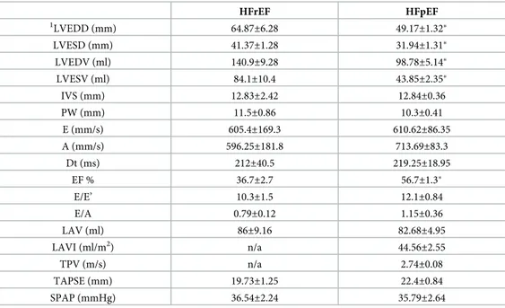
what already reported in numerous reports (see [18] and references therein). Echocardio-graphic parameters of HF patients were reported inTable 2. As shown, the ejection fraction is, by definition, significantly different between the two groups. Other significant differences were found in LVEDD, LVESD, LVEDV, LVESV all higher in HFrEF, as a distinctive feature of this disease.
Table 2. Echocardiographic parameters of HFrEF and HFpEF patients. Data are presented as mean± standard error of the mean (SEM).
HFrEF HFpEF 1 LVEDD (mm) 64.87±6.28 49.17±1.32� LVESD (mm) 41.37±1.28 31.94±1.31� LVEDV (ml) 140.9±9.28 98.78±5.14� LVESV (ml) 84.1±10.4 43.85±2.35� IVS (mm) 12.83±2.42 12.84±0.36 PW (mm) 11.5±0.86 10.3±0.41 E (mm/s) 605.4±169.3 610.62±86.35 A (mm/s) 596.25±181.8 713.69±83.3 Dt (ms) 212±40.5 219.25±18.95 EF % 36.7±2.7 56.7±1.3� E/E’ 10.3±1.5 12.1±0.84 E/A 0.79±0.12 1.15±0.36 LAV (ml) 86±9.16 82.68±4.95 LAVI (ml/m2) n/a 44.56±2.55 TPV (m/s) n/a 2.74±0.08 TAPSE (mm) 19.73±1.25 22.4±0.84 SPAP (mmHg) 36.54±2.24 35.79±2.64 1
Left ventricular end-diastolic diameter (LVEDD), left ventricular end-systolic diameter (LVESD), left ventricular end-diastolic volume (LVEDV), left ventricular end-systolic volume (LVESV), septal thickness (IVS), posterior wall thickness (PW), peak E-wave velocity (E), peak A-wave velocity (A), deceleration time (Dt), ejection fraction (EF %), pulsed-wave TDI E’ velocity (E’), E/E’ ratio, E/A ratio, left atrial volume (LAV), indexed left atrial volume (LAVI), tricuspidal peak velocity (TPV), tricuspid annular plane systolic excursion (TAPSE), and systolic pulmonary artery pressure (SPAP).
�p < 0.05
https://doi.org/10.1371/journal.pone.0210320.t002
Table 1. Baseline characteristics of patients with heart failure with reduced (HFrEF) and preserved (HFpEF) ejection fraction. Data are presented as mean± standard error of the mean (SEM).
HFrEF HFpEF
Number of patients 18 (15 males) 22 (14 males)
Age 69.2± 2.8 75.7± 1.8 NYHA class II (n = 9) III (n = 9) II (n = 16) III (n = 6) BMI (Kg/m2) 26.54 ± 0.95 28.9± 1.30 NT-proBNP (pg/ml) 6000.07± 2297.22 2548.40± 551.11� HOMA-IR 2.30± 0.38 2.73± 0.54 TAC (sec) 68.7± 4.7 75.8± 7.5 �p < 0.05 https://doi.org/10.1371/journal.pone.0210320.t001
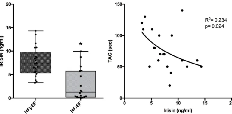
As shown inFig 1(left panel), circulating irisin levels in HFpEF were significantly higher (7.72± 0.76 ng/ml) than in HFrEF patients (2.77 ± 0.77 ng/ml). Moreover, a logarithmic corre-lation between TAC and irisin parameters was found in HFpEF patients (R2= 0,234 and p = 0,024) (Fig 1, right panel). Conversely, in HFrEF patients, irisin levels and TAC did not sig-nificantly correlate both in linear and in logarithmic regression analysis.
Finally, no significant correlation was found between irisin and BMI or HOMA-IR for both HFrEF and HFpEF groups. Moreover, in both groups of patients, there was no significant cor-relation between irisin and each of the echocardiographic parameters reported inTable 2.
At the logistic regression, only irisin levels show a significant association with HFpEF (OR = 1.76; 95% CI: 1.18–2.61; p < 0.01). The Hosmer-Lemeshow test supports the goodness of fit of the final model (p = 0.83).
To the best of our knowledge, this is the first study comparing irisin levels between HFpEF and HFrEF patients that shows higher irisin levels in HFpEF, despite a higher average age of this group.
The increased levels of irisin observed in HFpEF patients might be due, rather than a pas-sive release, to an enhanced secretion aimed to compensate for the development of a putative “irisin resistance” [12] and to maximize the beneficial effects of irisin on metabolic comorbidi-ties as well as on endothelium dysfunction.
Irisin, indeed, in addition to regulating energy metabolism, improves endothelial function [19,20] by its anti-inflammatory and anti-oxidizing properties.
Moreover, it has been suggested that blood levels of irisin may be regulated by oxidative stress that increases irisin secretion whereas antioxidants decrease it [4]. Our findings, con-firming this suggestion, show an inverse correlation between irisin and TAC in HFpEF patients.
Fig 1. Irisin levels in patients with heart failure with reduced (HFrEF) and preserved (HFpEF) ejection fraction. Left panel shows a Box-Plot comparing irisin levels in HFpEF with that in HFrEF (�p < 0.05). Right panel shows the correlation between irisin levels and TAC (in sec) in HFpEF.
Nevertheless, there are some potential limitations of the present study as expected by a pilot study. Firstly, the number of subjects in both groups is slightly small, so its statistical power is limited, and our findings will need to be confirmed in a larger population. Secondly, this pilot study and the power analysis cannot draw a cause-effect conclusion about irisin/TAC correla-tion in heart diseases patients. Further studies are needed to explain the physiological role of irisin in myocardium and its correlation to HF. Finally, we cannot discriminate the source of circulating irisin (skeletal muscle vs heart myocytes) in such patients that remain a pilot study.
In conclusion, this pilot study represent the first observation about irisin in different mod-els of HF. Moreover, despite these limitations, the present preliminary data are in favour of the concept that a different pathogenetic model is involved in the two HF subclasses and suggest that irisin levels in HFpEF could be an index of multi-systemic disease, according to the recent paradigm for HF [10], rather than a primitive heart disease.
Author Contributions
Conceptualization: Andrea Silvestrini, Angela Venuti, Angela Maria Rita Favuzzi, Elisabetta
Meucci, Alvaro Mordente, Antonio Mancini.
Data curation: Andrea Silvestrini, Carmine Bruno, Edoardo Vergani, Angela Venuti, Angela
Maria Rita Favuzzi.
Formal analysis: Andrea Silvestrini, Nicola Nicolotti. Funding acquisition: Andrea Silvestrini, Alvaro Mordente.
Investigation: Andrea Silvestrini, Edoardo Vergani, Angela Maria Rita Favuzzi, Francesco
Guidi, Elisabetta Meucci, Alvaro Mordente.
Methodology: Andrea Silvestrini, Carmine Bruno, Elisabetta Meucci. Project administration: Antonio Mancini.
Resources: Alvaro Mordente. Software: Carmine Bruno.
Supervision: Andrea Silvestrini, Antonio Mancini.
Writing – original draft: Andrea Silvestrini, Antonio Mancini.
Writing – review & editing: Andrea Silvestrini, Alvaro Mordente, Antonio Mancini.
References
1. Aronis KN, Moreno M, Polyzos SA, Moreno-Navarrete JM, Ricart W, Delgado E, et al. Circulating irisin levels and coronary heart disease: Association with future acute coronary syndrome and major adverse cardiovascular events. Int J Obes. 2015; 39: 156–161.https://doi.org/10.1038/ijo.2014.101PMID:
24916788
2. Bostro¨m P, Wu J, Jedrychowski MP, Korde A, Ye L, Lo JC, et al. A PGC1-α-dependent myokine that drives brown-fat-like development of white fat and thermogenesis. Nature. 2012; 481: 463–468.https:// doi.org/10.1038/nature10777PMID:22237023
3. Huh JY, Panagiotou G, Mougios V, Brinkoetter M, Vamvini MT, Schneider BE, et al. FNDC5 and irisin in humans: I. Predictors of circulating concentrations in serum and plasma and II. mRNA expression and circulating concentrations in response to weight loss and exercise. Metabolism. 2012; 61: 1725–1738.
https://doi.org/10.1016/j.metabol.2012.09.002PMID:23018146
4. Gouni-Berthold I, Berthold HK, Huh JY, Berman R, Spenrath N, Krone W, et al. Effects of Lipid-Lower-ing Drugs on Irisin in Human Subjects In Vivo and in Human Skeletal Muscle Cells Ex Vivo. Luque RM, editor. PLoS One. 2013; 8: e72858.https://doi.org/10.1371/journal.pone.0072858PMID:24023786 5. Aydin S, Kuloglu T, Aydin S, Eren MN, Celik A, Yilmaz M, et al. Cardiac, skeletal muscle and serum irisin
irisin than skeletal muscle. Peptides. 2014; 52: 68–73.https://doi.org/10.1016/j.peptides.2013.11.024
PMID:24345335
6. Bui AL, Horwich TB, Fonarow GC. Epidemiology and risk profile of heart failure. Nature Reviews Cardi-ology. 2011.https://doi.org/10.1038/nrcardio.2010.165PMID:21060326
7. Yancy CW, Jessup M, Bozkurt B, Butler J, Casey DE, Drazner MH, et al. 2013 ACCF/AHA guideline for the management of heart failure: A report of the american college of cardiology foundation/american heart association task force on practice guidelines. Circulation. 2013;https://doi.org/10.1161/CIR. 0b013e31829e8776PMID:23741058
8. Reddy YN V, Borlaug BA. Heart Failure With Preserved Ejection Fraction. Curr Probl Cardiol. 2016;
https://doi.org/10.1016/j.cpcardiol.2015.12.002PMID:26952248
9. Borlaug BA, Kass DA. Ventricular-Vascular Interaction in Heart Failure. Cardiology Clinics. 2011. pp. 447–459.https://doi.org/10.1016/j.ccl.2011.06.004PMID:21803232
10. Paulus WJ, Tscho¨ pe C. A novel paradigm for heart failure with preserved ejection fraction: Comorbidi-ties drive myocardial dysfunction and remodeling through coronary microvascular endothelial inflamma-tion. J Am Coll Cardiol. 2013; 62: 263–271.https://doi.org/10.1016/j.jacc.2013.02.092PMID:23684677 11. Polyzos SA, Anastasilakis AD, Efstathiadou ZA, Makras P, Perakakis N, Kountouras J, et al. Irisin in
metabolic diseases. Endocrine. 2017: 1–15.https://doi.org/10.1007/s12020-017-1476-1PMID:
29170905
12. Perakakis N, Triantafyllou GA, Ferna´ndez-Real JM, Huh JY, Park KH, Seufert J, et al. Physiology and role of irisin in glucose homeostasis. Nature Reviews Endocrinology. 2017.https://doi.org/10.1038/ nrendo.2016.221PMID:28211512
13. Ponikowski P, Voors AA, Anker SD, Bueno H, Cleland JGF, Coats AJS, et al. 2016 ESC Guidelines for the diagnosis and treatment of acute and chronic heart failure. European Heart Journal. 2016. p. 2129– 2200m.https://doi.org/10.1093/eurheartj/ehw128PMID:27206819
14. Rice-Evans C, Miller NJ. [241 Total antioxidant status in plasma and body fluids. Methods Enzymol. 1994; 234: 279–293.https://doi.org/10.1016/0076-6879(94)34095-1PMID:7808295
15. Mancini A, Leone E, Festa R, Grande G, Di Donna V, De Marinis L, et al. Evaluation of antioxidant sys-tems (coenzyme Q10 and total antioxidant capacity) in morbid obesity before and after biliopancreatic diversion. Metabolism. 2008; 57: 1384–1389.https://doi.org/10.1016/j.metabol.2008.05.007PMID:
18803943
16. Polyzos SA, Mantzoros CS. An update on the validity of irisin assays and the link between irisin and hepatic metabolism. Metabolism. 2015; 64: 937–942.https://doi.org/10.1016/j.metabol.2015.06.005
PMID:26130607
17. Hosmer DW, Hosmer T, Le Cessie S, Lemeshow S. A comparison of goodness-of-fit tests for the logis-tic regression model. Stat Med. 1997;https://doi.org/10.1002/(SICI)1097-0258(19970515)16:9<965:: AID-SIM509>3.0.CO;2-O
18. Tromp J, Khan MAF, Mentz RJ, O’Connor CM, Metra M, Dittrich HC, et al. Biomarker Profiles of Acute Heart Failure Patients With a Mid-Range Ejection Fraction. JACC Hear Fail. 2017; 5: 507–517.https:// doi.org/10.1016/j.jchf.2017.04.007PMID:28624483
19. Han F, Zhang S, Hou N, Wang D, Sun X. Irisin improves endothelial function in obese mice through the AMPK-eNOS pathway. Am J Physiol Circ Physiol. 2015; 309: H1501–H1508.https://doi.org/10.1152/ ajpheart.00443.2015PMID:26371167
20. Lu J, Xiang G, Liu M, Mei W, Xiang L, Dong J. Irisin protects against endothelial injury and ameliorates atherosclerosis in apolipoprotein E-Null diabetic mice. Atherosclerosis. 2015; 243: 438–48.https://doi. org/10.1016/j.atherosclerosis.2015.10.020PMID:26520898