UNIVERSITÀ DI PISA
Dipartimento di Farmacia
Corso di Laurea Specialistica in Chimica e Tecnologia Farmaceutiche
Tesi di Laurea:
Design, synthesis and biological evaluation of new
heterocyclic derivatives as serine palmitoyltransferase
inhibitors
Relatore: Relatore:
Dott. Giuseppe Saccomanni Dott.ssa Sara Del Carlo
Candidato:
Rosita Basile
(N° matricola 292690)
Anno Accademico 2012/2013 SSD: CHIM08
“Il contenuto di questa tesi è strettamente riservato, essendo presenti argomenti tutelati dalla legge come segreti. Pertanto tutti coloro che ne prendono conoscenza sono soggetti all’obbligo, sanzionato anche penalmente dagli articoli 325 e 623 del codice
Alla mia famiglia
Index
GENERAL INTRODUCTION………..……….………..
.2
1 Introduction ... 3 1.1 Sphingolipids ... 3 1.2 Ceramide ... 4 2 Serine palmitoyltransferase (SPT) ... 5 2.1 Bacterial SPT ... 6 2.2 Eukaryotic SPT ... 7 2.3 SPT substrates ... 8 3 Reaction mechanism ... 9 4 Spt inhibitors ... 12 4.1 Penicillamine ... 13 4.2 Cycloserine ... 15 4.3 Myriocin ... 20 5 Retinitis Pigmentosa ... 27 6 Alzheimer ... 297 Hereditary Sensory Neuropathy Type 1 (HSAN 1) ... 32
8 Insulin Resistance ... 35
INTRODUCTION TO EXPERIMENTAL PART….………...………..38
EXPERIMENTAL PART………...49
1 SPT assay ... 75
1.1 HPLC-FL ... 75
1.2 Cell culture and SPT cell lysate preparation ... 75
1.3 Incubation and sample extraction procedure ... 76
2
3
1 Introduction
More than 100 years ago, Johann L. W. Thudicum described a not well identified aliphatic alkaloid in the brain called sphingosine.1 The structure of this substance was characterized only 50 years later by Carter at al.2
In the last years, structural and distribution studies have highlighted the critical roles of sphingolipids in membranes micro domains formation, skin barrier function and modulate on cellular events like apoptosis, proliferation and differentiation3-5.
These discoveries along with bioactive signal function observation, increase research attraction and attention on sphingolipids metabolism.6-11
1.1 Sphingolipids
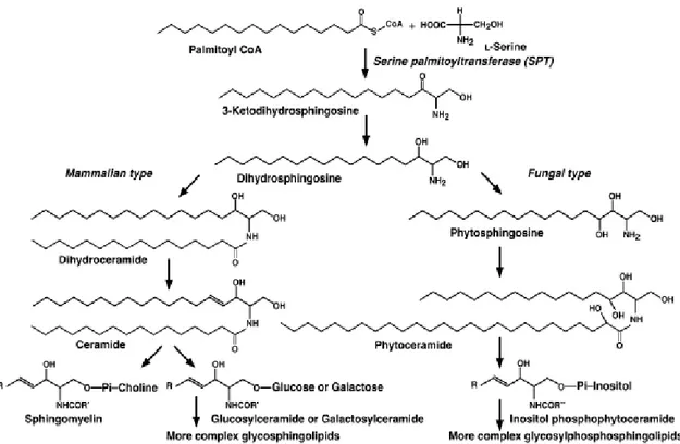
Sphingolipids (SPL) are one of eight categories of the LIPID MAPS classification system and they are described as lipids containing a sphingoid base (1,3-dihydroxy-2-amino alkane)linked through an ammidic bound to a fatty acid and to polar headgroup, like phosphate and carbohydrate.12 SPL distribution appears ubiquitous in eukaryotic cells. In particular, SPL alkane group is usually an eighteen carbons chain; C-2 and C-3, in the polar headgroup, are chiral and the configuration is 2S, 3R, D-Eritro. Indeed, in mammal cells, sphingosine appears to be the major sphingoid base, followed by dihydrosphingosine, while phytosphingosine, the third major type of sphingoid base, has been found mainly in plants, although it was revealed in large amount in mammal tissues like stomach and kidney. Sphingolipids containing cholinephosphoceramide, known as sphingomielyn, represent 5-10% of mammalian membrane’s phospholipids. SPL are produced also by some prokaryotes, plants, bacteria and fungi through different biosynthetic pathways. 1,12 In fungal and plants inositol-phosphoceramide is the most abundant constituent of the membrane phospholipids; these observations suggest a relevant structural dissimilarity in the polar headgroup in different species. Conversely the polar headgroup of glycerophospholipids are structurally very similar between mammal cells and fungi (Fig.1).12
4
Fig. 1: Biosynthetic pathway of sphingolipids in mammalian and fungal cells.
1.2 Ceramide
Several studies performed by Braun13 and Stoffel 14, independently, have demonstrated in 1968 that the second step of sphingolipid biosynthesis is the NADPH-dependent reduction of a C-3 carbonyl group leading to dihydrosphingosine.15 In mammalian cells, dihydrosphingosine undergoes to an N-acylation reaction forming dihydroceramide. Afterwards, an alkyl chain oxidation leads to ceramide which successively functionalized at the polar headgroup to give sphingomielyn or different glycosphingolipids. Conversely, in fungi, dihydrosphingosine undergoes to oxidation and then to N-acylation forming phytoceramide which is further converted in inositol phosphophytoceramide and more structurally complex derivates. In fungi, the acyl-chain is mainly represented by C-26 hydroxy fatty acid, whereas mammal ceramide derivatives are saturated C-16/C-24 fatty acid chain.16,17 Cells produce sphingolipids through two different metabolic ways: catabolic and anabolic pathways. The catabolic pathway involves hydrolyses of complex molecules, as sphingomielyn and glucosphingolipid, to obtain ceramides. The anabolic pathway (also called de novo biosynthesis), provides an enzymatic cascade which leads to the formation of ceramides starting from simple and abundant substrates like L-serine and palmitoyl-CoA. It takes placein the endoplasmatic reticulum (ER).1
5
2 Serine palmitoyltransferase (SPT)
Sphingolipids biosynthesis is different between species (i.e. S. cerevisiae produces phosphoinositol headgroup sphingolipids only, while P. pastoris both glucosylceramide and phosphoinositol sphingolipids headgroup), whereas the first enzymatic step of de
novo biosynthesis is conserved across all species producing sphingolipids (Fig.2).2 The common step of the de novo synthesis is an enzymatic condensation carried out by serine palmitoyltransferase (SPT), a pyridoxal-5’-phosphate (PLP) dependent enzyme. SPT is an enzyme belonging to alfa-oxoamine synthase (AOS) family which is a PLP dependent family.
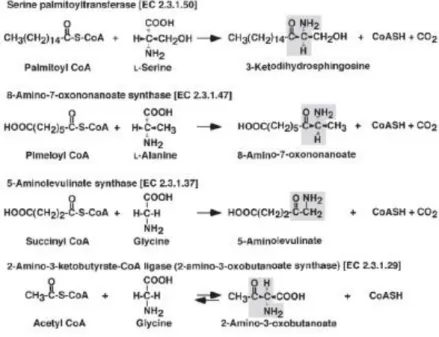
Fig. 2: POAS family. Reactions catalyzed by POAS family members.
SPT catalyzes a condensation between L-serine and typically C16 acyl-CoA thioester (palmitoyl-CoA), to give a Claisen-like C18 condensation product: 3-ketodihydrosphingosine (KDS) (or 3-ketosphinganine). Successively KDS is rapidly reduced to dihydrosphingosine (o sphinganine) in presence of NADPH.1,2 SPT plays a key role in the biosynthesis of sphingolipids; indeed regulation of SPT catalyzed step prevents accumulation of sphingolipids metabolites (i.e. sphingoid base) while inhibition of later biosynthetic steps lead to metabolite accumulation which are death effectors in various experimental models and pathological conditions. Therefore, SPT condensation reactions represent the rate-limiting step of sphingolipids biosynthesis.1
6
2.1 Bacterial SPT
The first three dimensional structure of SPT was obtained by the isolation of the homodimeric (and water soluble) SPT from the Gram-negative bacterium S.
paucimobilis (spSPT)18. The crystal structure reveals that the holo-SPT monomer (or internal aldimine) consists of three domains: the N-terminal, the central catalytic domain and the C-terminal domain. The N-terminal domain consists of 80 residues (an α-helix followed by 3 β-sheets). This domain is linked with the central domain also called catalytic domain. In the central domain is recognized the Lys265 residue responsible of PLP cofactor binding, while 7 β-sheet is composed by about 200 residues. The C-terminal domain is strictly linked with N-terminal domain by binding interactions19. Three dimensional crystal of the holo-enzyme revealed a homodimeric structure containing a conserved Lys265-binding-PLP residue. However, for the correct positioning of the PLP cofactor (crucial for the catalytic activity), π-stacking interactions between the pyridine ring and His159 are required. PLP interacts also with other residues such as Asn138, Asp231, His234, Thr262, Gly134 and Tyr135. Furthermore, an Arg379 is needed for the positioning of the L-serine in the catalytic site because of crucial interaction with the carboxy moiety of the substrate.1
Other SPT homologues, like Sphingobacterium multivorum SPT (SmSPT) and
Sphingobacterium wittichii (SwSPT), exhibit difference at few levels. SmSPT has a
38% homology and in active site the external aldimine (formed by the PLP and serine) is linked via two water molecules to two aminoacid residues, Ser81 and Met271 (and not to a Lys)20. Three-dimensional structure of SwSPT reveals a larger active site than in SpSPT, probably because of the use of a larger acylated-ACP thioester substrate.21,22
7
2.2 Eukaryotic SPT
Eukaryotic SPT is a heterodimer consisting in two subunits, LCB1 and LCB2, both linked to endoplasmatic reticulum (ER) and they are encoded by two different genes, lcb1 (SPTLC1) and lcb2 (SPTLC2), composed by 15 exons of 85 kbp size localized on chromosome 9, arm q21-q22, and 12 exons of 110 kbp size localized on chromosome 14, arm q24.3-q3 respectively. These genes encode for proteins of 53 and 63 kDa, with 20 % sequence identity that is probably critical for dimerization.23,24 LCB proteins have 95% identity between mammals and 40% between mammals and yeast. The sequencing of SPTLC1 and SPTLC2 genes in Saccharomyces cerevisiae revealed that the active site of eukaryotic SPT is localized in the LCB2 subunit; this observation is suggested by detection of conserved residues of AOS’s family (one lysine, two hystidine and an aspartate) which are essential to lock the PLP cofactor in the active site1,2. LCB1 subunit appears crucial for SPT’s catalytic activity. The lack of this subunit expression or a missense mutation in SPTLC1 gene causes alteration of SPT’s catalytic activity.25
Furthermore, CHO (Chinese hamster ovary) cells line defective in SPTLC1 transcription express lower levels of LCB2. To obtain an overexpression of LCB2 is required an overexpression of LCB1 and LCB2. Moreover, it appears to be unstable when it is not associated with the LCB1. On contrary an high level of LCB1 do not require an overexpression of LCB2. Both monomers have only one highly hydrophobic transmembrane domain (TMD). The catalytic site has a cytosolic orientation indeed indirect immunocytochemical analysis indicated that C-termini and N-termini of LCB1 have cytosol and lumen orientation respectively.26 LCB1 and LCB2 are ER membrane integrated proteins type I. Another isoform of LCB2 called LCB3 is expressed only in certain tissues.
In yeast this enzyme is constituted by the so called “SPOTS COMPLEX” that’s encodes for SPT–ORM1/2-Tsc3-Sac1 (phosphatase).27 Therefore, LCB1 and LCB2 subunits are associated with a third subunit, Tsc3, which is required for the maximal SPT activity. This heterodimeric structure is associated with oromucosoids proteins (ORM1/2) which are able to negatively regulate SPT.
Recently for human SPT two novel small subunits named ssSPTa and ssSPTb were discovered. These subunits can enhance activity >10 fold when bound to LCB1-LCB2 heterodimer. Moreover orosomucoid-like (ORMDL) proteins appear to be able to interact with the LCB1-LCB2 heterodimer.28
8
2.3 SPT substrates
Palmitoyl-CoA is the more kindred acyl-CoA thioester substrate of SPT in mammalian cells; on the other hand pentadecanoyl-CoA and heptadecanoyl-CoA are also good substrates less abundant. L- and D- serine are SPT amino acid substrates. SPT is able to form the Schiff’s bases with both enantiomers but the alfa-deprotonation step (before acylation) proceeds only with L-serine substrate. Thus L-serine is the common SPT’s substrate in the de novo synthesis. The hydroxyl, carboxyl and amino groups of L-serine appear to be necessary for interaction with SPT; neither L-alanine, L-serinamide, D,L-serinol and nor L-serine methyl ester not in the formation of [3H]KDS from L-[3H]serine, indicating that the hydroxyl, amino and carboxyl groups of L-serine are responsible for the recognition of the amino acid substrate by the SPT enzyme.29
9
3 Reaction mechanism
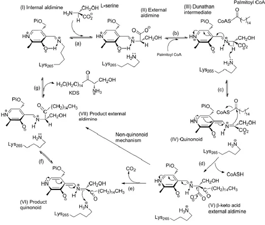
SPT catalyzes a Claisen like condensation of L-serine and palmitoyl-CoA which leads to 3-KDS as final product. SPT reaction mechanism proceeds across six step: 1) formation of the Schiff base between L-serine and PLP, 2) L-serine α-hydrogen removing; 3) nucleophilic attack to palmitoyl-CoA (formation a transient acylated adduct), 4) decarboxylation, 5) protonation of α-carbanion and 6) KDS release (Fig.3). Assays conducted with purified enzyme from chinese hamster ovary cell, suggested that one molecule of mammalian SPT is capable of catalyzing maximally of 80 cycles of these steps per minute.30
Fig. 3: Catalytic mechanism of SPT.
The PLP cofactor catalyses all the reaction mechanism. Interaction of PLP aldehyde group and Lys265 leads to a Schiff base (internal aldimine) in the active site. Spectrophotometric analysis of holo SPT revealed two UV absorption peaks which represent two tautomers of the Schiff base PLP-Lys265: the enolamine (338 nm) and ketoenamine (426 nm). Moreover, crystalline structure of S. multivorum and S.
paucimobilis SPT highlighted that PLP has Van der Walls interaction with His159 and Ala233.
10
Fig. 4: Spectrophotometric analysis of holo SPT
Addiction of L-serine to the holo-enzyme causes drastically changes in the UV spectra due to the formation of a new Schiff base between PLP and L-serine (external aldimine). Two intermediate adducts are identified: first adduct has an absorption similar to holo-SPT while the other one has a maximum peak at 426 nm (which represents the ketoenaminic tautomer of the new Schiff base) (Fig.4). The interaction of PLP with L-serine causes a rotation of pyridine ring or a torsion of the Schiff base C4-C4’ bound.
Once the external aldimine is formed, L-serine is conformationally stabilized by interaction of the carboxyl group with His159 Nε2 (hydrogen bond) and by interaction of the hydroxylic group with the PLP phosphate group and a molecule of water. These interactions fixed the PLP-L-serine complex allowing a perpendicular orientation (80°) of Cα-COO bond to imine-pyridine plane.
The next step of the reaction mechanism is represented by the formation of a new C-C bond via a Claisen-like condensation. Two different intermediate may be involved in the nucleophilic substitution: the decarboxylated intermediate or that one generated after removal of the -H. In order to understand the intermediate involved in the nucleophilic substitution reaction some experiments have been developed in presence of a palmitoyl-CoA analogue S-(2-oxoheptadecyl)-palmitoyl-CoA. This compound has a methylene group inserted between the sulphur atom and the carbonyl groups which doesn’t allow the nucleophilic substitution. Data obtained performing the enzymatic reaction in presence
11
of S-(2-oxoheptadecyl)-CoA and deuterated solvent showed the α-hydrogen switch with the deuterated solvent increase of about 100 times (high peak) at 426 nm. Therefore, α-deprotonation of external aldimine is improved in presence of palmitoyl-CoA substrate and a significative amount of quinoid adduct have been formed. This suggests that the quinoid adduct attack palmitoyl-CoA thioester to form the C-C bond. A faster α-deprotonation of external aldimine in presence of palmitoyl-CoA reduce the risk of intermediates accumulation which can form pyridoxamine-5’-phosphate (PMP) after transamination reaction (PMP can’t be converted to PLP thereby the enzyme is inactivated). According to Dunathan hypothesis, the -deprotonation process takes place when the bond involved in the nucleophilic substitution is perpendicular to the imine-pyridine place. The reaction takes place thanks to the complete overlap between the -orbital (occupied) of the designed bond and the free -orbital of the conjugate imine-pyridine system. The increase of -deprotonation process in presence of palmitoyl-CoA is related to a strictly stereochemical control of the reaction. In the external aldimine conformation the C-H bond is 40° rotated with respect to the imine-pyridine plane avoiding the -deprotonation process. This conformation is generated by interaction of the carboxyl group of L-serine with His159 as depicted in the SPT-PLP-serine crystal structure. Computational study showed that palmitoyl-CoA induced conformational change is related to the displacement of the hydrogen interaction of L-serine with His159: palmitoyl-CoA interacts with His159 (hydrogen bond) whereas L-serine performs a new interaction with the guanidine group of Arg390 rotating the C-N bond of 50°. In this new conformation the C-H bond of L-serine is perpendicular to the imine-pyridine plane promoting the -deprotonation.
The nucleophilic substitution of quinoid intermediate to the acyl-CoA thioester carboxylic group leads to a β-keto-acid derivates. The interaction between palmitoyl-CoA and His159 promotes the C-C bond formation by acidic catalysis played by His159: this residue exchange an hydrogen with the carboxylic group of palmitoyl-CoA inducing the right positioning. The product forms an hydrogen bond with His159 (the oxygen of the carbonyl group interact with the hydrogen of His159). Then β-keto-acid derivates undergoes to decarboxylation and release of the final adduct (KDS).
Generally PLP catalyzed reactions occur through decarboxylation process catalyzed by the electron withdrawing immine-pyridine system. Instead SPT decarboxylation reaction is promoted by the correct position of the carboxyl group by His159.1
12
4 Inhibitors
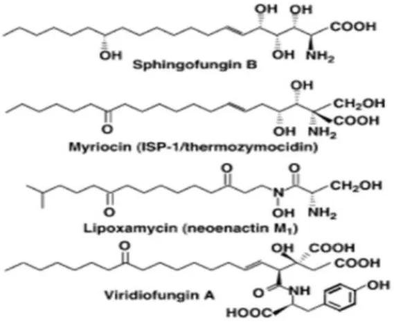
Natural inhibitors of SPT have been discovered. Sphingofungine, lipoxamicina (neoenactin M1) and myriocin (ISP-1/thermozymocidin) (Fig.5) are potent and highly selective inhibitors of both fungine and mammals SPT in cell-free models with nanomolar IC50. These compounds are structurally similar to the postulated transitory adduct formed by the L-serine and palmitoyl-CoA during the condensation suggesting the crucial role of a transitory adduct of these compound with PLP in the strong inhibition activity.
Indeed, the inhibitory activity of sphingofungine B is highly dependent on stereochemistry.31, 32
The crucial role of sphingofungine B stereochemistry has been demonstrated: the C14 hydroxyl group of sphingofungine B yields potent inhibitory activity but it’s not crucial for the activity. On the other hand, the configuration of the stereogenic centers in the α, β, γ and δ positions from carbonilic group are essential for the inhibitory activity.32,33
Viridiofungins were first isolated by Harris and co-workers in 1993 from the fungus,
Trichoderma viride. This family of alkyl citrates exhibited broad spectrum of anti-fungal properties with minimum fungicidal concentrations in the range of 1–20 μg/mL against a number of species. Furthermore, viridiofungins inhibited rat and yeast squalene synthesis. This antifungal activity is unrelated to the inhibition of ergosterol
biosynthesis. Instead, viridiofungins showed very potent (nanomolar range) inhibitory activity against serine palmitoyltransferase.34
13
4.1 Penicillamine
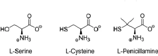
Penicillamine (Pen) is an α-amino acid and a characteristic degradation product of penicillins, used in Wilson's disease as copper chelator to form mixed disulfides with cystein. The enantiomer used as pharmaceutical drug is D-Pen. It has been well established that both enantiomers and racemic mixture exert anti-PLP activity by inhibiting several PLP-dependent enzymes such as alanine aminotransferase, aspartate aminotransferase, glutamate decarboxylase, hystidine decarboxylase and serine hydroxymethyl transferase. L-Pen has the stronger inhibitory activity thanks to the structure similarity with the enzyme’s naturally substrate (L-amino acids) (Fig.6).
Fig. 6: L-amino acids inhibitors of SPT.
A recent research developed by Campopiano group has clarified Pen inhibitory action and mechanism on SPT from Sphingomonas paucimobilis (spSPT). Incubation of D-Pen and L-Pen with spSPT results in different percentage of enzyme inhibition: incubation of 5 mM L-Pen with spSPT reduces enzyme activity to 3% while incubation with D-Pen decreases enzyme activity to 34%. Pen inhibition is reversible and the mechanism of SPT inactivation occurs by disabling the PLP cofactor. Indeed, incubation of enzyme inactivated by D-Pen and L-Pen with a buffer containing 50 µM of PLP, restores the SPT activity to 80% and 57 % respectively. Inability to restore 100% of the activity using a dialyzing buffer is due, probably, to possible additional competitive reactions that irreversibly modify the proteins. The Pen thiol-group plays a crucial role in the inhibition mechanism. The thiol-group is more nucleophilic than the hydroxyl group of the L-Ser and it’s able to interact with PLP aldehyde leading to PLP:thiazolidine (PLP:TA) adduct (Fig.7).
14
Fig. 7: Addition of L-Pen to SPT leads to formation of a PLP:TA adduct via an external aldimine intermediate (P
represents group phosphate).
The inhibition reaction could be monitored observing the disappearance of free PLP peaks at 390 nm and appearance of a new peak at 333 nm (thiazolidine adduct). Using the same spectrophotometric approach, a time dependent disappearance of holo SPT ketoenamine (420 nm) and appearance of a thiazolidine adduct have been observed after addiction of L-Pen to holo-SPT. ESI-MS analysis of these samples confirmed the formation of a PLP:TA adduct. Moreover L-Pen and L-cystein activities have been compared highlighting a faster inhibitory activity of L-Pen that could be related to the presence of gem-dimethyl group enhancing cycle formation through a Thorpe-Ingold effect (Fig.8).35
Fig. 8: (A) UV-visible spectrum of 20 mM holo-SPT (solid line) shows typical peaks at 335 nm and 420 nm due to
enolimine and ketoenamine forms of the PLP cofactor respectively. Addition of 10mM L-Cys to holo- SPT (broken lines) led to formation of a thiazolidine adduct (333 nm peak) over a 30 minute period, with concomitant loss of the 420 nm peak (B) improved thiazolidine formation in SPT over 30 minutes by addition of 10 mM L-Pen.
15
4.2 Cycloserine
β-chloro-L-alanine and L-cycloserine has been used as SPT inhibitors. These compounds are potent inhibitors of several PLP-dependent enzymes, therefore their use as specific inhibitors is limited.36,37
Cycloserine (Fig.9a) is cyclic α-aminoacid well known to inhibit many PLP-dependent enzymes (transaminase, racemase and decarboxylase).38-40 It exists as two enantiomers: D-cycloserine (DCS) and L-cycloserine (LCS).
Fig. 9.“Conformations” and “ structures” of D- and L- α-amino acids inhibitors of SPT.
The mechanism of antibacterial activity is based on the inhibition of alanine racemase, a PLP-dependent enzyme that synthesize D-alanine for the formation of D-alanyl-D-alanine dipeptide (an essential component of the peptidoglycane layer of the M. tuberculosis).41 Furthermore, DCS is an agonist of N-methyl-D-aspartic acid (NMDA) receptors that is implicated in several CNS pathologies and it’s used in neurological models of these pathologies;42 LCS has been prepared synthetically and used as a modulator of lipids metabolism in biological research, indeed it is a potent inhibitor of SPT activity. Furthermore, it has been demonstrate that LCS inhibits mouse brain SPT activity in vivo after intraperitoneal injection43. Some authors propose a cycloserine inhibition mechanism based on aromatization of the PLP: cycloserine external aldimine (“aromatization mechanism”) by α-deprotonation that give rise to a 3-hydroxyisoxazole–PMP adduct: the cycloserine ring remains intact and covalenty linked to the PLP.
16
A first study developed by Ikushiro44 revealed a change in SPT absorption 10 minutes later the addiction of LCS: the peak corresponding to the ketoenamine tautomer of the internal aldimine (426 nm) decreased whereas a new peak at 380 nm increased. On the basis of this data a mechanism of action was hypothesized: in a first attend LCS reacts with PLP forming and external aldimine that undergoes to decyclization of the isoxazolidone ring (forming a transient oxime intermediate with absorption at 380 nm) leading to pyridoxamine 5’-phosphate (PMP) and β-aminooxypyruvate (Fig.9b).
Fig. 9b: Proposed mechanism of LCS inactivation of SPT by Ikushiro et al.
In 2010 the research group of Campopiano45 has highlighted the enantiospecific inhibition of LCS and DCS and has identified a novel decarboxylative ring-opening mechanism for inactivation of SPT. In particular the data show that in order to obtain the same inhibition of SPT activity (to the same extent and over the same time) 15-fold higher concentration of DCS is needed in comparison with LCS showing a clear enantiospecific difference of the enzyme active site (Fig.10).
17
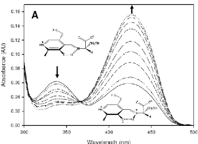
SPT activity (monitored by continuous DTNB assay) is reduced to 1% and 22 % after 2 hours incubation with 5 mM of DCS and LCS respectively. Furthermore incubation of treated samples in a buffer containing 25 M of PLP caused the recovery of SPT activity (83 % and 79 % of DCS and LCS inactivated samples respectively). Incubation of the inhibited enzyme in buffer (without PLP) doesn’t cause any further change of activity (the enzyme is still inactivated). These data confirm the disabling of PLP activity and the absence of further covalent modifications occur. The incubation of enzyme with DCS and LCS showed different UV-spectra. After 30 minutes from the addiction of 5 mM LCS new peaks appear at 330 and 380 nm and no more changes were observed when the incubation time was about 8 hours. Incubation of DCS with SPT causes the loss of the 425 nm peak and the appearance of a new peak at 380 nm. This peak shifted to 365 nm over time and at the same time a broad shoulder at 330 nm appeared (Fig.11A). These results suggest the formation of different species and the displace of the new PLP-Lys265 bond. In contrast, the UV-VIS is spectra of free PLP in the presence of both enantiomers show the same peak at 360 nm (Fig.11B).
Fig. 11: (A) SPT and 5 mM of LCS at time 0 (solid line), 30 seconds (long dash), 1 minute (dotted line) and 30
minutes (dash dot). (B) SPT and 5 mM of DCS at time 0 (solid line), 30 seconds (long dash), 2 hours (dotted line) and 10 hours (dash dot).
18
This last one result suggests that enzyme not only accommodated the PLP-cycloserine aldimine adduct but it’s also implicated in the formation of new different species. The reaction intermediates have been identified by LC ESI-MS analyses of SPT samples incubated with LCS: a peak at 2.8 minutes (m/z = 248) corresponding to PMP and another peak at 3.1 minutes (m/z = 256) corresponding to [M + H] ion of a hydrazine adduct formed by the derivatisation of the β-aminooxyacetaldehyde with 2,4-dinitrophenylhydrazine (2,4-DNP) have been identified. These peak wasn’t observed in control experiments with free PLP and 2,4 DNP derivative. These results suggest that the mechanism of LCS inhibition is based on the ring-opening of LCS by cleavage of the amide bond: this phenomenon take place because the C-α proton in the PLP:LCS external aldimine cannot be in the optimal orientation to be removed by the lysine. Successively two different decarboxilative pathways can take place: a) the ring-opened intermediate is acylated (enzyme mediated mechanism) and hydrolysed to form the carboxylated PLP intermediate (path [a] Fig.12); b) ring-opened adduct could proceeds through acid catalysis with water nucleophilic attack on the CS ring (path [b] Fig. 12).
Fig. 12: Novel ring-opening, decarboxylative mechanism for inactivation of SPT by LCS. Path (a) denotes an
enzymatic, nucleophile (Nu)- mediated mechanism with an acylated intermediate. Path (b) is the direct hydrolytic mechanism.
These pathways may come to form an oxime intermediate (observed as a 380 nm peak in the UV-visible spectrum) or decarboxylated adduct that is followed by the hydrolyzed immine form which conduct to PMP and β-aminooxyacetaldehyde .
Using a SPT R378N (a mutant which contain in the active site an alterated Arg378 residue that form a salt bridge with the carboxylate of the external aldimine) permits to
19
isolate intermediate and 40 fold reduce rate when compared with the wild-type enzyme, it was observed transient formation of a quinonoid species (formed by decarboxylation) at 510 nm during DCS inactivation, while inactivation with LCS (much faster inactivator in comparison with DCS) show a similar spectrum to the wild-type enzyme at 380 nm45.
In addition these researches have been conducted with modeling to isolate the PMP adduct (Fig.13).
Fig.13: Left: Fo-Fc electron density for the PMP molecule. This was calculated from molecular replacement model
which was refined and had omitted the co-factor. The map is contoured in green at 3s (0.2 Å3). Also shown are the side chains of Lys265, His159, Asp231 and His234. Monomer A is shown in ribbon in cyan and monomer B in magenta. The additional Fo Fc electron density „„blob‟‟ is shown in blue, contoured at 2.7s (0.2 Å 3
). A molecule of the β-aminooxyacetalaldehyde identified by mass spectrometry is placed in the density. Carbons are colored yellow, nitrogen blue, oxygen red and phosphorous orange. Right: Overlay of SPT:L-ser (2bwj) and the LCS inactivated form. The loop containing R378 adopts the „„swung-out‟‟ conformation in the LCS form, in contrast to the „„swung in‟‟ conformation in SPT:L-ser. The color scheme for the LCS inhibited form is as (left). For the SPT:L-ser structure, monomer A is colored light blue. Carbons are colored green, and other atoms are colored the same as in (left). The main chain of R378 adopts a very different conformation from the SPT:L-ser structure because the salt contact with L-ser is missing. A well-ordered molecule (red sphere) in the LCS structure is found in the same location as the L-ser carboxylate. The side chain of L-ser points towards the unfitted blob at the active site.
20
4.3 Myriocin
Natural products and their derivatives are known to be excellent tools for biochemicals and pharmacologicals studies. Myriocin [(2S, 3R, 4R, 6E)-2-amino-3,4-dihydroxy-2-(hydroxymethyl)-14oxo-6-eicosenoic acid] (Fig.14a) also known as thermozymocidin and ISP-1, has potent immunosuppressant properties in addition to antibacterial and antifungal activity.
Fig. 14a: myriocin structure
Firstly isolated by thermophilic moulds Myriococcum albomyces and Mycelia sterilia, myriocin remains the most valuable and widely used chemical probes in sphingolipids research as demonstrated by several studies: the identification of the two SPT subunits (SPTLCB1 and SPTLCB2) by Screiber et al.27 and of multi-protein membrane-bound
SPT complex (SPOTs complex) by Breslow at al.28 has been performed using myriocin. Kawasaki and collagues determined an IC50 value of 15 nM using cytotoxic T-cell line
(CTLL-2).46 Moreover, a recent study has demonstrated that myriocin reduces ceramide levels in rd10 mouse model of retinitis pigmentosa (RP) and therefore can rescue photorecepetors death47. Despite its use, the molecular basis of myriocin inhibition of SPT is largely unknown. Campopiano and coworkers have recently shed light in molecular mechanism of SPT inhibition by myriocin using a soluble and recombinant form of the enzyme from Sphingomonas paucimobilis (spSPT). Uv-vis analyses after addition of five-fold molar excess of myriocin to holo-SPT revealed that the two characteristic peak at 333 nm and 420 nm (corresponding to the ketoenamine and enolimine forms of the external aldimine) led to an increase absorbance with maximum at 430 nm and the disappereance of the peak at 333 nm. This data reflects a transamination reaction between PLP and myriocin that lead to the formation of a stable an external aldimine mimicking the β-keto acid intermediate (Fig.14b) and confirmed by LC ESI-MS analyses (Fig.14.b-B).
21
Fig. 14b: SPT inhibition occurs via formation of a PLP-myriocin aldimine. (A) UV-Vis spectrum of 40 μM SPT
before (solid line) and after 200 μM myriocin addition (dotted line). (B) The proposed structure of the inhibitory complex - a PLP-myriocin aldimine, 11. (C) Detection of the PLP-myriocin aldimine by LC-MS. Top, Extracted Ion Chromatogram at m/z 631. Bottom, high resolution mass spectrum of the PLPmyriocin aldimine, obtained by summing the spectra between t = 8-12 minutes. ([M+H]+, C29H48N2O11P; predicted m/z 631.29902; observed error 4.0 ppm). * denotes a contaminant.
22
Interstingly PLP-myriocin aldimine (11) is relatively stable at 25°C. However, after 16 hours of incubation a decrease of 430 nm peak and increase of 331 nm and 400 nm peaks is revelead (Fig.15).
Fig.15: UV-vis analysis of the degradation of the myriocin external aldimine in wild-type SPT. The
PLP-myriocin external aldimine (solid line) is stable for 90 minutes, before a decrease at 430 nm is observed, which is accompanied by a concomitant increase at 331 and 400 nm over 16 hours (dotted and dashed lines).
SPT:PLP-myriocin complex has a noncovalent, reversible nature and very slow off rate (koff) when incubated with myriocin for 10 minutes: sample dialysis in 25 µM PLP
containg buffer for 24 hours restored enzyme activity of 60% (accompanied with UV-Vis spectrum back to the internal aldimine). On the other hand a covalent and irreversible nature of myriocin-enzyme interaction has been observed when samples were incubated for 16 hours (Fig.16). Moreover, experiments demonstrated that myriocin is a competitive inhibitor for both L-serine and palmitoyl-CoA and a Ki of 967±98 nM.
Fig.16: Relative enzymatic activity after removal of inhibiting species by dialysis. SPT was inhibited with 200 μM
myriocin and incubated for 10 minutes (white bars) or 16 hours (grey bars) at 25 °C before removal of myriocin by extensive dialysis. The enzymatic activity was then determined at 0 hours, 3 hours, and 24 hours after dialysis.
23
To better understand the role of the Lys 265 in the dual reaction mechanism of myriocin, an incubation with five-fold molar excess of myriocin and catalytically-inactive SPT (K265A SPT) which presents active site without Lys residue was performed. After 16 hours the UV-Vis spectra remained unchanged in contrast with wild-type SPT suggesting that the initial SPT:PLP-myriocin inhibitor complex breaks down to form a second species that also inhibits wild-type SPT (Fig.17).
Fig. 17: UV-vis analysis of SPT K265A (40 μM) showed two absorbance maxima at 326 and 402 nm (solid line).
Upon addition of 200 μM myriocin, an immediate shift to a single peak at 425 nm occurred (dotted line), indicating the formation of a PLP-myriocin aldimine complex. Over 16 hours this spectrum remains unchanged (dashed line), indicating that the PLP-myriocin aldimine complex is not degraded by this mutant enzyme.
A retro-aldol like mechanism of SPT:PLP:myriocin complex which selectively and covalently modifies the Lys265 (crucial for the enzyme-catalysed reaction) leading to irreversible inactivation of the enzyme was hypothesized (Fig.18).
Mass spectrometry analyses showed a covalent adduct (14 in Fig.18) of SPT displaying a mass of 47.509 Da susceptible to NaBH4 of the covalent SPT-octadecenal imine
adduct and ketone group. Peptide mass fingerprinting identified Lys265 as the site of modification. Trypsin digests and mass spectrometry analysis identify three peptide species which displayed monoisotopic masses consistent with Lys265 modified by Δ mass +282.24 (Fig.19).
24
Fig. 18: Myriocin reacts with PLP in the active site to form the inhibitory PLP-myriocin aldimine 11, this species is
stable for greater than an hour at physiological temperature with inhibition being reversible upon addition of excess PLP.
Fig. 19: PLP-myriocin aldimine 11 (Fig.18) decomposes over 16 hours, at physiological temperature, to produce a
long chain aldehyde 12 (Fig.18) that react with the active site lysine to form an imine, thus rendering the enzyme inactive. This covalent modification can be classed as suicide inhibition.
25
Due to the timescale of crystallization, the PLP-myriocin aldimine degrades into the wilde-type enzyme and catalytically inactive K265A has been used to capture external aldimine which results stable and decompose only after seven days with a decarboxylation reaction. This slowness relative to wild-type complex is due to the not optimal orientation (“Dunathan conformation”) suggesting the crucial role of the Lys265
in the decarboxylation of the SPT:PLP-myriocin complex (Fig.20).
Fig. 20: Decarboxylation mechanism to account for PLP-decarboxymyriocin external aldimine observed in the
crystal structure of SPT K265A.
The crystal structure obtained highlights that the conserved residues His159, Asp231 and His234 are all in the same relative positions within the active site. Moreover, the CH2OH
head group of myriocin interacts with the 5′-phosphate of PLP. The 3,4-cis-diol of decarboxymyriocin makes hydrogen bonds to the protein, notably the 3-hydroxy group of myriocin with the important catalytic residue His159; this interaction would be expected to be preserved in the wild type SPT:myriocin complex. The hydrogen bond network that surrounds and includes the 4-hydroxy group of decarboxymyriocin may be changed by the presence of Lys265 but at least some of the same network seems certain to persist and this too involves the same residues that interact with the carboxylate of the PLP-L-serine external aldimine. These interactions rationalize the competitive inhibition with L-serine. Accompanying these interactions are movements of the side chains of Tyr73, Arg378 and Arg390 as well as a displacement of a key conserved stretch of amino acids (RPPATP) that constitute a mobile loop that undergoes conformational changes during the catalytic cycle. The 6,7 trans double bond geometry of myriocin is clearly defined and we can see electron density for the carbon chain up to C9 which sits in the hydrophobic cleft adjacent to PLP. The carbon tail of myriocin binds in a similar orientation to the decanoyl-tail of the PLP-product external aldimine observed bound in the crystal structure of the related AOS enzyme CqsA from Vibrio cholera consistent with hypothesis that myriocin mimics the condensation intermediate (Fig. 21).
26
Fig. 21: The structure of SPT K265A PLP-decarboxymyriocin aldimine inhibitory complex. (A) The biological SPT
dimer of the decarboxylated myriocin complex. The protein is shown as a cartoon with one subunit colored pale green and the other pale blue. The PLP external aldimine of the decarboxylated myriocin (15) is shown in space fill with carbons colored yellow, nitrogen blue, phosphorous orange. The N terminii are marked as dark blue spheres and C-terminii as red spheres. (B) Left, The Fo-Fc map (blue chicken wire contoured at 1.8σ, carve radius 1.5A) calculated from a model which had never contained either PLP or myriocin. Atoms are colored as figure 6A. Right, the final Fo-Fc map contoured at 0.85σ with a carve radius 1.8 Å. (C) Detailed representation of the active site interactions, carbon atoms are colored yellow and shown in sticks for the PLP-decarboxymyriocin aldimine 15. Carbon atoms in protein side chains are shown as white sticks, others atoms are colored as figure 6A. The hydrocarbon chain of the myriocin inserts into a hydrophobic pocket. (D) Superposition of the PLP-decarboxymyriocin external aldimine (colored as before) with the PLP-L-serine external aldimine 2W8J (cartoon pale orange, protein side chain carbons dark grey, aldimine carbons colored green, other atoms are colored as before). The key catalytic Lys265 residue is mutated to Ala in the decarboxylated myriocin complex.
This structure rationalizes the retro-aldol degradation of the PLP-myriocin external aldimine 11 into corresponding to the C18 aldehyde 12. This mechanism requires a base to abstract the proton from the 3-hydroxy group of myriocin but Lys265 would be in wrong face to perform this role. However, the absolutely conserved His159 is positioned 2.6 A away from the 3-hydroxy of myriocin and probably initiates the cleavage of the C2-C3 bond with the electrons sinking into the PLP ring. In the other hand deprotonated Lys265 is positioned to attack the newly formed C18 aldehyde species 12 to form a covalent aldimine adduct 14 and modifies the key catalytic lysine residue irreversibly and block access to the active site preventing regeneration by PLP on a biologically-relevant timescale.46
27
5 Retinitis Pigmentosa
Retinitis pigmentosa (RP) is a general term related to a wide range of rod and cone dystrophies characterized by progressive night blindness, visual field constriction and loss of acuity leading to an altered electroretinogram (ERG).48 Currently, there is no therapy able to stop the disease’s evolution based more frequently on mutations of genes involved in rod photoreceptors function and metabolism. Rods and cone photoreceptors cells death occurs through both apoptotic and non-apoptotic mechanisms.49
In many neurodegenerative and inflammatory diseases, an increase of the intracellular levels of ceramide, a well-characterized death effector, has been revealed. A direct genetic link between retinal degeneration and sphingolipid mediated apoptosis has been highlighted by the discovery of a mutation in CERKL (a gene expressing ceramide kinase-like protein) which causes loss of function and autosomal recessive RP.50
Ceramide levels are increased by de-novo biosynthesis or by activated intracellular sphingomyielinase which hydrolyze complex sphingolipids leading to ceramide. The
Drosophilla model of RP (knock out of the gene encoding for one subunit of SPT) is
characterized by a decrease of ceramide intracellular levels and protective effects in retinal function and morphology. Also the injection of ceramidase, a ceramide hydrolyzing enzyme, results in a decrease of ceramide intracellular levels and protective effects.51 In the murine 661W photoreceptor cell line, oxidative stress can increase ceramide levels leading to cell death via mitochondrial apoptotic pathway activation and caspase cascade.52 Therefore, inhibition of ceramide biosynthesis, accordingly with biochemical analyses, may represent a therapeutic approach for the treatment of this disease in humans.
A useful model for human RP is represented by rd10 mice line53 which presents a missense mutation of the beta subunit of the rod-specific phosphodiesterase gene, and mimics a form of human autosomal RP. Rd10 mice retinal ceramide levels increased at the third week of life, which correspond to the maximum photoreceptor death period as well as the human RP. These high levels are constant for the following period, whereas in wild-type mice ceramide levels decrease during the same period reaching a plateau after full retinal maturity. Rod death starts about at 12 days of life (P12) and reach the maximum peaks at 24 days of life (P24). Indeed ERG (electroretinogram) generated by
28
rods can be recorded up to 25 days of life (P25) until 45 days of life (P45) due to death of the retinal cones. Rod degeneration presents the characteristic features of apoptosis.54 In rd10 mice model of RP, single intraocular injections of 0.5 nmol (1 μL of 3.77 mM solution in DMSO) of myriocin, significantly decrease ceramide levels (17.5%) than in control, and rescued photoreceptors from apoptotic death whereas the ceramide levels in wild type are not significantly reduced (determined with the diacylglycerol kinase test). Eye drops consisting in a suspension of solid lipid nanoparticles (SLNs) loaded with myriocin, carried the drug across ocular tissues permitting trans-ocular drug administration: the non-invasive and long term treatment of these drops ameliorate the reduction of function loss and of ceramide levels (40.6%). Histological analyses (Fig. 22) demonstrate that myriocin treated rd10 mice presents normal retinal morphologies and excellent maintenance of ganglion cell morphology and structure: prolonged treatment (over 20 days) with solid lipid nanoparticles increase photoreceptor survival, preserving photoreceptor morphology (rhodopsin and cone immunoreactivity in well-organized outer segments of rods and cones and presence of well-well-organized dendrites in rod bipolar cells) and extends the ability of the retina to respond to light (demonstrated by the ERG).55 These data highlight that SPT inhibitors could be an useful tool for the
treatment of RP.
Fig. 22: Effects of myriocin-SLNs on retinal morphology. (A and B) Vertical retinal sections from rd10 mice treated
with control SLNs (A) andmyriocin-SLNs (B) for 10 d (from P14 to P24). The outer nuclear layer (ONL) of the myriocin-treated retina is thicker because it contains more photoreceptor rows than the control retina. These micrographs are from the same animals whose ERG data are shown in Fig. 4B. INL, inner nuclear layer; OPL, outer plexiform layer. (C) Quantification of photoreceptor rows at P24 and P30 in rd10 mice treated with control SLNs or myriocin-SLNs. Data are mean and SE. *P = 0.002, **P = 0.003, t test.
29
6 Alzheimer
Alzheimer's disease (AD) is the most common form of dementia. It was firstly described in 1906 by the german neuropathologist Alois Alzheimer. AD is characterized by short-term memory impairment, language disturbance (aphasia), confusion, irritability and loss of both judgment and reasoning ability, including deficiency of acetylcholine. The anatomical consequence of the cholinergic deficit is the atrophy and degeneration of subcortical cholinergic neurons (especially in the basal forebrain). This brain region provide cholinergic innervation to the cerebral cortex involving multiple neurotransmitter systems, including serotonin, glutamate, and neuropeptides. Not only cholinergic neurons are involved in AD, cortical and hippocampal targets are also interested. In the early stages (clinically silent stages) neurons degeneration and plaques formation in the hippocampal cortex take place and in late stages limbic and neocortical sites (conclamated AD) are involved.
Microscopic analyses of the affected parts of the brain have demonstrated the presence of a large amount of intracellular neurofibrillary tangles (NFT) consisting in microtubule-associated protein tau in the hyperphosphorilated and insoluble form. Moreover there are extracellular amyloid in the form of senile plaques (SP) consisting of a core of amyloid β-peptides (Aβ). For that reason AD represent one of the 20 clinically defined amyloidosis disease. The severity of impairment is roughly proportional to tangles and plaques abundance. The observed deposited fibrils were in a misfolded form β-sheet called β-amyloid. These components appear elevated in the hippocampus and in the associative regions of the cortex. Indeed, neural injury is most severe in the hippocampus and neocortex. Astroglia and hippocampal neurons exposed to β-amyloid show characteristic changes of apoptosis. The loss of neurons is not uniform but it varies dramatically in relation to functional regions. Mutations in the genes encoding for the amyloid precursor protein (APP), a type 1 cell surface glycoprotein which has neurotrophic and neuroprotective activity, and proteins known as the presenilins (PS1 and PS2), which may be involved with secretases (α, β and γ) enzyme in APP processing, lead to the formation of the Aβ. The presenilins and their mutated forms (in the PS1 gene on chromosome 14 and in the PS2 gene on chromosome 1) participate in APP processing leading to production, among others, of 4-kDa β-amyloid peptide of varied amino acid lengths, predominantly 40-42 (called Aβ40 and
30
proteolytic cleavage of APP by the β-site APP-cleaving enzyme 1 (BACE 1).56-60 Apolipoprotein E (apo E) has been identified as the first of what are likely to be many genetic risk factors for AD, involved in transport of cholesterol and lipids in blood. The mechanism by which the apo E 4 protein increases the risk of AD is unknown, but a secondary function of the protein in β-amyloid aggregation or processing of APP has been suggested. Lipid metabolism and high fat diet are risk factors for AD. Indeed, adequate intracellular ceramide is required for dendritic differentiation and survival of Purkinje cells61,62 while exogenous ceramides induce neurons and astroglia death in culture63-66. Elevated ceramide levels are show to be a risk factor for AD.67-71
Significant increase of the death-effector ceramide has been observed in AD patients neurons exposed to Aβ compared with control suggesting a molecular interaction between these two species. Aβ accumulation may cause oxidative stress as demonstrated by increased levels of the lipid peroxidation product 4-hydroxynonenal (4-HNE) in neurons exposed to Aβ72-74 and alteration of membrane lipid metabolism increas ceramide and sphingomielyn levels; this phenomen lead to synaptic function alteration, degeneration and neurons death (Fig.23). Moreover, alteration of the ceramide and cholesterol metabolism increases the γ-secretase’s cleavage of APP and enhances the production of Aβ42.75
Fig.23:Pathways of metabolism of sphingomyelin, ceramide and cholesterol, their modulation by oxidative stress, and their possible roles in neuronal death in AD. The production of sphingomyelin from serine and palmitoyl CoA is catalyzed by the enzyme serine palmitoyltransferase (SPT). Ceramides are synthesized as precursors to sphingomyelin and are also generated by the hydrolysis of sphingomyelin by sphingomyelinases (SMase). Exposure of cells to reactive oxygen species and Aβ induces ceramide production. CAPK, ceramide-activated protein kinase; CAPP, ceramide-activated protein phosphatase
31
In pathological conditions, ceramide facilitate the mislocation of BACE 1 and γ-secretase from outside to the lipid rafts, where γ-secretase cleavage of APP leading to the Aβ. Membrane ceramide stabilizes BACE 1 affecting the activity of the γ-secretase.76-80 Moreover, SPT appears increase and positively correlates with Aβ in human autopsy brain cortices, directly regulates their concentration in the serum and brain. Utilizing wild-type hybrid mice (C57/Bl6 x C3H), during a high-fat-diet of 5 months (starting at 4 months age) it was show an increase of SPT levels whereas in the same model a high-fat-diet of 3 months has not showed the same increase suggesting that the duration of high-fat diet consumption could have effect on metabolic processes.81
Also in these studies L-cycloserine (LCS) has demonstrated in vitro and in vivo reduction of cerebroside levels which largely consist in ceramide with polar head and single glucidic residue82. Furthermore, cognitive enhancement has been observed in AD patients in a duble-blind controlled trial with treatment of cycloserine (100 mg/day for 14 days).83
In an early-onset transgenic mice model, TgCRND8, encoding a double mutant form of APP 695 (KM670/671NL1V717F) under the control of the PrP gene promoter84, LCS has demonstrated to reduce Aβ42 ceramide levels in comparison with
mice fed a high fat diet and also in comparison with mice fed a control chow diet. LCS administration cause a decrease of cortical SPT protein levels with a significant positive correlation with ceramide and Aβ42 levels in all the study groups. Furthermore, SPT
appear to surround the SP in TgCRND8 mice and humans supporting the involvement of the enzyme in the Aβ formation. Moreover, LCS causes a decrease of the hyperphosphorilated tau protein reducing levels GSK3b (a kinase that mediates phosphorylation of tau). The administration of large doses (100 mg/kg) of LCS reduces immediately brain SPT levels with weight loss while low dose (25 mg/kg) show side effects without weight loss. Chronic LCS (by an intraperitoneal surgically implanted osmotic pump) administration doesn’t change brain histology, morphology, myelination, or memory in healthy mice with the LDH serum levels unchanged.85
32
7 Hereditary Sensory Neuropathy Type 1 (HSAN 1)
HSAN 1 is a neuropathy characterized by loss of pain, temperature sensation in hands and feet accompanied with skin ulcers, infections and high pain.86 In addition, degeneration of motor neuron occurs with consequent atrophy and weakness of distal muscles of hands and legs.87,88 Today it has been identified missense mutation at the SPTLC1 gene encoding the first subunits of SPT.89-90 Analysing 24 HSAN1 families patients, four missense mutations have been reported corresponding to C133W, C133Y, V144D, and G387A but recently have been reported two others mutations associated with HSAN 1. The most frequent mutation observed is an C133W, while G387A is a relatively not common mutation and doesn’t look like disease-causing mutation92
. Heterozygous SPTLC1 and SPTLC2 knock-out mice doesn’t develop the neuropathy. This data changed the common think that HSAN 1 is due to a loss of SPT function and haploinsufficiency should be reflected in reduced total sphingolipid levels. Indeed, the total sphingolipids levels are not increased in SPTLC133W transgenic mice that develop an age-dependent peripheral neuropathy with motor and sensory impairments93. Cells can also generate ceramide by the degradation of sphingomyelin from external sources and therefore they are principally able to compensate reduced de novo ceramide synthesis94. To analyze total sphingolipids levels and the correlation with de novo synthesis of ceramide, it has been utilized the SPT inhibitor myriocin and the ceramide synthase (CerS) inhibitor fumonisin B1 (FB1) which lead to accumulation of sphingolipids (permitting to observe the type of sphingolipids accumulated). In the HEK133W and HEK133Y expressing the SPTLC 1 mutant line cell, results in 50% reduction of the sphingolipids levels utilizing FB-1 in comparison to wild-type cell not expressing the SPTLC1 mutant. Furthermore, presence of unusually peak in cell lines expressing the mutant SPTLC1 but not in the wild-type line cells have been revealed. This peak disappears of cell lines treatment with myriocin suggesting direct correlation between SPT and the peak revealed in mutant cell lines. When extracted and analyzed the peak revealed two different metabolites with mass to charge ratio (m/z) of 462.3 and 448.3, these correspond to 16 and 30 Da smaller than sphiganine (SA); m/z=478.3) with loss mass of oxygen (16 Da) or hydroxymethyl group (30 Da), respectively. Several analyses reveal that these metabolites have a sphingoid backbone with lack of the hydroxyl group at the C1 therefore called deoxy-sphingoid bases (DSBs). The two identified metabolites originate from the conjugation of palmitoyl-CoA with alanine and
33
glycine instead of serine. This reaction would result in the formation of the two atypical sphingolipids with the lack of the hydroxyl and hydroxymethyl groups at C1 (Fig.24).
Fig. 24: (A) Products of the SPT reaction using serine, alanine, or glycine as substrates. The conjugation of
palmitoyl-CoA with alanine and glycine leads to the formation of the two DSBs: m18:0 and m17:0. (B) Chemical structure of the DSBs. The numbers of hydroxyls are designated by m (for mono-) and d (for di-) followed by the number of carbons. The second number indicates the double bonds. For example, d18:0 stands for sphinganine, and d18:1 stands for sphingosine. All shown metabolites were also found in the N-acetylated form.
Indeed, when HEK133W and HEK133Y cells treated with alanine (10 mM) and glycine (10 mM), and the de novo synthesis is blocked with FB1, the levels of 1-deoxy-sphinganine and 1-deoxymethyl-1-deoxy-sphinganine were increased, respectively, 4-fold and 10-fold suggesting that theHSAN1 mutations induce a shift in the substrate affinity of SPT from serine toward alanine and glycine (Fig.25).
Fig. 25: (C) Accumulation of m18:0 and m17:0 in HEKC133W cells after supplementing the culture medium with
alanine or glycine. HEKC133W cells were cultured using either standard medium (-) or medium that was supplemented with 10mM alanine (-ala) or 10mM glycine (-gly). De novo synthesis was blocked with FB1 for 24 h, and the accumulated lipids were analyzed by LC-MS. (D) Accumulation of DSB in HEK cells expressing mutant forms of SPT. HEKempty, HEKL1, HEKC133W, and HEKC133Y cells were treated with FB1 for 24 h, and the extracted lipids were quantified by LC-MS. Error bars in C and D indicate S.E. p values ≤ 0.01 were labeled with **.
34
DSBs are successively metabolized but not in the classical pathway. The lack of the hydroxyl and hydroxymethyl groups inhibit the formation of higher substituted sphingolipids, such as phospho- and glycosphingolipids, but these substrate can’t be degraded by the classical pathway because of the inability to format a phosphoester bond at C1. Furthermore, DSBs are substrate for ceramide synthase, N-acylated and also desaturated by ceramide desaturase (DES), which results in the formation of ceramide and methyl-ceramide (demonstrated by the elevated levels of deoxy-sfingosine and deoxy-methyl-sphingosine in lipid extraction). Indeed, in the human HSAN 1 plasma higher levels of unsaturated DSBs (than saturated type) has been detected. Furthermore, the highest DSBs levels are correlates with the most severe HSAN1 phenotype while moderately elevated DSBs levels are correlated with moderate HSAN 1 phenotype. Quantitative analyses revealed a dose-dependent effect of DSBs on neuritis growth and also in a significant dose-dependent reduction of neuritis length. Indeed, when DRG (dorsal root ganglia) cultured neurons are added of SA (1 µM), 1-deoxy-sphinganine (1 µM) and 1-deoxy-methylsphinganine (1 µM) it results in a reduction of cells that presents 1 or more neuritis (30% reduction) compared with the control take place suggesting that DSBs disturb neuritis formation. Furthermore, when DSBs are added to SA cultured cell, it results in significant regression of the already formed neuritis. Immune-fluorescence analyses suggest that DSBs change the stability and dynamics of neurofilament formation. Indeed, actin and neurofilament are co-locatizated over the whole length of the neuritis in neurons which are cultured in the presence of SA while the neuritis of neurons that are cultured in the presence of 1-deoxy-sphinganine has a clearly disturbed cytoskeletal structure. The neurofilament straining is significantly shortened and only partly co-localized with the actin, whereas the actin is detected over the whole length of the neuritis suggesting that the pathological mechanism in HSAN1 is the accumulation of these neurotoxic metabolites rather than the reduced de novo sphingolipid synthesis
35
8 Insulin Resistance
Insulin resistance (IR) is a physiological condition in which cells fail to respond to the normal actions of the hormone insulin. It is defined clinically as the inability of a known quantity of exogenous or endogenous insulin to increase glucose uptake and utilization in an individual as much as it does in a normal population. Insulin is a peptide hormone secreted by the pancreatic islets of Langerhans, as proinsulin (its precursor), maintaining normal blood glucose levels by facilitating cellular glucose uptake, regulating carbohydrate, lipid and protein metabolism and promoting cell division and growth through its mitogenic effects. Insulin action is the consequence of insulin binding to its plasma membrane receptor whose signal is transmitted through the cell by a series of protein-protein interactions. Two major cascades of protein-protein interactions mediate intracellular insulin action: one pathway is involved in regulating intermediary metabolism and the other one plays a role in controlling growth processes and mitoses.91
In IR condition, β pancreatic cells produce insulin, but cells become resistant to this hormone and are unable to use it leading to hyperglycemia. Subsequently cells in the pancreas increase their production of insulin contributing to hyperinsulinemia. Several mechanisms have been proposed as possible causes underlying the development of insulin resistance: the most important factors are genetic abnormalities of one or more proteins of the insulin action cascade, fetal malnutrition and increases in visceral adiposity.
IR syndrome describes the cluster of abnormalities which may lead to the development of type 2 diabetes, accelerated atherosclerosis, hypertension or polycystic ovarian syndrome depending on the genetic background of the individual developing the insulin resistance.
Recently, sphingolipids (SLs) have emerged as important mediators of insulin resistance. Sphingolipids are major constituents of cell membranes and for more than a century they were mainly considered to play a role in membrane integrity. Ceramide, a core of all complex sphingolipids, plays a vital role in numerous fundamental cellular processes, including growth, differentiation, cell cycle arrest, senescence, survival and apoptosis. It can accumulate in cells via two main routs: the hydrolysis of the membrane sphingomyelin, or its de novo synthesis from long chain fatty acids (LCFAs). In de novo synthesis, the enzyme serine palmitoyltransferase (SPT) is responsible for condensation
36
of a fatty acyl-CoA, usually palmitoyl-CoA, with serine to form 3-ketosphinganine. The final two steps of this pathway involve the generation of dihydroceramide from sphinganine (SFA) by the action of dihydroceramide synthase which is transformed into ceramide by dihydroceramide desaturase. Several studies have shown that the excessive delivery of palmitate (PA, a saturated fatty acid) causes IR by a substantial accumulation of ceramide, which interferes with insulin signaling pathways. Thus Watson et al.(2009) have shown that de novo ceramide generated from palmitate is a major factor promoting insulin resistance in muscle cells.92 Inhibition of SPT, by myriocin (SPT inhibitor), was sufficient to prevent ceramide accumulation in cultured L6 myotubes and simultaneously reverse palmitate induced inhibition of insulin-stimulated glucose transport.
These beneficial effects of myriocin, in relation to both short (2h) and prolonged (18h) time of inhibition (Fig 26), are associated with reduced level of SFA and ceramide and are reproduced when alternative inhibitors of de novo ceramide synthesis such as L-cycloserine (which also inhibits SPT) and fumonisin B1 (dihydroceramide synthase inhibitor) are used.
The inhibition of serine palmitoyltransferase (SPT) on palmitate induced insulin resistance in vitro skeletal muscle cells were compared with effects of inhibition of sphingosine kinase 1 (SphK1) which catalyzes sphingosine’s phosphorylation, leading to Sphingosine 1- Phosphate (S1P). SphK1 exists in two distinct isoforms: SphK1 and SphK2, but SphK1 is dominant in skeletal muscle. Because breakdown of S1P is the only way for cellular lipids to exit the sphingolipid pathway, sphingosine kinase (SphK) plays an important role in regulating the relative levels of ceramide (CER), sphingosine (SFO) and sphingosine-1-phosphate (S1P). These molecules are implicated in a variety of cellular and physiological processes. Despite their close structural homology, the biological role of these lipids is different and in most cases even opposite. In contrast with myriocin action, the prolonged inhibition of SphK by its reference inhibitors SKI II, enhances the PA effect on insulin-stimulated glucose uptake in L6 myotubes (Fig 26)93 by accumulation of sphingosine. In addition, it is showed that under potentially PA treatment, SKI II significantly augmented the toxic effect of PA followed by inhibition of insulin stimulated glucose uptake.
37
Fig. 26: Net insulin-stimulates glucose uptake in L6 myotubes cultured with palmitic acid (PA) and (A) serine
palmitoyltransferase inhibitor-myriocin and (B) sphingosine kinase 1 inhibitor-SKI II
On the basis of this data, inhibition of ceramide synthesis has emerged as a promising target for improving skeletal muscle insulin sensitivity: myriocin improved insulin sensitivity both in the short- and long-term treatment, whereas incubation with SKI II resulted in marked accumulation of sphingosine, a compound with the potential to promote IR within skeletal muscle.
38
INTRODUCTION TO
39
Sphingolipids (SLs) are components of membrane lipid layer ubiquitously distributed in mammalian, bacteria and fungi cells. SLs metabolites modulate different cellular events, such as proliferation, differentiation and apoptosis. Therefore, SLs metabolism modulators may represent powerful tools in biochemical and pharmacological studies. The first step of sphingolipid biosynthesis is represented by the condensation of L-serine and palmitoyl-CoA, catalysed by L-serine palmitoyltransferase (SPT). Several natural products have been identified as SPT inhibitors even if these compounds are able to interfere in other biochemical pathways. L-cycloserine and β-chloro-L–alanine have been studied as SPT inhibitors in intact cells. Unfortunately these compounds inhibit several PLP-dependent enzymes avoiding their clinical use. Natural compounds such as myriocin and viridofungines have been studied as powerful SPT inhibitors. Myriocin inhibits sphingolipids biosynthesis through inhibition of SPT rate and it is a potent immunosuppressant (more than 100 fold than cycloserine according to several studies) which may preclude a long term use in chronic pathologies. However, myriocin has been revealed an important tool which allows investigation of SPT as new therapeutic target for treatment of atherosclerosis, insulin resistance and retinitis pigmentosa (RP). Viridofungines are natural SPT inhibitors less active than myriocin but these derivatives are able to inhibit several enzymes susceptible to tri- and di-carbossilic acids like squalene synthase.34 Looking at non-natural aromatic SPT inhibitors the only data reported are represented by a patent developed by Bolton and colleagues.94
Neurodegenerative disease treatment represents a huge focus of pharmaceutical research. In the last few years SPT role as innovative target in neurodegenerative disease treatment has been highlighted.
Our research group has developed different compounds as new SPT inhibitors. Referring to literature compounds of formula A (described in the patent of Bolton)94 has
been evaluated as starting point to advance new SPT inhibitor (Fig. 27). Several structural analogues (not present in the patent) have been synthesized. Biological data showed interesting indications of substituent’s type on the central core. One of the most interesting derivative obtained is represented by compound B which has inhibitory activity in the order of micromolar range (IC50 = 19.424.9 M) (Fig. 27).


![Fig 10: Secondary plot of 1/k app versus 1/[inhibitor] for LCS and DCS.](https://thumb-eu.123doks.com/thumbv2/123dokorg/7951061.120271/19.892.311.569.856.1039/fig-secondary-plot-app-versus-inhibitor-lcs-dcs.webp)

