Alma Mater Studiorum – Università di Bologna
DOTTORATO DI RICERCA IN
SCIENZE FARMACEUTICHE
Ciclo XXI
Settore scientifico disciplinare di afferenza: CHIM/08
TITOLO TESI
CHARACTERIZATION OF L-TYPE CALCIUM CHANNEL
BINDING-SITE OF A NEW CLASS OF CALCIUM
MODULATORS BY A MULTIDISCIPLINARY APPROACH
Presentata da:
Dott. Maria Paola Ugenti
Coordinatore Dottorato
Relatore
Prof. Maurizio Recanatini
Prof. Alberto Chiarini
TABLE OF CONTENTS 1. INTRODUCTION……….1 1.1. Cardiac L-VDCC structure……….2 1.1.1.
α
1 subunits……….3 1.1.2. Accessory subunits………6α
2/δ
subunits……….6 Theγ
subunits………7β
subunit structure……….8The roles of
β
auxiliary subunits in influencing channel functions……….81.1.3. Regulation of intracellular Ca2+: Cardiac Dysfunction……….10
1.1.4. Ca2+ channel antagonist (blockers, “CCB”) and Ca2+ binding domains………11
Use-dependence………..14
1.1.5 Voltage-gate cation channels………..15
2. MOLECULAR BIOLOGICAL METHODS……….17
2.1. cDNA constructs and mRNA……….17
2.1.2. In vitro RNA synthesis……….18
3. MATERILAS AND METHODS……….20
3.1. Agarose gel electrophoresis……….20
3.2. Spectroscopic measurement of nucleic acid concentration……….20
3.3. Oocyte preparation and injection……….20
3.3.1 Frog surgery………20
3.3.2. Defolliculation and selection………..21
3.3.4. Oocyte microinjection………21
4. ELECTROPHYSIOLOGY……….24
4.1. Reagents and solutions………24
Solutions for voltage-clamp measurements………24
4.2. General procedures……….24
4.2.1. Electrical and mechanical isolation………25
Ag/AgCl electrodes………26
4.2.3. Digitizing, recording and analysis………26
4.3. Patch clamp………..27
4.3.1. Two-electrode voltage-clamp on Xenopus laevis oocytes……….28
Experimental setup……….28
Experimental procedures……….29
Advantages of the oocytes system……….30
Disadvantages of the oocytes system……….30
4.4. Voltage-clamp protocols used to characterize calcium channels……….31
4.4.1. Voltage dependence of activation………31
Whole-cell Ba2+ current recordings………31
Voltage dependence of steady-state inactivation……….31
Inactivation time constants and persistent current……….32
Recovery from inactivation………32
Use-dependent block………32
4.4.2. Isolation of ventricular myocytes and electrophysiology………..33
4.4.3. Statistics………..34
4.5. Isolated retrograde perfuse heart preparation……….34
Drug infusion………35
Statistical analysis………35
4.6. Isolation of adult cardiomyocytes for Ca2+ measurements………36
5. RESULTS………37
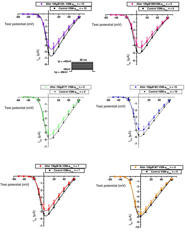
5.1. Effect of Diltiazem and Diltiazem analogs on the characteristics of Ba2+ currents through L-VDCC……….37
The Cav1.2 calcium channel in cardiac and smooth muscle……….38
Current-voltage relationships(I-V curve)………..40
Voltage-dependent tonic block………46
Use-dependent block (UDB) by Diltiazem and Diltiazem analogs………..46
Action of Diltiazem and Diltiazem analogs on the time course of IBa………54
Effect of drugs on the steady-state inactivation of HHT-Cav1.2 channel………..58
Effects of Diltiazem and a novel Diltiazem analog M8 on mouse ventricular cardiomyocytes………..66 5.2. Langendorff hearts……….70 5.2.1. Antiarrhythmic effect of Diltiazem and selected Diltiazem analogs………..76
5.3. Electrically evoked cystolic Ca2+ transients in single cardiac mouse myocytes………78
6. DISCUSSION………..82 7. Limitations of the study future plans and final conclusion……….97 8. BIBLIOGRAPHY………..102
1
1. INTRODUCTION
Ca2+ ions play crucially important roles in regulating a variety of cellular functions. They provide the basis for excitability in nerve and muscle cells. In neurons, they trigger the release of neurotransmitters from nerve the terminal. In heart and skeletal muscle, they regulate excitability and transform the action potential in mechanical contraction. In addition, Ca2+ ions are involved in a broad range of cell regulatory processes – e.g. hormone secretion and volume regulation, gene expression and mediating cell death. Several hereditary diseases have been linked to mutations in genes encoding ion channels. Timothy’s syndrome [1], which is a novel disorder characterized by multiorgan dysfunction including lethal arrhythmias, webbing of fingers and toes, congenital heart disease, immune deficiency, intermittent hypoglycemia, cognitive abnormalities, and autism. In every case, Timothy syndrome results from the identical, de novo CaV1.2 missense mutation G406R in exon 8a. CaV1.2 is expressed in all affected tissues. Functional expression reveals that G406R produces maintained inward Ca2+ currents by causing nearly complete loss of voltage-dependent channel inactivation. This likely induces intracellular Ca2+ overload in multiple cell types. In the heart, prolonged Ca2+ current delays cardiomyocyte repolarization and increases risk of arrhythmia, the ultimate cause of death in this disorder. These discoveries establish the importance of CaV1.2 in human physiology and development and implicate Ca2+ signaling in autism. Molecular electrophysiological investigation of these so-called channelopathies elucidate the pathophysiology of these rare diseases and help to understand disease-causing mechanisms in other, more common disorders with similar symptoms. Furthermore, insight into the molecular basis of these disorders allows a more rational approach to therapy. On the other hand, disease-causing mutations point to functional important parts of the protein and help to understand ion channel gating on a molecular level.
2
1.1. Cardiac L-VDCC Structure
The L-VDCCs are heterotetrameric polypeptide complexes comprised of the
α
1,α
2/δ
, andβ
(and in some tissues
γ
) subunits (See Figure 1 [2]) that allow depolarization-induced calcium influx into the cytosol. In all excitable tissues, Ca2+ channels invariably containα
1,
α
2/δ
, andβ
subunits. These are considered the functional minimum “core” for Ca2+ channel assembly. The accessory subunits (β
,α
2/
δ
) are tightly, but not covalently, bound to theα
1 subunitand modulate the biophysical properties and trafficking of the
α
1 subunit to the membrane.The stoichiometric ratio between the
α
1,α
2/
δ
and theβ
2 subunits is 1:1:1 [3].Figure 1. Structural organization of L-VDCC. Sites of interaction between subunits are indicated. The numbers point to areas found to be important to specific channel functions. The EF hand, A, c, and IQ motifs represent specific peptide sequences involved in CaM binding. Key amino acids required for Ca2+ antagonist binding are
represented in red letters. At least 5 consensus sites for phosphorylation by cAMP-dependent PKA have been discovered within the C-terminal tail of α1C. AKAP79 (79 kDa A-kinase-anchoring protein) helps to target PKA to its specific substrate. COOH, carboxy-terminal.
3
1.1.1.
αααα1
subunits
The founding members of this superfamily are the voltage-gated sodium channels (Na+). The Ca2+ channel
α
1 subunit (170-240 kDa) consists of four homologous motifs (I-IV) each
composed of six membrane-spanning
α
-helices (termed S1 to S6) linked by variable cytoplasmic loops (linkers) between the S6 and S1 segments. The different types ofα
1-subunits differ most strikingly in two principal regions: the cytoplasmic loop linking motifs II and III and the carboxy-terminal region. The currents that are supplied by the voltage-dependent calcium channels (VDCCs) are called: L-, N-, P-, Q- (or P/Q) - R- and T-types. To date 10
α
1 subunit genes have been identified (Table 1 and 2 [4,5]), separated into fourclasses. The first class is the L-type channels, consisting of Cav1.1 (
α
1S), 1.2 (α
1C), 1.3 (α
1D),1.4 (
α
1F). Only theα
1C (DHP-sensitive) is expressed in high levels in cardiac muscle. Thesecond class contains Cav2.1 (
α
1A), 2.2 (α
1B), 2.3 (α
1E) which form P/Q-, N- and possiblyR-type channels, respectively and are all found in brain. They are primarily responsible for initiation of synaptic transmission at fast synapses in the nervous system. They have a larger intracellular loop connecting domains II and III, which contains a synaptic protein interaction site that binds SNARE proteins involved in exocytosis [6]. The third class of channels are the T-type channels, Cav3.1 (
α
1G), 3.2 (α
1H), 3.3 (α
1I) which are localized to thebrain, kidney and heart and originally called low voltage activated (LVA) channels and unlike L-type channels, they are relatively insensitive to DHPs. Cav3 channels conduct T-type Ca2+ currents which are important in wide variety of physiological functions, including neuronal firing, hormone secretion, smooth muscle contraction, cell proliferation of some cardiac tissue and myoblast fusion. In the heart, T-type channels are abundant in sino-atrial pacemaker cells and Purkinje fibers of many species and are important in pacemaker activity by setting the frequency of action potential firing. It has been reported that the T- type channels are re-expressed in the ventricle of some animal models of heart failure (HF) suggesting that T-channels play a role in cardiac disease. The neuronal T-type channels can generate low-threshold spikes that lead to burst firing and oscillations which are prominent in the thalamus and implicated in variety of neurological disorders [5]. In addition to these well characterized Ca2+ channels, the cloning of a single Cav-like protein suggest the
4
existence of a fourth subfamily [7]. This novel protein contains conserved amino acids found in Ca2+ (EEEE) or Na+ channels (DEKA), including EEKE residues in the corresponding region of its pore loops. However, this new putative channel has not been functionally expressed in a heterologous expression system (Xenopusleavis oocytes).
The
α
1 subunit incorporates the ion-selective pore, voltage sensor, gating machinery, andthe binding sites for channel-modulating drugs [3,8,9], and is autoregulatory. The
α
1-subunitis a substrate for protein kinase A, protein kinase C, and Ca2+-calmodulin dependent kinase. The pore is asymmetric, with conserved glutamate (EEEE) or aspartate residues comprising the ion-selectivity filter(s) [10-13], located between segments S5 and S6 of each motif. Cloning and analysis of the VDCC have revealed that the positively charged fourth transmembrane segment (S4) of each motif is highly conserved and is likely to form an
α
-helix in which every thirdor fourth residue is basic (Arg or Lys). It is thought that the S4α
-helices traverse the membrane electric field and that, as a response to a depolarizing stimulus, they move outwardinto the extracellular space and initiate conformational changes from non-conducting to conductingstates of the channel (‘sliding –helix model’) (reviewed in ref. [14]). However, the emerging models are still controversial. Most structure-function studies support a spiral or rotational motion of the S4 or S3 plus S4α
-helices through the channel protein in order to move gating charges across the membrane electric field.5
6
1.1.2. Accessory subunits
αααα
2222////δδδδ
subunits
The
α
2δ
subunits are closely associated with theα
1 subunit by surface interaction andintracellularly linked through a disulfide bridge to a small protein, the
δ
subunit. Theα
2subunit is entirely extracellular and
δ
has a single transmembrane region with a very short Tab.27
intracellular part. The
α
2 andδ
subunits are encoded by the same gene which is separatedby proteolytic cleavage [15]. Ellis et al. [16] first cloned the
α
2/δ
subunit from rabbit skeletal muscle thinking there was only one product from the gene. Presently, at least 4 isoforms encoded by separate genes and have been identified (α
2/δ
1, 2, 3, 4) [15,17,18]. Theissue of in vivo structure-function has yet to be resolved. In heterologous expression systems, co-expression of the
α
2/δ
subunit affectsα
1 function by increasing channeldensity, charge movement and Bmax of drug binding (e.g. the DHP, isradipine), with smaller effects on KD and variable minor effects on channel kinetics [19-21] (and references cited
therein). It is probable that
α
2/δ
andβ
subunits “drive" theα
1 subunit to the membrane inthe correct insertion mode. The
α
2/δ
1 subunit is ubiquitously distributed, and possesses a stereo-selective high-affinity binding site for certain GABA-antagonists, such as the drug gabapentin, (1-aminomethyl cyclohexane acetic acid) which is widely used to treat epilepsy, pain, sleep disorders, and many other paroxysmal neurological conditions [22-24]. Theα
2/δ
2subunit also binds gabapentin but at a low affinity, while
α
2/δ
3 andα
2/δ
4 do not bind thisdrug. Mice deficient in
α
2/δ
2 exhibit neurological dysfunction such as enhanced seizure susceptibility and cardiac abnormalities, viz. a tendency to bradycardia [25]. The recently cloned humanα
2/δ
4[17]is localized to fetal liver, colon, pituitary, and adrenal gland, and is associated with theα
1C-subunit (Cav 1.2) and theβ
3 subunit. This reinforces the complexityof L-VDCCs, since subunit association appears to confer biophysical properties [10,20]. This rich diversity opens avenues for exciting physiological and pathological discoveries [26].
The
γγγγ
subunits
It was originally thought that the
γ
subunit was the product of a single gene and only existed in skeletal muscle [18]. It is interesting that characterization of a genetic defect that induces epileptic seizures in stargazer mice [27] led to the detection of a family of at least 5 novel isoforms of theγ
subunit that are almost exclusively expressed in brain. To date, 8 genes, encoding a variety ofγ
subunit isoforms have been identified [28]. Although Cavγ
1is associated specifically with skeletal muscle Cav1.1 channels, there is evidence that theγ
2 subunit interacts with AMPA receptor subunits [29] and possibly othermembrane-8
signaling proteins. Unlike other auxiliary subunits (
β
andα
2/δ
), theγ
subunits do not have a significant role in the membrane trafficking of the calcium channels.γ
1 modulates thebiophysical properties of the Ca2+ channel. Clearly this is a very important subunit, but since it does not appear to be expressed in heart, it is not discussed further.
ββββ
Subunit structure
Four
β
subunit (β
1−β
4) isoforms have been described. All are hydrophilic, non-glycosylated,and located within the cell and only
β
2 has been reproducibly shown to form cardiacL-VDCCs. The
β
subunit does not have a membrane spanning region. It is tightly bound to a highly conserved motif in the cytoplasmic linker between repeats I and II of all cloned high voltage-activatedα
1 subunit isoforms, called theα
-interacting domain (AID) [30,31], andalso to a secondary site [32]. Just recently, elucidation of the high-resolution 3D structure [32-34] of a
β
subunit has shed light on the molecular mechanism of binding of theα
1 toβ
subunits. Previous work suggested that the
β
subunit interacts withα
1 primarily through ahighly conserved 30 amino acid motif within the
β
-subunit, called theβ
-interaction domain (BID), which binds directly to the AID. However, Van Petegem and colleagues [33] reported that the BID engages the AID through a conserved hydrophobic cleft, and named theα
-binding pocket (ABP). Interference with AID-ABP -binding might provide a novel way to modulate Ca2+ channel function in pathological states. The I-II loop of theα
1-subunit
contains an endoplasmic reticulum retention signal that restricts cell surface expression. The
β
-subunit reverses the inhibition imposed by the retention signal [35].The roles of the
ββββ
auxiliary subunits in influencing channel function
Though much knowledge has been accumulated about the multiple roles for the
β
-subunit in the processing and functioning of L-VDCC using heterologous recombinant co-expression of the different subunits [20,36], the physiological significance of subunit interaction in the context of native tissue is only just beginning to be studied via transgenic approaches and adenovirus-mediated intracellular incorporation of genes, encoding Ca2+ channelβ
-subunits into single cells [3,37,38]. In general, co-expression of theβ
-subunits modulates the9
biophysical properties of the L-VDCC
α
1 subunit, producing a substantial increase in Ca2+current amplitude and/or changes in the current kinetics (leftward shift of the current-voltage (I-V) relationship) which is consistent with the involvement of the S4 region of the
α
1 subunit voltage sensor region. Moreover, it has been shown thatα
1 subunitsexpressed in the absence of
β
subunits are not regulated by theβ
-adrenergic system [39] or by pH changes [40]. Frequency- and prepulse-dependent facilitation of L-VDCC activity is regulated by certain classes ofβ
subunits [41]. By employing an antisense strategy to lower the numbers of endogenousβ
subunits in oocytes, leaving onlyα
1 subunits intact, a loss ofcurrent occurred, implying that the
β
subunit functions in the assembly and expression of theα
1 subunit [42]. Dr. Schwartz’s group provided evidence that theβ
subunits have achaperone-like role, in trafficking
α
1C subunits from the ER to the plasma membrane, andinserting in its proper geometry [43]. Viard et al. [44] demonstrated that a region of this
β
2a subunit is involved in phosphatidylinositol 3 kinases (PI3K)-induced increases of Cav1.2(rat brain) channel density. The PIK3-induced regulation is mediated by PIP3–activated Akt/PKB and requires phosphorylation of Cav
β
2 subunits on Ser574, which is common to allsplice variants of the
β
2 subunits. These results indicate that PIK3 regulates Ca2+ channeltrafficking to the plasma membrane and may be a general mechanism for the regulation of Ca2+ entry into excitable cells [44]. The
β
1-subunit also has a crucial role in EC coupling as
proven by
β
1-KO mice with impaired EC coupling and early lethality [45]. The exactmechanism for the lack of EC coupling is not known but it is possible that the deficiency in the assembly process of the
α
1/β
complex results in the degradation of theα
1 subunit. Therole of the
β
2 subunit in EC coupling is unclear [46].β
3 null mice have no detectableabnormalities in the heart [47].
Hullin et al. [48] cloned two distinct
β
subunits,β
2 andβ
3, from rabbit heart and showed anassociation with the
α
1 subunit of the L-VDCC. The amino acid homology of these subunitswas similar to that of
β
1, originally cloned from skeletal muscle [9]. It should be noted that10
associated with membrane targeting of non-transmembrane proteins (such as the
β
subunit) to specific areas including the plasma membrane. The role of theβ
subunits in L-VDCC expression is well characterized although we still do not know how theβ
subunits modulate preexistingα
1 subunit expression. Colecraft et al. [51] devised a novel system in whichrecombinant adenoviruses were used to express GFP-fused
β
1–4 subunits in cultured adultrat cardiomyocytes. While all four subunits (
β
1b,β
2a,β
3,β
4) increased ICa density, theireffects on inactivation kinetics were non-uniform. The conclusion of this study was that overexpression of the newly cloned rat splice variant of a
β
2b in adult rat heart cells yieldedchannels that were identical to that in the native unmodified rat heart cells.
β
3 is most abundant in brain but also is expressed in heart, aorta, lung, trachea, and skeletalmuscle. Expression levels of various
β
subunit isoforms [48,51,52] indicate that altered single-channel behavior in human heart may be due to differential effects and changes inβ
subunit gene products.1.1.3. Regulation of intracellular Ca
2+: Cardiac Dysfunction
The control of myocardial Ca2+ is crucial for the maintenance of normal rhythm, regulation of contraction, enzymatic reactions, as well as, growth and development. For many years, drugs modifying cardiac function by affecting myocardial Ca2+ have been used to modulate Ca2+ fluxes, levels of Ca2+ at storage sites, or increase the Ca2+ sensitivity of the contractile proteins. In general, drugs that reduce Ca2+ loading of the cardiac myocyte serve to “protect” the heart from the ultimate cause of myocardial cell death, i.e., Ca2+ overload. Among the many problems that arise from Ca2+ overload, ventricular tachycardia is the most prominent and is often life threatening. Both early and delayed after-depolarizations can be treated with Ca2+ channel-blockers (CCBs). Likewise, arrhythmias ascribable to disorders of conduction are also treated with CCBs that function presumably by inhibiting conduction disturbances in the sinoatrial or atrioventricular junction. Clinical trials show that CCB’s do not reduce mortality of heart failure. The state of art in pharmacotherapy of heart failure, considering its epidemic nature in the world, is very disappointing. Recent reviews have
11
emphasized that we are dealing with a highly complex, multifaceted disease almost similar to the cancers [53-57].
Although considerable information is known regarding the L-VDCC and its role in EC coupling, the consequence of increased Ca2+ channel density remains speculative and controversial. In Dr. Schwartz’s laboratory a transgenic mouse model was designed overexpressing the L-VDCC
α
1C subunit (α
1C-Tg) as a way to increase [Ca2+]i. The transgenicmouse model exhibits a sustained, low level [Ca2+]i increase in cardiac cells throughout the life of the animal; hypertrophy develops slowly and the ensuing failure occurs at 9-12 months. This model mimics human dilated, hypertrophic-(ischemic) cardiomyopathy.
1.1.4. Ca
2+channel antagonists (blockers, “CCB”) and Ca
2+binding
domains
L-VDCC is an important pharmacologic target in the treatment of a number of conditions. In the late 1960s, Fleckenstein showed that Ca2+-antagonists like verapamil protected the rat heart against structural damage associated with prolonged [Ca2+]i overload. Among the many problems that arise from Ca2+ overload, ventricular ectopic rhythm is the most prominent (ventricular fibrillation). In pharmacological models, the CCBs are considered promising drugs to treat supraventricular arrhythmias, hopefully preventing lethal ventricular fibrillation (VF).
Using a variety of techniques, several laboratories have identified individual amino acids within the L-VDCC
α
1 subunit that participate in the formation of the major drug-bindingdomains. Three classes of organic CCBs, include: Dihydropyridines (DHPs), phenylalkylamines (PAAs), and benzothiazepines (BTZs). Each drug type, DHP, PAA and BTZ has separate but overlapping, or allosterically-linked, Ca2+ channel-binding sites at IIIS6 and IVS6, IVS6 and IIIS6, and IVS6 motifs (Figure 2), respectively. Nine amino acid residues in segments IIIS5 [58], IIIS6 [59] and IVS6 contribute to the “DHP pocket”. The four amino acid region (YMAI) in motif IVS6 is a common binding site for both BTZ [60] and PAA [61,62]. DHPs block the calcium channel at the extracellular level and have greater vascular selectivity than other classes of CCBs. When used clinically, as a result of vigorous peripherial vasodilatation, they tend to cause reflex tachycardia and often increase
12
contractility, as catecholamines activate the calcium channel intracellularly, on a different site from that blocked by DHPs. Tissue selectivity is considered to be one of the most beneficial properties of CCBs as it diminishes the likelihood of undesirable side-effects. In general, vascular selectivity permits coronary and peripherial dilatation in the absence of significant myocardial depression. Nifedipine is at least 10 times more vascular than myocardial-selective compared to Verapamil and Diltiazem. Nisoldipine and felodipine are at least 1000 times more selective than nifedipine. On the other hand it can be a disadvantage, causing adaptive reactions. Verapamil and Diltiazem have prominent effects on nodal tissues. The BTZs are thought to approach their binding site on Cav1.2 from the extracellular face of the plasma membrane [63]. Verapamil and Diltiazem have a use-dependent or a frequency-dependent effect. The more frequently the calcium channel opens, the better the penetration to the binding site. This explains their effect on nodal tissue in paroxysmal supraventricular tachycardia. The general absence of ‘use-dependence’ and the presence of voltage-sensitivity of DHP binding explain their vascular selectivity. The use-dependent blockade by diltiazem of L-VDCCs would be expected to inhibit large increases in Ca2+ influx accompanying a sudden increase in heart rate. In addition, the acceleration of inactivation would be expected to reduce Ca2+ influx during action potentials that arise from normal negative resting potentials. Both of these effects likely contribute to the beneficial effect of Diltiazem when used as a prophylactic or depressant of supraventricular tachycardia [64,65]. Diltiazem facilitates the inactivation of the channel both by accelerating the transition from the activated state to the inactivated state and by decelerating the transition from the inactivated state to the available state. However, these drugs have not emerged unequivocally favorable in all clinical studies to date. Verapamil and Diltiazem can, in some cases, prevent episodes of acute ischemic VF in humans, but they do not have much of a beneficial effect on overall mortality as do the
β
-blockers, and the ACE inhibitors. The clinical implications of this finding reported in different clinical trials are similar to that reported for encaidine and flecainidine in the Cardiac Arrhythmia Suppression Trial [66]. Patients suffering from coronary disease may die of either heart failure or arrhythmias. Likewise, arrhythmias ascribable to disorders of conduction are also treated with CCBs that function, presumably, by inhibiting conduction disturbances in the sinoatrial or atrioventricular junction. Although CCBs bind specifically to regions of theα
1Csubunit of13
the L-VDCC, these drugs are not currently judged as being helpful in the setting of congestive heart failure. In fact, all clinical trials to date, with the exception of the Prospective Randomized Amlodipine Survival Evaluation (PRAISE I) study in where the patents had congestive heart failure, were a failure. However, a sub-group analysis (PRAISE II) revealed that improved clinical symptoms were seen only in patients with heart failure of a non-ischemic cardiomyopathic nature. A favorable effect on survival was found only in patients without a history of angina [67]. In the Third Vasodilator-Heart Failure Trial [68] (V-HeFT III), felodipine was administered to patients with congestive heart failure in a setting of stable therapy with enalapril, diuretics and digoxin. The drug had neither a beneficial or deteriorating effect despite the improvement in exercise performance and LV function, as reported previously in the V-HeFT II trial in patients with chronic CHF [69]. Chronic Nifedipine (has a strong peripheral vasodilating effects) therapy caused a higher incidence of clinical deterioration and worsening of HF [70,71]. The DEFIANT-I study (Doppler Flow and Echocardiography in Functional Cardiac Insufficiency: Assessment of Nisoldipine Therapy) was a double-blind randomized study of the effects of the DHP nisoldipine on left ventricular (LV) size and function after acute myocardial infarction (MI). Diastolic LV function improved in patients recovering from acute MI [72]. The Danish Verapamil Infarction Trial II (DAVIT II) demonstrated that long term treatment with verapamil significantly improved reinfarction survival after acute MI. Verapamil significantly reduced (35%) the 18 month mortality rate, but produced no change in mortality in the heart failure (HF) group [73]. Hypertensive patients are often treated with CCBs to reduce cardiovascular disease risk, but the overall benefit compared with atenolol and hydrochlorothiazide and ACE inhibitors is controversial and problematic. The Controlled Onset Verapamil Investigation of Cardiovascular End Points (CONVINCE) trial indicated that the effectiveness of CCB therapy was comparable to diuretic and
β
-blocker treatment in reducing cardiovascular disease [74]. According to the Multicenter Diltiazem Postinfarction Trial [75] analysis diltiazem exerted no overall effect on mortality or cardiac events in a large population of patients with previous infarction, but in patients with pulmonary congestion diltiazem was associated with an increased number of cardiac events and mortality. Interestingly, the increase in mortality was not accompanied by a worsening of HF. Despite these concerns, in the Studies of Left Ventricular Dysfunction Trial14
(SOLVD), 3-35% of the patients were treated with CCBs in addition to digitalis and diuretics. CCB use was associated with significantly increased risks of fatal and nonfatal MI [76]. CCBs have a favorable systemic vasodilator effect and should improve diastolic relaxation. One would assume that CCBs inhibiting Ca2+ influx into myocardial cells might be beneficial because theoretically they could reduce Ca2+ overload, an important trigger for activating certain downstream Ca2+-dependent pathways involved in hypertrophy and cardiac dysfunction. Clinical trials to date however have been disappointing. In fact, it has been suggested that the effects of CCBs on mortality in patients with HF may be associated with increased sympathetic activity. Summing up the therapy of heart failure, the
β
-AR blockers, ACE Inhibitors (angiotensin-converting enzyme inhibitors)), diuretics, and aldosterone receptor(s) inhibitors (spironolactone and new derivatives) have achieved therapeutic success probably through multiple actions that culminate in a positive "remodeling" of the diseased heart ([77] Review). The state of art in the pharmacotherapy of heart failure, considering its epidemic nature in the world, is also disappointing.As discussed above, although CCBs bind specifically to regions of the
α
1C subunit of theL-VDCC, these drugs are not currently judged as being helpful in the setting of congestive heart failure, and should be if used at all, with caution. All clinical trials underscore the use of drugs that inhibit the sympathetic nervous system and reduce the load on the heart.
Use-dependence
Less detailed structural information is known regarding the mechanism of use-dependent block, a feature that is critical to the activity of therapeutically successful L-VDCC antagonists. The modulated receptor theory states that drug affinity for a specific receptor on the channel is modulated by the channel state. The model has been proposed (modulated receptor hypothesis) that can account for the voltage and use-dependent block of Na+ channels by local anesthetics [78,79]. Each channel (sodium and calcium) blocker has a characteristic association and dissociation rate constant for channels in each of these states. The more frequently the Ca2+ channel opens, the better penetration is for the drug to reach the binding site. Verapamil and Diltiazem preferentially interact with the open and inactivated states of the channel (reviewed in ref.[80], explaining their preferential effect on nodal tissue. Single amino acids have now been identified as inactivation determinants in
15
motifs IIIS6, IVS6 and IVS5 with some of them also serving as high affinity determinants for the DHP receptor site [80-82].
Diltiazem is the prototype benzothiazepine and is the only drug of this type currently in clinical use (Cardizem). Diltiazem exhibits modest selectivity for block of vascular muscle over cardiac muscle L-type channels, but is also useful in treatment of arrhythmias due to the block of L-type channels in the heart. The BTZ binding site on L-VDCCs is allosterically linked to the binding sites for PAAs and DHPs. BTZ binding inhibits PAA binding but stimulates DHP binding to L-type channels in a temperature dependent manner. Diltiazem was found to produce more tonic block than methoxyverapamil but less than nifedipine and nitrendipine. The frequency-dependent block by Diltiazem is due to rapid binding to the open state and preferential binding to the inactivated channel.
1.1.5. Voltage-gated cation channels
The gating of most ion channels is regulated by different specific mechanisms, which change their permeation properties: membrane potential (voltage-gated ion channels), specific chemical signals such as Ca2+ or synaptic transmitters (ligand gated ion channels), or changes in the membrane conformation (mechanosensitive or volume sensitive ion channels etc.). Voltage-gated cation channels are responsible for the generation and propagation of action potentials. Whereas potassium channels form tetramers of four identical domains, the main molecule of the Na+ and Ca2+ channels is one large subunit. The pore of the voltage-gated cation channels is formed by the loops, which are located in between the S5 and S6 transmembrane regions of the four domains/motifs. The S4 segments contain four to eight positively charged residues conferring voltage dependence to the channel protein (for a review see [3,83]. At the resting membrane potential (-60 to –90mV) the channels usually reside in closed state and are not permeable for ions (Figure 2 [83]). At very hyperpolarized potentials, where the channels are in their resting state, inactivation is removed (steady-state inactivation curve). This means that the inactivation gate is not occluding the channel and if nothing else was in the way ions could flow through the pore. However at those very negative potentials, the activation gates are all closed. Those closed gates block the pore and prevent conduction. Upon depolarization, they open their gate and can conduct ions, a process called activation. This is triggered by the movement of the voltage sensors. Some
16
of the channels, after the opening of the activation gate, can close another existing gate upon ongoing depolarization a process called inactivation. The inactivation gates begin to close, but with a slower time course than the activation gates. The inactivated channels require time at hyperpolarized potentials before they can be activated again (recovery from inactivation). During this refractory period the channels recover from the inactivated state. Voltage gated ion channels can have more inactivated states which are kinetically distinct. Upon hyperpolarization, the opened activation gate closes in a process called deactivation. In general, voltage-gated ion channels are characterized by at least one open (O) and one closed (C) state and may have one or more inactivated (I) states (Figure 2A [83]).
Figure 2. Gating transitions in voltage-gated cation channels. (A) Schematic representation of the main conformational states of voltage-gated channels and the transitions between them. (B) Mechanistic view of channel gating [83].
17
2. MOLECULAR BIOLOGICAL METHODS
2.1. cDNA constructs and mRNA
Restriction enzymes were used to linearize plasmid cDNA for in vitro cRNA synthesis. All restriction enzymes were used according to the manuals of the suppliers. The cDNA for the human heart
α
1C (HHT-α
1C, Accession number L04569) [84] subunit used in this project wassubcloned into the ΗindIII/NotI sites of the pBS SK- (Stratagene) vector. Rabbit lung
α
1Cb (VSM-α
1Cb Accession number X55763) [85] was subcloned into the pSP72 vector(Promega). The cDNAs must be linearized prior to generating mRNA. This allows the RNA polymerase to transcribe the cDNA only once, at the end of the cDNA the RNA polymerase “falls off” the DNA.
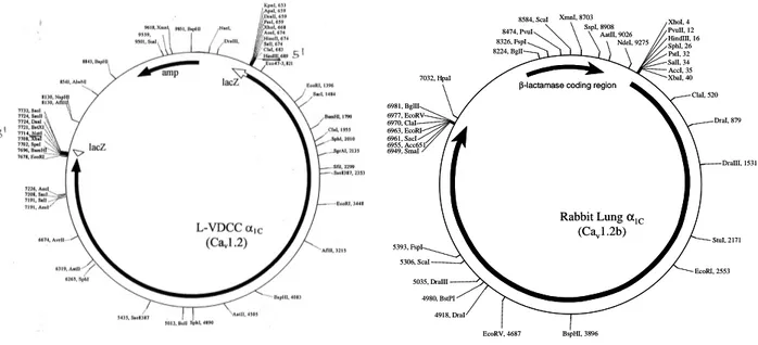
Figure 3. The map of pBS SK-vector and the pSP72-vector with the insertion of recombinant HHT-α1Cand
Lung-α1Cb, respectively. The vectors are showing the positions of the ampicillin resistance gene or the β−lactamase
coding region and selection of the most important restriction sites.
All restriction enzymes that were used to linearize the plasmid cDNAs for in vitro mRNA synthesis were used according to the protocols of the supplier.
For these experiments, 30-50
µ
g of DNA was digested overnight (to a final volume of 400µ
l) with specific restriction enzymes. HHT-α
1C was linearized with XbaI (NEB),VSM-α
1Cb linearized with Acc651 (a neoschizomer of KpnI, NEB), rabbit skeletal muscleα
2/δ−1
XbaI, 40 ClaI, 520 DraI, 879 DraIII, 1531 StuI, 2171 EcoRI, 2553 BspHI, 3896 EcoRV, 4687 4918, DraI 5035, DraIII 4980, BstPI 5306, ScaI 5393, FspI 6955, Acc651 7032, HpaI 8224, BglI 8326, FspI 8474, PvuI 8584, ScaI XmnI, 8703 SspI, 8908 AatII, 9026 NdeI, 9275 Rabbit Lungα1C (Cav1.2b)
β-lactamase coding region
6949, SmaI 6961, SacI 6963, EcoRI 6970, ClaI 6977, EcoRV 6981, BglII AccI, 35 SalI, 34 PstI, 32 SphI, 26 HindIII, 16 PvuII, 12 XhoI, 4 XbaI, 40 ClaI, 520 DraI, 879 DraIII, 1531 StuI, 2171 EcoRI, 2553 BspHI, 3896 EcoRV, 4687 4918, DraI 5035, DraIII 4980, BstPI 5306, ScaI 5393, FspI 6955, Acc651 7032, HpaI 8224, BglI 8326, FspI 8474, PvuI 8584, ScaI XmnI, 8703 SspI, 8908 AatII, 9026 NdeI, 9275 Rabbit Lungα1C (Cav1.2b)
β-lactamase coding region
6949, SmaI 6961, SacI 6963, EcoRI 6970, ClaI 6977, EcoRV 6981, BglII AccI, 35 SalI, 34 PstI, 32 SphI, 26 HindIII, 16 PvuII, 12 XhoI, 4
18
[16](SK-
α
2, Accession number M21948) was linearized with NheI (NEB) and human heartβ
3-subunit (Accession number L06112) [86] was linearized with NotI (NEB). These enzymeswere chosen because they have unique restriction sites (cut only once) and generate 5’ overhangs (Figure 4).
5’-CCCGGGGGTACCGAGCTC--GGGCCCCCATGGCTCGAG-3’
SmaI
Acc651
SacI
5’-CCCGGGGGTACCGAGCTC--GGGCCCCCATGGCTCGAG-3’
SmaI
Acc651
SacI
5’-ACTAGTTCTAGAGCGGCCGC--TGATCAAGATCTCGCCGGCG-3’
SpeI
XbaI
NotI
5’-ACTAGTTCTAGAGCGGCCGC--TGATCAAGATCTCGCCGGCG-3’
SpeI
XbaI
NotI
Human Heart
α
1CRabbit Lung
α
1CFigure 4. Enzyme restrictions of the Lung-α1Cb and HHT-α1CDNA molecules, respectively. Figure illustrates the
detailed positions of the recognition sequences for Acc651 and Xba1, respectively. Before transcription of cDNA to mRNA the circular DNA has to be linerized using restriction enzymes. These endonucleases break the internal phosphodiesterase bonds at a very specific position which is characteristic for every enzyme.
After overnight digestion, the samples were phenol/chloroform extracted and then precipitated with Ammonium Acetate and Ethanol to remove impurities. The DNA pellet was resuspended in 30
µ
l RNase-free water and the concentration was determined by the Optical Density at wavelengths 260 and 280 (OD260/280),2.1.2. In vitro RNA synthesis
All reagents and materials were kept on ice. The commercial kit mMessageTM mMachine (Ambion) was used to generate mRNA using the T7 promoter for HHT-
α
1C,α
2-1[72]
andh
β
3 and the SP6 promoter kit was used for RL-α
1Cb following the manufacturer’s protocol.1
µ
l GTP was added to the reaction mixture to compensate for the long message. In brief, the linearized plasmid DNA was incubated for 2 hours with the transcription buffer, the ribonucleotide mix and the enzyme mix at 37°C. Additionally 1µ
l RNAse-free DNAse type I19
(2U/
µ
l) was added and the mixture was incubated for 15 more minutes at 37°C to remove DNA. After mRNA synthesis, the mRNA was precipitated with Lithium Chloride (as stated in the manufacturer’s protocol) followed by a resuspension in 100µ
l RNase-free water. The mRNA was then precipitated with Ammonium Acetate and Ethanol to further remove any unincorporated nucleotides. The removal of unincorporated nucleotides is essential because their presence will artificially increase the mRNA concentration as determined by OD260/280. After precipitation with ammonium acetate, the mRNA is resuspended in RNase-free water. The amount of water used depended on the size of the mRNA pellet, usually between 50-80µ
l. The concentration of mRNA was determined by OD260/280.20
3. MATERIALS AND METHODS
3.2. Spectroscopic measurement of nucleic acid concentration
To determine the concentration of the DNA and RNA probes the absorption at 260nm (A260) optical wavelength was measured on an automated spectrophotometer (UV-Visible Spectrophotometer, UV-1650 PC, SHIMADZU). Appropriate nucleic acid dilutions were prepared to obtain absorption between 0 and 1. An Optical Density (OD) of 1 corresponds to a concentration of 50
µ
g/ml for DNA and 40µ
g/ml for RNA. A qualitative test was done by measuring the ratio A260/A280 (Absorption at 260 and 280nm wavelengths). A ratio between 1.6 and 2 was considered to indicate purified DNA or RNA.3.1. Agarose gel electrophoresis
The quality of the mRNA is determined by running formaldehyde based RNA gel electrophoresis on the samples. The mRNA is place into a solution containing formaldehyde, formamide and MOPS. This is run on a 1% agarose gel also containing formaldehyde, formamide and MOPS. Staining the gel with ethidium bromide after electrophoresis (followed by destaining with water) allows the mRNA to become visible under UV light. Any unincorporated nucleotides show as a blurry band at a lower molecular weight.
3.3. Oocyte preparation and injection
The use of Xenopus laevis oocytes as an expression system for ion channels was first described by Miledi et al. [87] in 1983 and since then has become a standard technique for investigation of ion channels. Oocytes are especially suitable for electrophysiological recordings, since they have only a few endogenous channels, and faithfully express foreign RNA that has been injected.
3.3.1 Frog surgery
All the experiments were carried out in accordance with the University of Cincinnati Institutional Animal Care and Use Committee. Adult female Xenopus laevis (XenopusI, Ann Arbor, MI) frogs with a length of at least 9cm were anesthetized in 1L 0.5% Tricaine water
21
solution (3-aminobenzoic acid ethyl ester methanesulfonate salt (MS-222), Sigma-Aldrich, USA) for about 15-20min. The frog was put on its back on ice after achieving the necessary anesthetic grade checked by the loss of the righting response and the loss of response to painful stimuli. Extremities and head were covered with ice in order to prolong the effect of the anesthetic and to prevent excessive bleeding. The surgical area and all surgical instruments were sterilized with 70% ethanol solution. Instruments were kept on a sterile wrap during the procedure. Skin, muscle fascia and muscle tissue were cut at a length of 1cm. A portion of the ovary was removed through this opening with forceps and scissors and placed in OR-2 medium.
3.3.2. Defolliculation and selection
Before defolliculation, oocytes were extensively washed with OR-2 medium (in mM: 82.5 NaCl, 2 KCl, 1 MgCl2, 5 HEPES, pH 7.5). Defolliculation was achieved by treatment with 2mg/ml collagenase (type IA; Sigma-Aldrich, USA) dissolved in OR-2 Ca2+ free medium. Usually 2 hours of gently shaking at room temperaturewere enough to defolliculate the oocytes completely without damaging the vitelline membrane. Defolliculated stage V-VI oocytes were incubated in P/S medium (in mM): 96 NaCl, 2.0 KCl, 1.0 MgCl2, 1.8 CaCl2, 5.0 HEPES, 2.5 sodium pyruvate, and 0.5 theophylline at pH 7.5. The P/S medium was supplemented with 100
µ
g/ml streptomycin and 100 units/ml penicillin at 19°C in low temperature aseptic incubator (VWR Scientific, WestChester, USA). Medium was changed every day. Before injection, a selection was donefor healthy stage V and VI oocytes [88].3.3.4. Oocyte microinjection
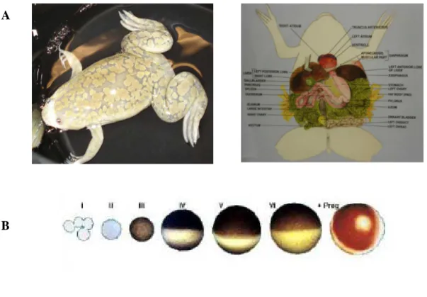
In vitro synthesized mRNA can be injected and expressed in Xenopus laevis oocytes. Borosilicate glass pipettes (210/310/510, Drummond Scientific, Broomall, PA USA) were pulled on a vertical pipette puller, Model 720 (David Kopf Instruments, Tujunga, California). The tips of the pipettes were broke under a StereoZoom 3 microscope (Nikon,, Japan) to obtain optimal diameter and sharpness (around 20
µ
m tip diameter). Pipettes were filled with heavy mineral oil (Sigma-Aldrich, USA) and were mounted in the head-stage of a Drummond Digital Microdispenser, Cat. #3-000-510 (Drummond Scientific Co., Broomall, PA USA). The pipette tip was filled with 2µ
l RNA.22
Figure 5. The Xenopus oocyte expression system. (A) Adult female Xenopus leavis frog (B) Developmental states of Xenopus leavis oocyte: healthy stage V and VI oocyte can be recognized by their size and by their clear differentiation of the animal (dark) and vegetal poles (light).
Oocytes were lined up on a plastic grid previously sterilized with 70% ethanol and covered with culture medium. 50nl undiluted mRNA were injected in the vegetal pole of a single oocyte. After injection oocytes were stored in groups of 20 per plates and incubated at 19°C. The amount of the injected RNA correlated well with expression levels, i.e. current size was dependent on the expression rate. With a concentration of 1
µ
g/µ
l mRNA, the amount of mRNA injected to each oocyte is about 50ng. In order to compare current amplitudes of Cav1.2 L-VDCC channels HHT-α
1C [84] and rabbit lung VSM-α
1Cb (a generousgift from Dr. R. Hullin) [85], RNA preparations were injected on the same day into the same batch of oocytes and measured in parallel on days 2-4 after injection. In order to increase current density and stabilize gating, the calcium channel
α
1C-subunit encoding mRNA wasco-injected with mRNA encoding the
β
3 [86] andα
2/δ−1
[16] calcium channel subunits in a 1:1:1 molar ratio (the final concentration remains 1µ
g/µ
l or 1ng/nl). To minimizeA
23
contamination with chloride current, oocytes were microinjected with 50nl of a 40mM K4-BAPTA solution (potassium 1,2-bis(2-amino-phenoxy)ethane-N,N,N’,N’-tetraacetate in 10mM HEPES, pH 7.05) 60min prior to current recording.
24
4. ELECTROPHYSIOLOGY
4.1. Reagents and solutions
Solutions for voltage-clamp measurements
All reagents were ordered from Sigma-Aldrich (St. Louis, MO USA).
4.2. General procedures
Most of the current knowledge about ion channels in cell membranes derived from experiments using voltage clamp. In the voltage clamp technique, the membrane potential is held constant with a feedback amplifier while the current flowing through the membrane is measured. Current is measured because the investigator has no direct way of measuring conductance. The method was developed by Cole [89] in 1949 and Hodgkin and Huxley [90]. Since that time many variants of the technique have evolved and voltage clamp measurements have been extended to a wide range of tissues.
Figure 6. (A) Simplified electrical circuit of membrane that contains only voltage-gated Na+ channels. Cm, membrane conductance; gNa, Na+ conductance. (B) The current elicited by a voltage clamp depolarization consist of
a brief initial capacity current followed by an ionic current. In this case, the ionic current only flows through voltage-gate channels. The Na+ current flows from the outside to the inside of the membrane. Edited by Ashcroft
FM, Ion Channels and Disease (Book), Academic Press, 2000, UK.
The current that flows through the membrane at any particular potential can be measured (It). This current is the sum of the ionic current (Ii), which represents current flow through open ion channels and the capacitive current (Ic), which is largely due to charging of the membrane capacitance. The magnitude of the capacity current is determined by the
25
capacitance of the cell membrane (Cm), which is normally 1µF/cm2, and it flows only while the voltage is changing (Figure 6).
Thus,
I
t=I
i+C
mx dV/dt
. Since IC =C dV/dt, once the potential is clamped, no capacitivecurrent flows. Hence, IC flows only during the membrane-clamping process. Clearly, the ionic current will be the same as the total membrane current. The good voltage clamp requires that the membrane potential be changed rapidly, so that the capacity current is over before the ionic current starts to flow. The speed of the voltage response is limited by the rate at which current can be injected to the microelectrode. This depends on the electrode resistance.
4.2.1. Basic apparatus with electrical and mechanical isolation
A patch-clamp set-up may consist of a chamber, a microscope (for cell visualization) placed on an anti-vibration table (pneumatic support for the patch clamp setup and passive support for the two electrode voltage clamp measurements), a patch clamp amplifier and pulse generator for voltage-clamping the cells, a micromanipulator holding the amplifier probe for positioning the attached patch pipette, and data-recording devices (oscilloscope, computer). A Faraday cage was build around the experimental setup and all metal parts near the head-stage were grounded, using the virtual ground input of the amplifier. An example of patch-clamp set-up is shown in Figure 7.
Figure 7. A common electrophysiology setup. To reduce vibration, the microscope with the recording chamber, manipulator and headstage should be mounted on a massive antivibration table (A and B). The glass microelectrode
(C) is connected to a feedback amplifier (D). The amplified and filtered signal is digitized by the AD converter (E).
The digitized signal is visualized and stored on a PC (F). Single-Channel Recording Second Edition (Book), Edited by Sakmann B. and Neher E., Plenum Press, 1995, USA
26
4.2.2. Glass electrodes
Glass microelectrodes were pulled with David Kopf vertical Pipette Puller Model 720 (David Kopf Instruments, Tujunga, California USA) from thin wall borosilicate glass pipettes with filament (1.2mm OD, 0.90mm ID; A-M Systems, INC. Everett, WA, USA). The microelectrodes for patch-clamp recordings were pulled with Sutter and fire polished (Garner Glass Company, Claremont, CA USA ID 1.10, OD 1.60, Glass type 7052). The resistance of the whole-cell patch clamp electrodes ranged between 2 and 4MΩ. Sharp glass electrodes for two-electrode voltage clamp (TEVC) were pulled in a way to minimize the access resistance while minimizing cell damage – 0.5 to 1.5MΩ with the standard 3M KCl electrode solution.
Ag/AgCl electrodes
Ag/AgCl pellet holder’s electrodes were purchased from Warner Instruments (Catalog# 64-1301). The bathing solution was connected to the ground through a 3M KCl/agar bridge (2% agarose in filtered 3M KCl).
4.2.3. Digitizing, recording and analysis
In the cardiomyocytes, the Axopatch-200A patch-clamp amplifier was used to record membrane currents. The voltage clamp amplifier output was digitized by a DIGIDATA 1200 AD/DA converter (Axon Instruments, Union City, USA), connected to a personal computer (PC 486) running Clampex 5.6 (Axon Instruments, Union City, USA) acquisition software under Microsoft DOS 6.2 (Microsoft Corp. Redmond, USA). In the two electrode voltage-clamp experiments in oocytes, we used Axovoltage-clamp-2A amplifier with pCLAMP software (version 5.5, Axon Instruments, USA) to generate voltage clamp protocols, acquire data and analyze current traces. Whole cell leakage currents were subtracted on-line using the P/4 procedure. Current were digitized at 1kHz after being filtered 1kHz. The data were stored on the computer hard drive for later analysis and a hard copy record was done for every protocol. Analysis was done on a PC using combination of Clampfit 6.03 (Axon), Origin (Version 7.0 Microcal, Northampton, USA) and Excel (Microsoft XP 2003) software.
27
4.3. Patch clamp
Instead of using sharp microelectrodes to puncture the membrane and penetrate the cell in traditional voltage clamping, patch-clamping uses a fire-polished pipette of about 1µm that is sealed to a ‘patch’ of membrane. After forming the cell-attached seal the membrane patch is destroyed by strong suction, electrically or it may be permeabilized with pore-forming antibiotic nystatin. This provides electrical continuity with the cell interior allowing currents through the whole of the cell membrane to be measured under of the whole-cell current. The tightness of the gigaseal prevents the leak currents flowing between the pipette and the reference electrode. However, the current through the series resistance of the pipette and the residual resistance of the ruptured patch is often sufficiently large to introduce significant voltage errors. It has an advantage that the intracellular solution can be manipulated however, but it has a disadvantage since the cytosolic constituents are lost from the cell by dialysis. A typical patch clamp circuit shown in Figure 8. The key difference between the two electrode voltage clamp is that the patch clamp uses a single electrode both to control the membrane potential and to measure the current.
Figure 8. One electrode continuous voltage clamp method (whole cell patch clamp circuit). A typical patch-clamp circuit is shown in Figure. One advantage of this system is that the patch-clamp uses a single electrode both to control the membrane potential and to measure current. The sensitive current-to-voltage converter is fabricated using a high megaohm resistor (Rf) and an operational amplifier (Feedback amplifier, A1). The patch pipette is
connected to the inverting input of a feedback amplifier and the command voltage is connected to the positive input. Since the A1 has extremely high gain, the potential at the negative input is forced to follow the command potential (Vcmd) at the positive input. All current flowing in the micropipette also flows through Rf. This current is
proportional to the voltage across Rf, which is measured at the output of the differential amplifier (A2). Edited by
28
4.3.1. Two-electrode voltage-clamp on
Xenopus laevis
oocytes
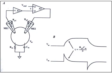
Experimental setup
Figure 9 shows a schematic diagram for the two electrode voltage clamp (TEVC) circuit. The oocyte is penetrated with two microelectrodes filled with electrolyte solution (typically 3M KCl), connected to the recording system through Ag-AgCl electrodes. In the two-electrode voltage clamp recording from oocytes, one intracellular electrode measures the membrane potential (voltage electrode-ME1) and the second passes sufficient current (current electrode-ME2) to maintain the desired voltage clamp, using a feedback circuit. The amount of current passed through the current electrode is determined by the discrepancy between the membrane potential and the command potential. When these two are equal, no current flows through the current electrode. A good voltage clamp requires that the membrane potential be changed rapidly so that the capacity current is over before the ionic current starts to flow. The main disadvantage of the TEVC is the relative slow response due to the high value of the capacitance of the oocyte membrane. The response time (
τ
) of a voltage clamp to a step voltage change is:τ
=RICm/A where RI is the resistance of the current-passing electrode, Cm is the membrane capacitance, and A is the gain of the command amplifier. Since not that much can be done about Cm, the only two things one can do in order to achieve fast clamping of the cell is to use the lowest RI and the largest A possible. The higher the electrode resistance, the more difficult it is to inject sufficient current fast. When studying voltage gated channels, one wants the fastest clamp attainable. A well-tuned, TEVC can clamp an oocytes fast enough to study the gating kinetics of most voltage-gated channels except Na+. A faster clamp can be achieved by recording from smaller oocytes.29
Figure 9. Conventional Two-Electrode Voltage Clamp. (A) The membrane potential (Vm) is recorded by a
unity-gain buffer amplifier (A1) connected to the voltage recording microelectrode (ME1). Vm is compared to the
command potential (Vcmd) in a high-gain differential amplifier (A2; gain=µ). The output of A2 is proportional to the
difference (ε) between Vm and Vcmd. The voltage at the output of A2 forces current to flow through the
current-passing microelectrode (ME2) into the cell. The polarity of the gain in A2 is such that the current in ME2 reduces
ε. (B) unlike the ideal case, there is a finite time is required to charge the cell capacitance. If µ (the gain of the clamp amplifier) was infinite, or if Rp2(the resistance of the output ME2) was zero, the response would approach the ideal case. The Axon Guide for Electrophysiology & Biophysics Laboratory Techniques (1993, USA)
In the oocyte set-up, the Plexiglas recording chamber was placed under a stereo microscope (StereoZoom 3, Nikon, Japan). Head-stages (HS-2, Gain: x10MG, Axon Instruments, USA) and pipette holders (MEH3S12 Microelectrode Holder Half-Cells-World Precision Instruments, Inc. Sarasota, FL USA) were fixed on Narishige manipulators. Axoclamp-2A amplifier was used for all TEVC measurements.
Experimental procedures
The voltage electrode was wrapped in aluminum foil in order to reduce the capacity coupling between current and voltage electrodes. In order to minimize the capacity Cm (Figure 9), the
recording chamber was filled with solution just enough to cover the oocyte. The cell was fixed on a grid in the recording chamber. The tips of the two electrodes were inserted in the bath solution. Electrode potentials were compensated, while the amplifier was switched to current clamp mode. The voltage electrode was inserted in the oocyte and the resting membrane potential was measured. Cells with a resting membrane potential more negative
30
than –20mV were only measured. In whole cell patch clamp experiments Series resistance and capacity compensation were performed, using the circuits present in the amplifier.
Advantages of the oocytes system:
(Single-Channel Recording Second Edition (Book), Edited by Sakmann B. and Neher E., Plenum Press, 1995, USA)1. Hundreds of viable cells can be isolated from a given donor frog. 2. The cells are quite hardy and can survive for up to 2 weeks in vitro.
3. The cells are big (up to 1.3mm in diameter) and can be easily injected with RNA 4. The oocytes faithfully express foreign RNA that has been injected into them.
5. The oocytes have only a few endogenous channels (the major one being a Ca2+-activated Cl- channels) that are found in high density at the animal pole, and which usually carry only a small fraction of the current expressed. In voltage-clamp recordings, the Cl- current shows voltage-dependency, but this is a result of Ca2+ influx through endogenous voltage-gated Ca2+ channels followed by activation of the Cl- channels. The Ca2+ activated Cl- current can be inhibited by intracellular injection of Ca2+ chelators (BAPTA) or pharmacological blockers (Niflumic acid).
Disadvantages of the oocytes system:
1 Because of its large size, whole-cell patch-clamp experiments, where one can control the intracellular ionic compositions by dialysis across the patch pipette, are not possible.
2. The endogenous channels, although few, can interfere with current measurements if they are small.
3. Posttranslational modifications may be different in the oocytes compared with the native cells. Hence, channels may actually function differently in their native environment.
4. Ocyte exhibit seasonal variation such that channel expression and ability to obtain seals are more difficult in the summer months.
5. Xenopus is an amphibian, and the cells should be studied at room temperature (18-23°C).
31
4.4. Voltage-clamp protocols used to characterize calcium channels
4.4.1. Voltage dependence of activation
Whole-cell Ba
2+current recordings
The recording medium was a Ca2+- and Cl- free solution composed of (in mM): 40 Ba(OH)2, 50 N-methyl-glucamine, 2.0 KOH, 5.0 HEPES, Niflumic acid (282mg/2l) pH adjusted to 7.4, with methanesulfonic acid. The voltage dependence of activation for HHT-
α
1C andRL-VSM-α1Cb calcium channels was obtained by 350ms depolarizing pulses to different potentials
from a holding potential of –60mV for the whole-cell measurements, –80mV for the TEVC experiments) at 15s. The exact holding potential used in the protocol is further specified in the results section. I-V curves were obtained by plotting the maximal current amplitude of every trace vs. voltage. I-V curves were fitted to standard Boltzmann equation in the form
IBa= Gmax(Vm-Vrev)/(1+exp(-(Vm-V0.5)/K)), where K is the slope factor, V0.5 is the voltage that
causes half maximal activation, Gmax is the maximal conductance, Vm is membrane voltage, IBa is the current measured at the same voltage , and Vrev is the reversal potential of IBa. In order to avoid the IBa run-down, we determined the I-V curves before and after drugs by using 80ms long test pulse from -50mV to +60mV from a holding potential at -80mV at 10s (0.1Hz). In cardiomyocytes, ICa currents were activated by depolarizing steps (400ms) from -40mV to +50mV in 10mV increments from a holding potential of -50mV at 40s (0.025Hz) or 5s (0.2Hz).
Voltage dependence of steady state inactivation
Steady-state inactivation was determined using a double pulse protocol: 5s prepulse to the various potentials from -80mV to +50mV at a holding potential of -80mV followed by a 30ms test pulse to –80mV. Finally a 400ms long test pulse was applied at +10 mV at 30s (0.033Hz). The normalized peak current at this potential is proportional to the percentage of calcium channels that are still available for activation. The data points were fit to a standard Boltzmann function: I/Imax=1/(1+exp[(V-V0.5)/kV]), with V0.5 being the voltage of
32
dependence of steady-state ICa inactivation, a gapped double-pulse voltage-clamp protocol was used. Ca2+ currents recorded at +10mV for 200ms after 1-s prepulse to increasing values of voltages between -80mV and +50mV at holding potential of -80mV at a frequency of 0.033Hz (30s). Channel availability was calculated by dividing the remaining current (I) at +10mV by Imax and expressed as a function of the prepulse potential.
Inactivation time constants and persistent current
The time course of decay for IBa in oocytes was determined using least-squares fit of the inactivating section of the current trace (5000ms) with a first exponential function by using the Chebyshev algorithm of Clampfit. C is the non-inactivating current.
I(t)=A exp[-(t-t0)/τ] + C
The amplitude A of the time constant τ contributes to more than 90% to the total current amplitude.
Recovery from inactivation
Recovery of IBa from inactivation was studied after depolarizing the calcium channels during a 3s prepulse to +10mV. The time course of IBa recovery from inactivation was estimated at a holding potential of -80mV by applying a 400ms test pulse to +10mV at various time intervals after the conditioning prepulse (from 20ms to 28s) at 30s. After the double-pulse protocol oocytes were hyperpolarized to -100mV for 3min to permit the channels to recover from inactivation resulting in complete unblock of the channels. Peak IBa values were normalized to the peak current amplitude measured during the prepulse. The time course of recovery from inactivation was best fit to a second order exponential function with an initial delay.
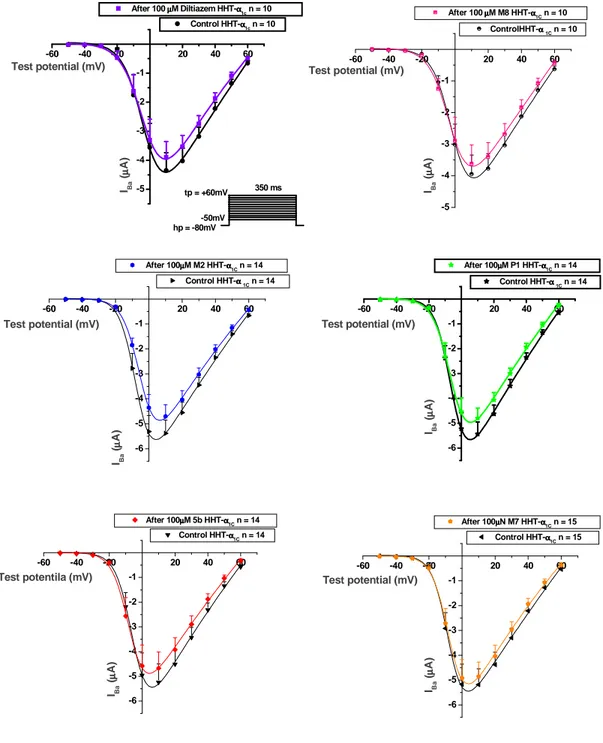
Use-dependent block
Use-dependent block was determined as the inhibition of peak IBa during trains of 15 test pulses lasting 80ms applied at 0.5Hz from a holding potential of -60mV to test potentials +10mV positive to the peak potential of the I-V curves. An identical pulse protocol was used in the presence of drugs. Diltiazem (racemic) and the new Diltiazem analogs were perfused