To my mum
To my mum
To my mum
To my mum
1
1GGEENNEERRAALLPPAARRTT
1.1INTRODUCTION 2
1.2FUNGAL PLANT PATHOGENS 5
1.2.1AIR-BORNE FUNGI 5
1.2.1.1 Botrytis cinerea 6
1.2.1.2 Ascochyta rabiei 9
1.2.1.3 Phytophthora infestans 10
1.2.2SOIL-BORNE FUNGI 13
1.2.2.1 Fusarium oxysporum 14
1.2.2.1.1 Fusarium oxysporum f.sp. basilici 15
1.2.2.1.2 Fusarium oxysporum f.sp. lycopersici 17
1.2.2.1.3 Fusarium oxysporum f.sp. melonis 20
1.2.2.2 Phytophthora spp. 23
1.2.2.2.1 Phytophthora capsici 24
1.2.2.2.2 Phytophthora cactorum 26
1.2.2.3 Pythium spp. 29
1.2.2.4 Monosporascus cannonballus 32
1.3BIOLOGICAL CONTROL OF FUNGAL PLANT PATHOGENS 36
1.3.1PLANT-DERIVED PRODUCTS 38
1.3.1.1 Novel plants for extracts to control fungal plant pathogens 46
1.3.1.1.1 Boerhavia spp. 46 1.3.1.1.2 Cordia spp. 47 1.3.1.1.3 Phyllanthus spp. 48 1.3.1.1.4 Siparuna spp. 49 1.3.1.1.5 Vitex spp. 50 1.3.2ANTAGONISTIC MICROORGANISMS 51 1.3.2.1 Fungal antagonists 51 1.3.2.2 Bacterial antagonists 53
1.4SCREENING METHODS OF ANTIFUNGAL ACTIVITY 54
1.4.1LABORATORY SCREENING METHODS 54
1.4.2IN VIVOSCREENING METHODS 56
1.5.2 PATHOGENESIS-RELATED PROTEINS 62
1.5.2.1 PR-1 proteins 63
1.5.2.2 PR-4 proteins 64
1.5.2.3 PR-5 proteins 64
1.5.2.4 PR-6 proteins 63
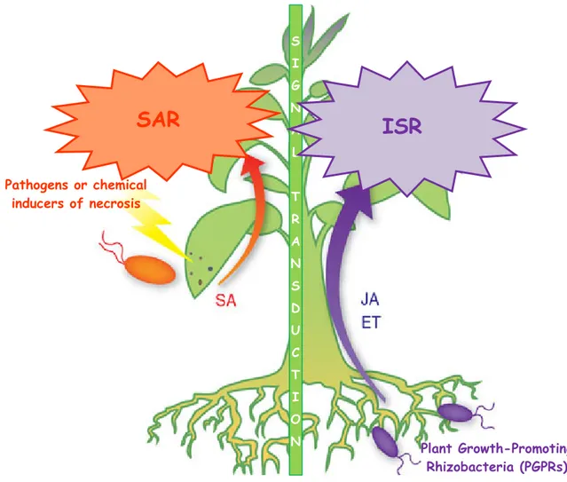
1.5.3 SIGNALS INVOLVED IN TRANSCRIPTIONAL INDUCTION
OF DEFENCE-RELATED GENES 65
1.5.3.1 Biological induction 66
1.5.3.2 Induction by chemicals 67
1.5.3.3 Induction by plant extracts 68
1.5.4 SYSTEMIC RESISTANCE MECHANISMS 68
2
2EEXXPPEERRIIMMEENNTTAALLPPAARRTT
2.1INTRODUCTION 73
2.2ANTIFUNGAL SCREENING OF PLANT EXTRACTS 74
2.2.1 SIPARUNA GUIANENSIS EXTRACT/ BOTRYTIS CINEREA
AND ASCOCHYTA RABIEI 74
2.2.1.1 Abstract 74
2.2.1.2 Introduction 74
2.2.1.3 Materials and methods 77
2.2.1.4 Results and discussion 82
2.2.1.5 Conclusions 91
2.2.2 CORDIA LEUCOCEPHALAEXTRACT/ FUSARIUMSPP. 92
2.2.2.1 Abstract 92
2.2.2.2 Introduction 92
2.2.2.3 Materials and methods 94
2.2.2.4 Results and discussion 98
2.2.2.5 Conclusions 106
2.2.3 BOERHAVIA DIFFUSAEXTRACT/ PHYTOPHTHORASPP. 107
2.2.3.1 Abstract 107
2.2.3.2 Introduction 107
2.2.3.3 Materials and methods 109
2.2.4 VITEX AGNUS-CASTUSEXTRACT/ PYTHIUM ULTIMUM 116
2.2.4.1 Abstract 116
2.2.4.2 Introduction 116
2.2.4.3 Materials and methods 117
2.2.4.4 Results and discussion 119
2.2.4.5 Conclusions 121
2.2.5 PHYLLANTHUS NIRURIEXTRACT/ MONOSPORASCUS CANNONBALLUS 122
2.2.5.1 Abstract 122
2.2.5.2 Introduction 122
2.2.5.3 Materials and methods 123
2.2.5.4 Results and discussion 126
2.2.5.5 Conclusions 129
2.3 INNOVATIVE METHODS TO STUDY PLANT/PATHOGEN/EXTRACT INTERACTIONS 130 2.3.1DETERMINATION OF CORDIA LEUCOCEPHALA EXTRACT
ANTIFUNGAL ACTIVITY BY FLOW CYTOMETRY 130
2.3.1.1 Abstract 130
2.3.1.2 Introduction 130
2.3.1.3 Materials and methods 132
2.3.1.4 Results and discussion 133
2.3.1.5 Conclusions 140
2.3.2MOLECULAR ANALYSIS OFPRGENE ACTIVATION BY TREATMENT WITHVITEX AGNUS-CASTUSEXTRACT ANDPYTHIUM ULTIMUM
INOCULATION IN TOMATO 141
2.3.2.1 Abstract 141
2.3.2.2 Introduction 141
2.3.2.3 Materials and methods 143
2.3.2.4 Results and discussion 146
2.3.2.5 Conclusions 152
2.4FINAL CONCLUSIONS 153
BIBLIOGRAPHY
G
1.1 INTRODUCTION
Plants and their pathogens have evolved together for millions of years. Losses of crop yield from diseases have had severe effects on the human race, for example the Irish potato famine in the 1800's, caused by late blight, a fungal disease of potato; in the mid 19th century, downy mildew grape disease was accidentally introduced in Europe and almost destroyed the vineyards in many countries; massive European epidemics caused by ergot of rye grain frequently occurred during medieval times; and in the early 20th century, the American Chestnut tree was wiped out by an Asian blight disease.
For disease to occur, three critical factors or conditions must exist: (i) a pathogen, (ii) a susceptible host plant, and (iii) the right mix of environmental conditions. The relationship of these factors is represented by theDISEASE TRIANGLE(Fig. 1.1), which represents a central
concept of plant pathology. It is based on the principle that infectious diseases develop when a susceptible host and a disease-causing pathogen meet in a favorable environment. If any one of these three conditions were not met, there would be no disease.
Fig. 1.1: Disease triangle
PATHOGENS (fungi, bacteria, viruses, mycoplasma) are microorganisms causing
disease and, because they are living, they are called biotic agents. Each of them has a different life cycle, which includes an infectious stage. Most pathogens are host-specific to a particular plant species, genus or family, while some diseases, such as powdery mildews, produce similar symptomes on different plants. Fungi are usually host-specific.
A SUSCEPTIBLE HOST has a genetic background that permits the development of a
particular disease. The genetic defence against a disease is called disease resistance. This resistance can be represented by physical, chemical, and growth patterns of the plant. For disease to occur, the host plant must be at a specific stage of development.
SpecificENVIRONMENTAL CONDITIONSmust exist for pathogens to cause infection and
they vary for different pathogens. High moisture and specific temperature ranges, for example, are necessary for many fungal diseases. These conditions must continue for a critical period of time, while the pathogen is in contact with the host, for infection to occur. Each pathogen has a specific temperature range for growth and activity. Soil temperature can also be critical for pathogen infection. Cool, wet soils promote fungal root diseases. Moisture, temperature, wind, sunlight, nutrition and soil quality affect plant growth. If one of these factors is out of balance for the culture of a specific plant, that plant may have a greater tendency to become diseased.
TheDISEASE CYCLE is characterized by five stages: (i) inoculation, (ii) incubation, (iii)
penetration, (iv) infection, and (v) symptoms. Pathogen is carried on the plant (inoculation) by rain, wind, insects, birds, people or it is transmitted by seeds or plant debris. During the second stage of disease development (incubation), pathogen changes or grows into a form that can enter the new host plant; in many fungal diseases, it arrives on the plant as a spore, which must germinate before it can grow into the plant, where it sends out the hyphae penetrating the plant through wounds or natural pores. In the fourth stage of the disease cycle (infection), the pathogen grows within the plant and begins damaging the plant tissue. Then, as the pathogen consumes nutrients, the plant reacts by showing symptoms. Symptoms are the evidence of the damages caused by the pathogen to the plant. In response to disease, the plant defence response often involves a hypersensitive response (HR), visible as flecks of dead cells at sites of attempted entry.
Successful DISEASE CONTROL requires knowledge of the causal agent and the disease
cycle, the host-pathogen interactions in relation to environmental factors, and cost. Disease control starts with the best variety, seed, or planting stock available and continues throughout the life of the plant. For harvested crops, disease control must be extended through transport, storage, and marketing. Relatively few diseases are controlled by a single method; the majority require several approaches. These often need to be integrated into a broad program of biological, cultural, and chemical methods to control as many different diseases on a given crop as possible.
There are at least 50,000 diseases of crop plants and new diseases are discovered every year. Pathogens multiply and mutate rapidly. The use of appropriate agronomical practices and an understanding of plant pathology are the first line of defence against pathogens. Almost all control methods are aimed at protecting plants from becoming diseased but only few plant diseases can be controlled satisfactorily by therapeutic means in the field. The
various control methods can be classified as regulatory, cultural, biological, physical, and chemical, depending on the nature of the agent employed (Agrios, 2005). Regulatory control measures aim at excluding a pathogen from a host or from a certain geographic area. Most cultural control methods help plants to avoid contact with a pathogen, so creating environmental conditions unfavorable to the pathogen, as well as eradicating or reducing the amount of a pathogen in a plant (host eradication), a field (crop rotation), an area (sanitation) and improving plant growing conditions. Most biological methods use living organisms or natural plant-derived products to reduce the pathogen inoculum. Other type of biological control consist in the use of transgenic plants that exhibit resistance to a certain disease. Finally, physical (i.e. sterilization, heat treatment of plant organs, refrigeration, and radiations) and chemical methods (i.e. soil treatment, soil fumigation, and seed treatment with chemicals) aim at protecting plants from pathogen inoculum or curing an infection that is already in progress. Some chemicals operate by activating the defences of the plant (systemic resistance) against pathogens (Agrios, 2005). Synthetic fungicides are still the most important components in the management of fungal diseases. More than 100 fungicides have been developed, and several hundreds of fungicide formulations are available (Vidhyasekaran, 2004).
Resistance of numerous fungi to some systemic fungicides, all of which containing a benzene ring, began to appear in the 1960s. To date, fungicide resistance has been reported in
Phytophthora infestans, Peronospora parasitica, and Bremia lactucae against phenylamides
(metalaxyl compounds); Ustilago nuda and Ustilago maydis against carboxamides (carboxin compounds); Erysiphe graminis f. sp. hordei against hydroxypyrimidine; E. graminis f. sp.
tritici, E. graminisf. sp. hordei, and Pyrenophora teres against triazoles (propiconazole); and Botrytis cinerea, Botrytis fabae, Septoria tritici, Pyrenopeziza brassicae, Cercospora beticola, Fusarium nivale, Fusarium culmorum, and Pseudocercosporella herpotrichoides
against benzimidazoles (carbendazim, benomyl, thiophanate-methyl compounds). Some other important pathogens such as Alternaria, Colletotrichum, Verticillium, Sphaerotheca, Mycosphaerella, Aspergillus, Penicillium, Pythium, and Venturia inaequalis are known to have produced strains resistant to one or more of the systemic fungicides (Agrios, 2005; Vidhyasekaran, 2004).
In addition, some efficient fungicides may be phased out globally because they are seriously harmful for the environment, e.g. methyl bromide that, since 2005, was banned in European Union countries (Batchelor, 2002) because considered as ozone-depleting compound (Anonymous, 1998). Therefore, integrated control methods has been widely
involved in plant disease management and are usually aimed against all diseases affecting a crop (Agrios, 2005). As there is a great interest to decrease the impact of chemicals applied to the crops, novel natural products are evaluated for possible use in plant disease control. Such products can be either of microbial or plant origin.
Thousands of plant species are known to have medicinal properties due to their chemical compounds. Plant-derived products such as plant extracts are traditionally used in local medicine all over the world. Therefore, these extracts are supposed to have antimicrobial properties in plant disease management as well. Such preparations may have many advantages for both grower and consumer, e.g. in the crop culture the environment is less stressed by natural products and fungi and resistance against such products has not been acquired. In this way, natural products are often prefered by consumers as they are considered helthier due to minor content of chemical residues.
1.2 FUNGAL PLANT PATHOGENS
Fungi are one of the most important groups of plant pathogens. To date, there are about 10,000 species of fungi, that can cause diseases in plants. All plants are attacked by some kinds of fungi, and each of the parasitic fungi can attack one or many kinds of plants (Vidhyasekaran, 2004; Agrios, 2005). Some species of fungi, the mycorrhizae, live symbiotically on or in the roots of many plants. This relationship is basically parasitic but in many situations is probably beneficial to both the plant and the fungus. The growth of the plant is promoted by the improved uptake of some mineral nutrients while the fungus gains access to organic nutrients and shelter.
The majority of phytopathogenic fungi belong to the Ascomycetes and the Basidiomycetes. Fungi are reproduced both sexually and asexually by production of spores that may be spread long distances by air or water or may be soil borne. Fungi are common in soil, in air (mainly as spores) and on plant surfaces throughout the world.
1.2.1
AIR-
BORNE FUNGIMany Ascomycetes and imperfect fungal pathogens are airborne and attack plant shoots. Air-borne fungi are transmitted mainly by air currents or water droplets and they attack plant penetrating through aerial organs.
1.2.1.1 Botrytis cinerea
Botrytis cinerea Pers. is a necrothrophic fungus, probably the most common and
widely distributed among vegetable, ornamental, and fruit greenhouse and field-grown crops throughout the world. Botrytis cinerea is pathogenic on over 200 species of plants.
Botrytis diseases appear primarily as blossom blights and fruit rots, but also as damping-off, stem cankers or rots, leaf spots, and tuber, corm, and bulb rots. Some of the most serious diseases caused by Botrytis include gray mold of strawberry, grapes and of many vegetables, calyx end rot of apples, onion blast and neck rot, blight or gray mold of many ornamentals, bulb rot of amaryllis, corm rot of gladiolus, and others. Botrytis also causes secondary soft rots of fruits and vegetables in storage, transit, and market (Agrios, 2005; Webster and Weber, 2007; Beever and Weeds, 2007). In greenhouse culture, Botrytis cinerea is well-known cause of considereable damages in tomato.
The Pathogen
The pathogen Botrytis cinerea, a Deuteromycete, produces abundant gray mycelium and long branched conidiophores with rounded apical cells bearing clusters of colorless or gray, one-celled, ovoid conidia. The conidiophores (Fig. 1.2) and clusters of conidia resemble a grape-like cluster. Hyaline conidia (asexual spores) are
released readily in humid weather and are carried by air currents. The fungus frequently produces black, hard, flat, irregular sclerotia as survival structures in older cultures. It overwinters as sclerotia or intact mycelia, both of which germinate in spring to produce conidiophores. The conidia are dispersed by wind and rain-water and cause new infections.
It is usually referred to the fungus with its anamorph (asexual form) name, because the sexual phase is rarely observed. The teleomorph (sexual form) is an ascomycete, Botryotinia
fuckeliana (Jarvis, 1980). Some species of Botrytis occasionally produce a Botryotinia
perfect stage in which ascospores are produced in an apothecium (Agrios, 2005).
Symptoms
In the field, blossom blights often precede and lead to fruit rots and stem rots. The fungus becomes established in flower petals and there it produces abundant mycelium with
conidia forming whitish-gray or light brown mold. The fungus later moves from the petals into the fruit and causes a blossom end rot of the fruit, which may destroy part or all of the fruit (Fig. 1.3). Rot development is rapid and quite common in the field. Infected fruit and succulent stems become soft, watery, and light brown. As the tissue rots, the epidermis cracks and the fungus fruits abundantly. Flat black sclerotia may appear on the surface. Damping-off of seedlings due to
Botrytis occurs primarily in cold frames, where humidity is high. Some species of Botrytis
cause leaf spots on their hosts e.g. on gladiolus, onion, and tulip. Infection of below ground parts, such as bulbs, corms, tubers, and roots, may begin while these organs are still in the ground or at harvest (Agrios, 2005; Maas, 2004).
Disease development
Botrytis overwinters in the soil as mycelium in decaying plant debris and as conidia
and sclerotia (Fig. 1.4). Each part of the fungus thallus can serve as a survival structure. Mycelia, sclerotia and conidia have different abilities for survival and dispersal, and the relative roles of these structures will vary greatly depending on ecosystem and season. Sclerotial types are over winter favoured on perennial hosts while conidial types on annual hosts with abundant susceptible flowers (Beever and Weeds, 2007; Holz et al., 2007). The fungus requires cool (18–25°C) and damp weather (humidity >90%) for best growth, sporulation, spore release and germination, and establishment of infection. The pathogen, active at low temperatures, may cause considerable losses on crops kept for long periods in storage, even at the temperatures between 0 and 10°C. Botrytis sclerotia usually germinate by producing mycelial threads that can infect directly but, in a few cases, sclerotia germinate by producing apothecia and ascospores (Agrios, 2005; Ponti and Laffi, 1985).
Fig. 1.3: B. cinerea infection on strawberry
Fig. 1.4:Life cycle of Botrytis cinerea (from Agrios, 2005)
Control
The control of Botrytis diseases is difficult to obtain. It is aided by the removal of infected debris from the field and by providing storage conditions with proper aeration. Other measures that can be taken only to minimize losses due to Botrytis fruit rot include prevention of excessive vegetative growth by regulating plant density, timely nitrogen applications, harvesting fruit before it is fully ripe to avoid injuries and prompt transfer of harvested fruit to refrigerated storage. Storage organs such as onion bulbs can be protected by keeping them at 32 to 50°C for 2 to 4 days to remove excess moisture and then keeping them at 3°C in dry environment. In greenhouses, humidity should be reduced by ventilation and heating.
Biological control of Botrytis gray mold may be obtained with spray application of spore suspensions of certain antagonistic fungi and with mixtures of several biocontrol fungi and bacteria. A number of microbial products available commercially for control of Botrytis diseases indicates that biocontrol can succeed. Especially Trichoderma and Ulocladium have given the greatest success in Botrytis control. Sprays with a number of broad-spectrum or systemic fungicides control Botrytis on a wide variety of crops. The most efficient fungicides are Vinclozolin, Procimidone, and Iprodione. Other fungicides in use against Botrytis are Captano, Folpet, Captafol, Clorotalonil, and Tiram. However, control of Botrytis in the field through chemical sprays has been only partially successful. Application of botryticides just
before or after harvest is desirable but this practice is restricted by the toxicological risks of their residues. Botrytis strains resistant to several systemics and even to some broad-spectrum fungicides have been found in various crops, in particular against benzimidazoles, phenylcarbamates, and dicarboximides (Agrios, 2005; Vidhyasekaran, 2004; Leroux, 2007; Maas, 2004; Ponti and Laffi, 1985).
1.2.1.2 Ascochyta rabiei
Ascochyta blight is a serious disease of cool-season grain legumes (chickpea, faba bean, lentil, and pea) caused by fungal species of the anamorphic genus Ascochyta and related genera (White and Chen, 2007). Ascochyta rabiei (Pass.) Labr. causes the most destructive disease (Fig. 1.5) in many chickpea growing countries (Basandrai et al., 2007).
The pathogen
This pathogen undergoes heterothallic sexual reproduction on infested residues, resulting in air-borne ascospores, which are capable of spread over long distances. Rapid polycyclic spread within crops occurs through splash-borne asexual conidia (pycnidiospores) (Davidson and Kimber, 2007).
Symptoms
The disease affects all above ground parts of the plant. Symptoms are characterised by necrotic lesions (Fig. 1.6), which under favourable conditions, can girdle stems leading to breakage and severe yield reduction on susceptible cultivars. Wilting causes the death of the plant. Seed quality may also be reduced through seed discolouration or retardation of seed development (Davidson and Kimber, 2007).
Fig. 1.6:Disease symptoms of A. rabiei
Fig. 1.5:Chickpea field damaged by A. rabiei
Disease development
Under favourable conditions, conidia germinate forming new hyphae which penetrates through epidermal cells into the plant tissue. In consequence, this necrotrophic pathogen in advance kills the plant cells of further mycelial development. Therefore, toxins and cell-wall degrading enzymes are often presumed to be important biochemical determinants of pathogenesis. The necrotic lesions develop after 4-5 days in susceptible varieties (Alam et al., 1989; Pandey et al., 1987). The lesions on the stems and branches enlarge, sometimes covering entire plant organ. Such infected tissues cause death of the plant as the tissues break easily and are not any more able to give the mechanical support to the plant. Sometimes, numerous picnidia with abbundant conidia are formed on the necrotic lesions. In resistant varieties, fungus penetration is limited and it is followed by much rapid necrosis of the cells with consequent suppression of disease development (Porta-Puglia, Crinò, 1993). The rapid development of the disease is favoured by temperatures between 10 and 20oC with relative humidity over 60%. The temperatures higher than 25oC reduce the pathogen attack (Reddy and Singh, 1990; Weltzien and Kaack, 1984).
Control
The control methods include destroying inoculum sources, manipulating sowing times, using crop rotations, disease-free seed, application of seed and foliar fungicides, and adopting cultivars with improved resistance. Implemented fungicide strategies differ according to cultivar resistance and the control efficacy of fungicides. The effectiveness of genetic resistance varies according to seasonal conditions. The combination of strategies is determined by economics, availability of cultivar resistance and disease epidemiology (Davidson and Kimber, 2007).
1.2.1.3 Phytophthora infestans
An oomycete, Phytophthora infestans (Mont.) De Bary is the causal agent of late blight both of potatoes and tomatoes. The crop cultures are attacked by the fungus both under field and greenhouse conditions. The losses in yield caused by P. infestans are of a world-wide importance as potato and tomato make, an essential part of human alimentation throughout the world (Crinò, 1993; Ponti and Laffi, 1985).
The Pathogen
The mycelium produces branched sporangiophores with lemon-shaped sporangia at their tips (Fig. 1.7). At the places where sporangia are produced, sporangiophores form swellings that are characteristic for this oomycete (Agrios, 2005). This organism is heterothallic and requires two mating types for sexual reproduction by gametangia contact. Sexual
reproduction is not necessary for the survival of the organism as the mycelium may survive in infected tubers. Several biflagellated zoospores are formed and are liberated from each sporangium. Zoospores on leaves encyst and germinate via germ tubes, which form appressoria (Ament and Trigiano, 2004). Although there are potato and tomato strains of P.
infestans, each is capable of infecting the other host (Horst, 2008).
Symptoms
Symptoms (Figs. 1.8, 1.9) appear at first as water-soaked spots, usually at the edges of the lower leaves. In moist weather, the spots enlarge rapidly and form brown, blighted areas with indefinite borders. A white zone appears at the border of the lesions on the undersides of the leaves. Under continuously wet conditions, blighted plant organs give off a characteristic odor due to secondary bacterial and fungal invaders. In dry weather, existing lesions stop enlarging, turn black, curl, and wither. Infected potato tubers are small with sunken lesions, stained purple or brown. On tomato seedlings, small and dark spots on stems or leaves are followed by death within 2 or 3 days. On mature tomato plants, blight starts with dark,
Fig. 1.9:Disease symptoms of P. infestans in tomato
Fig. 8:Disease symptoms of P. infestans in potato
watersoaked leaf spots and large dark brown spots on fruit, with most of the leaves soon hanging lifeless and fruit rotting on the ground. The rot, however, continues to develop after the tomato fruit or potato tuber is harvested (Agrios, 2005; Ament and Trigiano, 2004; Horst, 2008).
Disease development
Entire plants and entire fields may become blighted and die in a few days or a few weeks. The development of epidemics depends on the prevailing humidity and temperature during the different stages of the life cycle of the oomycete (Fig. 1.10).
Fig. 1.10:Life cycle of P. infestans (from Agrios, 2005)
P. infestansgrows and sporulates abundantly at a relative humidity near 100% and at
temperatures between 15 and 25°C (optimal temperature is 20-23°C). Temperatures above 30°C slow or stop the growth of the oomycete in the field, and the oomycete can start to sporulate again when the temperature and humidity become favorable. Sporangia germinate by the zoospores at temperatures up to 12 or 15°C, whereas above 15°C sporangia may germinate directly by producing a germ tube. Oospores may survive in the soil for 3–4 years.
Mycelium from infected potato tubers or from germinating oospores and zoospores spreads into shoots produced from infected or healthy tubers and cause discoloration and collapse of the cells. When the mycelium reaches the aerial parts of plants, it produces sporangiophores, which emerge through the stomata of the stems and leaves, and produce sporangia. The sporangia, when ripe, become detached and are carried off by the wind or are dispersed by rain and infect new plants. A large number of asexual generations and new infections may be produced in one growing season (Agrios, 2005; Ponti and Laffi, 1985).
Control
Late blight can be controlled successfully by a combination of sanitary measures, partially resistant varieties, and chemical sprays. Only disease-free plant material should be used for seed. All volunteer potato plants in the area should be destroyed, as they can be a source of late blight infection (Agrios, 2005). Only the most resistant varieties should be planted. However, no cultivars of both tomato and potato are completely resistant to this fungus (Crinò, 1993). Several potato varieties resist to one or more races of oomycete. Many varieties possess so-called field resistance, which is a partial resistance of varying degrees effective against all races of the fungus. Even resistant varieties should be sprayed regularly with fungicides to eliminate the possibility of becoming suddenly attacked by races of P.
infestansto which they are not resistant. Several broad-spectrum and systemic fungicides are
used for late blight control. The most effective are the copper fungicides. Protective spraying of foliage usually affects a considerable reduction in tuber infection. However, the new strains of P. infestans are resistant to some of the systemic fungicides (metalaxyl) (Agrios, 2005; Ponti and Laffi, 1985).
1.2.2
SOIL-
BORNE FUNGISoil-borne fungi survive in the soil and and in residues on the soil surface; they attack plant penetrating through lesions into the roots or collets. Thus the soil is a reservoir of inoculum of these pathogens, the majority of which are widely distributed in agricultural soils.
These diseases are difficult to control because they are caused by pathogens which can survive for long periods in the absence of the normal crop host and often have a wide host range including weeds; chemical control often does not work well or is not practical or too
expensive and it is difficult to develop resistant varieties of plants. These diseases are often very difficult to diagnose accurately. Damages to root and crown tissues is hidden in the soil. Thus, these diseases may not be noticed until the above-ground (foliar) parts of the plant are affected severely showing symptoms such as stunting, wilting, chlorosis and death.
1.2.2.1 Fusarium oxysporum
Fusarium vascular wilts are F. oxysporum is a widespread and destructive fungus that causes Fusarium wilt disease in more than a hundred species of plants, many ornamental and horticultural crops included. It does so by colonizing the water-conducting vessels (xylem) of the plant. As a result of this blockage and breakdown of xylem, symptoms appear in plants such as leaf wilting, yellowing and eventually plant death.
In solid media culture, such as potato dextrose agar (PDA), the different special forms of F. oxysporum can have varying appearances. In general, the aerial mycelium first appears white (Fig. 1.11), and then may change to a variety of colors ranging from violet to dark purple -according to its special form.
The fungus can persist in the soil also for prolonged periods. It
is characterized by production of three types of asexual spores (microconidia, macroconidia and chlamydospores). Microconidia (Fig. 1.12), which have one or two cells, are the most frequently and abundantly produced spores under all conditions within the wessels of infected plants. Macroconidia are the typical “Fusarium” spores; they are three to five celled, have gradually pointed and curved ends, and appear commonly on the surface of plants killed by the pathogen. Chlamydospores are one-or two celled, thick-walled, round spores produced either within or terminally on older mycelium or in macroconidia. Only chlamydospores can survive in the soil for a long time (Agrios, 2005). The life cycle of this fungus is divided in two phases: (1) saprofitical and (2) parasitical.
Morphological characterization of F. oxysporum is based on the shape of macroconidia, the structure of microconidiophores, and on the formation and disposition of the chlamidydospores. Asexual reproduction of Fusarium is accomplished by macroconidia and microconidia.
Fig. 1.11:Mycelium of Fusarium sp.
Fig. 1.12:Micro and macroconidia produced by F. oxysporum
1.2.2.1.1 Fusarium oxysporum f.sp. basilici
Fusarium wilt, caused by F. oxysporum f. sp. basilici (Dzidzariya) Arms. et Arms. (FOB), is one of the main problems for sweet basil because of its very destructive nature (Fig. 1.13) under field, hydroponic, and greenhouse conditions. The pathogen has a narrow host range and is considered specific for basil (Keinath, 1994; Garibaldi et al., 1997; Reuveni et al., 1997). FOB has been disseminated to new growing areas throughout the world via imported seeds (Elmer, 2001) and is now present in most countries where sweet basil is cultivated.
Unfortunately, there is no treatment for infected basil plants. Fungicides are ineffective and although a few biological control organisms have been tested, none have reliably provided control. Infected plants should be removed immediately because spores from infected stems can move on air currents or splashing water to infect nearby basil plants. Prevention is the only way to control Fusarium wilt of basil.
Typically, basil Fusarium wilt management relies on the integration of different control measures, such as soil and substrate disinfestation, raised bench cultivation, seed dressing, and use of antagonistic Fusarium spp. (Garibaldi et al, 1997; Minuto et al., 1994, 1995, 1997). However, considerable potential for soil contamination and reinfestation through infected seed and airborne propagules makes soil disinfestation only partially effective against
F. oxysporum f. sp. basilici (Chiocchetti et al., 1999). The low efficacy of chemical control
measures, the limited availability of resistant cultivars (Reuveni et al., 1997), and the
Fig. 1.13:Basil plant damaged by Fusarium wilt
unsatisfactory level of control sometimes offered by the commercially available formulations of biocontrol agents (Keinath, 1994; Minuto et al., 1997) boost the urgency for seed and transplant certification procedures on sweet basil.
Probably the best option is to use a variety that is tolerant or resistant to the disease. ‘Nufar’ is a variety resistant to fusarium wilt originated from a few healthy plants found in the middle of a diseased field in Israel. The variety is now commercially available in the United States through several seed companies.
The pathogen
The mycelium is colorless at first, but later it becomes cream-colored, pale yellow or pink. Microconidia are the most frequent primary inoculum.
Symptoms
This pathogen causes wilt of plants at all growth stages, brown and black discoloration of roots and lower stems, black lesions on stems, root and crown rot, blackening and drying of vegetative apices, growth retardation, and xylem discoloration (Figs. 1.14, 1.15). The stems are covered withpink-orange layer consisting mainly of macroconidia (Gamliel et al., 1996).
Fig. 1.15:Symptoms caused by F. oxysporum f. sp. basilici on basil.
(a) internal discolouration of basil stems infected with fusarium wilt. (b)young basil plant with Fusarium wilt
Disease development
The fungus penetrates into the roots from the soil. First, the pathogen causes leaf chlorosis. It is followed by wilting of the leaves, epinasty and stunting which lead to death of the plant. The temperatures favourable to the disease development are rather higher, about 27oC. Although, Fusarium is a soil-borne pathogen, Rekah (2000) and Gamliel et al. (1996) reported the importance of air-borne inoculum as well.
Control
Currently, no efficient fungicide is available to control the disease. Also the availability of commercially acceptable resistant cultivars is limited. Therefore, there is still a need for alternative ways to control the disease. Moreover, the current trend to near-zero market tolerance for pesticide residues in fresh herbs also leads to search for non-chemical means to control the disease. For instance, in the plants grown in soilless culture, the choice of the medium can affect the disease proliferation rate. Also the use of compost can induce protection against F. oxysporum f. sp. basilici and reduce the severity of the visual symptoms of Fusarium wilt (Reuveni et al., 2002; Minuto et al., 1997). Moreover, Reuveni et al. (2002) suggest that under practical conditions, compost, combined with the resistant cultivar ‘Nufar’ could provide full protection for basil plants against Fusarium wilt.
1.2.2.1.2Fusarium oxysporum f. sp. lycopersici
Fusarium oxysporum f. sp. lycopersici (Sacc.) W.C. Snyder and H.N. Hans causes
Fusarium wilt specifically in tomato for which it is one of the most prevalent and damaging diseases. This disease, first described in England in 1895, it is of worldwide importance because at least 32 countries had reported the disease.
Fusarium wilt is most destructive in warm climates and sandy soils of temperate regions. The disease causes great losses when soil and air temperatures are rather high during
much of the season. Infected plants become stunted and soon wilt and finally die. Occasionally, entire fields of tomatoes are killed or damaged severely before a crop can be harvested (Agrios, 2005). The development and use of resistant cultivars have nearly eliminate the concern over this disease.
The Pathogen
The mycelium of F. oxysporum f. sp. lycopersici is colorless at first, but with age it becomes cream-colored, pale yellow, pale pink, or purplish (Agrios, 2005). Three physiological races of the fungus have been reported. Race 1 is the most widely distributed and has been reported from most geographical areas. Although race 2 was first reported in Ohio in 1940, it did not become widespread or of economic concern until its discovery in Florida in 1961. Since then, it was rapidly reported in several other countries, including Australia, Brazil, Great Britain, Israel, Mexico, Morocco, the Netherlands, and Iraq. Race 3 was reported in 1966 in Brazil. Thereafter, it has been found in Australia and in Florida and California.
The mycelium is delicate white to pink, often with purple tinge, and is sparse to abundant. The fungus produces the three types of spores, microconidia, macroconidia, and chlamydospores (Fig. 1.16). Microconidia are borne on simple phialides arising laterally and are abundant, oval-ellipsoid, straight to curved, 5-12 x 2.2-3.5 µm, and nonseptate. Macroconidia, sparse to abundant, are borne on branched conidiophores or on the surface of sporodochia and are thin walled, three- to five-septate, fusoid-subulate and pointed at both ends, have pedicellate base. Three-septate spores are more common. Chlamydospores, both smooth and rough walled, are abundant and form terminally or on an intercalary basis. They are generally solitary, but occasionally form in pairs or chains. No perfect stage is known.
Fig. 1.16:Main traits of F. oxysporum f. sp. lycopersici. (a) mycelium; (b) microconidia; (c) macroconidia; (d) chlamydospores
Fig. 1.17:Symptoms of Fusarium wilt in tomato
Symptoms
Fusarium wilt can affect tomato plants in every developmental stage (Ponti and Laffi, 1985). The first symptoms appear as slight vein clearing on the outer and younger leaflets (Fig. 1.17). Subsequently, the oldest and lowest leaves show yellowing and epinasty caused by drooping of the petioles. Plants infected at the seedling stage usually wilt and die soon.As the disease progresses,
growth is typically stunted, and little or no fruit develops. Older plants in the field may wilt and die suddenly, if the infection is severe and if the weather is favorable for the pathogen. However, in older plants, vein clearing and leaf epinasty are more commonly followed by stunting of the plants, yellowing of the lowest leaves, occasional formation of adventitious roots, wilting of leaves and young stems, defoliation, marginal necrosis of the remaining leaves, and finally death of the plant. After an initial period of stunting, the smallest side roots rot. Also fruit may occasionally become infected and then it rots and drops off (Agrios, 2005).
If the main stem is cut, dark brown streaks may be seen running lengthwise through the stem. This discoloration often extends far up the stem and is especially noticeable in a petiole scar. The browning of the vascular system is characteristic of the disease and generally can be used for its identification.
Disease development
The disease development depends on the plant growth stage. In young plants, the disease development is fast and leads to early death. The pathogen is soilborne and remains for up to ten years in infested soils whereit survives as mycelium and in all its spore forms in infected plant debris in the soil; as chlamydospores, it can survive for many years in the soil. In contaminated soil, the germ tube of spores or the mycelium penetrates root tips directly or enters the roots through wounds or at the point of formation of lateral roots. The mycelium travels through the vessels toward the stem and crown of the plant; it spread throughout the plant by the vascular system. In the vessels, the mycelium branches and produces microconidia, which are detached, carried upward in the sap stream and then germinate. When the leaves transpire more water than the roots and stem can transport to them, the stomata close, the leaves wilt and finally die, followed by death of the rest of the plant. The fungus then invades all tissues of the plant extensively, reaches the surface of the dead plant, and there sporulates profusely.
The spores may be disseminated to new plants or air by wind or water (Agrios, 2005).
Dissemination of the pathogen is via seed, tomato stakes, soil, and infected transplants or infested soil adhering to transplants. Local dissemination is by transplants, tomato stakes, windborne and waterborne infested soil, and farm machinery.
The optimal temperatures for development of Fusarium wilt in tomato are between 27 and 30oC. For this reason the disease developes rather in the summer (Ponti and Laffi, 1985).
Too warm (34°C) or too cool (17-20°C) soils retard wilt development. If soil temperatures are optimum but air temperatures below optimum, the pathogen will extend into the lowest parts of the stem, but without exhibiting external symptoms. In general, factors favoring wilt development are: soil and air temperatures of 28°C, soil moisture optimum for plant growth, plants pre-conditioned with low nitrogen and phosphorus and high potassium, low soil pH, short day length, and low light intensity. Virulence of the pathogen is enhanced by micronutrients, phosphorus, and ammonium nitrogen and decreased by nitrate nitrogen.
Control
Use of tomato varieties resistant to the fungus and solarization of field soil by covering with transparent plastic film are the most practical measures for controlling the disease in the field. Soil sterilization should be always practiced also for greenhouse-grown tomato plants. Use of healthy seed and transplants is mandatory, and hot-water treatment of seed should precede planting (Agrios, 2005). The seed can be treated also with benzimidazole fungicides. Crop rotation of 4-5 years is recommended (Ponti and Laffi, 1985). Recently, biological control of Fusarium wilt seems to be successful. Such control methods include prior inoculation of plants with nonpathogenic strains of F. oxysporum or the use of antagonistic fungi, such as Trichoderma and Gliocladium, Pseudomonas fluorescens and Burkholderia cepaciabacteria. It was shown that spraying tomato plants with a suspension of zoospores of the oomycete Phytophthora cryptogea induces systemic acquired resistance. Although promising, none of these methods have been used for control of Fusarium wilt in practice so far (Agrios, 2005).
1.2.2.1.3 Fusarium oxysporum f.sp. melonis
Fusarium wilt [Fusarium oxysporum Schlecht f.sp. melonis Snyder and Hansen. (Fom)] is one of the most devasting and widely distributed diseases of melon both under greenhouse and field conditions (Fig. 1.18). It can attack the plant at any developmental stage.
The pathogen
The fungus produces pink or purplish mycelium and three types of asexual spores: macroconidia, microconidia, and chlamydospores (Fig. 1.19). Conidia are produced on monophialiades and in sporodochia, and are scattered loosely over the surface of a mycelium. Microconidia are predominantly uninucleate and germinate poorly and variably. The macroconidia are produced abundantly, are multinucleate and germinate rapidly, thereby reproducing the fungus efficiently. Chlamydospores are viables, asexually produced accessory spores resulting from the structural modification of a vegetative hyphal segment or conidia cell possessing a thick wall. Its function is primarily survival in soil.
macroconidia microconidia chlamydospores
Fig. 1.19:Three kinds of spores ofF. oxysporumf. sp. melonis
Four fysiological races of this fungus are known as 0, 1, 2, and 1.2 (Risser et al., 1976) based on the interaction between two major genes of resistance of the host and variants of the pathogen. The dominant resistance genes Fom-1 and Fom-2 provide resistance to Fom races 0 and 2, and to races 0 and 1, respectively. Race 1.2 of Fom is able to overcome these two resistance genes, and was classified into pathotype Y, which causes symptoms of yellowing, and pathotype W, which causes wilt symptoms.
Symptoms
The first symptoms, leaf-yellowing and wilting, appear before flowering. In some cases, wilting proceeds so fast that the leaves conserve a part of the chlorophyl that gives them a grey-greenish colour. When the stem of diseased plant is cut horizontaly, the browning of the vascular system is visible (Fig. 1.20). Usually, yellowing proceeds slowly starting from
Fig. 1.18:Melon plants damaged by Fusarium wilt
the nerves. The brown-orange layer of exudates covering the stems is the most characteristic symptom. Fruit rot usually begins at leaf-stalk. On the dead plants or gravely damaged plants, the white-pink mould can be observed (Ponti and Laffi, 1985).
Disease development
The fungus survives in the soil mainly as chlamydospores, which can remain vital up to ten years. It can be transmitted also by infected seeds. Propagules can be moved from field to field in soil on farm machinery, infected crop debris, and irrigation water.
In optimal conditions (temperatures about 20oC), the fungus penetrates into the roots through microlesions. In the xylem vascular tissues, the fungus grows fast and the first symptoms are visible in 10-15 days (Ponti and Laffi, 1985; Ferrari, 1998).
Control
Use of resistant varieties, when available, must be preferred.Until now many sources of resistance to Fom races 0, 1, and 2 have been reported (Alvarez et al., 2005), but the same does not occur for race 1.2. Only partial resistance to race 1.2 of Fom have been found in several accessions (Risser and Rode 1973; Perchepied and Pitrat 2004). These genotypes allowed breeding for partially resistant lines to Fom race 1.2, such as ‘Isabelle’ and, recently, two double-haploid lines ‘Nad-1’ and ‘Nad-2’, which show higher resistance than other genotypes (Ficcadenti et al., 2002). However, Herman and Perl-Treves (2007) found recently that, in genotype BIZ, two complementary recessive genes are required to obtain full resistance to race 1.2 of Fom. Herman and Perl-Treves (2007) demonstrated that alleles encoding for Fom race 1.2 resistance may behave as dominant, co-dominant, or recessive depending on the environment conditions.
Apart use of resistant varieties, also grafting results as an efficient measure to control this fungus. In any case, seed treatment with benzimidazole is recommended, as well as soil disinfestation. The lesions on the roots should be avoided during transplanting (Ponti and Laffi, 1985). Dong and Cohen (2001) showed that dry mycelium of Penicillium chrysogenum is an inducer of resistance against F. oxysporum f.sp. melonis. Also movement field to field by washing equipment has to be minimized.
Fig. 1.20:Browning of melon vascular system caused by
1.2.2.2
Phytophthora spp.
Phytophthora spp. belong to Oomycetes, Peronosporales. This important genus contains many species causing destructive diseaseson different types of plants ranging from seedlings of annual vegetables or ornamentals to fruit and forest trees. Most species cause root rots, damping-off of seedlings, and rots of lower stems, tubers, and corms similar to those caused by Pythium spp. Others cause rots of buds or fruits, and some cause blights of the foliage, young twigs, and fruit. Some species attack only one or two species of host plants, but others may cause similar or different symptoms on many different kinds of host plants. The best known species is Phytophthora infestans, the causal agent of late blight of potatoes and tomatoes, but several other species also cause extremely destructive diseases on their hosts.
Phytophthora cactorum, P. cambivora, P. cinnamoni, P. citrophthora, P. fragariae, P. palmivora, and P. syringae cause primarily root and lower stem rots, but also some cankers,
twig blights, and fruit rots of woody ornamentals and of fruit and forest trees as well as of vegetables and other herbaceous plants. Several other species, such as P. capsici, P.
cryptogea, P. megasperma, and P. parasitica, cause root, stem, and fruit rots of many
vegetables, ornamentals, and field crops, but also of some woody plants (Agrios, 2005). The life history of Phytophthora species differs in detail from that of most Pythium species. The most notable difference between the two genera is that zoospores of
Phytophthoraspecies are delimited and functional within the sporangium, whereas in Pythium
species, zoospores are formed from the cytoplasm of sporangia that have migrated into a vesicle. Phytophthora species also have indeterminate sporangia, but many species have very differentiated sporangiophores or sporangia. For example, P. infestans produces sympodially branched sporangiophores, which have swollen nodes, and produce lemon-shaped, papillate sporangia. Some Phytophthora species produce haustoria unlike closely related Pythium species (Ament, Trigiano, 2004). Sporangia, formed successively on sporangiophores, slender, sparsely branched hyphae emerging from stomata, germinate either by a germ tube or by zoospores. The sexual spore is an oospore (Horst, 2008).
The oomycete requires two mating types for sexual reproduction. When the two mating types grow adjacently, the female hypha grows through the young antheridium (male reproductive cell) and develops into a globose oogonium (female reproductive cell) above the antheridium. The antheridium then fertilizes the oogonium, which develops into a thick-walled and hardy oospore. Oospores germinate by means of a germ tube that produces a sporangium, although at times the germ tube grows directly into the mycelium(Agrios, 2005).
1.2.2.2.1 Phytophthora capsici
P. capsici Leon. is an oomycete plant pathogen that infects a wide host range that
including cucumber, squash, melon, pumpkin, pepper, tomato, eggplant (Roberts et al., 2004). On pepper, the disease was first reported in New Mexico, USA in 1922 (Leonian, 1922). The disease is now distributed worldwide throughout North and South America, Europe, and Asia. Extensive losses of pepper occur during an epidemic (Roberts et al., 2004).
The pathogen
P. capsici is reproduced asexually by
producing ellipsoid to pyriform sporangia with papilla (Fig. 1.21). The sporangium is usually oblong, average 30-60 µm. It is nearly spherical to pyriform (pear shape), hyaline (colorless), papillate (pointed at the tip), deciduous (spores fall from the colony) and have a long pedicel (stalk) attached to the base of the spore., zoospores. In water, sporangia form and release several (15-25) spores called zoospores that
are flagellated and are chemotacticallly attracted to plant and root exudates. Sporangia also germinate directly by producing several germ tubes that begin new fungal colonies. Hyphae are fairly coarse and irregularly branched.
P. capsici is heterothallic and has two mating types (A1 and A2). Both mating types
are needed for the abundant production of sexual spores called oospores. These are thick walled and results from the pairing of an oogonium with an antheridium. Some species of
Phytophthora are induced to form oospores when the mating type of another species is
present.
The pathogen does not produce chlamydospores (Roberts et al., 2004). It grows well between 25 and 30 CoC.
Symptoms
The pathogen is capable of infecting all parts of the pepper plant (Fig. 1.22). It causes seedling death, crown and stem lesions, root rot, leaf blight and fruit rot. Stem lesions,
Fig. 1.21:Sporangia and spores of P. capsici
Fig. 1.22:Pepper plant damaged by Phytophthora blight
whether at the soil line or higher, initially appear first dark green and water-soaked, but then turn dark brown to black and girdle the stem. Infected roots are dark brown and rotten.
Leaf blights begin as small, irregular to round shaped and water-soaked areas on the underside of leaves, which rapidly enlarge. Later, the lesion becomes dry, papery, and turns a light tan to grayish brown. During warm, wet weather, infected areas may be bordered by white fungal growth. Entire branches may wilt and die from infection at the forked part of stems. Pepper fruit are frequently infected through wounds. The fruit tissue becomes dark, shrunken, and white fungal growth of mycelium and sporangia is apparent on its surface. Infected fruit remain attached to the stem (Roberts et al., 2004). Symptoms on squash are similar; green leaf lesions spreading over the blade, a basal stem rot, and wilting (Horst, 2008).
The pathogen moves into stems from infected leaves and sections of the plant that are killed. Rots which develop at the soil line or affect major branches cause the plant to wilt or die. Root rots can be severe and stunt the plant or cause plant decline. Fruit rots are irregular in shape and olive green or light green with water soaked borders. Rots expand rapidly and fruits can be completely diseased and desiccated, causing the formation of "mummified" fruits. Infected seeds are brown and shriveled.
Disease development
Phytophthora capsici survives on plant debris and in the soil as oospores, mycelium,
sporangia or zoospores. Phytophthora blight is a polycyclic disease in which the pathogen reproduces and infects new plants several times within a single season. Epidemics of pepper fields are caused by P. capsici during wet weather. Environmental conditions such as water temperature, nutrition, pH, and other factors determine the length of time in which zoospores continue to swim. Encysted zoospores germinate by producing a thin fungal hypha or thread. The germ tube commonly penetrates the leaf through stomates which are natural opening in the leaf epidermis. Larger germ tubes produced by sporangia also penetrate the leaves.
In pepper fields, the fungus is soil borne and initial infections of roots, collars, and lower leaves occur. The fungus grows within the host and produces sporangia on the surface of diseased tissue, especially leaves. Sporangia are spread by splashing water from irrigation or rain. With moisture present, zoospores are formed and released. Released zoospores swim in a film of water for a few minutes to more than an hour to reach host tissue before encysting. Periodic flooding and saturated soil conditions from either rainfall or irrigation stimulates the release of zoospores from sporangia (Bowers and Mitchell, 1990). The
pathogen and its sporangia are dispersed by splashing rain and wind or overhead irrigation (Bowers et al., 1990, Ristaino, 1991). Sporangia are also moved within the field by contact with field equipment, clothing, gloves, tools, etc. Movement of soil from one field to another on equipment or boots will move this pathogen. Dissemination is also through infected transplants and contaminated soil and equipment. Root to root contact spreads the disease. Wet soil and high temperatures encourage blight. The application of water made frequently through drip irrigation causes earlier onset of the disease compared to applications of drip irrigation made less frequently (Ristaino, 1991; Ristaino et al., 1993; Horst, 2008). Optimum temperature for growth of P. capsici is about 27oC (Cristinzio, 1993).
Control
The control of this oomycete is difficult due to its great adaptability on many different hosts. In addition, it has developed a resistance against some systemic fungicides such as Metalaxyl. Therefore, it is necessary to applicate an integral disease management such as crop rotation with non-susceptible crops rather than exclusive use of chemical fungicides (Cristinzio, 1993) to reduce the amount of P. capsici surviving in soil. Disinfested equipment and fungicide-treated seed as well as sterile potting media should be used to establish healthy transplants. Fields should be well drained without volunteer cucurbit and solanaceous plants. Soil should be fumigated before planting. The use of black plastic mulch can be used as a barrier to dispersal of inoculum (Ristaino et al., 1997). Fungicides, both contact and systemic, should be applied to the foliage and used preventively. However, some isolates are insensitive to mefenoxam. Soil solarization may reduce populations of the pathogen (Yucel, 1995). Soil amendments, such as compost and manure, are a possibility for disease management, as they can develop suppressive soils. Resistant varieties should be planted, where available (Roberts
et al., 2004). Some cultivars are more tolerant, and better resistance to P. capsici is being
developed in pepper.
1.2.2.2.2 Phytophthora cactorum
Crown rot, caused by Phytophthora cactorum (Leber et Cohn) Schroeter, is an important strawberry disease in Europe and other temperate to subtropical regions. First identified on cacti, this oomycete is capable of infecting an extremely large number of hosts and is problematic in low-lying or wet field conditions. It can limit production for many economically important crops such as strawberry (Fig. 1.23), apple, pear, rhododendron, and azalea. The disease is apparently caused by a distinct pathotype of P. cactorum (Maas, 2004).
P. cactorum can cause root, collar, and crown rots, as well
as foliar and fruit infections. The fungus causes also increased culling of seedlings in forest nurseries (Lilja et al., 2006).
The pathogen
P. cactorumforms a white mycelium. Sporangia are
distinctively papillate, and are usually borne terminally. Sporangial shapes vary widely, and can be broadly ellipsoidal, obpyriform, ovoid or spherical (Ellis, 1998; Erwin and Ribeiro, 1996). A distinctive characteristic of this pathogen, compared to other Phytophthora, is represented by the caducous sporangia with pedicels less than 40 µm. Each sporangium may contain more than 50 zoospores. Chlamydospores are generally terminal but may occasionally be intercalary. P. cactorum is homothallic, and oospores can be found on the plant debris. All antheridia are paragynous and are generally spherical or club-shaped. Oogonia are usually hyaline and smooth-walled, and oospores are plerotic (Fig. 1.24; Ellis, 1998).
Symptoms
Although P. cactorum can parasitize a wide range of plant species, pathogenicity may vary widely across hosts. In general, this pathogen can cause a number of symptoms and diseases depending on the host. In strawberry, P. cactorum causes crown and root rot, and fruit leather rot (Fig. 1.25).
Symptoms typically develop during early-mid summer, when the disese often appears in the field as a sudden wilting or
collapse of the plants. The youngest leaves turn bluish-green. When lifted, diseased plants may easily break at the crown. Wilting will quickly spread throughout the plant, leading to plant death. Extensive internal dark-brown crown necrosis and vascular disintegration are characteristics of this disease. P. cactorum crown rot symptoms are most evident at the basal stolon attachment area or at the crown. Roots typically are not infected but die when the upper
Fig. 1.24:Oospores of P. cactorum in infected strawberry root tissue
Fig. 1.25:Fruit leather rot caused by P. cactorum Fig. 1.23:Strawberry affected by
part of the plant dies. Plants that do not succumb and appear to recover remain stunted and unproductive (Pettitt and Pegg, 1994; Maas, 2004).
Disease development
The most important propagule for this pathogen is represented by the zoospores, which originate from hyphae or germinating oospores and sporangia (Fig.
1.26). Oospores in soil or in strawberry
plant debris provide initial sources of inoculum. The oospores germinate to produce zoosporangia and zoospores which infect plants, usually at wound sites. In many cases, this pathogen may enter a field through infected transplants.
The fungus requires warm temperatures and prolonged wetness periods for inoculum production and for infection development. Motile zoospores are released from sporangia during saturated soil conditions and enter through wounds (Seemuller, 1998). Once the zoospore reaches a host, it infects and developing hyphae of the fungus colonize the host.
Crowns damaged by frost injury prior to plant harvesting or by low temperatures during cold storage are predisposed to infection (Bell et al., 1997; Pettitt and Pegg, 1994). Planting time also affects symptom development; plantings established in spring to early summer are more quickly affected than those established in mid-summer. Disease development is accelerated by high temperatures and water stress (Molot and Nourrisseau 1974; Lederer and Seemüller, 1992). Low temperatures delay disease progression, but does not halt it. Infections may become latent as temperatures become lower, especially during cold storage (Maas, 2004).
Control
An integrated approach is the most effective way to reduce damages caused by P.
cactorum. Prevention and sanitation are extremely important as this pathogen is often
introduced through infected propagative material (Fig. 1.27).
a b c
Fig. 1.27:Phytophthora blight in production of strawberry plugs: (a) plug production (b) plugs infected with P. cactorum,(c) in-line chemigation.
Cultural control may be beneficial as adequate soil drainage and low soil pH may help to reduce disease. In general, site and soil conditions should be managed to ensure rapid water run-off and prevention of soil water-logging. Planting in low, wet areas should be avoided as fields that have a history of crown rot disease. Healthy runner plants should be used for transplantation (Maas, 2004). Cultivars vary in their resistance to crown rot, but the degree of resistance is slight and depend on predisposing factors and resistance screening methods (Bell et al., 1997; Hancock et al., 1990). Systemic fungicides such as metalaxyl can be used as a preventive measure. Also mefenoxam is recommended for strawberry (Louws, 2004). However, there is the risk of development of the resistant strains of P. cactorum to fungicides. Biological control with either Enterobacter aerogenes or Trichoderma has been successful as well (Erwin and Ribeiro, 1996).
1.2.2.3 Pythium spp.
Pythium sp. is a genus of parasitic oomycete and one of the most common and most
important causes of seed rot, seedling damping-off, and root rot of all types of plants, and also of soft rots of fleshy fruits in contact with the soil. Pythium damping off is a very common problem in fields and greenhouses, where the organism kills newly emerged seedlings (Fig.
1.28). The greatest damage, is done during germination on seed and seedling roots either
before or after emergence. This disease complex usually involves other pathogens such as
Phytophthora and Rhizoctonia. Pythium wilt is
caused by zoospore infection of older plants leading to biotrophic infections that become necrotrophic in response to colonization/reinfection pressures or environmental stress. Crop rotation alone is often not capable of eradicating the pathogen.
In field crops, damages by Pythium spp. are often limited to the area affected, as the motile zoospores require ample surface water to travel long distances. Additionally, the capillaries formed by soil particles act as a natural filter and effectively trap many zoospores. However, in hydroponic systems inside greenhouses, where extensive monocultures of plants are maintained in plant nutrient solution that is continuously recirculated to the crop, Pythium spp. cause extensive and devastating root rot and is often difficult to prevent or control. The root rot affects entire operations within two to four days due to the inherent nature of hydroponic systems where roots are exposed to the water medium, in which the zoospores can move freely.
Losses vary considerably with soil moisture, temperature, and other factors. Older plants develop root and stem lesions and root rots; their growth may be retarded considerably (Agrios, 2005). The genus Pythium is best known for its saprotrophic soilinhabiting members, many of which are opportunistic pathogens especially in young plants. There are also obligately pathogenic Pythium spp. Many Pythium species, along with their close relatives,
Phytophthora species are plant pathogens of economic importance in agriculture. Pythium
spp. tend to be very generalistic and unspecific in their host range. Generally, Pythium spp. parasitizes a wider diversity of hosts than Phytophthora (Webster and Weber, 2007).
The pathogen
Pythium produces a white and rapidly growing mycelium, usually characterized by
production of coenocytic hyphae with sporangia. Sporangia germinate directly or by a short hypha with a balloon-like secondary sporangium called a vesicle containing 100 or more zoospores. When released, they form a cyst, and then germinate by producing a germ tube. The germ tube penetrates the host tissue and starts a new infection.
Mycelium also gives rise to spherical oogonia (Fig. 1.29) and club-shaped antheridia. Oospores serve as survival stage of the fungus. The type of germination of both sporangia and oospores is determined primarily by the temperature; those above 18°C favor germination by germ tubes, whereas temperatures between 10 and 18°C induce germination by zoospores.
Pythiumspecies occur in surface waters and soils throughout the world. They live on
dead plant and animal materials as saprophytes or as parasites of fibrous roots of plants. The pathogen needs free water for its zoospores to swim and infect (Agrios, 2005).
Fig. 1.29:Oogonia produced by Pythiumspp.
Symptoms
When seeds are attacked by Pythium, they fail to germinate, become soft and mushy, and then turn brown, shrivel, and finally disintegrate. Young seedlings can be attacked before emergence, the invaded cells collapse, and the seedling dies (pre-emergence damping-off). Seedlings that have already emerged are usually attacked at the roots and sometimes in the stems or below the soil line. The invaded areas become water soaked and discolored and they soon collapse. The basal part of the seedling stem becomes softer and thinner and at the end the seedling falls over on the soil. The fungus continues to invade the fallen seedling, which quickly withers and dies (postemergence damping-off) (Agrios, 2005).
Symptoms include black discolorations on roots (Fig. 1.30). The entire root often darken, look unhealthy, and lack new growth. Foliage is reduced in size, has poor color, and wilts in warm weather. Although many roots rot away completely, they lack the red core discoloration typical of red stele.
Development of Disease
The fungus enters the seeds or seedling tissues by direct penetration. Enzymes secreted by the oomycete macerate the tissue and then the pathogen grows among and through the cells. As a result, infected seeds and young seedlings are killed and turn into a rotten mass. When the invasion of Pythium is limited to the cortex of the belowground stem of the seedling, it can still live for a short time before it falls over and dies. If the infection occurs in well developed seedlings, the advance of the oomycete is stopped and only small lesions develop. Rootlets can be attacked at any stage of plant growth. Invasion of older roots is usually limited to the cortex. Pythium can also infect fleshy vegetable fruits and other organs in the field or in storage. Infections of the fruit begin at the point of contact with wet infested soil. As the infection progresses, sporangia begin to appear, followed by the production of oospores, inside and outside the host tissues (Fig. 1.31; Agrios, 2005).
Fig. 1.31 :Disease cycle of damping-off and seed decay caused by Pythium spp. (from Agrios, 2005)
Control
So far, no commercial varieties of plants resistant to Pythium are available (Agrios, 2005). Management of Pythium diseases is primarily carried out through cultural practices and fungicides, although some biocontrol tactics are used to control some oomycetes species. Good soil drainage and management of soil moisture are important cultural practices used to control diseases caused by Pythium species in both field and greenhouse situations. Some other practices such as crop rotation and avoiding excess of nitrogen fertilization can be effective. Seeds may be treated with captan or other contact fungicides to prevent root rots. Plants may be drenched or sprayed with one of several systemic acylalanine fungicides (metalaxyl or mefenoxam) either to prevent or control infection (Trigiano et al., 2004).
1.2.2.4 Monosporascus cannonballus
Monosporascus cannonballus Pollack & Uecker is a soilborne, root infecting
Ascomycete causing root rot and vine decline in melons and watermelons and less commonly in other members of the Cucurbitaceae family.
The disease occurs in areas with semiarid climate, high summer temperatures, and saline and alkaline soils. Such areas include for example the southwestern United States, north Africa, Spain, Israel, Iran, India and Japan. The disease affects primarily muskmelon and watermelon. It appears as a root rot and a sudden collapse causing successively death of the plants in the field (Fig. 1.32). Losses in melon producing areas infested with M. cannonballus fluctuate from year to year from 10 to 25% of the crop, but the crop may be also destroyed completely (Agrios, 2005).
M. cannonballus has only recently emerged as important problem in melon
production, but is thought to have been present for many years. Proper identification of the pathogen and shifts in cultural production of melons, such as changing from overhead to drip irrigation and planting of hybrid cultivars, has resulted in a high importance of this disease (Martyn, 2002).
The Pathogen
Root rot and vine decline of melons is caused by M. cannonballus and by another related species, M. eutypoides, that seems to be responsible for the disease in southeast Asia.
The fungus produces dark spherical perithecia (Fig. 1.33) that contain 200 or more asci each, but each ascus contains only one, spherical, cannonball-like ascospore (Fig. 1.34). This represents the unique feature of the fungus. The fungus does not have an imperfect stage, i.e. it does not produce conidia (Agrios, 2005).
Fig. 1.33:Dark spherical perithecia of the fungus Fig. 1. 34:Immature ascospores inside the asci of M. cannonballus