Annals of Medicine and Surgery 62 (2021) 278–282
Available online 24 January 2021
2049-0801/© 2021 The Authors. Published by Elsevier Ltd on behalf of IJS Publishing Group Ltd. This is an open access article under the CC BY-NC-ND license (http://creativecommons.org/licenses/by-nc-nd/4.0/).
Otitis media with effusion: The role of Helicobacter Pylori in
its pathogenesis
Laith Khasawneh
a,*, Adi H. Khassawneh
b, Khalid A. Kheirallah
b, Giampiero Neri
c,
Giulio Filograna Pignatelli
d, Hasan Ibrahim Al-Balas
e, Stefano Martinotti
f,
Abdel-Hameed Al-Mistarehi
b,**aDepartment of Surgery, Faculty of Medicine, The Hashemite University, Zarqa, Jordan
bDepartment of Public Health and Family Medicine, Faculty of Medicine, Jordan University of Science and Technology, Irbid, Jordan cDepartment of Neuroscience, Imaging e Clinical Sciences, University G. D’Annunzio of Chieti-Pescara, Italy
dDepartment of Head and Neck, AUSL della Romagna, S. Maria delle Croci Hospital, Ravenna, Italy eFaculty of Medicine, Yarmouk University, Irbid, Jordan
fDepartment of Medical, Oral and Biotechnological Sciences, University G. D’Annunzio of Chieti-Pescara, Italy
A R T I C L E I N F O Keywords:
Helicobacter pylori Otitis media with effusion RT-PCR
A B S T R A C T
Background: Otitis Media with Effusion (OME) is the most common disease of the middle ear. Different factors
play a role in its pathogenesis, such as viral and bacterial infections, allergy, morphological and functional changes of nasal passage, Eustachian Tube (ET), and cleft palate. This study aims to investigate the Helicobacter Pylori presence in middle ear effusions from patients with OME through RT-PCR and compare our results with results from other published articles.
Methods: The study was carried out from October 2007 to February 2009, in the Department of
Otorhinolar-yngology of SS. Annunziata Hospital, Chieti, Italy. 132 consecutive patients with OME were included in the study. Fluid in the middle ear was assessed for the presence of Helicobacter Pylori through RT-PCR.
Results: 132 consecutive patients with OME were included in the study. The patients were between ages 8 and 78
(median 50); 62 were males (47%), 70 were females (53%), and 53 patients had bilateral OME (40%). 185 samples were collected from 132 patients. Of the 185 samples taken from the ear, 21 (11.35%) were not adequate for the correct execution of the DNA extraction procedure. The remaining 167 samples, subjected to RT-PCR, did not show in any case an increase in fluorescence linked to the FAM fluorophore, thus demonstrating the complete absence of Helicobacter Pylori.
Conclusion: Based on the results obtained, we can affirm that although a third of the cases of OME is correlated to
the presence of reflux, Helicobacter Pylori does not seem to play any role in the pathophysiology of OME as it cannot be found in endo-tympanic exudate.
1. Introduction
Otitis Media with Effusion (OME) is the most common disease of the middle ear. Different factors play a role in its pathogenesis, such as viral and bacterial infections, allergy, morphological and functional changes of nasal passage, Eustachian Tube (ET), and cleft palate [1,2]. These factors determine a common pathogenetic mechanism, resulting in a loss of ET opening/closing system and the mucociliary clearance dysfunction
through the ET.
Although Politzer et al. already described the disease in 1867 [3], there still are some questions about its pathogenesis. Tasker et al. and Crapko et al. found that patients suffering from gastroesophageal reflux (GER) could have an OME episode because of gastric acid juice’s irri-tating action on the ET mucosa developing edema and impaired tube function [4,5]. Gastric acid and Pepsin can disturb the functions of the ET. In particular, Pepsin maintains a specific proteolytic activity up to * Corresponding author. Department of Surgery, Faculty of Medicine, The Hashemite University, P.O. Box 330127, Zarqa, 13133, Jordan.
** Corresponding author. Department of Public Health and Family Medicine, Faculty of Medicine, Jordan University of Science and Technology, P.O.Box 3030, Irbid, 21110, Jordan.
E-mail addresses: [email protected] (L. Khasawneh), [email protected] (A.-H. Al-Mistarehi). Contents lists available at ScienceDirect
Annals of Medicine and Surgery
journal homepage: www.elsevier.com/locate/amsuhttps://doi.org/10.1016/j.amsu.2021.01.056
pH 6.5 and could be the main harmful factor compared with gastric HCl. Tasker et al. (2002) founded a high level of pepsinogen in middle ear effusions up to 1000 times higher than serum pepsinogen levels [6]. Therefore, the pro-inflammatory chemical effect directed on the eusta-chian tubes’ mucous lining leads to OME development [6].
The presence of bacteria or viruses in the nasopharynx is considered the first essential step in OME’s etiology, migrating into the ME via the ET and subsequently initiating immune reactions in the middle ear or disturbing Eustachian tube function [7,8]. As a consequence of these studies, it has been hypothesized that Helicobacter Pylori (HP) could play a role in the OME pathogenesis as HP reaches the middle ear and causes inflammation. In 2005, Yilmaz et al. studied HP’s presence in middle ear effusions of 18 OME and adenoid hypertrophy patients through real-time-polymerase chain reaction (RT-PCR), showing a pos-itive result in 12 (67%) of patients and 16 of 34 ear effusion samples (47%) [9].
This study aims to investigate the HP presence in middle ear effu-sions from patients with OME through RT-PCR and compare our results with results from other published articles.
2. Materials and methods
2.1. Study design and participants recruitment
A cross-sectional study was carried out from October 2007 to February 2009, in the Department of Otorhinolaryngology of SS. Annunziata Hospital, Chieti, Italy. This study included all individuals presented to the assigned department of the hospital during the period mentioned above, regardless of their age, with the following inclusion criteria: 1) had symptoms and signs of otitis media after history taking and clinical examination, 2) confirmed diagnosis of OME, and 3) signed a written consent to participate in this study after introducing a piece of information clarified the purpose and protocol of this study to each participant. The diagnosis has been reached through otoscopic exams, audiometry, and tympanometry. Myringotomy and ventilation tube insertion were performed in all patients who have shown OME effusion persistence after three weeks despite the medical therapy.
Exclusion criteria included patients who: 1) had acutely infected middle ears at the time of myringotomy procedure; 2) refused to un-dergo the procedures conducted in this study that included myr-ingotomy and urea breath test (UBT), 3) did not consent to participate in this study. Also, patients with a history of neurological disorders, genetic syndromes or craniofacial abnormalities such as Down syndrome, and other causes of upper airway obstruction such as the deviated septum, nasal polyps, and turbinate hypertrophy were excluded.
Patients were examined to establish the presence of symptoms (hearing loss, fullness in the ear, tinnitus, earache, balance disorder) and signs (tympanic membrane modification, air-fluid levels, effusion, fever) caused by OME. Moreover, patients were evaluated for the presence of symptoms and signs related to GER (Globus sensation, dental erosions, sinusitis, hoarseness, chronic cough, dysphagia, laryngitis, broncho-constriction, heartburn, regurgitation, noncardiac chest pain) with Reflux symptom index, and we have considered as pathological the score ≥13 [10,11].
Written informed consent was obtained from the recruited patients. All procedures performed in this study involving human participants were reviewed and ethically approved by the Institutional Review Board (IRB) and the research and ethics committee at SS. Annunziata Hospital, Chieti, Italy. This study was conducted following the 1975 Helsinki declaration, as revised in 2008 and its later amendments or comparable ethical standards [12]. This work has been reported in line based on STROCSS 2019 guidelines (Strengthening the Reporting of cohort studies in surgery) [13]. The protocol had been registered at research Registry with the unique identification number researchregistry6309 [14].
2.2. Myringotomy and samples collection
None of the included patients in this study had an acute infection on the day of the operation. The local anesthesia induction was performed via lidocaine, except for two patients who underwent general anesthesia to complete the myringotomy with the adenotonsillectomy procedure.
After clearing the ear canal, a myringotomy was done using a Carl Zeiss microscope and a Tympovent kit. Middle ear fluid was aspirated with a micro-aspiration cannula and was collected under sterile condi-tions in an Eppendorf test-tube. The operation has been completed with the insertion of a ventilation tube-device. 185 samples were collected, frozen immediately after aspiration, and stored at − 80 ◦C until the DNA extraction.
2.3. DNA extraction and real Time-PCR
After thawing samples, DNA has been extracted with DNA Extraction Kit II (Biodiversity, Brescia, Italy). Then the amplification has been performed with BioDetect H. pylori Kit (Biodiversity, Brescia, Italy). It has been amplified an HP DNA sequence belonging to the UreaC gene. The mixture reaction utilized a DNA probe HP-specific, fluorescent- labeled, combined with two oligonucleotides with primer-function for PCR; this method made it possible to monitor the reaction’s progress in real-time, following the increase in fluorescence for each sample at the end of the extension phase of each PCR cycle.
The kit included a positive control related to Helicobacter pylori’s specific target (to guarantee the PCR reaction’s correct outcome) and a negative amplification control. As an internal control, the system also provided the amplification of an exogenous gene in each mixture to verify the correct extraction of the nucleic acid (a negative result in the amplification of the exogenous gene indicates PCR inhibitors’ presence in the extractor of highly degraded DNA).
Each extracted DNA sample (5 μL) was added to the total mix (Total
Mix) containing the reagents and previously centrifuged (45 μL); inside
the mixture, there was the enzyme for the polymerase of the hot start type, which can be activated only after denaturation at 95 ◦C for 10 min. The test was performed by Real-Time PCR Thermal Cycler. The first amplification cycle included a single repetition in a single step at a 95 ◦C temperature for 10 min. The second cycle was divided into two steps: the first step of 50 repetitions at 95 ◦C for 6 min and 30 s; a second step at 56 ◦C for 1 min, at the end of the real-time fluorescence data were ac-quired. In this way, each sample’s amplification traces were obtained utilizing the fluorescence signal of two fluorophores: a fluorescence signal related to the FAM fluorophore for HP positivity and a fluores-cence signal related to the JOE/VIC fluorophore for the amplification control internal (and also extraction). A JOE/VIC fluorescence had been observed in each sample and a second eventual FAM in only HP positive samples. The data were then analyzed using the thermal cycler software. 2.4. Urea breath test (UBT)
Patients underwent UBT to investigate the HP presence in the stomach. It is a non-invasive test consisting of ingesting an isotopically labeled (13C) urea hydrolyzed in the stomach to ammonia and labeled CO2 by HP urease activity. Exhaled 13CO2 is then detected in the breath of tested patients by isotope ratio mass spectrometry. The excess of delta (δ) 13CO2 excretion can measure intragastric HP bacterial load. 2.5. Statistical analysis
The IBM Statistical Package for Social Sciences Software (SPSS) for Windows, version 25.0, was used for data processing and analysis. The characteristics of patients were described using frequency and percent-age for categorical variables and median for continuous variables. A chi- square test or Fisher’s exact test was used to assess the association be-tween categorical variables.
3. Results
132 consecutive patients with OME were included in the study. The patients were between ages 8 and 78 (median 50); 62 (47%) were males, 70 (53%) were females. 53 (40%) patients had bilateral OME. 185 samples were collected from 132 patients. In 100 cases (76%), we found a type B tympanogram, while a type C tympanogram was detected in 32 cases (24%). The presence of hearing loss and sensation of ear occlu-sion/ovulation was found in all patients (100% of cases). Tinnitus was present in 105 patients (80%), while otalgia was reported by 69 patients (53%); autophony occurred in 96 patients (73%), while the dizzying sensation was present in 36 patients (27%) (Table 1).
All patients (100% of cases) showed opacity and retraction of the tympanic membrane and the presence of a hydro-air level (in both ears in cases of bilaterality). In only nine cases (6.6%), the patient com-plained of thermal rise. The samples drawn from the middle ear had a mucous character in 69 cases (52%), serous in 50 patients (38%), and purulent in 13 subjects (10%).
38.6% of all patients reported one or more symptoms linked to the presence of GER, 26 patients (20%) complained of retrosternal pain, hoarseness was observed in 18 cases (13.3%), while pharyngeal globe sensation and cough were reported in 26 cases (20%). Only nine patients (6.6%) had a history of peptic ulcers linked to HP positivity, for which eradication therapy was performed (Table 2). Urea Breath Test, showing positivity for HP’s presence in the stomach in 18 cases (13.3%).
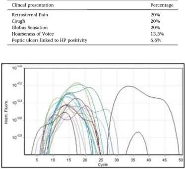
Of the 185 samples taken from the ear, 21 (11.35%) were not adequate for the correct execution of the DNA extraction procedure. The remaining 167 samples, subjected to RT-PCR, did not show in any case an increase in fluorescence linked to the FAM fluorophore, thus demonstrating the complete absence of HP in the material under ex-amination (Fig. 1). On the other hand, there was an expected increase in the JOE/VIC fluorophore fluorescence signal related to the internal control (Fig. 2) and of the fluorescence signal related to the positive control (Fig. 1), testifying to the successful outcome of the method.
4. Discussion
Even a significant number (38.6%) of our patients with OME had a history of gastric acid reflex, and 13.3% were positive for the Urea Breath Test, we did not detect the presence of HP in the exudate of the middle ear of our patients. Thus, we could not suggest an association between HP and OME.
HP is one of the most popular infectious agents in the world. Its virulence is recognized not only in gastritis but also in systemic diseases. HP’s involvement has been demonstrated in the pathogenesis of atherosclerosis, hepato-biliary, intestinal, hematological, pulmonary, and head-neck diseases [15]. The stomach represents the first reservoir for HP, but its presence has been demonstrated in the dental plaque, tonsils, adenoids, and squamous carcinomas of the larynx, even if the literature results are contradictory [16–18]. The authors have found the bacterium’s involvement in the OME pathogenesis and hypothesize that GER could cause inflammation of the mucosa that lines the Eustachian tube and the production of pro-inflammatory cytokines, such as TNF-α.
This subsequently would induce the secretion of mucins (MUC1 and MUC5AC), secretory type mucins that constitute the stomach’s primary
HP receptor, allowing the bacterium to readily colonize the middle ear [16–18].
HP could also increase inflammation through the expression of the CagA and VacA subunits, which, through an up-regulation mechanism of mucin expression, would amplify the pathogenetic process of the OME [9,19]. According to the authors, HP would find the micro-aerophilic environment in these patients’ middle ear (5% of CO2) necessary for its duplication [9]. In this case, the myringotomy inter-vention would create an environment with a high oxygen concentration that would prevent the bacterium’s further growth.
In our experience, despite that there are 18 patients (13.3%) included in the study were positive for the Urea Breath Test, which in-dicates the presence of gastric HP, and 38.6% were symptomatic for GER, the presence of HP in the exudate of the middle ear of patients with OME has not been found although the method we used based on the chain polymerization reaction (RT-PCR), a test with high sensitivity and specificity in demonstrating the presence of bacterial DNA [20].
Our findings are concordat with Jeyakumar et al., who investigated 48 children with OME and found no evidence of H pylori presence in any Table 1
The clinical presentation of 132 patients with OME and its frequency.
Clincal presentation Percentage
Hearing loss 100% Tinnitus 80% Autophony 73% Otalgia 53% Dizzeness 27% Table 2
Gastroesophageal reflux symptoms for the 132 patients with OME and its frequency.
Clincal presentation Percentage
Retrosternal Pain 20%
Cough 20%
Globus Sensation 20%
Hoarseness of Voice 13.3%
Peptic ulcers linked to HP positivity 6.6%
Fig. 1. Fluorescence signal determined by FAM fluorophores (colored lines)
and positive control (black lines).
Fig. 2. Fluorescence signal determined by JOE/VIC fluorophores related to
middle ear fluid specimen using the PCR test [21]. Also, Bitar et al. failed to detect HP in all 28 middle ear effusion samples from 18 children with otitis media using culture and PCR [22]. Pitk¨aranta et al. reported HP negative cultures of 12 middle ear fluid samples in all 8 examined children with recurrent otitis media [23]. Yilmaz et al. showed no sig-nificant difference regarding the positivity for H pylori in either middle ear mucosa culture results or middle ear mucosa PCR results between 22 patients with a diagnosis of OME, adenoid hypertrophy (AH), and chronic tonsillitis (CT) versus 20 patients with a diagnosis of adenoid hypertrophy and chronic tonsillitis without OME [24]. However, when culture and PCR results were combined, the middle ear was positive for H pylori in 10 (45%) patients in the study group and 2 (10%) patients in the control group (p = 0.011) [24]. Boronat-Echeverría et al. found that two out of 69 patients (2.9%) had positive results for H. pylori detection using ELISA and PCR and reported a significant association between the results of ELISA, PCR, and GERD questionnaire [25]. Karlidag et al.’s study showed a positive HP presence in the middle ear exudate of nine (16.3%) samples (total number of samples = 55) [26]. However, Agirdir et al. used the Campylobacter-like organism test (CLO-test) to detect HP presence in middle ear effusions of 45 patients diagnosed with chronic OME and adenoid hypertrophy and reported positive HP results in 66.6% of patients [27]. Skinner et al. did not find any positive results in 50 tonsillar samples through CLO-test [28]. Moreover, Yilmaz et al. did not find any positive results through CLO-test in 50 patients who un-derwent adenotonsillectomy procedure [29], confirming what other authors have already observed [30].
Despite the belief that refluxes are a trigger for the OME, repre-senting 38.6% of cases in our case history, the data observed by us excluding HP’s direct involvement in otitis media’s pathogenesis. It should be noted that the high level of hygiene and health, the favorable socio-economic conditions, and the widespread use of antibiotics, as occurs in our country, could reduce the exposure of the population to the bacterium, the transmission of which most likely occurs via the gold channel-fecal; in this way, the reduced prevalence of carriers of gastric HP infection, the early diagnosis of the latter and the aggressive eradi-cating antibiotic-based therapies (clarithromycin, amoxicillin, metro-nidazole), inhibitors of gastric secretion (omeprazole, ranitidine) and medical devices mechanical action containing various substances such as hyaluronic acid, sodium alginate, potassium bicarbonate or chon-droitin sulfate associated with components with high bioadhesive ca-pacity such as poloxamer 407 or aloe vera gel, could prevent the transmission of the pathogen to the middle ear.
Our group of patients had been selected neither by age nor by the positivity for gastroesophageal reflux, but only for the presence of OME in the active phase and rebellious to medical treatment; thus, an extremely homogeneous population from the epidemiological point of view was preserved.
However, there are some limitations in this study that should be mentioned. The cross-sectional design of this study without patients’ follow-up and the lack of control group, and the relatively small number of specimens limit the power of this study; similarly, with this being a single-center study, its generalizability to other populations is limited and would require further studies in other regions and other pop-ulations. The data presented in this study were collected from 2007 to 2009; while it may look outdated, the results are still in line with the literature. Our findings, therefore, are still valid and applicable regardless of the data collection time. Also, we did not monitor the esophageal pH to confirm the presence of reflux. However, we did not consider this procedure necessary for two reasons, firstly because the primary objective of our study was the demonstration of a possible correlation between OME and middle ear infection by HP and secondly because, as already observed by Yilmaz et al. [9], the pH-meters, which uses probes placed at 5 and 20 cm above the lower esophageal sphincter, is useful in the demonstration of gastroesophageal reflux but not of laryngopharyngeal reflux as it could not demonstrate the possible rise of gastric material at the level of the pharynx and even less of the
nasopharynx. Detection of H pylori can be difficult; we elected to use PCR to process OME since most other studies using it. PCR performance is dependent on the target gene, the primers used, and the conditions of the reaction, but in general, it is highly sensitive and specific for HP detection [20].
5. Conclusion
Based on the results obtained, we can affirm that although a third of OME cases are correlated to the presence of gastric acid reflux, Heli-cobacter Pylori does not seem to play any role in the etiology of the pathology as it cannot be found in endo-tympanic exudate. Therefore, further studies are needed to define the actual mechanism that de-termines all those forms of OME associated with reflux, the nature of which remains to be defined.
Ethical approval
Institutional approval was obtained from the Institutional Review Board at SS. Annunziata Hospital, Chieti, Italy.
Funding
No Funding.
Author contribution
All authors contributed significantly and in agreement with the content of the article. All authors were involved in project design, data collection, analysis, statistical analysis, data interpretation and writing the manuscript. All authors presented substantial contributions to the article and participated of correction and final approval of the version to be submitted.
Registration of research studies
UIN: researchregistry6309 https://www.researchregistry.com/register-now#home/registrat iondetails/5fc410d3863c4e001baeb54d/ Guarantor Laith Khasawneh. Email: [email protected]. Consent
Informed consent was obtained from each participant.
Provenance and peer review
Not commissioned and externally peer-reviewed.
Declaration of competing interest
The authors declare that they have no competing interests.
Acknowledgment
None.
Appendix A. Supplementary data
Supplementary data to this article can be found online at https://doi. org/10.1016/j.amsu.2021.01.056.
References
[1] M. François, New views on the pathogenesis of acute otitis media and its complications, Clin. Microbiol. Infect. 3 (Suppl 3) (1997 Jun) S5–S12. 11869222. [2] S.P. Wiertsema, A.J. Leach, Theories of otitis media pathogenesis, with a focus on Indigenous children, Med. J. Aust. 191 (S9) (2009 Nov 2) S50–S54. 19883357.
[3] A. Politzer, Diagnose und Theraphaie der AnsammIung seroeser Fluessigkeit in der
Thrommelhoehle, Wien Med. Wochenschr. 17 (1867) 224–247.
[4] A. Tasker, P.W. Dettmar, M. Panetti, J.A. Koufman, J.P. Birchall, J.P. Pearson, Reflux of gastric juice and glue ear in children, Lancet 359 (9305) (2002) 493,
https://doi.org/10.1016/S0140-6736(02)07665-1.
[5] M. Crapko, J.E. Kerschner, M. Syring, N. Johnston, Role of extra-esophageal reflux
in chronic otitis media with effusion, Laryngoscope 117 (2007) 1419–1423.
[6] A. Tasker, P.W. Dettmar, M. Panetti, J.A. Koufman, J.P. Birchall, J.P. Pearson, Is gastric reflux a cause of otitis media with effusion in children? Laryngoscope 112 (11) (2002) 1930–1934, https://doi.org/10.1097/00005537-200211000-00004. [7] M. Straetemans, N. van Heerbeek, E. Tonnaer, K.J. Ingels, G.T. Rijkers, G.
A. Zielhuis, A comprehensive model for the aetiology of otitis media with effusion, Med. Hypotheses 57 (6) (2001 Dec) 784–791, https://doi.org/10.1054/
mehy.2001.1494. 11918448.
[8] L. Lau, P. Mick, D.A. Nunez, Grommets (ventilation tubes) for recurrent acute otitis media in children, Cochrane Database Syst. Rev. 4 (4) (2018 Apr 6) CD004741,
https://doi.org/10.1002/14651858.CD004741.pub3. PMID: 29624209; PMCID:
PMC6494442.
[9] M.D. Yilmaz, O. Aktepe, Y. Cetinkol, A. Altuntas¸, Does Helicobacter pylori have role in development of otitis media with effusion? Int. J. Pediatr. Otorhinolaryngol. 69 (6) (2005) 745–749, https://doi.org/10.1016/j.ijporl.2004.12.009.
[10] C.A. Leone, F. Mosca, R. Grassia, P. Capasso, La diagnosi clinico strumentale del
reflusso laringofaringeo, in: C.A. Leone, E. Cunsolo (Eds.), Il Reflusso
Faringolaringeo, Torgraf, 2013, pp. 11–18.
[11] P.C. Belafsky, G.N. Postma, J.A. Koufman, Validity and reliability of the reflux
symptom index (RSI), J. Voice 16 (2) (2002) 274–277.
[12] Wma, WMA Declaration of Helsinki – Ethical Principles for Medical Research Involving Human Subjects – WMA – the World Medical Association, World Med. Assoc., 1964. https://www.wma.net/policies-post/wma-declaration-of-helsinki-e
thicalprinciples-for-medical-research-involving-human-subjects/. (Accessed 8
April 2020).
[13] R. Agha, A. Abdall-Razak, E. Crossley, N. Dowlut, C. Iosifidis, G. Mathew, for the
STROCSS Group, The STROCSS 2019 guideline: strengthening the reporting of
cohort studies in surgery, Int. J. Surg. 72 (2019) 156–165.
[14] ResearchRegistry, Available at: https://www.researchregistry.com/regist
er-now#home/registrationdetails/5fc410d3863c4e001baeb54d/.
[15] F. Franceschi, A. Gasbarrini, Helicobacter pylori and extragastric diseases, Best
Pract. Res. Clin. Gastroenterol. 21/2 (2007) 325–334.
[16] E. Aygenc, A. Selcuk, S. Celikkanat, C. Ozbek, C. Ozdem, The role of Helicobacter pylori infection in the cause of squamous cell carcinoma of the larynx, Otolaryngol. Head Neck Surg. 125 (5) (2001) 520–521, https://doi.org/10.1067/
mhn.2001.119438.
[17] J.R. Grandis, G.I. Perez-Perez, V.L. Yu, J.T. Johnson, M.J. Blaser, Lack of serologic evidence for Helicobacter pylori infection in head and neck cancer, Head Neck 19 (3) (1997) 216–218, https://doi.org/10.1002/(sici)1097-0347(199705)19:
3<216::aid-hed9>3.0.co;2-5.
[18] A. Kizilay, L. Saydam, A. Aydin, M.T. Kalcioglu, O. Ozturan, N.E. Aydin, Histopathologic examination for Helicobacter pylori as a possible etiopathogenic factor in laryngeal carcinoma, Chemotherapy 52 (2) (2006) 80–82, https://doi.
org/10.1159/000091727.
[19] B. Jelavic, J. Petricevic, I. Marijanovi´c, M. Bevanda, Helicobacter pylori in Otorhinolaryngology: cause or bystander, Eurasian J. Med. 51 (2) (2019) 196–202,
https://doi.org/10.5152/eurasianjmed.2018.18192.
[20] J.J. Lu, C.L. Perng, R.Y. Shyu, Comparison of five PCR methods for detection of
Helicobacter pylori DNA in gastric tissues, J. Clin. Microbiol. 37 (1999) 772–774.
[21] A. Jeyakumar, R.E. B´egu´e, Otitis media with effusion and Helicobacter pylori, OTO Open 2 (3) (2018 Jul 26), https://doi.org/10.1177/2473974X18792489, 2473974X18792489. PMID: 31535067; PMCID: PMC6737865.
[22] M. Bitar, R. Mahfouz, A. Soweid, et al., Does Helicobacter pylori colonize the nasopharynx of children and contribute to their middle ear disease? Acta Otolaryngol. 126 (2) (2006) 154–159, https://doi.org/10.1080/
00016480500312679.
[23] A. Pitk¨aranta, K.L. Kolho, H. Rautelin, Helicobacter pylori in children who are prone to upper respiratory tract infections, Arch. Otolaryngol. Head Neck Surg. 131 (3) (2005) 256–258, https://doi.org/10.1001/archotol.131.3.256. [24] T. Yilmaz, M. Ceylan, Y. Aky¨on, O. Ozçakýr, B. Gürsel, Helicobacter pylori: a
possible association with otitis media with effusion, Otolaryngol. Head Neck Surg. 134 (5) (2006) 772–777, https://doi.org/10.1016/j.otohns.2006.02.002. [25] N. Boronat-Echeverría, H. Aguirre-Mariscal, M. Carmolinga-Ponce, et al.,
Helicobacter pylori detection and clinical symptomatology of gastroesophageal reflux disease in pediatric patients with otitis media with effusion, Int. J. Pediatr. Otorhinolaryngol. 87 (2016) 126–129, https://doi.org/10.1016/j.
ijporl.2016.06.023.
[26] T. Karlidag, Y. Bulut, E. Keles, et al., Detection of Helicobacter pylori in children with otitis media with effusion: a preliminary report, Laryngoscope 115 (7) (2005) 1262–1265, https://doi.org/10.1097/01.MLG.0000165697.83921.2B.
[27] B.V. Agirdir, S. Bozova, A.T. Derin, M. Turhan, Chronic otitis media with effusion and Helicobacter pylori, Int. J. Pediatr. Otorhinolaryngol. 70 (5) (2006) 829–834,
https://doi.org/10.1016/j.ijporl.2005.09.026.
[28] L.J. Skinner, D.C. Winter, A.J. Curran, et al., Helicobacter pylori and tonsillectomy, Clin. Otolaryngol. Allied Sci. 26 (6) (2001) 505–509, https://doi.org/10.1046/
j.1365-2273.2001.00513.x.
[29] M. Yilmaz, C.O. Kara, I. Kaleli, et al., Are tonsils a reservoir for Helicobacter pylori infection in children? Int. J. Pediatr. Otorhinolaryngol. 68 (3) (2004) 307–310,
https://doi.org/10.1016/j.ijporl.2003.10.016.
[30] G. di Bonaventura, M. Neri, G. Neri, G. Catamo, R. Piccolomini, Do tonsils represent an extragastric reservoir for Helicobacter pylori infection, J. Infect. 42 (3) (2001) 221–222, https://doi.org/10.1053/jinf.2001.0815.