Synthesis, Structural Elucidation, and Biological Evaluation of NSC12,
an Orally Available Fibroblast Growth Factor (FGF) Ligand Trap for
the Treatment of FGF-Dependent Lung Tumors
Riccardo Castelli,
‡Arianna Giacomini,
§Mattia Anselmi,
‡Nicole Bozza,
‡Federica Vacondio,
‡Silvia Rivara,
‡Sara Matarazzo,
§Marco Presta,*
,§Marco Mor,*
,‡and Roberto Ronca*
,§ ‡Dipartimento di Farmacia, Universita
̀ degli Studi di Parma, Parco Area delle Scienze 27/A, I-43124, Parma, Italy
§
Dipartimento di Medicina Molecolare e Traslazionale, Universita
̀ degli Studi di Brescia, Via Branze 39, I-25123, Brescia, Italy
*
S Supporting InformationABSTRACT:
NSC12 is an orally available pan-FGF trap able to inhibit FGF2/FGFR interaction and endowed with promising
antitumor activity. It was identi
fied by virtual screening from a NCI small molecule library, but no data were available about its
synthesis, stereochemistry, and physicochemical properties. We report here a synthetic route that allowed us to characterize and
unambiguously identify the structure of the active compound by a combination of NMR spectroscopy and in silico
conformational analysis. The synthetic protocol allowed us to sustain experiments aimed at assessing its therapeutic potential for
the treatment of FGF-dependent lung cancers. A crucial step in the synthesis generated a couple of diastereoisomers, with only
one able to act as a FGF trap molecule and to inhibit FGF-dependent receptor activation, cell proliferation, and tumor growth
when tested in vitro and in vivo on murine and human lung cancer cells.
■
INTRODUCTION
Fibroblast growth factors (FGFs) are members of a large family
of structurally related proteins that a
ffect growth,
differ-entiation, migration, and survival of a variety of cell types by
leading the formation of productive ternary complexes with
signaling FGF receptors (FGFRs) and cell-surface heparan
sulfate proteoglycans (HSPGs).
1−3FGFs are highly expressed in epithelial human tumors where
they modulate growth, neovascularization, metastatic spreading,
and drug resistance.
4In lung cancer, the activation of the FGF/
FGFR signaling pathway promotes cancer cell survival,
resistance to chemotherapy, and growth of small- and
non-small-cell lung tumors.
5Elevated serum concentrations of
FGF2 represent an unfavorable prognostic factor in lung
cancer,
6while ampli
fication of the FGFR1 gene is a frequent
feature of squamous cell carcinomas in smoking patients.
7Thus, the FGF/FGFR system represents a privileged target for
the therapeutic approach of lung tumors in which the
FGF-dependent activation of FGFR is an oncogenic factor.
Current pharmacological approaches to address the
inhib-ition of the FGF/FGFR system include tyrosine-kinase
inhibitors (TKIs) and anti-FGFR antibodies or peptides.
8Multitarget TKIs that inhibit FGFR signaling are used in the
clinic and selective FGFR TKIs are evaluated in clinical trials.
However, both selective and nonselective TKIs show signi
ficant
side e
ffects.
9An alternative approach aimed at preventing FGF
binding to its cognate receptors is the use of ligand traps.
10Recently, we reported the characterization of the interaction
between FGF2 and its endogenous binder long-pentraxin 3
(PTX3)
11and identi
fied a 5-mer peptide belonging to the
amino-terminal sequence of PTX3 as the minimal motif needed
to bind FGF2 and to block its biological activity.
12The
pentapeptide was used for the modeling of a pharmacophore,
which in turn was used to screen the NCI2003 library of small
molecules.
134,4,4-Tri
fluoro-1-(3-hydroxy-10,13-dimethyl-
2,3,4,7,8,9,11,12,14,15,16,17-dodecahydro-1H-cyclopenta[a]-phenanthren-17-yl)-3-(tri
fluoromethyl)butane-1,3-diol
(com-pound 1 in
Scheme 1
, deposited at NCI with the code number
NSC172285 and named NSC12) was identi
fied from this
screening as a low molecular weight compound able to act as a
pan-FGF trap. This property resulted in a potent antiangiogenic
and antitumor activity when tested in various FGF-dependent
tumor models, designating 1 as the
first nonpeptidic, orally
Received: December 29, 2015Published: May 3, 2016
pubs.acs.org/jmc
available small molecule FGF trap endowed with potential
implications for the therapy of FGF-dependent cancers.
14However, a survey of the available information provided
minimal data for this compound, with no details regarding its
stereochemistry and its preparation, puri
fication, and
character-ization. We report here the synthesis of 1, designed on the sole
structure provided by the NCI, the assignment of its absolute
con
figuration, in vitro and in vivo experiments that confirmed
its biological activity, and characterization of its
physicochem-ical properties and oral bioavailability.
■
RESULTS
Synthesis. Retro-synthetic analysis of 1 dissects the
molecule into the steroidal portion and the 1,1-bis-tri
fluor-omethyl-1,3-propanediol motif, deriving in turn from the
reduction of the
β-hydroxy ketone 2 (
Scheme 1
) and some
minor protecting group manipulation. Following the
dis-connection at C21, compound 2 derives its carbon backbone
from the aldol condensation of a suitably protected derivative of
commercially available pregnenolone acetate 3 and hexa
fluor-oacetone (HFA) 4.
15We were con
fident that reduction of the
β-hydroxy ketone would furnish the 1,3-propanediol fragment
in high stereoselectivity, assuming as a
first hypothesis that 1
had been produced employing conditions that favor attack of
the reducing nucleophile on the si-face of the keto group.
16Before engaging it in the aldol condensation, pregnenolone
acetate 3 was converted to the corresponding benzoate
(
Scheme 2
, compound 6) by means of transesteri
fication and
subsequent benzoylation of the hydroxyl group of intermediate
5.
The optimized protocol featured the generation of the
lithium enolate of pregnenolone benzoate 6 with freshly
prepared LiHMDS in anhydrous THF at
−78 °C and warming
to 0
°C for a few minutes
17and then promptly delivering HFA
to the cold (
−78 °C), stirred solution of lithium enolate.
18After quenching of the reaction and aqueous workup, the
product could be isolated in up to 76% yield. The reduction of
the carbonyl function was straightforwardly accomplished by
treatment of
β-hydroxy ketone 7 with NaBH
4in a mixture of
methanol and THF (
Scheme 2
) to furnish 8 as a single
isomer.
19After saponi
fication of the benzoate ester, compound
9
was tested in vitro to evaluate its cytotoxicity, resulting in it
being devoid of signi
ficant biological activity.
To rationalize why 9 behaved di
fferently from the compound
provided by the NCI, we set out to perform structural analysis.
Whereas ESI-MS spectra showed identical molecular ion peak
and fragmentation pattern for the two substances, HPLC
analysis showed a di
fferent retention time of 9 with respect to 1
(co-injection as shown in
Figure 1
). Inspection of the
1H NMR
spectrum of both compounds revealed few small but
appreciable di
fferences in some key regions of the spectrum.
Remarkably, the chemical shift of the H18 methyl group is
about 0.3 ppm di
fferent and a doublet of triplet at 2.10 ppm
(H12eq) is clearly visible in the spectrum of compound 9 while
absent in that of 11 (see
Supporting Information Figure 4
).
The signal of H20 for both compounds is almost perfectly
superimposable. We hypothesized that compounds 9 and 1
were diastereoisomers, and given that the only stereocenter
generated during the synthesis was C20, we located the origin
of the epimeric nature of the two compounds at the reduction
step.
20After screening of a variety of reducing agents (LiAlH
4,
Luche reduction, DiBAL-H, among others), the only one
capable of delivering useful quantities (dr 87:13, 8/10) of both
diastereoisomers was sodium triacetoxyborohydride, by virtue
of an intramolecular rather than intermolecular reaction
(
Scheme 3
).
21All the other common reducing agents
consistently furnished exclusively the major isomer 8, precursor
to 9. Attempts to invert the con
figuration at C20 of compound
8
(sulfonylation/nucleophilic substitution, cyclic sulfate
for-mation/nucleophilic substitution, Mitsunobu conditions) failed
to deliver any useful precursor of 1.
Scheme 1. Retrosynthetic Analysis for Compound 1
Scheme 2
aaReagents and conditions: (a) MeONa, MeOH, rt; (b) BzCl, pyridine,
DMAP cat., rt, 86% yield over two steps; (c) (i) (Me3Si)NLi, THF, −78 to 0 °C (ii) HFA, −78 °C to rt; 78% yield; (d) NaBH4, MeOH/ THF 1:1; (e) MeONa, MeOH, rt, 68% yield over two steps.
Figure 1.HPLC trace of a co-injection of compounds 9 and 1 showing different retention times.
After careful chromatographic separation with standard
flash
column chromatography, followed by separation of the mixture
enriched of the minor isomer with an automated MPLC
system, reasonable quantities of the two epimers 8 and 10
could be obtained and then deprived of the benzoate ester by
transesteri
fication with catalytic MeONa in MeOH to afford
products 9 and 11. We could
finally prove the identity of 11
and 1 spectroscopically by means of
1H and
13C NMR analysis
and chromatographically (HPLC
−MS/MS; see
Supporting
Information
).
NMR Analysis. After establishment that compound 11 is
identical to 1, the substance provided by the NCI, and that
compound 9 is the corresponding C20 epimer, the absolute
con
figuration of the two compounds at C20 was assigned.
Inspection of
1H NMR spectra revealed the peculiar
multi-plicity of H20, a triplet signal for both compounds 9 and 11.
The coupling constant
3J is 10.3 Hz. H20 is neighbored by
three vicinal protons (H17, H21a, H21b), meaning that such an
unexpected low multiplicity arises from (i) a null-coupling
constant with one of the three protons and (ii) two couplings
of equal magnitude.
1H TOCSY experiment with selective band
center irradiation on H20 allowed observing that H21a has only
one geminal (
2J) coupling with H21b (J = 15.0 Hz), in turn
resonating as canonical doublet-of-doublet AB system with
coupling constants of 15.0 and 10.3 Hz, respectively. Therefore,
H21a has the null coupling with H20.
22According to the
Karplus equation,
23the dihedral angle between these protons
should be close to 90
°, meaning that H20 should assume an
anti arrangement with both H17 and H21b and suggests a
conformationally locked, six-membered ring induced by an
intramolecular hydrogen bond involving the hydroxyl groups of
the 1,3-diol portion (
Figure 2
).
The coupling constants calculated by the software
Schro
̈dinger Maestro 9.7
24for H20 on the minimum-energy
geometries represented in
Figure 2
, in which the hydrogen
bond between the two hydroxyl groups is formed, were
consistent with the observed triplets. In particular, the
calculated values for H20
−H17, H20−H21a, and H20−H21b
were 9.9, 1.0, and 11.0 Hz for 9 and 9.7, 1.5, and 11.3 Hz for
11, respectively. From the minimized geometries of the
compounds with 20R and 20S stereochemistry (
Figure 2
) it
is possible to appreciate the enantiomorphic arrangement of
functional groups, in which the remarkable di
fference in the
distance between the C18 methyl group and the C21
methylene suggested the use of nuclear Overhauser e
ffect
(NOE) NMR experiments to identify the two epimers.
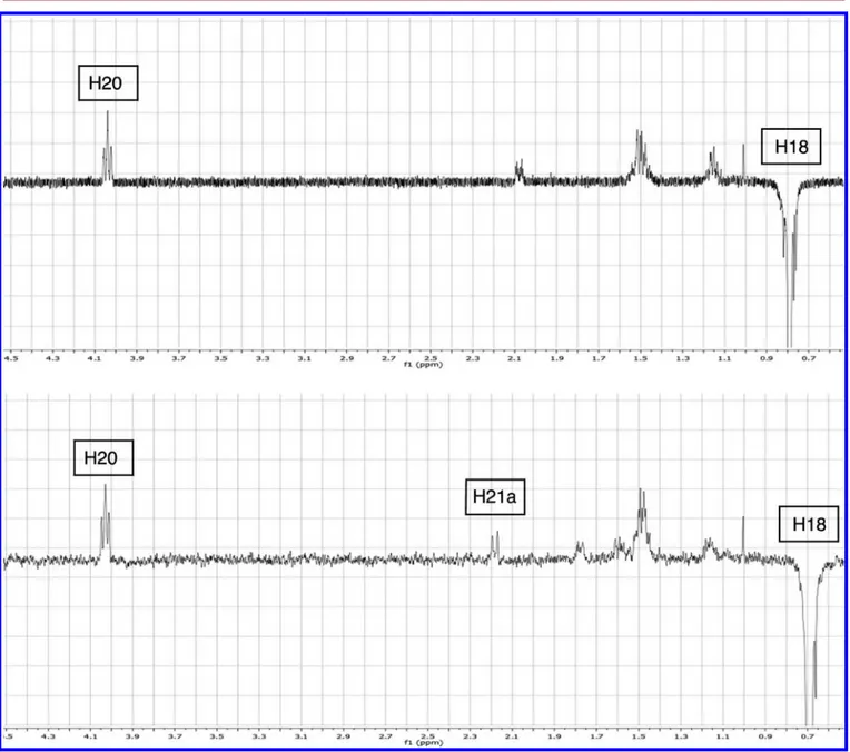
25NOE-NMR spectra of both compounds showed a clear
contact between H18 hydrogens and H20, indicating a
comparable relative distance. While compound 11 showed a
NOE contact between H18 and H21a at 2.18 ppm, compound
9
showed a signal at 2.05 ppm that could not be unequivocally
identi
fied by 2D-COSY nor by HSQC-NMR experiments, due
to extensive signal overlapping from protons of the steroidal
skeleton (
Figure 3
). We thus decided to engage the C20 and
C22 hydroxyl groups in a six-membered cyclic carbonate
(
Scheme 4
) to shift some of the critical signals in the NMR
spectra and
“freeze” the conformation accessible to the 1,3-diol
portion.
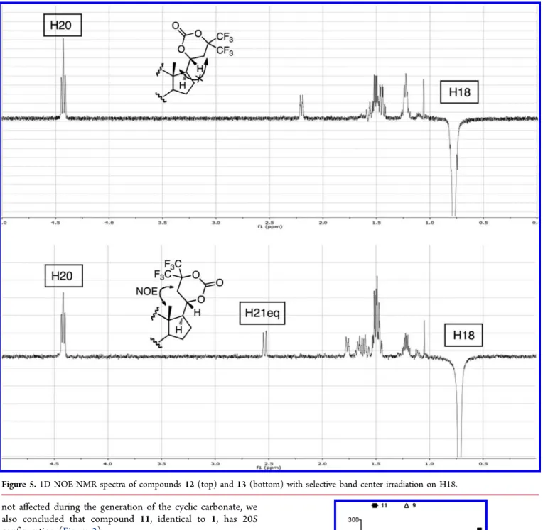
26,27The energy-minimized geometries of carbonates
12
and 13 (
Figure 4
) allowed the calculation of the distance
between H21a and H18, found to be 4.33 and 2.46 Å for the
20R and 20S carbonates, respectively.
1H NMR analysis of
cyclic carbonates showed that H20 retained the triplet
multiplicity and that the critical pseudoequatorial H21a signal
was in a region of the spectrum free of other resonance peaks.
The results of NOE experiments are depicted in
Figure 5
. H18
of compound 12 showed NOE contacts with H19 (d = 2.95 Å,
calculated from molecular model), H16 (d = 2.43 Å), H11ax (d
= 2.18 Å), H8 (d = 2.17 Å), H12eq (d = 2.53 Å), and H20 (d =
2.35 Å); the unequivocal assignment of these signals was
accomplished by 2D-COSY and HSQC-NMR experiments, and
these results are consistent with the proposed conformation
(
Supporting Information Figure 8
). As expected, no contact
was observed between H21eq (2.42 ppm) and H18 of
compound 12. On the contrary, H18 of compound 13, in
Scheme 3
aa
Reagents and conditions: (a) Na(AcO)3BH, MeOH/THF MeOH, rt; (b) MeONa, MeOH, rt, 74% yield for 9, 11% for 11 over two steps.
Figure 2. Proposed preferential conformations and geometries for compounds 9 and 11.
addition to showing the expected NOE contacts with H19 (d =
2.96 Å), H8 (d = 2.17 Å), H16 (d = 2.43 Å), H11ax (d = 2.18
Å), and H20 (d = 2.41 Å), showed a distinct NOE contact with
H21eq (d = 2.46 Å), as predicted from the molecular models
(see
Supporting Information Figures 12 and 13
). We therefore
concluded that the cyclic carbonate 12, deriving from
compound 8, the major isomer obtained from the reduction
step, has 20R con
figuration, whereas 13, the cyclic carbonate
obtained from compound 10, the minor isomer, has 20S
con
figuration. Given that the absolute configuration at C20 was
Figure 3.1D NOE-NMR spectra of compounds 9 (top) and 11 (bottom) with selective band center irradiation on H18.
Scheme 4
aaReagents and conditions: (a) triphosgene, pyridine, −50 °C, 77%
yield for 12, 68% yield for 13.
Figure 4. Minimized geometries for compounds 12 (top) and 13 (bottom). Distances between H21eq and the closest C18 hydrogen are the following: 12, 4.33 Å; 13, 2.46 Å. 3-Benzoyl esters are omitted for clarity.
not a
ffected during the generation of the cyclic carbonate, we
also concluded that compound 11, identical to 1, has 20S
configuration (
Figure 2
).
Molecular Interactions and Docking Simulations.
Compound 1 exerts its FGF2 trap activity by binding to the
growth factor molecule, thus hampering FGF/FGFR
inter-action. On this basis, DMSO stock solutions of 9 and 11 were
diluted in PBS, keeping the
final concentration of DMSO at
3%, and their capacity to bind FGF2 was assessed by surface
plasmon resonance (SPR) spectroscopy. In parallel, their
solubility was evaluated under the same experimental
conditions. Similar to 1,
14compound 11 binds FGF2
immobilized to a BIAcore sensor chip in a dose-dependent
manner with an apparent K
dvalue equal to
∼40 μM (
Figure 6
).
When assessed for its solubility, 11 was fully soluble in the 3%
DMSO solution up to the concentration of 30
μM, its solubility
decreasing at higher concentrations, whereas compound 9 was
significantly less soluble. When compared to 11, compound 9
appeared to be unable to interact with immobilized FGF2 at
any concentration tested, even when dissolved at a nominal 100
μM concentration, which corresponds to an actual
concen-tration of 25
μM in 3% DMSO solution (
Figure 6
and
Supporting Information Figure 14
).
A binding mode for compound 1-FGF2, based on docking
and molecular dynamics simulations and supported by NMR
data, has been described.
14Thus, docking simulations that took
into account published information were used to explore the
Figure 5.1D NOE-NMR spectra of compounds 12 (top) and 13 (bottom) with selective band center irradiation on H18.
Figure 6.SPR analysis of compounds 9 and 11 on a FGF2-coated sensor chip.
possibility that this binding mode could explain the di
fferent
FGF2-binding capacity of the two diasteroisomers. To this aim,
starting from a crystal structure of FGF2 (see
Experimental
Section
), only solutions involving the surface around key
residues were considered. Among the
first 100 nonredundant
docking poses of compound 11 having polar interactions with
Glu96, one presented all the features previously described, i.e.,
the butyl chain and tri
fluoromethyl groups within the
hydrophobic patch de
fined by Leu55, Ala57 and Val63; the
hydroxyl group on C20 taking a hydrogen bond with Glu96;
packing of rings A and B of the steroid sca
ffold with Leu107. In
this pose, the ligand occupies the portion of FGF2 surface that
faces the linker between FGFR1-D2 and D3 in the crystallized
structure (
Figure 7
).
Application of the same docking protocol to compound 9
gave a similar pose, with both the hydroxyl groups on the ligand
chain interacting with the carboxylate group of Glu96, while the
steroid nucleus projected toward the FGFR1-D2-interacting
side. In both cases, the docking solutions presented docking
scores signi
ficantly lower than the best poses (−1.6 vs −3.5 for
compound 20S and
−2.1 vs −3.5 kcal/mol for 20R) that
pointed the steroid nucleus toward the FGFR1-D3-interacting
area and were therefore less consistent with reported NMR
data.
In conclusion, compounds 9 and 11 can be docked on the
FGF2 surface in a binding mode that, even though consistent
with previous observations, does not explain the di
fferent
FGF2-binding a
ffinity of the two diasteroisomers. Given the
uncertainty of virtual docking models, further structural or
structure
−activity information will be required to elucidate
unambiguously the mode of interaction of the two compounds
with the FGF2 molecule.
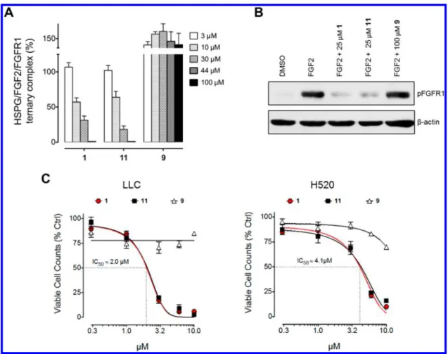
In Vitro Studies. Given their different FGF2-binding
potential, compounds 9 and 11 were compared to 1 for their
capacity to prevent the formation of HSPG/FGF2/FGFR1
ternary complexes in a FGF2-mediated cell
−cell adhesion
assay.
12bThe results demonstrate that 1 and 11 dissolved in 3%
DMSO hamper the formation of HSPG/FGF2/FGFR
complexes with an IC
50equal to
∼10 μM, whereas 9 was
inactive also when tested at a nominal 100
μM concentration
(
Figure 8
A), corresponding to an actual 25
μM concentration
(see above). Accordingly, when tested under the same
experimental conditions, compound 11, but not 9, inhibited
FGFR1 phosphorylation triggered by FGF2 in Lewis lung
carcinoma (LLC) cells in a manner similar to 1 (
Figure 8
B). As
a consequence, compounds 11 and 1 e
fficiently impaired the
proliferation of FGF-dependent murine (LLC) and human
(H520) lung carcinoma cell lines in vitro with an identical IC
50value, equal to 2.0
μM and 4.1 μM, respectively (
Figure 8
C). In
contrast, compound 9 did not exert a signi
ficant inhibitory
activity on both tumor cell lines. In the latter experiment, the
final concentration of DMSO was maintained equal to 1%,
higher concentrations of DMSO being cytotoxic in cell
proliferation assays.
Physicochemical Characterization. To assess whether
di
fferences in the physicochemical properties of the two
diastereoisomers might account, at least in part, for their
di
fferent antiproliferative activity, we measured the actual
concentration of 9 and 11 when dissolved at 10
μM in cell
culture medium plus 1% DMSO. While a concentration of 10.7
(
±0.8) μM was obtained for compound 11, compound 9 was at
least 5-fold less soluble, with a recovered concentration of 2.1
(
±0.2) μM.
Moving from these results, we investigated whether the
observed di
fference in solubility could be ascribed to a different
ionization for the two compounds. The values of aqueous
dissociation constants (pK
a) were extrapolated by the
potentiometric pH-metric method, starting from mixtures of
methanol and water in the 40
−10% range. Nonlinear
multiparametric analysis of the titration curves revealed the
presence of one ionization site, with pK
avalues of 9.08 (
±0.05)
for compound 9 and 9.04 (
±0.08) for compound 11, ruling out
this property as the basis for the di
fferences in solubility.
The role of divalent cations (such as Ca
2+or Mg
2+) that are
present in the biological assay conditions and could negatively
influence the solubility through the formation of complexes was
then investigated. The kinetic solubility of compound 9 was
determined in the cell culture medium, adding 2 mM EDTA as
chelating agent. No e
ffect of EDTA chelation was observed, as
the kinetic solubility value for compound 9 was 1.73 (
±0.15)
μM, superimposable to the result obtained in absence of
EDTA.
As the kinetic solubility value is a nonequilibrium
measure-ment, its dependence on the concentration within the stock
solution was evaluated. Two di
fferent starting concentrations of
compound 9 were tested (i.e., 600
μM and 300 μM in DMSO
stock solution), corresponding to nominal concentrations of
the
final solutions of 6 and 3 μM, also employed in the cell
proliferation assay. In both cases, we could recover
concen-trations close to 2
μM, i.e., 1.7 (±0.3) μM and 1.4 (±0.2) μM,
respectively. It therefore appears that 2
μM represents the limit
of kinetic solubility for compound 9, a concentration
corresponding to the IC
50of compound 11 for LLC cells.
Thus, solubility may represent a determinant in the
interpretation of the cell proliferation assay data when 9 is
dissolved in cell culture medium at concentrations higher than
2
μM. However, its incapacity to affect LLC cell proliferation at
this and lower concentrations con
firms the lack of FGF trap
activity of 9 when compared to 11.
In Vivo Study. Compound 1 was identified as the first small
molecule FGF trap able to inhibit the growth of human
FGF-Figure 7.(A) Docking solution of compound 11 (orange carbons) on FGF2 surface. Amino acids reported to interact with the ligand are shown (green carbons). The colors of FGF2 surface are related to molecular electrostatic potential. (B) Docking solution of compound 9 (orange carbons) on FGF2 surface. (C) View of the FGF2-FGFR1 (green ribbons) complex, as observed in the crystallized structure 1fq9, with docked structures of compounds 9 and 11.
dependent tumor cells following oral administration in
tumor-harboring immunode
ficient nude mice.
14In order to extend
these observations in an immunocompetent syngeneic animal
model, we designed a study to evaluate whether the different
physicochemical and biological properties of compounds 9 and
11
also resulted in a di
fferent in vivo antitumor FGF trap
activity when administered by oral gavage to syngeneic mice
grafted subcutaneously with FGF-driven murine LLC lung
carcinoma cells.
28In keeping with the in vitro observations,
daily treatment by oral gavage with compound 11 at the dose of
7.5 mg/kg for 7 days signi
ficantly reduced the growth of LLC
tumor grafts, while administration of compound 9 did not affect
the rate of tumor growth that was indistinguishable from that
observed in vehicle-treated animals (
Figure 9
A). Accordingly,
the weight of tumors harvested at the end of experimentation
was signi
ficantly reduced in the group treated with compound
Figure 8.(A) Inhibition of HSPG/FGF2/FGFR1 ternary complexes formation exerted by 1 and compounds 9 and 11. (B) Western blot analysis of FGFR1 phosphorylation (pFGFR1) on murine Lewis lung carcinoma (LLC) cells upon stimulation with FGF2 and treatment with compounds 1, 9, and 11.β-Actin was used for loading normalization. (C) Antiproliferative effect of 1 and compounds 9 and 11 on murine LLC and human lung carcinoma H520 cells.
Figure 9.(A) Left panel: inhibition of murine Lewis lung carcinoma tumor growth exerted by oral daily administration of compounds 9, 11, or vehicle. Right panel: tumor picture and weight after administration of 9, 11, or vehicle. (B) Mice plasma concentrations after a single oral administration of 7.5 mg/kg of 9 or 11: (∗) p < 0.05, (∗∗) p < 0.01, (#) p < 0.001.
11
when compared to the one treated with 9 and control
animals (
Figure 9
A). In addition, in line with the antiangiogenic
activity of 1, all tumors of animals treated with compound 11
were less vascularized, as shown by their pale appearance when
compared to the reddish appearance of the grafts of the other
experimental groups (
Figure 9
A). Both compounds were well
tolerated, as treated mice did not show signi
ficant clinical signs
of toxicity and body weight changes throughout the whole
experimental procedure. The bioavailability of compounds 11
and 9 following gavage administration in mice was assessed at
di
fferent time points after administration. The two compounds
(
Figure 9
B) showed a signi
ficant difference in their
bioavailability. Indeed, the plasma levels of compound 11
measured 30 min after a single oral administration of the
molecule in adult mice were signi
ficantly higher (Conc(11,30
min) = 316
± 45 nM) than those measured in the plasma of
age-matched animals treated with compound 9 (Conc(9,30
min) = 128
± 28 nM). At later time points (2 and 8 h after
gavage), plasma levels of compound 11 decreased to values that
remained higher than those measured for 9.
■
DISCUSSION AND CONCLUSIONS
Previous observations led to the identi
fication of 1 as the first
low molecular weight FGF trap molecule. Given its novel
mechanism of action and its e
fficient in vitro and in vivo activity
on di
fferent FGF-dependent tumor cell lines, 1 may represent a
prototype for the development of novel orally available
therapeutic agents targeting those tumors where
ligand-dependent FGFR activation represents a key factor. In this
paper we described the synthetic procedure for the preparation
of 1 that allowed definition and confirmation of its chemical
structure, permitting at the same time the identi
fication,
isolation, and physicochemical characterization of the two
C20 epimers of this steroidal derivative (compounds 11 and 9).
The results demonstrate that compound 11 recapitulates all
the anti-FGF properties described for 1, while 9, obtained as
the major isomer, is devoid of a signi
ficant FGF trap activity. In
particular, 11 acts as an FGF trap molecule able to inhibit
FGFR activation, cell proliferation, and tumor growth when
tested in vitro and in vivo on FGF-dependent murine and
human lung cancer cells, whereas 9 was ine
ffective. In addition,
a relevant point descending from the characterization of the
two C20 epimers is that solubility is an issue for the biological
activity and bioavailability of these steroidal derivatives.
Despite our e
fforts, the inability to increase the yield of
compound 11 still represents a limitation in the synthetic
protocol. Further chemical exploration will be required based
on the novel observation that the absolute con
figuration of C20
represents a crucial point for the structure
−activity relationship
of the compound.
In conclusion, this
first chemical investigation traces the way
for the synthesis of new FGF trap small molecules,
characterized by improved solubility, to devise relevant
structure
−activity relationships aimed at the design of new
compounds with optimized physicochemical properties and
improved potency while retaining the critical oral e
fficacy and
lack of toxicity. These data will constitute the starting point for
chemical optimization and structure
−activity relationship
studies of a promising class of compounds acting as FGF
traps with a genuine protein
−protein interaction-inhibitor
mechanism of action.
■
EXPERIMENTAL SECTION
Chemistry. All chemicals were used as received unless stated otherwise. All reactions were performed under a steady overpressure of nitrogen delivered through a balloon. Tetrahydrofuran was distilled over sodium/benzophenone prior to use. Anhydrous solvents such as dichloromethane, N,N-dimethylformamide, and pyridine were pur-chased stored over 3 Å molecular sieves and packed under argon. They were subsequently manipulated by syringe under a steady pressure of nitrogen. Column chromatography was performed on silica gel 60 (0.040−0.063 mm) under forced flow of the appropriate solvent mixtures. TLC analysis was conducted on HPTLC aluminum sheets (Sigma-Aldrich, silica gel 60, F254). Compounds were visualized by UV absorption (245 nm) and/or by dipping in a solution of (NH4)6Mo7O24·4H2O 25 g/L and (NH4)4Ce(SO4)4·2H2O 10 g/L, in 1 L of 10% aqueous H2SO4.1H,13C, and19F NMR spectra were recorded with Bruker AV 300, 400 and with a Varian 600. Chemical shifts (δ scale) are reported in parts per million (ppm) relative to the residual hydrogen peak of the deuterated solvent. Optical rotation was measured with a PerkinElmer 341 polarimeter, the concentration c of the analytes being reported as mg/mL. Mass spectra were recorded on an Applied Biosystem API-150 EX system spectrometer with an ESI or an APCI interface. Purity offinal compounds was analyzed by HPLC with UV detection at λ = 220 nm employing a Shimadzu HPLC gradient system (Shimadzu Corp., Kyoto, Japan) on a Supelco Discovery C18 column (150 mm× 4.6 mm, 5 μm particle size) by gradient elution, equipped with two LC-10AD solvent delivery modules, a Rheodyne 7125 sample injector, and a SPD-10A UV−vis detector. Prior to analysis, samples were prepared in MeOH at afinal concentration of 0.1 mg/mL. Flow rate was 1 mL/min, and injected volume was 10 μL. Solvent A: MeCN with 0.1% v/v of HCOOH. Solvent B: H2O with 0.1% v/v HCOOH. Isocratic conditions were employed for allfinal compounds: 75% A/25% B. Purity results are presented as tR(minutes) and relative chemical purity (%). All tested compounds were >95% pure.
3β-Hydroxypregn-5-en-20-one (5).29
To a solution of pre-gnenolone acetate (2.0 g, 5.6 mmol) in a mixture of MeOH/THF (1:1, 60.0 mL), Na (25.8 mg, 1.12 mmol 0.2 equiv) was added at room temperature. The reaction was stirred for a total of 4 h, when TLC analysis showed complete consumption of the starting material. The reaction mixture was neutralized with Amberlite IR-120 resin (H+ form). The solids werefiltered and the solvents removed in vacuo to afford 5 (1.7 g, quant) as a white powder used without any further purification. 1H NMR (300 MHz, CDCl 3) δ: 5.33 (bd, 1H, J = 5.2 Hz); 3.50 (m, 1H); 2.51 (t, 1H, J = 9.0 Hz); 2.32−2.15 (m, 3H); 2.11 (s, 3H); 2.05−1.97 (m, 2H); 1.88−1.81 (m, 2H); 1.71−1.37 (m, 9H); 1.29−0.93 (m, 7H); 0.62 (s, 3H).13C NMR (100 MHz, CDCl 3)δ: 209.7; 140.9; 121.4; 71.6; 63.7; 56.9; 50.0; 44.1; 42.3; 38.9; 37.3; 36.6; 31.9; 31.8; 31.6; 24.5; 22.8; 21.1; 19.4; 13.3. ESI-MS calcd for C21H32O2: 316.24. Found: 317.1 [M + H]+.
3β-Benzoyloxypregn-5-en-20-one (6).30
Pregnenolone 5 (2.5 g, 8.0 mmol) was dissolved in pyridine (30 mL) at 0 °C. Benzoyl chloride (1.2 mL, 10.3 mmol) was added, and the reaction mixture was stirred at room temperature for 24 h. The reaction was quenched with 1 M HCl solution, and the mixture was diluted with DCM (50 mL). The organic phase was washed with HCl 1 M (20 mL) and brine. After drying over Na2SO4, the solvent was evaporated under reduced pressure. The crude product was triturated with EtOAc to afford 6 (2.7 g, 86%) as a white crystalline solid. Analytical data correspond to those reported in the literature.30 Mp 192−193 °C (lit. 192−193 °C).1H NMR (300 MHz, CDCl3)δ: 8.05 (d, 2H, J = 7.1 Hz); 7.57 (t, 1H, J = 7.4 Hz); 7.45 (t, 1H, J = 7.7 Hz); 5.44 (bd, 1H, J = 4.3 Hz); 4.87 (m, 1H); 2.57 (t, 1H, J = 9.1 Hz); 2.50 (d, 2H, J = 7.8 Hz); 2.26−1.92 (m, 8H); 1.83−1.48 (m, 8H); 1.30−1.19 (m, 3H); 1.14−1.05 (m, 4H); 0.66 (s, 3H).13C NMR (100 MHz, CDCl 3) δ: 209.7; 166.1; 139.8, 132.9; 130.9; 129.6; 128.4; 122.6; 74.5; 63.8; 56.9; 50.0; 44.1; 38.9; 38.2; 37.1; 36.8; 31.9; 31.9; 31.7; 27.9; 24.6; 22.9; 21.2; 19.5; 13.3. ESI-MS calcd for C28H36O3: 420.26. Found: 421.1 [M + H]+.
Preparation of Hexafluoroacetone. HFA is a colorless, hygroscopic, nonflammable, and highly reactive gas at standard
pressure and temperature. The only commercial form that we could reasonably access, however, was the liquid trihydrate. A survey of the patent literature suggested the use of strong mineral acids to sequester water from the trihydrate.31After minor optimization, we successfully prepared gaseous HFA by dropping the trihydrate in warm (50°C) 98% sulfuric acid under stirring and delivering the liberated gas to the reaction flask through a Teflon tubing by means of a slight overpressure of nitrogen. A picture of the reaction setup is given in theSupporting Information.
3 β-Benzoyloxy-21-(bis(trifluoromethyl)hydroxymethyl)-pregn-5-en-20-one (7). To a stirred solution of bis-trimethylsilyl-amine (550μL, 2.62 mmol) in THF (50 mL) at −78 °C in a two-neck flask, BuLi (1.55 M in hexane, titrated with diphenylacetic acid prior to use,321.6 mL, 2.48 mmol) was added. The mixture was allowed to stir for a total of 30 min. Compound 6 (1003.1 mg, 2.39 mmol) was dissolved in dry THF (10 mL) and added with a syringe to the−78 °C cold solution of LiHMDS thus formed under nitrogen atmosphere and allowed to stir for 40 min. In a separate two-neck flask containing H2SO4 (98%, 10.0 mL) warmed to 50 °C, hexafluoroacetone trihydrate (3.0 mL, 21.5 mmol) was added dropwise from a pressure equalizing dropping funnel, under a positive pressure of nitrogen. The gas was delivered to theflask containing the enolate through a Teflon tube with both extremities secured to the second neck of theflasks with a septum. The reaction mixture was allowed reaching room temperature over the course of 4 h. The reaction was quenched with AcOH (5% v/v in H2O, 10 mL) and diluted with EtOAc (50 mL). The organic layer was washed with H2O (30 mL) and brine (30 mL), dried over Na2SO4, and the solvent was evaporated under reduced pressure. The residue was purified by flash column chromatography (SiO2Hex/DCM 50:50) to give 7 (1093.6 mg, 78%) as a white solid. Mp 181°C.1H NMR (400 MHz, CDCl3)δ: 8.04 (d, 2H, J = 7.1 Hz); 7.55 (t, 1H, J = 7.4 Hz); 7.43 (t, 1H, J = 7.8 Hz); 7.08 (s, 1H); 5.41 (bd, 1H, J = 4.7 Hz); 4.86 (m, 1H); 2.94 (d, 1H, J = 17.2 Hz); 2.80 (d, 1H, J = 17.2 Hz); 2.61 (t, 1H, J = 9.0 Hz); 2.47 (bd, 2H, J = 7.0 Hz); 2.18 (q, 1H, J = 9.3 Hz); 2.04−1.90 (m, 4H); 1.79−1.49 (m, 8H); 1.30−1.20 (m, 3H); 1.09−1.04 (m, 4H); 0.69 (s, 3H).13C NMR (100 MHz, CDCl3) δ: 212.1; 166.1; 139.8; 132.9; 130.8; 129.6; 128.4; (127.0; 123.9; 121.1; 118.4 (CF3)2); 122.3; (76.8; 76.5; 76.2; 75.9 (COH(CF3)2); 74.4; 64.9; 57.0; 49.9; 45.3; 38.7; 38.3; 37.8; 37.1; 36.7; 32.0; 31.8; 27.9; 24.4; 22.9; 21.1; 19.5; 13.3.19F NMR (376 MHz, CDCl3)δ: −78.03 (q, J = 9.4 Hz), −78.24 (q, J = 9.4 Hz). ESI-MS calcd for C31H36F6O4: 586.25. Found: 585.16 [M− H]−.
( 2 0R ) 3 β B e n z o y l o x y 2 1 ( b i s ( t r i fl u o r o m e t h y l ) -hydroxymethyl)pregn-5-en-20-ol (8). Compound 7 (110.2 mg, 0.19 mmol) was dissolved in MeOH/THF (1:1 v/v, 20 mL), and NaBH4(10.3 mg, 0.27 mmol) was added at 0°C. The reaction was warmed to room temperature and stirred for 1 h, when complete conversion of the starting material was observed by TLC analysis. The reaction was quenched with HCl solution (1 M, 5.0 mL), and stirring was continued for 15 min. Then reaction was diluted with aqueous NaOH (2 M, 30 mL) and stirred for an additional 2 h. After addition of EtOAc (50 mL), the organic layer was washed with water and brine, dried over Na2SO4 and the solvent was evaporated under reduced pressure, affording a white solid. The residue was purified by flash chromatography (SiO2Hex/DCM 30:70) to furnish 8 (80.5 mg, 72%) as a white powder. Mp 252°C.1H NMR (400 MHz, CDCl3)δ: 8.03 (d, 2H, J = 7.1 Hz); 7.56 (t, 1H, J = 7.4 Hz); 7.44 (t, 1H, J = 7.8 Hz); 6.77 (s, 1H); 5.42 (bd, 1H, J = 4.8 Hz); 4.88 (m, 1H); 4.21 (q, 1H, 9.3 Hz); 2.98 (d, 1H, J = 7.7 Hz); 2.48 (m, 2H); 2.18 (q, 1H, J = 9.3 Hz); 2.09−1.92 (m, 6H); 1.81−1.35 (m, 9H); 1.26−1.15 (m, 3H); 1.09− 1.040 (m, 4H); 0.84 (s, 3H).13C NMR (100 MHz, CDCl3)δ: 166.7; 139.8; 133.2; 130.4; 129.7; 128.4; (128.0; 127.3; 125.1; 124.4; 122.3; 121.5; 119.2; 118.4; (CF3)2); 122.6; (77.1; 76.8; 76.5; 76.2 (COH(CF3)2); 56.9; 56.1; 50.0; 42.6; 39.9; 38.2; 37.2; 36.8; 33.7; 31.9; 31.7; 28.3; 27.8; 25.4; 24.5; 21.0; 19.5; 12.6.19F NMR (376 MHz, CDCl3)δ: −75.68 (q, J = 9.8 Hz), −79.53 (q, J = 9.8 Hz). ESI-MS calcd for C31H38F6O4: 588.27. Found: 587.2 [M− H]−.
(20 R)-21-(Bis(trifluoromethyl)hydroxymethyl)pregn-5-en-3β,20-diol (9). Compound 8 (85.3 mg, 0.14 mmol) was dissolved in a mixture of MeOH/THF (2:1 v/v). Na (4.9 mg, 0.21 mmol) was
added and the reaction stirred for 1 h. After neutralization with a 1 M HCl solution, the mixture was diluted with EtOAc and the organic phase was washed with brine (5 mL), dried over Na2SO4 and the solvent was evaporated under reduced pressure. The residue was purified by flash column chromatography (SiO2Hex/EtOAc 70:30) to afford 9 (64.4 mg, 95%). Mp 203 °C; [α]D20−28.8 (c 2.88, CHCl3/ MeOH = 1/1).1H NMR (400 MHz, CD3OD)δ: 5.34 (bd, 1H, J = 2.8 Hz); 4.06 (t, 1H, J = 10.3 Hz); 3.39 (m, 1H, J = 4.9 Hz); 2.21 (m, 2H); 2.10 (dt, 1H, J = 3.5, 12.8); 2.02−1.92 (m, 3H); 1.85 (dt, 1H, J = 3.54, 13.4); 1.78 (m, 1H); 1.69 (m, 2H); 1.57−1.45 (m, 6H); 1.29− 1.14 (m, 4H); 1.09−1.02 (m, 4H); 0.96 (td, 1H, J = 5.2, 6.2); 0.81 (s, 3H).13C NMR (100 MHz, CD3OD)δ: 141.0; (128.2; 127.5; 125.4; 124.6; 122.5; 121.8; 119.7; 119.0; (CF3)2); 121.0; 76.9; 76.8; 76.4 (COH(CF3)2); 71.1; 70.9; 56.7; 56.3; 50.5; 42.4; 41.7; 39.2; 37.3; 36.4; 33.5; 31.8; 31.7; 31.0; 25.1; 24.2; 20.7; 18.6; 11.2.19F NMR (376 MHz, CD3OD)δ: −77.18 (q, J = 9.9 Hz), −80. (q, J = 9.8 Hz). ESI-MS calcd for C24H34F6O3: 484.24. Found: 483.3 [M− H]−. HPLC− UV purity: tR= 6.6 min, 96%.
( 2 0S ) 3 β B e n z o y l o x y 2 1 ( b i s ( t r i fl u o r o m e t h y l ) -hydroxymethyl)pregn-5-en-20-ol (10). Compound 7 (191.6 mg, 0.33 mmol) was dissolved in THF (20 mL), and Na(OAc)3BH (190 mg, 0.9 mmol) was added at 0°C. The reaction was warmed to room temperature and stirred for 6 h, when complete conversion of the starting material was observed by TLC analysis. The reaction was quenched with HCl solution (1 M, 5.0 mL), and stirring was continued for 15 min. The mixture was diluted with aqueous NaOH (2M, 30 mL) and stirred for additional 2 h. After addition of EtOAc (50 mL), the organic layer was washed with water and brine, dried over Na2SO4and the solvent was evaporated under reduced pressure, affording a white solid. The residue was purified by flash column chromatography (SiO2 Hex/EtOAc 80:20). The fractions containing both epimers 8 and 10 were subjected to MPLC (Isolera Dalton, Biotage, gradient t(0) Hex/EtOAc 98:2, t(20) Hex/EtOAc 80:20, giving 10 (21.9 mg, 11%) as a white amorphous solid. Mp 256°C.1H NMR (400 MHz, CDCl3)δ: 8.03 (d, 2H, J = 7.2 Hz); 7.55 (t, 1H, J = 7.4 Hz); 7.43 (t, 1H, J = 7.7 Hz); 6.42 (bs, 1H); 5.42 (bd, 1H, J = 4.4 Hz); 4.85 (m, 1H); 4.21 (t, 1H, 9.6 Hz); 2.47 (d, 2H, J = 7.8 Hz); 2.23 (d, 1H, J = 15.2 Hz); 2.04−1.43 (m, 13H); 1.35−1.07 (m, 8H); 0.85 (m, 1H); 0.72 (s, 3H).13C NMR (100 MHz, CDCl3)δ: 116.1, 139.7, 132.8, 130.7, 129.6, 128.3, 125.0, 122.4, 122.1, 74.5, 72.0, 57.3, 56.4, 49.8, 41.8, 39.0, 37.0, 36.6, 33.6, 31.7, 31.5, 29.7, 27.8, 24.8, 23.9, 20.7, 19.4, 12.5.19F NMR (376 MHz, CDCl3)δ: −75.45 (q, J = 9.8 Hz), −79.50 (q, J = 9.8 Hz). ESI-MS calcd for C31H38F6O4: 588.3. Found: 587.2 [M− H]−.
(20 S)-21-(Bis(trifluoromethyl)hydroxymethyl)pregn-5-en-3β,20-diol (11). Compound 10 (85.3 mg, 0.14 mmol) was dissolved in a mixture of MeOH/THF (2:1 v/v). Na (4.9 mg, 0.21 mmol) was added and the reaction stirred for 1 h. After neutralization with a 1 M HCl solution, the mixture was diluted with EtOAc and the organic phase was washed with brine (5 mL), dried over Na2SO4 and the solvent was evaporated under reduced pressure. The residue was purified by flash column chromatography (SiO2 Hex/EtOAc 80:20) giving 55.4 mg of 11 as a white powder (96%). Mp 205°C; [α]D20 −34.5 (c 5.73, CHCl3/MeOH = 1/1).1H NMR (400 MHz, MeOD) δ: 5.35 (bd, 1H, J = 5.5 Hz); 4.05 (t, 1H, J = 10.3 Hz); 3.39 (m, 1H, J = 4.6 Hz); 2.24−2.19 (m, 3H); 1.98 (m, 1H); 1.94−1.86 (m, 2H); 1.80 (m, 2H); 1.72−1.43 (m, 8H); 1.28−1.04 (m, 5H); 1.02 (s, 3H); 0.96 (td, 1H, J = 11.4, 4.7); 0.72 (s, 3H). 13C NMR (100 MHz, MeOD)δ: 142.4; (129.6; 128.7; 126.8; 126.0; 124.0; 123.2; (CF3)2); 122.4; 76.9; 76.8; 76.4 (COH(CF3)2); 72.6; 71.8; 59.1; 58.1; 51.7; 43.1; 42.8; 40.6; 38.6; 37.8; 35.0; 33.0; 32.9; 32.4; 31.0; 26.6; 25.0; 22.1; 20.0; 12.9.19F NMR (376 MHz, CDCl3)δ: −76.95 (q, J = 10.0 Hz),−80.66 (q, J = 10.0 Hz). ESI-MS calcd for C24H34F6O3: 484.2. Found: 483.3 [M− H]−. HPLC−UV purity: tR= 5.2 min, 98%.
(17 R)-3β-Benzoyloxy-17-((4R)-6,6-bis(trifluoromethyl)-2-oxo-1,3-dioxan-4-yl)androst-5-ene (12). Compound 8 (50.1 mg, 0.08 mmol) was dissolved in anhydrous DCM (5 mL) under nitrogen atmosphere before anhydrous pyridine (70μL, 0.8 mmol) was added. The mixture was cooled to−50 °C, and triphosgene (25.3 mg, 0.08 mmol) was added and stirred for 2 h. Once the mixture was warmed to
room temperature, to the mixture were added saturated aqueous solution of NH4Cl and a 1 M HCl solution. The mixture was then diluted with EtOAc and the organic phase was washed with brine (5 mL) dried over Na2SO4and the solvent was evaporated under reduced pressure, affording a white solid (55 mg). The residue was purified by flash column chromatography (SiO2 Hex/DCM 50:50) giving 12 (39.8, 77%) as a white powder. Mp 268°C. 1H NMR (400 MHz, CDCl3)δ: 8.04 (d, 2H, J = 7.5 Hz); 7.55 (t, 1H, J = 7.4 Hz); 7.43 (t, 1H, J = 7.7 Hz); 5.41 (bd, 1H, J = 4.7 Hz); 4.86 (m, 1H); 4.44 (t, 1H, J = 10.9 Hz); 2.47−2.38 (m, 3H); 2.21 (m, 2H); 2.19 (m, 3H); 2.17− 1.45 (m, 9H); 1.28−1.03 (m, 8H); 0.80 (s, 3H).13C NMR (100 MHz, CDCl3)δ: 166.0, 144.5, 139.9, 132.8, 130.8, 129.5, 128.3, 122.2, 77.4, 74.4, 55.9, 53.6, 49.9, 42.9, 38.8, 38.2, 37.0, 36.7, 31.8, 31.7, 27.8, 27.3, 24.4, 24.2, 20.7, 19.4, 12.4.19F NMR (376 MHz, CDCl 3)δ: −77.00 (q, J = 9.4 Hz),−78.3 (q, J = 9.4 Hz). (17 R)-3β-Benzoyloxy-17-((4R)-6,6-bis(trifluoromethyl)-2-oxo-1,3-dioxan-4-yl)androst-5-ene (13). Compound 10 (68.8 mg, 0.1 mmol) was dissolved in dry DCM (5 mL) under nitrogen atmosphere before pyridine (7.0 mL) was added. The mixture was cooled to−50 °C, and triphosgene (42.1 mg, 0.1 mmol) was added and stirred for 2 h. Once the mixture was warmed to room temperature, to the mixture were added a saturated aqueous solution of NH4Cl and a 1 M HCl solution. The mixture was then diluted with EtOAc and the organic phase was washed with brine (5 mL) dried over Na2SO4and the solvent was evaporated under reduced pressure, affording a white solid (65 mg). The residue was purified by flash column chromatography (SiO2Hex/EtOAc 95:5) giving 13 (48.9 mg, 68%) as a white powder. Mp 220°C.1H NMR (400 MHz, CDCl3)δ: 8.04 (d, 2H, J = 7.3 Hz); 7.55 (t, 1H, J = 7.4 Hz); 7.43 (t, 1H, J = 7.6 Hz); 5.42 (bd, 1H, J = 4.6 Hz); 4.86 (m, 1H); 4.21 (t, 1H, J = 9.5 Hz); 2.56 (d, 1H, J = 13.9 Hz); 2.47 (m, 1H); 2.21 (t, 1H, J = 12.9); 2.10− 1.99 (m, 3H); 1.91 (dt, 1H, J = 13.3, 3.4 Hz); 1.82−1.43 (m, 10H); 1.34−1.01 (m, 7H); 0.74 (s, 3H).13C NMR (100 MHz, CDCl 3)δ: 166.0, 144.9, 139.6, 132.8, 130.8, 129.5, 128.3, 122.9, 122.7, 122.4, 120.7, 119.8, 79.3, 79.0, 78.0, 74.4, 56.1, 54.0, 49.8, 41.9, 38.8, 38.2, 37.0, 36.6, 31.7, 31.4, 27.8, 27.7, 25.1, 24.1, 20.7, 19.4, 12.6.19F NMR (376 MHz, CDCl3)δ: −77.2 (q, J = 9.3 Hz), −78.2 (q, J = 9.3 Hz). Kinetic Solubility Measurements. Kinetic solubility for com-pounds 9 and 11 was determined starting from freshly prepared DMSO stock solutions.33 To assess solubility in the cell-based assay buffer (DMEM buffer + 0.4% FBS), an amount of 2 μL of each stock solution was added to 198μL of buffer to reach the final nominal concentrations of 10, 6, 3μM (9) or 10 μM (11) in a 96-well plate. In the EDTA-treated samples, 10μM 9 was incubated in DMEM buffer + 0.4% FBS further added with 2 mM EDTA. Final DMSO concentration in samples was 1%. The plate was kept under stirring for 4 h (250 rpm, rt). To assess solubility for 9 and 11 in the SPR assay buffer (PBS buffer), DMSO stock solutions of 9 and 11 were added to PBS to reach thefinal nominal concentrations of 100, 44, 29, 16, 8.9 μM. Final DMSO concentration in samples was 3%. Samples were stirred for 5 min (250 rpm, rt). At the end of the reported stirring times, all samples were centrifuged (1000g, 3 min, 20°C) to separate undissolved compound, and an aliquot of the supernatant was further diluted 1 to 100 with MeCN containing the internal standard 8 at the final concentration of 100 nM. Samples were further centrifuged (14 000g, 10 min, 5°C), and an amount of 10 μL of the supernatant was injected into the HPLC−MS/MS system for quantification employing the described analytical method.
pKaMeasurements. The pKa values for compounds 9 and 11 were determined by the potentiometric pH-metric method,34 employing a Sirius GLpKa instrument (Sirius Analytical Instruments Ltd., Forrest Row, U.K.) equipped with a semimicro combined electrode, quartz precision dispensers, a temperature probe, and a micro mechanical stirrer. As the solubility of the compounds did not allow a direct aqueous determination of pKa, aqueous pKavalues were extrapolated starting from cosolvent pKavalues (psKa) obtained in mixtures of water (at 0.15 Mfixed ionic strength for KCl addition) and methanol in the 40%−10% w/v range. All potentiometric titrations were performed at 25.0± 0.1 °C under a nitrogen atmosphere.
HPLC−ESI-MS/MS Analytical Method. A HPLC−ESI-MS/MS method for the quantitative analysis of compounds 9 and 11 was developed employing a Thermo Accela ultra high performance liquid chromatography (UHPLC) gradient system coupled to a Thermo TSQ Quantum Max triple quadrupole mass spectrometer (Thermo Italia, Milan, Italy) equipped with a heated electrospray ionization (H-ESI) ion source. Chromatographic separation occurred on a Phenomenex Synergi Fusion column (100 mm × 2.0 mm, 4 μm particle size) by gradient elution. Eluent A was acetonitrile; eluent B was water. Gradient: t(0 min), 5%A/95%B. t(1 min): 5%A/95%B. t(6 min): 95%A/5%B. t(9 min): 95%A/5%B. t(11 min): 5%A/95%B, with a 3 min re-equilibration time. Mass spectrometric analyses were done in negative ion mode and in multiple reaction monitoring (MRM). H-ESI interface parameters were set as follows: probe middle (D) position; capillary temperature 270°C; spray voltage 3.0 kV. Nitrogen was used as nebulizing gas at the following pressure: sheath gas, 35 psi; auxiliary gas, 15 arbitrary units (a.u.). Argon was used as the collision gas at a pressure of approximately 1.5 mTorr. For quantitative analysis, the following parent ion→ product ions transitions were selected. 9: m/z 483.1 → m/z 413.3 + m/z 111.0 + m/z 69.1 (tube lens 74; collision energies 22, 25, 74 eV, respectively; retention time (tR), 7.50 min). 11: m/z 483.1→ m/z 413.3 + m/z 111.0 + m/z 69.1 (tube lens 74; collision energies 22, 25, 74 eV, respectively; retention time, 7.33 min). 8 (internal standard): m/z 587.1→ m/z 517.5 + m/z 499.6 + m/z 111.0 (tube lens 93; collision energies 22, 30, 25 eV). Calibration curves for the analyte were prepared by spiking blank mouse plasma with stock solutions of 9 or 11 in DMSO. Both calibration and unknown samples were processed by protein precipitation via organic solvent addition (acetonitrile containing the internal standard; ratio 1:2). Samples were centrifuged (13 000 rpm, 10 min, 4°C), and an amount of 10μL of the supernatant was injected into the HPLC−ESI-MS/MS system for quantification. Linearity was checked in the 500−1 nM concentration range, with a LOQ equal to 1 nM. The coefficients of correlation (r2) were >0.99 for all curves. The specificity of the assay was evaluated by comparison of HPLC−ESI-MS/MS chromatograms of compounds 9 and 11 at the LOQ to those of blank plasma samples. Extraction efficiency was determined by comparing the peak area ratio of spiked mouse plasma samples at three concentration levels (low, intermediate, and high) to those of extracted blank plasma spiked with the corresponding concentrations. The mean extraction recovery ranged between 90% and 95%.
Computational Methods. The structures of compounds 9, 11, 12, and 13 were built with Maestro 9.7,24 and their geometries were optimized with OPLS2005 forcefield in combination with an implicit solvent model (water) using a convergence criterion for energy minimization of 0.05 kcal mol−1Å−1.35
A model of FGF2 was built, by the Protein Preparation Wizard of Maestro 9.7, from the crystal coordinates of a ternary FGF2-FGFR1-heparin complex (PDB code 1fq9). In thefinal structure, the C and N termini were capped with a methylamino and an acetyl group, respectively; glutamate and aspartate residues were deprotonated, lysines and arginines were protonated, and histidines were in their neutral form. Amino acid polar side chains and water molecules were reoriented to optimize the overall hydrogen-bond network. Prior to docking simulations, all molecules different from FGF2 of the first subunit were removed.
Docking studies were performed with Glide 6.1.36The docking grid on the surface of FGF2 was centered on the residues that interact with the ligand in the binding mode described in a previous work,14 i.e., Leu55, Ala57, Val63, Glu96, and Leu98. The bounding box, enclosing ligand center of mass, was extended for 15 Å in each axis. Docking simulations were performed in standard precision mode, imposing a hydrogen bond constraint on the carboxylate group of Glu96. van der Waals radii of the ligand were scaled to 0.7. One-hundred poses were collected for each compound, avoiding multiple solutions of similar poses.
We adopted residue numbering from mature FGF2, which is also used in the PDB file. The previous work had adopted a different numbering, with a positive shift of nine units (Leu64, Ala66, Val72, Glu105, and Leu107).
Biology Reagents and Cell Cultures. Human recombinant FGF2 was from Tecnogen (Piana di Monteverna, Caserta, Italy). Murine Lewis lung carcinoma (LLC), cultured in DMEM plus 10% heat-inactivated FBS, were provided by R. Giavazzi (Istituto M. Negri, Milan, Italy). Human H520 cells were obtained from ATCC and cultured in RPMI plus 10% FBS. Cells were maintained at low passage, returning to original frozen stocks every 3−4 months, and tested regularly for Mycoplasma negativity.
Surface Plasmon Resonance (SPR). Compounds 9 and 11 were analyzed for their capacity to directly bind to immobilized FGF2 using a BIAcore X-100 apparatus (BIAcore Inc., Piscataway, NJ, USA). FGF2 (20μg/mL in 10 mM sodium acetate, pH 6.0) was allowed to react with aflow cell of a CM5 sensor chip previously activated with a mixture of 0.2 M N-ethyl-N′-(3-dimethylaminopropyl)carbodiimide hydrochloride and 0.05 M N-hydroxysuccinimide (35μL, flow rate 10 μL/min). After ligand immobilization, matrix neutralization was performed with 1.0 M ethanolamine (pH 8.5) (35μL, flow rate 10 μL/min) and the activated/deactivated dextran was used as reference (control) system. Increasing concentrations of 9 and 11 (ranging between 8 and 150μM) were injected over the FGF2-coated sensor chip, and the response was recorded as a function of time-tracking the SPR intensity change upon binding progression. Injection lasted for 4 min (flow rate 30 μL/min) to allow association to immobilized FGF2 and was followed by 10 min of dissociation; each run was performed in 3% DMSO in PBS and the sensor chip was regenerated with 10 mM NaOH. The equilibrium (plateau) values of the SPR sensorgrams were used to build the binding isotherms displayed, after normalization. Binding isotherm points werefitted with the Langmuir equation for monovalent binding to evaluate the mass surface dissociation constant, Kd. The best-fitting procedure was performed with the SigmaPlot 11.0 software package (Systat Software Inc.).
HSPG/FGF/FGFR1 Mediated Cell−Cell Adhesion Assay. This assay was performed as described with minor modifications.37Briefly,
wild-type CHO-K1 cells were seeded in 24-well plates at 150 000 cells/cm2. After 24 h, cell monolayers were washed with PBS and incubated with 3% glutaraldehyde in PBS for 2 h at 4°C. Fixation was stopped with 0.1 M glycine, and cells were washed extensively with PBS. Then, A745-CHO-flg-1A-luc cells (50 000 cells/cm2) were added to CHO-K1 monolayers in serum-free medium plus 10 mM EDTA with or without 30 ng/mL of FGF2 in the absence or presence of increasing concentrations of 1, 11, or 9 dissolved in 3% DMSO. After 2 h of incubation at 37°C, unattached cells were removed by washing twice with PBS, A745-CHO-flg-1A-luc cells bound to the CHO-K1 monolayer were solubilized, and luciferase activity was quantified. All experiments were performed in triplicate.
Western Blotting. LLC cells were treated with FGF2 (30 ng/mL) in the absence or presence of compound 1, 9, or 11 dissolved in culture medium and 3% DMSO. After 20 min of incubation, cell samples were washed in cold PBS and homogenized in RIPA buffer containing 1% Triton-X100, 0.2% BriJ, 1 mM sodium orthovanadate, and protease inhibitors cocktail. Protein concentrations were determined using the Bradford protein assay (Bio-Rad Laboratories, Milano, Italy). Blotting analysis was performed using antiphospho FGFR1 (Santa Cruz Biotechnology, Santa Cruz, CA, USA). Equal loading of the lanes was confirmed by immunoblotting with anti-β-actin antibody.
Viable Cell Counting. Cells were cultured under appropriate conditions for 48 h (LLC cells) or 72 h (H520 cells) in the absence or presence of compound 1, 9, or 11 dissolved in culture medium and 1% DMSO. Propidium iodide (PI) staining (Immunostep, Salamanca, SP, EU) was used to detect PI-negative viable cells byflow cytometry. Absolute cell counts were obtained by the counting function of the MACSQuant analyzer (Miltenyi Biotec).
In Vivo Studies. Animal experiments were performed in accordance with the institutional and national guidelines and regulations. Eight-week-old C57BL/6 mice were injected sc into the dorsolateralflank with 5 × 10⁵LLC cells in 200μL total volume of PBS. When tumors were palpable, 11 or 9 (both at 7.5 mg/kg) or vehicle was administered daily by gavage in a 200μL final volume of DMSO/H2O (1:1, vol:vol). Tumors were measured in two
dimensions, and tumor volume was calculated according to the formula V = (D× d2)/2, where D and d are the major and minor perpendicular tumor diameters, respectively.28 At the end of the experimental procedure, tumors were harvested, weighed, and photographed. For the determination of plasma levels of compounds 9and 11, eight-week-old C57BL76 mice were treated orally with 7.5 mg/kg of each compound and blood samples were collected 30 min, 2 h, and 8 h thereafter for plasma analysis.
Statistical Analyses. Statistical analyses were performed using the statistical package Prism 5 (GraphPad Software). Student’s t test for unpaired data (two-tailed) was used to test the probability of significant differences between two groups of samples. For more than two groups of samples, data were statistically analyzed with a one-way analysis of variance, and individual group comparisons were evaluated by the Bonferroni multiple comparison test. Tumor volume data were statistically analyzed with a two-way analysis of variance, and individual group comparisons were evaluated by the Bonferroni correction. Differences were considered significant when P < 0.05.
■
ASSOCIATED CONTENT
*
S Supporting InformationThe Supporting Information is available free of charge on the
ACS Publications website
at DOI:
10.1021/acs.jmed-chem.5b02021
.
NMR and mass spectra of relevant compounds, kinetic
solubility, and synthesis information (
)
Molecular formula strings (
CSV
)
■
AUTHOR INFORMATION
Corresponding Authors
*M.P.: phone, +39 030 3717311; fax, +39 030 3717747; e-mail,
[email protected]
.
*M.M.: phone, +39 0521 905059; fax, +39 0521 905006;
e-mail,
[email protected]
.
*R.R.: phone, +39 030 3717735; fax, +39 030 3717747; e-mail,
[email protected]
.
Author Contributions
The manuscript was written through contributions of all
authors. All authors have given approval to the
final version of
the manuscript.
Notes
The authors declare no competing
financial interest.
■
ACKNOWLEDGMENTS
This work was supported by grants from Ministero Istruzione,
Universita
̀ e Ricerca (FIRB Project RBAP11H2R9 2011),
Associazione Italiana Ricerca sul Cancro (AIRC Grant 14395)
to M.P., and S.M. was supported by Fondazione Italiana per la
Ricerca sul Cancro Fellowships.
■
ABBREVIATIONS USED
CHO, Chinese hamster ovary; DCM, dichloromethane;
DiBAL-H, diisobutylaluminum hydride; DMEM, Dulbecco
’s
modi
fied Eagle medium; EDTA, ethylenediaminetetraacetic
acid; FBS, fetal bovine serum; FGF2,
fibroblast growth factor 2;
FGFR,
fibroblast growth factor receptor; HFA,
hexafluoroace-tone; HMDS, hexamethyldisilylamine; HSPG, heparan sulfate
proteoglycan; LiHMDS, lithium hexamethildisilylamide;
MPLC, medium pressure liquid chromatography; NMR,
nuclear magnetic resonance; NOE, nuclear Overhauser e
ffect;
PBS, phosphate bu
ffered saline; RPMI, Rosewell Park
Memorial Institute; THF, tetrahydrofuran; TK, tyrosine kinase
■
REFERENCES
(1) Bikfalvi, A.; Klein, S.; Pintucci, G.; Rifkin, D. B. Biological roles of fibroblast growth factor-2. Endocr. Rev. 1997, 18, 26−45.
(2) Schlessinger, J.; Plotnikov, A. N.; Ibrahimi, O. A.; Eliseenkova, A. V.; Yeh, B. K.; Yayon, A.; Linhardt, R. J.; Mohammadi, M. Crystal structure of a ternary FGF-FGFR-Heparin complex reveals a dual role for heparin in FGFR binding and dimerization. Mol. Cell 2000, 6, 743−750.
(3) Beenken, A.; Mohammadi, M. The FGF family: biology, pathophysiology and therapy. Nat. Rev. Drug Discovery 2009, 8, 235−253.
(4) (a) Folkman, J.; Klagsbrun, M. Angiogenic factors. Science 1987, 235, 442−447. (b) Presta, M.; Dell’Era, P.; Mitola, S.; Moroni, E.; Ronca, R.; Rusnati, M. Fibroblast growth factor/fibroblast growth factor receptor system in angiogenesis. Cytokine Growth Factor Rev. 2005, 16, 159−178. (c) Brooks, A. N.; Kilgour, E.; Smith, P. D. Molecular pathways: fibroblast growth factor signaling: a new therapeutic opportunity in cancer. Clin. Cancer Res. 2012, 18, 1855− 1862.
(5) Morrison, R. S.; Shi, E.; Kan, M.; Yamaguchi, F.; McKeehan, W.; Rudnicka-Nawrot, M.; Palczewski, K. Inositol hexakisphosphate (InsP6): an antagonist of fibroblast growth factor receptor binding and activity. In Vitro Cell. Dev. Biol.: Anim. 1994, 30A, 783−789.
(6) Hu, M.−M.; Hu, Y.; Gao, G.−K.; Han, Y.; Shi, G.−L.; Li, B.−I. Basic fibroblast growth factor shows prognostic impact on survival in operable non-small cell lung cancer patients. Thorac. Cancer 2015, 6, 450−457.
(7) Weiss, J.; Sos, M. L.; Seidel, D.; Peifer, M.; Zander, T.; Hueckmann, J. M.; Ullrich, R. T.; Menon, R.; Maier, S.; Soltermann, A.; Moch, H.; Wagener, P.; Fischer, F.; Heynck, S.; Koker, M.; Schöttle, J.; Leenders, F.; Gabler, F.; Dabow, I.; Querings, S.; Heukamp, L. C.; Balke-Want, H.; Ansén, S.; Rauh, D.; Baessmann, I.; Altmüller, J.; Wainer, Z.; Conron, M.; Wright, G.; Russell, P.; Solomon, B.; Brambilla, E.; Brambilla, C.; Lorimier, P.; Sollberg, S.; Brustugun, O. T.; Engel-Riedel, W.; Ludwig, C.; Petersen, I.; Sänger, J.; Clement, J.; Groen, H.; Timens, W.; Sietsma, H.; Thunnissen, E.; Smit, E.; Heideman, D.; Cappuzzo, F.; Ligorio, C.; Damiani, S.; Hallek, M.; Beroukhim, R.; Pao, W.; Klebl, B.; Baumann, M.; Buettner, R.; Ernestus, K.; Stoelben, E.; Wolf, J.; Nürnberg, P.; Perner, S.; Thomas, R. K. Frequent and focal FGFR1 amplification associates with therapeutically tractable FGFR1 dependency in squamous cell lung cancer. Sci. Transl. Med. 2010, 2, 62ra93.
(8) (a) Burbridge, M. F.; Bossard, C. J.; Saunier, C.; Fejes, I.; Bruno, A.; Léonce, S.; Ferry, G.; Da Violante, G.; Bouzom, F.; Cattan, V.; Jacquet-Bescond, A.; Comoglio, P. M.; Lockhart, B. P.; Boutin, J. A.; Cordi, A.; Ortuno, J. C.; Pierré, A.; Hickman, J. A.; Cruzalegui, F. H.; Depil, S. S49076 is a novel kinase inhibitor of MET, AXL, and FGFR with strong preclinical activity alone and in association with bevacizumab. Mol. Cancer Ther. 2013, 12, 1749−1762. (b) Ho, H. K.; Yeo, A. H.; Kang, T. S.; Chua, B. T. Current strategies for inhibiting FGFR activities in clinical applications: opportunities, challenges and toxicological considerations. Drug Discovery Today 2014, 19, 51−62. (c) Ronca, R.; Benzoni, P.; Leali, D.; Urbinati, C.; Belleri, M.; Corsini, M.; Alessi, P.; Coltrini, D.; Calza, S.; Presta, M.; Dell’Era, P. Antiangiogenic activity of a neutralizing human single-chain antibody fragment against fibroblast growth factor receptor 1. Mol. Cancer Ther. 2010, 9, 3244−3253.
(9) Dieci, M. V.; Arnedos, M.; Andre, F.; Soria, J. C. Fibroblast growth factor receptor inhibitors as a cancer treatment: from a biologic rationale to medical perspectives. Cancer Discovery 2013, 3, 264−279. (10) (a) Harding, T. C.; Long, L.; Palencia, S.; Zhang, H.; Sadra, A.; Hestir, K.; Patil, N.; Levin, A.; Hsu, A. W.; Charych, D.; Brennan, T.; Zanghi, J.; Halenbeck, R.; Marshall, S. A.; Qin, M.; Doberstein, S. K.; Hollenbaugh, D. W.; Kavanaugh, W. M.; Williams, L. T.; Baker, K. P. Blockade of nonhormonal fibroblast growth factors by FP-1039 inhibits growth of multiple types of cancer. Sci. Transl. Med. 2013, 5, 178ra39.
(11) Inforzato, A.; Baldock, C.; Jowitt, T. A.; Holmes, D. F.; Lindstedt, R.; Marcellini, M.; Rivieccio, V.; Briggs, D. C.; Kadler, K. E.;
Verdoliva, A.; Bottazzi, B.; Mantovani, A.; Salvatori, G.; Day, A. J. The angiogenic inhibitor long pentraxin PTX3 forms an asymmetric octamer with two binding sites for FGF2. J. Biol. Chem. 2010, 285, 17681−17692.
(12) (a) Giacomini, A.; Matarazzo, S.; Pagano, K.; Ragona, L.; Rezzola, S.; Corsini, M.; Di Salle, E.; Presta, M.; Ronca, R. A long pentraxin-3-derived pentapeptide for the therapy of FGF8b-driven steroid hormone-regulated cancers. Oncotarget 2015, 6, 13790−13802. (b) Leali, D.; Bianchi, R.; Bugatti, A.; Nicoli, S.; Mitola, S.; Ragona, L.; Tomaselli, S.; Gallo, G.; Catello, S.; Rivieccio, V.; Zetta, L.; Presta, M. Fibroblast growth factor 2-antagonist activity of a long-pentraxin 3-derived anti-angiogenic pentapeptide. J. Cell. Mol. Med. 2010, 14, 2109−2121.
(13) Developmental Therapeutics Program, NCI/NIH.https://dtp. cancer.gov/.
(14) Ronca, R.; Giacomini, A.; Di Salle, E.; Coltrini, D.; Pagano, K.; Ragona, L.; Matarazzo, S.; Rezzola, S.; Maiolo, D.; Torrella, R.; Moroni, E.; Mazzieri, R.; Escobar, G.; Mor, M.; Colombo, G.; Presta, M. Long-pentraxin 3 derivative as a small-molecule FGF trap for cancer therapy. Cancer Cell 2015, 28, 225−239.
(15) On the basis of the NCI structure, only one option is available for the steroidal skeleton: a pregnane-type, with no ambiguities regarding the ring junction owing to theΔ5,6-unsaturation.
(16) Given the lower priority of the entering group, the attack on the si face leads to the 20 R diastereoisomer. Typical nucleophilic reducing agents (NaBH4, LiAlH4, borane−dimethylsulfide, etc.) provide the 20R product in high epimeric purity. Midland, M. M.; Kwon, Y. C. Stereochemistry of hydroboration ofα-chiral olefins and reduction of α-chiralketones. An unusual anti-Cram selectivity with dialkylboranes. J. Am. Chem. Soc. 1983, 105, 3725−3727.
(17) In no instances could we observe products arising from the enolization at C17 (thermodynamic enolate) despite the relatively high temperature needed for the enolization. When kept at lower temperature for longer times, the yield was consistently poor.
(18) Ikeda, M.; Matsumura, H.; Sawada, N.; Hashimoto, K.; Tanaka, T.; Noguchi, T.; Hayashi, M. Synthesis and biological evaluations of C-23-modified 26,26,26,27,27,27-F6-vitamin D3 analogues. Bioorg. Med. Chem. 2000, 8, 1809−1817.
(19) Fukase, H.; Horii, S. Synthesis of a branched-chain inosose derivative, a versatile synthon of N-substituted valiolamine derivatives from D-glucose. J. Org. Chem. 1992, 57, 3642−3650.
(20) We excluded different steroidal skeletons, as the 1H NMR spectra were extremely similar and the ESI-MS spectra matched perfectly.
(21) Evans, D. A.; Chapman, K. T.; Carreira, E. M. Direct reduction ofβ-hydroxy ketones employing tetramethylammonium triacetoxybor-ohydride. J. Am. Chem. Soc. 1988, 110, 3560−3578.
(22) Relevant NMR spectra are reported in the Supporting Information.
(23) Karplus, M. Vicinal proton coupling in nuclear magnetic resonance. J. Am. Chem. Soc. 1963, 85, 2870−2871.
(24) Maestro, version 9.7; Schrodinger LLC: New York, 2009. (25) Bachers, G. E.; Schaefer, T. Applications of the intramolecular nuclear Overhauser effect in structural organic chemistry. Chem. Rev. 1971, 71, 617−626.
(26) Nicolaou, K. C.; Li, Y.; Sugita, K.; Monenschein, H.; Guntupalli, P.; Mitchell, H. J.; Fylaktakidou, K. C.; Vourloumis, D.; Giannakakou, P.; O’Brate, A. Total synthesis of apoptolidin: completion of the synthesis and analogue synthesis and evaluation. J. Am. Chem. Soc. 2003, 125, 15443−15454.
(27) (a) Eckert, H.; Forster, B. Triphosgene, a crystalline phosgene substitute. Angew. Chem., Int. Ed. Engl. 1987, 26, 894−895. (b) Fujiwara, K.; Aki, Y.-I.; Yamamoto, F.; Kawamura, M.; Kobayashi, M.; Okano, A.; Awakura, D.; Shiga, S.; Murai, A.; Kawai, H.; Suzuki, T. Synthesis of the C8-C20 and C21-C30 segments of pectenotoxin 2. Tetrahedron Lett. 2007, 48, 4523−4527.
(28) Euhus, D. M.; Hudd, C.; La Regina, M. C.; Johnson, F. E. Tumor measurement in the nude mouse. J. Surg. Oncol. 1986, 31, 229−234.
(29) Shingate, B. B.; Hazra, B. G.; Salunke, D. B.; Pore, V. S.; Shirazi, F.; Deshpande, M. V. Stereoselective synthesis and antimicrobial activity of steroidal C-20 tertiary alcohols with thiazole/pyridine side chain. Eur. J. Med. Chem. 2011, 46, 3681−3689.
(30) Giral, F. Synthesis of desoxycorticosterone from pregnenolone. J. Am. Chem. Soc. 1950, 72, 1913−1914.
(31) Bonfield, J. H.; Karsay, B. I. Purification of crude hexafluoroacetone containing nitrogen oxides and sulfur dioxide. Patent US 4473712, 1984.
(32) Kofron, W. G.; Baclawski, L. M. A convenient method for estimation of alkyllithium concentrations. J. Org. Chem. 1976, 41, 1879−1880.
(33) Alsenz, J.; Kansy, M. High throughput solubility measurement in drug discovery and development. Adv. Drug Delivery Rev. 2007, 59, 546−567.
(34) Box, K.; Bevan, C.; Comer, J.; Hill, A.; Allen, R.; Reynolds, D. High-throughput measurement of pKa values in a mixed-buffer linear pH gradient system. Anal. Chem. 2003, 75, 883−892.
(35) Banks, J. L.; Beard, H. S.; Cao, Y.; Cho, A. E.; Damm, W.; Farid, R.; Felts, A. K.; Halgren, T. A.; Mainz, D. T.; Maple, J. R.; Murphy, R.; Philipp, D. M.; Repasky, M. P.; Zhang, L. Y.; Berne, B. J.; Friesner, R. A.; Gallicchio, E.; Levy, R. M. Integrated modeling program, applied chemical theory (IMPACT). J. Comput. Chem. 2005, 26, 1752−1780.
(36) Glide, version 6.1; Schrodinger, LLC: New York, NY, 2013. (37) Ronca, R.; Benzoni, P.; Leali, D.; Urbinati, C.; Belleri, M.; Corsini, M.; Alessi, P.; Coltrini, D.; Calza, S.; Presta, M.; Dell’Era, P. Antiangiogenic activity of a neutralizing human single-chain antibody fragment against fibroblast growth factor receptor 1. Mol. Cancer Ther. 2010, 9, 3244−3253.