Molecular Identification and Echinocandin
Susceptibility of Candida parapsilosis
Complex Bloodstream Isolates in Italy,
2007–2014
Grazia Lovero1, Elisa Borghi2, Stella Balbino1, Daniela Cirasola2, Osvalda De Giglio1, Federica Perdoni2, Giuseppina Caggiano1, Giulia Morace2, Maria Teresa Montagna1*
1 Department of Biomedical Science and Human Oncology, Hygiene Section, Università degli Studi of Bari “Aldo Moro”, Bari, Italy, 2 Department of Health Sciences, Università degli Studi di Milano, Milan, Italy *mariateresa.montagna@uniba.it
Abstract
The Candida parapsilosis group encompasses three species: C. parapsilosis, C. orthopsilo-sis, and C. metapsilosis. Here, we describe the incidence and echinocandin susceptibility pro-file of bloodstream isolates of these three species collected from patients admitted to an Italian university hospital from 2007 to 2014. Molecular identification of cryptic species of the C. parapsilosis complex was performed using polymerase chain reaction amplification of the gene encoding secondary alcohol dehydrogenase, followed by digestion with the restriction enzyme BanI. Minimum inhibitory concentrations were determined using the broth microdilu-tion method according to European Committee for Antimicrobial Susceptibility Testing (EUCAST EDef 7.2) and Clinical Laboratory Standards Institute (CLSI M27-A3) guidelines, and the results were compared with those obtained using the E-test and Sensititre methods. Of the 163 C. parapsilosis complex isolates, 136 (83.4%) were identified as C. parapsilosis, and 27 (16.6%) as C. orthopsilosis. The species-specific incidences were 2.9/10,000 admis-sions for C. parapsilosis and 0.6/10,000 admisadmis-sions for C. orthopsilosis. No resistance to echinocandins was detected with any of the methods. The percent essential agreement (EA) between the EUCAST and E-test/Sensititre methods for anidulafungin, caspofungin, and micafungin susceptibility was, respectively, as follows: C. parapsilosis, 95.6/97.8, 98.5/88.2, and 93.4/96.3; C. orthopsilosis, 92.6/92.6, 96.3/77.8, and 63.0/66.7. The EA between the CLSI and E-test/Sensititre methods was, respectively, as follows: C. parapsilosis, 99.3/100, 98.5/89.0, and 96.3/98.5; C. orthopsilosis, 96.3/92.6, 100/81.5, and 92.6/88.9. Only minor dis-crepancies, ranging from 16.9% (C. parapsilosis) to 11.1% (C. orthopsilosis), were observed between the CLSI and E-test/Sensititre methods. In conclusion, this epidemiologic study shows a typical C. parapsilosis complex species distribution, no echinocandin resistance, and it reinforces the relevance of using commercially available microbiological methods to assess antifungal susceptibility. These data improve our knowledge of the national distribu-tion of species of the psilosis group, as there are very few studies of these species in Italy. OPEN ACCESS
Citation: Lovero G, Borghi E, Balbino S, Cirasola D, De Giglio O, Perdoni F, et al. (2016) Molecular Identification and Echinocandin Susceptibility of Candida parapsilosis Complex Bloodstream Isolates in Italy, 2007–2014. PLoS ONE 11(2): e0150218. doi:10.1371/journal.pone.0150218
Editor: M. Hong Nguyen, University of Pittsburgh, UNITED STATES
Received: October 26, 2015 Accepted: February 10, 2016 Published: February 26, 2016
Copyright: © 2016 Lovero et al. This is an open access article distributed under the terms of the Creative Commons Attribution License, which permits unrestricted use, distribution, and reproduction in any medium, provided the original author and source are credited.
Data Availability Statement: All relevant data are within the paper.
Funding: This study was supported by an unrestricted educational grant from Pfizer Italia. The funder had no role in study design, data collection and analysis, decision to publish, or preparation of the manuscript.
Competing Interests: This study was supported by an unrestricted educational grant from Pfizer Italia. This does not alter the authors' adherence to PLOS ONE policies on sharing data and materials.
Introduction
Candida spp. are important causative agents of nosocomial fungal infections, and they are associated with significant morbidity, prolonged hospital stays, high mortality, and increased healthcare costs. While Candida albicans is the most common cause of such infections, recent epidemiologic studies have reported increasing candidemia due to the Candida parapsilosis complex [1–3], which has become the predominant yeast in some pediatric and hematology wards [4,5].
The C. parapsilosis complex historically has been categorized into groups I, II, or III. How-ever, molecular fingerprinting and mitochondrial genome architectures have demonstrated that these groups correspond to three different species: Candida parapsilosis sensu stricto (for-merly group 1, referred to hereafter as C. parapsilosis), Candida orthopsilosis (for(for-merly group 2), and Candida metapsilosis (formerly group 3) [6,7].
Although closely related, the members of the C. parapsilosis complex differ from each other regarding their virulence and echinocandin susceptibility. In vitro infection models have sug-gested that C. metapsilosis is the least virulent species [8,9], and this is in line with its low clini-cal relevance compared with C. parapsilosis and C. orthopsilosis [10,11]. In vitro susceptibility data on anidulafungin (AND), caspofungin (CSP), and micafungin (MCF) indicate that C. parapsilosis is less susceptible than C. metapsilosis and C. orthopsilosis [12,13].
Until now, there has not been an extensive study in Italy concerning the epidemiological data and the echinocandin susceptibilities of species of the psilosis group [11,14]. Therefore, the aim of this study was: i) to describe the candidemia incidences of C. parapsilosis, C. metap-silosis, and C. orthopsilosis in a large university hospital in Southern Italy; ii) to evaluate the sus-ceptibilities of these three species to AND, CSP, and MCF using both the European Committee for Antimicrobial Susceptibility Testing (EUCAST) and the Clinical Laboratory Standards Institute (CLSI) broth microdilution methods; and iii) to evaluate two commercially available methods (E-test and Sensititre) in terms of their agreement with the CLSI and EUCAST methods.
Materials and Methods
From January 2007 to December 2014, a prospective, observational, laboratory survey of Can-dida bloodstream infections (BSIs) was conducted in a large university hospital in Southern Italy. A case of candidemia was defined as having at least one positive blood culture yielding Candida spp. Only the first episode of candidemia was reported for patients with recurrent or subsequent episodes of infection. Detailed data (sex, age, hospital department, and underlying condition) for each case were analyzed. For the purpose of this study, all patients (n = 163) diagnosed with candidemia due to the C. parapsilosis complex (34.8% of all Candida BSIs) were investigated.
The local institutional review committee (Azienda Ospedaliero–Universitaria Policlinico of Bari, Italy) approved the study, and informed consent was not required because of the observa-tional nature of this study. Registered data were managed in accordance with Italian data pro-tection laws (privacy law).
Strain Collection
The isolates were phenotypically identified as the C. parapsilosis complex using the ID32C and VITEK-2 System (BioMérieux, Marcy l’Etoile, France), stored in glycerol at −80°C, and cul-tured on Sabouraud dextrose agar plates (BioMèrieux) to ensure purity and viability prior to molecular identification and antifungal testing.
Molecular Identification
Prior to nucleic acid extraction, all strains were grown overnight at 35°C in yeast extract-pep-tone-dextrose medium. Yeast genomic DNA was extracted using the PrepMan Ultra Sample Preparation Reagent (Applied Biosystems, Carlsbad, CA, USA) according to the manufactur-er’s protocol. The extracted DNA was quantified with a spectrophotometer by measuring its absorbance at 260 nm, and stored at−20°C until used.
BanI digestion patterns of the secondary alcohol dehydrogenase (SADH) polymerase chain reaction (PCR) products allow the identification of the three species [6]. Briefly, a 716-bp frag-ment of the SADH gene was amplified by PCR with the primers S1F (50-GTTGATGCTGTTG GATTGT-30) and S1R (50-CAATGCCAAATCTCCCAA-30), and then digested with the
restric-tion endonuclease BanI. C. parapsilosis, C. orthopsilosis, and C. metapsilosis isolates were iden-tified by differences in the number of restriction sites contained in their SADH amplicons, one, zero (no restriction site), and three BanI restriction sites, respectively. C. parapsilosis American Type Culture Collection (ATCC) 22019, C. orthopsilosis ATCC 96139, and C. metapsilosis ATCC 96143 were included as quality control strains.
Antifungal Susceptibility Testing
CLSI document M27-A3 [15] and EUCAST document EDef 7.2 [available on the EUCAST website:http://www.eucast.org] methods were described in a previous report [16]. Standard antifungal powders of AND (Pfizer Pharmaceuticals, Groton, CT, USA), CSP (Merck & Co., Inc., Whitehouse Station, NJ, USA), and MCF (Astellas Pharma, Tokyo, Japan) were provided by the respective manufacturers. The E-test assay (AB BIODISK, BioMérieux) using RPMI-1640 agar plates (Biolife, Milan, Italy), and Sensititre YeastOne technique (SYO-09 panel, Trek Diagnostic Systems, Ltd, East Grinstead, England) were performed as instructed in the com-mercial guidelines.
All tests were performed in duplicate and in case of discrepancies were repeated once more. Quality control was performed for each method and for each session of testing, using Can-dida krusei ATCC 6258 and C. parapsilosis ATCC 22019 according to the CLSI M27-A3 docu-ment [15].
Interpretation and Analysis of Results
Statistical analyses were performed with GraphPad Prism version 5.0 for Windows (San Diego, CA, USA). Comparison between the members of C. parapsilosis complex was based on Stu-dent’s t-test for continuous variables and on the chi-square test or Fisher’s exact test for cate-gorical variables. The level of significance was set at a p value less than 0.05.
Echinocandin susceptibilities were defined according to the species-specific clinical break-points (CBPs) proposed by EUCAST version 7.0 [available on the EUCAST website:http:// www.eucast.org/clinical_breakpoints] and by CLSI M27-S4 documents [17].
The minimum inhibitory concentrations (MICs) for each echinocandin obtained using the EUCAST and CLSI techniques were compared with those obtained using the E-test and Sensi-titre systems by assessing the essential agreement (EA) and categorical agreement (CA). MIC discrepancies of no more than ± 2-fold dilutions were used to calculate the EA. The E-test MICs were rounded to the next highest CLSI or EUCAST concentration to simplify the com-parison. For antifungal agents whose CBPs have been set, CA was defined as the percentage of isolates classified in the same category by the reference procedures and the E-test and Sensititre systems. Discrepancies were considered“very major errors” when the reference method cate-gorized the organism as resistant, but the E-test or Sensititre methods catecate-gorized the organism as susceptible.“Major errors” occurred when the reference method categorized the isolate as
susceptible, but the E-test or Sensititre methods categorized it as resistant.“Minor errors” (MiEs) occurred when the reference method categorized an organism as susceptible or resistant and the E-test or Sensititre methods categorized it as intermediate, or the reference method cat-egorized it as intermediate and the E-test or Sensititre methods catcat-egorized it as susceptible or resistant.
Results
According to the BanI restriction patterns of the PCR amplicons, 136 (83.4%) strains were identified as C. parapsilosis, and 27 (16.6%) were identified as C. orthopsilosis. No C. metapsilo-sis strains were identified. The species-specific average incidences were 2.9/10,000 admissions for C. parapsilosis (incidence range: 2–4.4) and 0.6/10,000 admissions for C. orthopsilosis (inci-dence range: 0.3–0.9), without any trend during the study period for both species.
The patients’ demographic and clinical information is summarized inTable 1. The intensive care unit (ICU) was the most frequent ward (55.2%, 90/163), especially for C. parapsilosis (82/ 163 cases vs. 8/27 cases for C. orthopsilosis; p<0.01). Oncohematological disorders were associ-ated more commonly with C. orthopsilosis than with C. parapsilosis (12/27 cases vs. 27/136 cases, respectively; p<0.01).
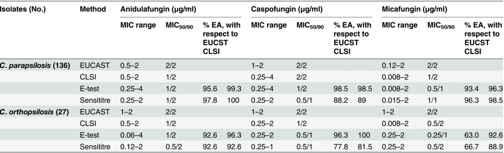
The antifungal susceptibility testing results, as well as the percent EA for each echinocandin, are summarized inTable 2. When the species-specific CBPs were applied, no resistance was detected with any of the tested methods. The geometric mean MICs (μg/mL) by both methods
Table 1. Characteristics of 163 patients with Candida parapsilosis complex candidemia.
Characteristic C. parapsilosis (n = 136) C. orthopsilosis (n = 27) p-value
Male, n (%) 98 (72.1) 14 (51.9) 0.039
Age (years) mean (SD) 44.5±23.7 46± 26.8 0.762
Hospital ward, n (%) Hematology 8 (5.9) 1 (3.7) 1.000 Adult ICU 57 (41.9) 7 (25.9) 0.123 Internal medicine 15 (11.0) 2 (7.4) 0.741 Neonatal ICU 25 (18.4) 1 (3.7) 0.081 Pediatric onco-hematology 19 (14.0) 11 (40.7) 0.001 Surgery 12 (8.8) 5 (18.5) 0.164 Underlying condition, n (%) Autoimmune disorder 2 (1.5) 0 (0.0) 1.000 Cancer 13 (9.6) 3 (11.1) 0.731
Central venous catheter 130 (95.6) 23 (85.2) 0.062
Gastrointestinal diseases 11 (8.1) 2 (7.4) 1.000 HIV infection 4 (2.9) 1 (3.7) 1.000 HSCT 4 (2.9) 0 (0.0) 1.000 Kidney diseases 6 (4.4) 0 (0.0) 0.591 Major surgery 21 (15.4) 5 (18.5) 0.774 Onco-hematological diseases 27 (19.9) 12 (44.4) 0.006 Premature birth 14 (10.3) 1 (3.7) 0.469 Pulmonary diseases 6 (4.4) 0 (0.0) 0.591 Sepsis 16 (11.8) 2 (7.4) 0.740 Trauma 12 (8.8) 1 (3.7) 0.698
HSCT, Hematopoietic Stem Cell Transplant; ICU, Intensive Care Unit. doi:10.1371/journal.pone.0150218.t001
(EUCAST/CLSI) for AND, CSP, and MCF was, respectively, as follows: C. parapsilosis 1.79/ 1.24, 1.84/1.74, and 1.77/1; C. orthopsilosis 1.85/1.08, 1.90/1.26, and 1.76/0.58.
Candida parapsilosis and C. orthopsilosis showed similar susceptibility patterns to all echi-nocandins, with MIC90s ranging from 1–2 μg/mL, depending on the method. The MICs of the
quality control strains fell within the established ranges that have been published for both methods [15,18].
For C. parapsilosis, the EAs between the echinocandin MIC results obtained by the E-test and Sensititre methods and the reference procedures (CLSI and EUCAST) were very high, ranging from 88.2% to 100% according to the technique–drug combination. For C. orthopsilo-sis, EAs between the E-test and Sensititre methods and the CLSI method were also high, rang-ing from 81.5% to 100% accordrang-ing to the technique–drug combination. In comparison to EUCAST, the EA was worse for MCF (E-test, 63.0%; Sensititre, 66.7%) than for AND (92.6% for the E-test and Sensititre methods) and CSP (E-test, 96.3%; Sensititre, 77.8%).
Regarding the CAs between the E-test and Sensititre techniques and the EUCAST method, for AND and MCF, discrepancies were not observed for each organism tested. Comparing the CLSI method with the E-test and Sensititre techniques, for C. parapsilosis the CAs (%) were 91.9/100, 94.9/96.3, and 100/100 for AND, CSP, and MCF, respectively. Regarding C. orthopsi-losis, the CAs were 100% for CSP and MCF, respectively with exception of AND (88.9%) when the E-test was compared with the CLSI method. Only MiEs occurred, and they were slightly greater for C. parapsilosis (23/136, 16.9%) than C. orthopsilosis (3/27, 11.1%). Consequently, 11 C. parapsilosis and three C. orthopsilosis isolates classified as AND susceptible by the CLSI method were considered to be intermediate by the E-test. For CSP, ten C. parapsilosis isolates were categorized as intermediate by the CLSI and susceptible by the E-test and Sensititre (five strains for each reference-commercial method comparison), and two were CLSI susceptible and E-test intermediate.
Discussion
Candidemia caused by the C. parapsilosis complex is increasing in Italy [1] and some other countries [2,3], and it is the second most commonly isolated Candida species. Genotypic dif-ferences allowed the taxonomic division of the C. parapsilosis complex into three groups that
Table 2. MICs and EAs of the E-test and Sensititre methods compared with those of the CLSI and EUCAST methods.
Isolates (No.) Method Anidulafungin (μg/ml) Caspofungin (μg/ml) Micafungin (μg/ml) MIC range MIC50/90 % EA, with
respect to EUCST CLSI
MIC range MIC50/90 % EA, with
respect to EUCST CLSI
MIC range MIC50/90 % EA, with
respect to EUCST CLSI C. parapsilosis (136) EUCAST 0.5–2 2/2 1–2 2/2 0.12–2 2/2 CLSI 0.5–2 1/2 0.25–4 2/2 0.008–2 1/2 E-test 0.25–4 1/2 95.6 99.3 0.25–4 1/2 98.5 98.5 0.008–2 0.5/1 93.4 96.3 Sensititre 0.25–2 1/2 97.8 100 0.25–2 0.5/1 88.2 89 0.015–2 1/1 96.3 98.5 C. orthopsilosis (27) EUCAST 1–2 2/2 1–2 2/2 1–2 2/2 CLSI 0.5–2 1/2 0.25–2 1/2 0.008–2 0.5/2 E-test 0.06–4 1/2 92.6 96.3 0.25–2 0.5/1 96.3 100 0.25–2 0.25/1 63.0 92.6 Sensititre 0.12–2 0.5/2 92.6 92.6 0.25–1 0.5/1 77.8 81.5 0.25–2 0.5/2 66.7 88.9 CLSI, Clinical and Laboratory Standards Institute; EUCAST, European Committee on Antimicrobial Susceptibility Testing; MIC, minimum inhibitory concentration; EA, essential agreement
were recognized as separate species: C. parapsilosis (group 1), C. orthopsilosis (group 2), and C. metapsilosis (group 3). The epidemiology of candidemia and the antifungal susceptibility of these species are scarcely defined in Italy. To our knowledge, only one national study of the epi-demiology of invasive candidiasis caused by the C. parapsilosis complex has been published [11], which showed that 95, 3.6, and 1.4% of the C. parapsilosis complex strains were identified as C. parapsilosis, C. orthopsilosis, and C. metapsilosis, respectively.
Based on these data, the present study was performed to simultaneously evaluate the epide-miology and echinocandin susceptibility patterns of C. parapsilosis complex BSIs in a univer-sity hospital in Southern Italy from January 2007 to December 2014. At our institution, the incidence of C. parapsilosis (83.4% of all C. parapsilosis complex isolates) was 2.9 cases per 10,000 admissions; while incidences of 2.2–2.9 and 3.4 were reported in Spain and Turkey, respectively [10,12,19]. The incidence of C. orthopsilosis candidemia was 0.6 per 10,000 admissions, which is similar to the incidences reported by other studies, which range from 0.2 to 0.9 [10,12]. Interestingly, the percentage of C. orthopsilosis observed (16.6%) was one of the highest published thus far [11–13,20,21], although higher percentages were reported in Spain (23.5%) [10] and Qatar (24%) [22]. There were no cases of C. metapsilosis candidemia, in keep-ing with studies performed in Portugal [23], Qatar [22], Scotland [24], and Kuwait [25]. C. metapsilosis has been reported to be rarely recovered from blood (incidence of 0.7–6.9%) [13, 20], which is in accordance with its low virulence [8,9]. It is noteworthy that some authors have reported that C. metapsilosis is more common than C. orthopsilosis [13,26]. This suggests a geographical variation in the distribution of species belonging to the C. parapsilosis complex.
The C. parapsilosis complex is particularly common in critically ill patients, probably because this yeast has a high affinity for vascular devices, medical instrumentation, and indwelling plastics [27]. In fact, in the present study, the majority of patients (55.2%) infected with the C. parapsilosis complex were from the ICU, and the indwelling catheter was the most frequent underlying condition (93.9%), and it was mainly observed in the ICU patients (58.8%). Of note, we observed that ICU patients were more likely to develop C. parapsilosis candidemia than C. orthopsilosis candidemia. This issue may be partially explained by the greater capacity of this species to form biofilms on central lines [28], compared with the closely related species C. orthopsilosis and C. metapsilosis [29].
Concerning the susceptibility results, both CLSI and EUCAST procedures were used in this study, and no echinocandin resistance by the species within the psilosis complex was observed using either method, as was reported by other studies [11,20]. In agreement with results previ-ously reported by Garcia-Effron et al. [10], our MICs showed a rank order of activity with MCF> AND> CSP against C. parapsilosis and C. ortopsilosis. However, some studies [13,14] showed that these species are more susceptible to CSP than to the other echinocandins. These data reflect the need of wider study regarding the echinocandin susceptibility. Of note, some authors have reported that C. parapsilosis is the only species that is resistant to echinocandins [11,23,30]. In the current study, the echinocandin MICs for C. parapsilosis were similar to those for C. orthopsilosis (MIC90of 2μg/mL for each organism according to the technique–
drug combination), which differ from those reported in a multicenter study conducted in Italy by Borghi et al. [11]. This difference may result from the fact that, in the present study, the iso-lates were only collected in one hospital; thus, the different results could be related to differ-ences in antifungal drug management between the hospitals analyzed in the two studies.
The present study compared, for the first time, the echinocandin MICs obtained by the E-test and Sensititre methods with those obtained by the CLSI and EUCAST procedures by deter-mining the species of the psilosis group. Our analysis showed that there were excellent EAs between these methods, except between the EUCAST and the E-test and Sensititre methods when testing the MCF susceptibility of C. orthopsilosis, which was less than 67%. The meaning
of this in vitro finding is not clear and needs to be clarified in more detail. A good CA was also observed for all organism-drug combinations, ranging from 88.9 to 100%. The MIC differences between the standard procedures and the commercially available assays are small enough that the choice of method should not result in susceptibilities that differ enough to affect treatment decisions.
As our study was an observational laboratory-based survey, some medical files were miss-ing: the severity of illness scores, the type and duration of antifungal therapy, and mortality data. Nevertheless, to the best of our knowledge, this study provides the first data on the inci-dence of species of the psilosis group that are responsible for candidemia in Italy. Although this study was conducted in a single hospital, we attained a large sample size (more than 45,000 patients). Moreover, this 8-year survey revealed no echinocandin resistance among the species within the C. parapsilosis complex using the CLSI and EUCAST methods, thereby suggesting that these strains are, in generally, highly susceptible to echinocandins [14,16]. Finally, a com-parison of the CLSI and EUCAST methods and the E-test and Sensititre methods revealed that they yielded similar MICs, which reinforces the relevance of using commercially available methods in clinical microbiology laboratories to test for antifungal susceptibility.
Acknowledgments
This study was supported by an unrestricted educational grant from Pfizer Italia.
Author Contributions
Conceived and designed the experiments: MTM GM GC. Performed the experiments: EB SB DC FP GL ODG. Analyzed the data: GL. Contributed reagents/materials/analysis tools: MTM GM. Wrote the paper: GL SB MTM GM.
References
1. Caggiano G, Coretti C, Bartolomeo N, Lovero G, De Giglio O, Montagna MT. Candida bloodstream infections in Italy: changing epidemiology during 16 years of surveillance. Biomed Res Int. 2015; 2015:256580. doi:10.1155/2015/256580PMID:26064890
2. Guinea J, Zaragoza Ó, Escribano P, Martín-Mazuelos E, Pemán J, Sánchez-Reus F, et al. Molecular identification and antifungal susceptibility of yeast isolates causing fungemia collected in a population-based study in Spain in 2010 and 2011. Antimicrob Agents Chemother. 2014; 58: 1529–1537. doi:10. 1128/AAC.02155-13PMID:24366741
3. Nucci M, Queiroz-Telles F, Alvarado-Matute T, Tiraboschi IN, Cortes J, Zurita J, et al. Latin American invasive mycosis network. epidemiology of candidemia in Latin America: a laboratory-based survey. PLoS One. 2013; 8: e59373. doi:10.1371/journal.pone.0059373PMID:23527176
4. Ozsevik SN, Sensoy G, Karli A, Albayrak C, Dagdemir A, Belet N, et al. Invasive fungal infections in children with hematologic and malignant diseases. J Pediatr Hematol Oncol. 2015; 37: 69–72. 5. Montagna MT, De Giglio O, Napoli C, Lovero G, Caggiano G, Delia M, et al. Invasive fungal infections
in patients with hematologic malignancies (aurora project): lights and shadows during 18-months sur-veillance. Int J Mol Sci. 2012; 13: 774–787. doi:10.3390/ijms13010774PMID:22312285
6. Tavanti A, Davidson AD, Gow NA, Maiden MC, Odds FC. Candida orthopsilosis and Candida metapsi-losis spp. nov. to replace Candida parapsimetapsi-losis groups II and III. J Clin Microbiol. 2005; 43: 284–292. PMID:15634984
7. Rycovska A, Valach M, Tomaska L, Bolotin-Fukuhara M, Nosek J. Linear versus circular mitochondrial genomes: intraspecies variability of mitochondrial genome architecture in Candida parapsilosis. Micro-biology. 2004; 150: 1571–1580. PMID:15133118
8. Németh T, Tóth A, Szenzenstein J, Horváth P, Nosanchuk JD, Grózer Z, et al. Characterization of viru-lence properties in the C. parapsilosis sensu lato species. PLoS One. 2013; 8: e68704. doi:10.1371/ journal.pone.0068704PMID:23874732
9. Orsi CF, Colombari B, Blasi E. Candida metapsilosis as the least virulent member of the 'C. parapsilo-sis' complex. Med Mycol. 2010; 48: 1024–1033. doi:10.3109/13693786.2010.489233PMID: 20507266
10. Garcia-Effron G, Canton E, Pemán J, Dilger A, Romá E, Perlin DS. Epidemiology and echinocandin susceptibility of Candida parapsilosis sensu lato species isolated from bloodstream infections at a Spanish university hospital. J Antimicrob Chemother. 2012; 67: 2739–2748. doi:10.1093/jac/dks271 PMID:22868644
11. Borghi E, Sciota R, Iatta R, Biassoni C, Montagna MT, Morace G. Characterization of Candida parapsi-losis complex strains isolated from invasive fungal infections. Eur J Clin Microbiol Infect Dis. 2011; 30: 1437–1441. doi:10.1007/s10096-011-1242-xPMID:21479840
12. Cantón E, Pemán J, Quindós G, Eraso E, Miranda-Zapico I, Álvarez M, et al. Prospective multicenter study of the epidemiology, molecular identification, and antifungal susceptibility of Candida parapsilo-sis, Candida orthopsiloparapsilo-sis, and Candida metapsilosis isolated from patients with candidemia. Antimi-crob Agents Chemother. 2011; 55: 5590–5606. doi:10.1128/AAC.00466-11PMID:21930869 13. Gomez-Lopez A, Alastruey-Izquierdo A, Rodriguez D, Almirante B, Pahissa A, Rodriguez-Tudela JL,
et al. Prevalence and susceptibility profile of Candida metapsilosis and Candida orthopsilosis: results from population-based surveillance of candidemia in Spain. Antimicrob Agents Chemother. 2008; 52: 1506–1509. doi:10.1128/AAC.01595-07PMID:18285486
14. Spreghini E, Orlando F, Tavanti A, Senesi S, Giannini D, Manso E, et al. In vitro and in vivo effects of echinocandins against Candida parapsilosis sensu stricto, Candida orthopsilosis and Candida metap-silosis. J Antimicrob Chemother. 2012; 67:2195–2202. doi:10.1093/jac/dks180PMID:22635526 15. Clinical and Laboratory Standards Institute. Reference method for broth dilution antifungal susceptibility
testing of yeasts; third edition, M27-A3. Wayne, PA, USA: CLSI; 2008.
16. Montagna MT, Lovero G, Coretti C, Martinelli D, De Giglio O, Iatta R, et al. Susceptibility to echinocan-dins of Candida spp. strains isolated in Italy assessed by European Committee for Antimicrobial Sus-ceptibility Testing and Clinical Laboratory Standards Institute broth microdilution methods. BMC Microbiol. 2015; 15: 106. doi:10.1186/s12866-015-0442-4PMID:25990252
17. Clinical and Laboratory Standards Institute. Reference method for broth dilution antifungal susceptibility testing of yeasts; fourth informational supplement, M27-S4. Wayne, PA, USA: CLSI; 2012.
18. Arendrup MC, Cuenca-Estrella M, Lass-Flörl C, Hope WW, European Committee on Antimicrobial Sus-ceptibility Testing-Subcommittee on Antifungal SusSus-ceptibility Testing (EUCAST-AFST). EUCAST tech-nical note on Candida and micafungin, anidulafungin and fluconazole. Mycoses. 2014; 57: 377–379. doi:10.1111/myc.12170PMID:24417759
19. Aydin F, Bayramoglu G, Guler NC, Kaklikkaya N, Tosun I. Bloodstream yeast infections in a university hospital in Northeast Turkey: a 4-year survey. Med Mycol. 2011; 49: 316–319. doi:10.3109/13693786. 2010.512023PMID:20807028
20. Bonfietti LX, Martins Mdos A, Szeszs MW, Pukiskas SB, Purisco SU, Pimentel FC, et al. Prevalence, distribution and antifungal susceptibility profiles of Candida parapsilosis, Candida orthopsilosis and Candida metapsilosis bloodstream isolates. J Med Microbiol. 2012; 61: 1003–1008. doi:10.1099/jmm. 0.037812-0PMID:22493277
21. de Toro M, Torres MJ, Maite R, Aznar J. Characterization of Candida parapsilosis complex isolates. Clin Microbiol Infect. 2011; 17: 418–424. doi:10.1111/j.1469-0691.2010.03302.xPMID:20636431 22. Taj-Aldeen SJ, Kolecka A, Boesten R, Alolaqi A, Almaslamani M, Chandra P, et al. Epidemiology of
candidemia in Qatar, the Middle East: performance of MALDI-TOF MS for the identification of Candida species, species distribution, outcome, and susceptibility pattern. Infection. 2014; 42: 393–404. doi:10. 1007/s15010-013-0570-4PMID:24352810
23. Silva AP, Miranda IM, Lisboa C, Pina-Vaz C, Rodrigues AG. Prevalence, distribution, and antifungal susceptibility profiles of Candida parapsilosis, C. orthopsilosis, and C. metapsilosis in a tertiary care hospital. J Clin Microbiol. 2009; 47: 2392–2397. doi:10.1128/JCM.02379-08PMID:19494078 24. Odds FC, Hanson MF, Davidson AD, Jacobsen MD, Wright P, Whyte JA, et al. One year prospective
survey of Candida bloodstream infections in Scotland. J Med Microbiol. 2007; 56: 1066–1075. PMID: 17644714
25. Asadzadeh M, Ahmad S, Al-Sweih N, Khan ZU. Rapid molecular differentiation and genotypic hetero-geneity among Candida parapsilosis and Candida orthopsilosis strains isolated from clinical specimens in Kuwait. J Med Microbiol. 2009; 58: 745–752. doi:10.1099/jmm.0.008235-0PMID:19429750 26. Chen YC, Lin YH, Chen KW, Lii J, Teng HJ, Li SY. Molecular epidemiology and antifungal susceptibility
of Candida parapsilosis sensu stricto, Candida orthopsilosis, and Candida metapsilosis in Taiwan. Diagn Microbiol Infect Dis. 2010; 68: 284–292. doi:10.1016/j.diagmicrobio.2010.07.004PMID: 20851551
27. Trofa D, Gacsera A, Nosanchuk JD. Candida parapsilosis, an emerging fungal pathogen. Clin Microbiol Rev 2008; 21: 606–625. doi:10.1128/CMR.00013-08PMID:18854483
28. Tumbarello M, Posteraro B, Trecarichi EM, Fiori B, Rossi M, Porta R, et al. Biofilm production by Can-dida species and inadequate antifungal therapy as predictors of mortality for patients with candidemia. J Clin Microbiol. 2007; 45: 1843–1850. PMID:17460052
29. Lattif AA, Mukherjee PK, Chandra J, Swindell K, Lockhart SR, Diekema DJ, et al. Characterization of biofilms formed by Candida parapsilosis, C. metapsilosis, and C. orthopsilosis. Int J Med Microbiol. 2010; 300: 265–270. doi:10.1016/j.ijmm.2009.09.001PMID:19932053
30. Treviño-Rangel Rde J, Garza-González E, González JG, Bocanegra-García V, Llaca JM, González GM. Molecular characterization and antifungal susceptibility of the Candida parapsilosis species com-plex of clinical isolates from Monterrey, Mexico. Med Mycol. 2012; 50: 781–784. doi:10.3109/ 13693786.2012.675526PMID:22493945