i
U
NIVERSITY
L
A
S
APIENZA OF
R
OME
,
F
ACULTY OF
M
EDICINE
A
NGIO
-C
ARDIO
-T
HORACIC
P
ATHOPHYSIOLOGY AND
I
MAGING
D
OCTOR OF
P
HILOSOPHY
,
XXXII^E
DITION
C
OORDINATOR
P
ROFESSOR
G.
V
ENUTA
_____________________________________________________________________________________
New Perspectives in the diagnosis of
Cardiac Allograft Vasculopathy:
the CT-scan role in the follow-up of
heart transplanted patients
CANDIDATE
Marzia Cottini, MD
Candidate's ID number: 1748036
INDEX
INDEX ... ii
LIST OF TABLES ... iv
LIST OF FIGURES ... v
LIST OF SYMBOLS AND ABBREVATIONS ... vi
Symbols ... vi
Abbreviations and Acronysms ... vi
LIST of PAPERS ... 2
INTRODUCTION ... 5
CHAPTER 1: The Heart Transplantation ... 7
1.1 Definition of the End Stage Heart Failure ... 8
1.2 The Heart Transplantation ... 12
1.2.1 Definition ... 12
1.2.2 Indications and contraindications of Htx ... 13
1.2.3 Surgical Techniques ... 15
1.2.4 Survival ... 17
1.2.5 Complications ... 18
CHAPTER 2: Cardiac Allograft Vasculopathy ... 21
2.1 Definition ... 22
2.2 Classification ... 22
2.3 Pathogenesis ... 24
2.4 Histopathological presentation ... 25
2.5 Epidemiology and Aetiology ... 27
CHAPTER 3: ... 30
Detection and imaging in Cardiac Allograft Vasculopathy ... 30
3.1 Detection of CAV ... 31
3.2 Non Invasive stress testing ... 33
3.3 Invasive methods... 39
CHAPTER 4: Prognosis ... 43
CHAPTER 5: Study Design and Development ... 47
5.1 INTRODUCTION OF THE STUDY DESIGN ... 48
5.2 FIRST STEP ... 48
iii
5.2.3 Results ... 50
5.3 SECOND STEP ... 56
5.3.1 Material and Methods ... 56
5.3.2. Statistical Analysis ... 57
5.3.3 Results ... 58
5.4 THIRD STEP ... 62
5.4.1 Material and Methods ... 62
5.4.2 Statistical Analysis ... 62 5.4.3: Results ... 63
CHAPTER 6: Discussion ... 66
CHAPTER 7: Conclusion ... 72
Bibliography ... 74
Acknowledgements ... 80
LIST OF TABLES
TABLE 1:DEFINITION OF THE HEART FAILURE ACCORDING TO THE 2016ESCGUIDELINES FOR THE DIAGNOSIS AND TREATMENT OF ACUTE AND CHRONIC HEART FAILURE:THE TASK FORCE FOR THE DIAGNOSIS AND TREATMENT OF ACUTE AND CHRONIC HEART FAILURE OF THE EUROPEAN SOCIETY OF CARDIOLOGY (ESC)DEVELOPED WITH THE SPECIAL CONTRIBUTION [2] 8 TABLE 2:HTX INDICATIONS AND CONTRAINDICATIONS ACCORDING TO ESC2016HFGUIDELINES [2]. ... 14 TABLE 3: THE MOST IMPORTANT LITERATURE EXPERIENCES OF IVUS IN THE DETECTION OF CAV[58][59][56][55][60]. 41 TABLE 4:STANFORD CLASSIFICATION OF IVUS/CAV[61] ... 41 TABLE 5:THE MOST IMPORTANT ARTICLES IN SCIENTIFIC LITERATURE ABOUT THE USE OF PCI IN THE TREATMENT OF CAV ... 45 TABLE 6:ASSESSMENT OF CCTA USED TO DESIGN THE STUDY ... 49 TABLE 7:BASELINE CHARACTERISTICS OF THE STUDY POPULATION.HTX: HEART TRANSPLANTATION;AMI: ACUTE MYOCARDIAL INFARCTION,PCI: PERCUTANEOUS CORONARY INTERVENTION. ... 60 TABLE 8:MEAN AGE OF THE PATIENTS AT THE HEART TRANSPLANTATION SURGERY. ... 51 TABLE 9:THE CAV PATIENT CONSIDERED IN THE STUDY ... 62 TABLE 10:MAIN DIFFERENCES BETWEEN CAV GROUP VS NO-CAV GROUP.CAV: CARDIAC ALLOGRAFT VASCULOPATHY,PCI
PERCUTANEOUS CORONARY INTERVENTIONS,PCR:C- REACTIVE PROTEIN. ... 52 TABLE 11:YEARS FROM HEART TRANSPLANTATION (FOLLOW UP). ... 52 TABLE 12: THE DISTRIBUTION OF THE PATIENTS ACCORDING TO THE “AGE AT THE HTX” AND THE “AGE AT THE FOLLOW UP”.53 TABLE 13:BASELINE CHARACTERISTICS OF THE STUDY POPULATION (CAV GROUP AND NO CAV GROUP).HTX: HEART
TRANSPLANTATION;AMI: ACUTE MYOCARDIAL INFARCTION,CREA: CREATININ,CLCR:CLEARANCE CREA,HT: HEMATOCRIT, LVEF: LEFT VENTRICLE EJECTION FRACTION,PCR:C REACTIVE PROTEIN,PCI: PERCUTANEOUS CORONARY INTERVENTION, CABG: CORONARIC ARTERY BYPASS GRAFT. ... 58 TABLE 14:THE 19HTX-PATIENTS CONSIDERED IN THE SECOND STEP OF THE STUDY ... 69 TABLE 15:MAIN DIFFERENCES BETWEEN CAV GROUP VS NO-CAV GROUP.CAV: CARDIAC ALLOGRAFT VASCULOPATHY,PCI
PERCUTANEOUS CORONARY INTERVENTIONS,PCR:C- REACTIVE PROTEIN. ... 59 TABLE 16:CAV CHARACTERISTICS IN STUDY PROTOCOL AND THEIR RESULTS. ... 60 TABLE 17:SCIENTIFIC LITERATURE DATA ABOUT THE SENSIBILITY, SPECIFICITY,NPV AND PPV IN THE COMPARISON OF CCTA TO
CCA. ... 68 TABLE 18:RESULTS OF THE SENSIBILITY, SPECIFICITY,NPV AND PPV IN THE COMPARISON OF CCTA TO... 69 TABLE 19:SCIENTIFIC LITERATURE DATA ABOUT THE SENSIBILITY, SPECIFICITY,NPV AND PPV IN THE COMPARISON OF CCTA TO
v
LIST OF FIGURES
FIGURE 1:INCIDENCE OF HF IN THE US AND THE FUTURE PERSPECTIVES ... 8
FIGURE 2:PREVALENCE AND INCIDENCE OF HF WORLDWIRE [9] ... 9
FIGURE 3:STAGES OF HF[14] ... 10
FIGURE 4:INTERNATIONAL SOCIETY HEART AND LUNG TRANSPLANTATION REPORT OF THE 2018.THE NUMBER OF HTX PROCEDURE SINCE THE LAST 2018 IN EUROPE,NORTH AMERICA AND OTHER COUNTRIES. ... 12
FIGURE 5:ADULT AND PEDIATRIC HEART TRANSPLANT KAPLAN-MEYER CUMULATIVE SURVIVAL CURVE ... 17
FIGURE 6:ADULT AND PEDIATRIC HEART TRANSPLANT KAPLAN-MEYER GROUP SURVIVAL CURVES ... 17
FIGURE 7:REJECTION ... 18
FIGURE 8:THE CAUSES OF DEATH AFTER HEART TRANSPLANTATION ACCORDING TO ISHLT REGISTRY AND REPORT 2018. .... 20
FIGURE 9:THE RELATIVE INCIDENCE OF LEADING CAUSES OF DEATH ... 19
FIGURE 10:THE CLASSIFICATION OF CAV ACCORDING TO GAO [19] ... 22
FIGURE 11:NOMENCLATURE FOR CARDIAC ALLOGRAFT VASCULOPATHY ACCORDING TO ISHLT ... 23
FIGURE 12: COLLABORATION AND INTERACTION OF ALLOIMMUNE-DEPENDENT AND -INDEPENDENT FACTORS INFLUENCING THE PATHOGENESIS OF TRANSPLANT VASCULOPATHY.AG INDICATES ANTIGEN;CD, CLUSTER OF DIFFERENTIATION; ENOS, ENDOTHELIAL NO SYNTHASE; AND SMC, SMOOTH MUSCLE CELL [20]. ... 24
FIGURE 13:HISTOLOGICAL VIEW OF THE CARDIAC ALLOGRAFT VASCULOPATHY.IT SHOWS THE TYPICAL CONCENTRIC LESION VERSUS THE ECCENTRIC ONE OF ATHEROMATOUS DISEASE. ... 25
FIGURE 14: ATHEROSCLEROTIC DISEASE OF CORONARY ARTERY. ... 26
FIGURE 15:COSTIMULATORY MOLECULES PLAY CRUCIAL ROLES IN THIS T CELL ACTIVATION.MANY COSTIMULATORY PATHWAYS HAVE BEEN DESCRIBED, AND SOME ARE INVOLVED IN THE PATHOGENESIS OF CAV, ATHEROGENESIS, AND SUBSEQUENT PLAQUE FORMATION.IN THIS REVIEW, WE SUMMARIZE THE PRESENT KNOWLEDGE OF THE ROLE OF THESE PATHWAYS IN CAV DEVELOPMENT AND THE POSSIBILITY OF MANIPULATING THESE PATHWAYS AS A MEANS TO TREAT HEART ALLOGRAFT VASCULAR DISEASE AND ATHEROSCLEROSIS [21] ... 27
FIGURE 16:IMMUNOLOGICAL EVENTS INVOLVE IN THE CAV PATHOGENESIS.ENVIRONMENTAL AND GENETIC FACTORS CONTRIBUTE TO THE ETIOLOGY OF SSC.THE PATHOGENESIS OF SSC INVOLVES AN INTERPLAY BETWEEN VASCULAR, IMMUNOLOGICAL, AND FIBROTIC PROCESSES.VASCULAR INJURY AND ENDOTHELIAL DAMAGE ARE THE EARLIEST EVENTS IN THE PATHOGENESIS OF SSC.ACTIVATED ENDOTHELIAL CELLS UPREGULATE THE EXPRESSION OF ADHESION MOLECULES AND SECRETE CHEMOKINES, LEADING TO INFLAMMATION AND AUTOIMMUNITY.MACROPHAGES AND T-CELLS ARE THE PREDOMINANT INFLAMMATORY CELL TYPES OF THE INFLAMMATORY INFILTRATES AND PRODUCE CYTOKINES AND GROWTH FACTORS THAT DRIVE THE SYNTHESIS OF EXTRACELLULAR MATRIX PROTEINS BY FIBROBLASTS, RESULTING IN PROGRESSIVE FIBROSIS.T-CELLS HAVE ALSO BEEN IMPLICATED IN AUTOANTIBODIES PRODUCTION. ... 29
FIGURE 17:THE ROLE OF IMAGING IN THE DETECTION OF CAV IN ITS DIFFERENT STAGES. ... 32
FIGURE 18:EXAMPLE OF THE CARDIAC COMPUTED TOMOGRAPHY OF THE LAST GENERATION. ... 34
FIGURE 19:IMAGES OF THE NEW GENERATIONS SCANNER IN CARDIAC COMPUTED TOMOGRAPHY DEVICES. ... 35
FIGURE 20:MULTI-SECTOR SCANNING AND ECG ... 36
FIGURE 21:THE ECG PHASES IN CARDIACCT SCAN ... 37
FIGURE 22:ALGORITHM FOR CAV SURVEILLANCE AND MANAGEMENT. ... 44
FIGURE 23:PREVENTIVE MEASURES EARLY POST-TRANSPLANT ... 46
FIGURE 24:INCIDENCE OF CAV IN THE POPULATION ... 53
FIGURE 25: SCHEME OF THE CALCULATION OF THE SENSITIVITY, SPECIFICITY ... 54
FIGURE 26:CRITERIA OF DIAGNOSIS OF CORONARY DISEASE BY IVUS IN THE LEFT MAIN CORONARY LESION AND INTERMEDIATE NON-LEFT MAIN CORONARY LESIONS. ... 57
FIGURE 27:INCIDENCE OF CAV IN THE STUDY POPULATION ... 60
LIST OF ABBREVATIONS and ACRONYMS
Abbreviations and Acronyms
16-MDCT = 16-slice multidetector computed tomography 64-MDCT = 64-slice multidetector computed tomography Ag = antigen
BMI = body mass index BMS = bare metal stent BNP = brain natriuretic peptide bpm = beats per minutes CAC = coronary artery calcium CAD = coronary atherosclerotic disease CAV = cardiac allograft vasculopathy CCA =coronary artery angiography CCTA = coronary CT angiography CD = cluster of differentation CI = confidence interval
CIN = contrast-material–induced nephropathy CMV = cytomegalovirus
CNIs = Calcineurin inhibitors CRP = C-reactive protein CT = computed tomography
CT-FFR = CT-derived fractional flow reserve CTDIvol = volume CT dose index
CTP = CT myocardial perfusion CX = left circumflex
D/GL = death or graft loss DA: descending anterior coronary DAP = dose area product
DES = drug eluting stent DLP = dose-length product DS = Dual-source
DSE = Dobutamine stress echocardiography ECG = electrocardiogram
ECM = extracellular matrix EEM = external elastic membrane EGF = endothelial grow factor
ELAM =endothelial cell adhesion molecule eNOS = endothelial NO synthase
FFR = fractional flow reserve FGF = fibroblast grow factor GFR = glomerular filtration rate GP130 = glycoprotein 130
HFmrEF = heart failure mid-range ejection fraction HFpEF= heart failure preserved ejection fraction HFrEF = heart failure preserved ejection fraction HR = hazard ratio
HTx = Heart transplantation HU = Hounsfield units
ii
ICA = invasive coronary angiography IFN =interferon
IFN-ɣ = interferon-gamma IGF = insulin-like grow factor IL = interleukin
IMR = index of microcirculatory resistance
ISHLT = International Society for Heart and Lung Transplantation IVUS = intravascular ultrasound
k = organ-weighting factor LAD = left anterior descending LM = left main
LVEF = left ventricle ejection fraction MD = multidetector
MDCT= multidetector computed tomography MHC = major histocompatibility complex MIT = maximal intimal thickness
MMF = mycophenolate mofetil MMP-1=matrix metalloproteinases-1 MPI = myocardial perfusion imaging mTOR = mammalian target-of-rapamycin MTS = metal stent
NF-MACE = non-fatal major adverse cardiac events NO = Nitric Oxide
NPV = negative predictive value NPV = negative predictive value OCT = optical coherence tomography OHT = orthotopic heart transplantation OPG = Osteoprotegerin
OR = odds ratio
PCI = percutaneous coronary intervention PDGF=platelet-derived growth factor PhD = doctor of philosophy
PPV = positive predictive value PPV= positive predictive value
PTDL = post-transplantation lymphoproliferative disorder
RCA = right coronary artery Se = sensibility
SMC = small muscle cell Sp = specificity
SPECT = single-photon emission computed tomography SSc = systemic sclerosis
TGFβ = transforming growth factor beta Th1= T helper type 1 cells
TNF-α = tumor necrosis factor-α Treg = T-regulatory cell
VCAM = vascular cell adhesion molecule VCAM-1= receptors cell adhesion molecule-1 VH = Virtual histology
vSMC = vascular small muscle cell vWf = von Willebrand factor
3
This is the summary of abstracts/posters presented at national and international congress and the article published regarding my PhD issue.
1. M Cottini, FB Fabio Sbaraglia, VB Vitaliano Buffa, PLM Paola Lilla Della Monica, GDS Giada
Distefano, AP Amedeo Pergolini, VP Vincenzo Polizzi, FM Francesco Musumeci, Cardiac
allograft vasculopathy: the way for early diagnosis, European Journal of Heart Failure, Volume
20, Issue S1, 2018.
2. M Cottini, FB Fabio Sbaraglia, VB Vitaliano Buffa, PLM Paola Lilla Della Monica, GDS Giada
Distefano, AP Amedeo Pergolini, VP Vincenzo Polizzi, FM Francesco Musumeci, Cardiac
allograft vasculopathy: the way for early diagnosis, Heart Failure 2018 ESC Wien, 26-29/03/2018, P1953
3. M. Cottini, Vitaliano Buffa, Vincenzo Polizzi, Fabio Sbaraglia, Giada Di Stefano, P. Myriam
Lo Presti, Amedeo Pergolini, Federico Ranocchi, Andrea Montalto, Riccardo Gherli, Emilio Ferretti, Paolo Giuseppe Pino, Paola Lilla Della Monica, Francesco Musumeci. Cardiac allograft
vasculopathy: new perspective in diagnostic workout. ESC Congress 2017, Barcellona
(Accepted abstract and poster presentation 82834)
4. Cottini M, Feccia M, Montalto A., Ranocchi F., Luzi G., Gherli R., Ferretti E., Fiorani B.,
Bergonzini M., Giacopino F., D'Alessandro C., Lo Presti M., Sbaraglia F, Di Stefano G, Pergolini A, Polizzi V, Pino G, Buffa V, Lilla della Monica P, Musumeci F. Il Trapianto
Cardiaco combinazione di arte chirurgica e mente cardiologica. Esperienza quindicennale di un singolo centro. (Abstract Session C24, 28 October 2016, SITO Congress, Rome)
5. Cottini M, Dominici T, Montalto A., Ranocchi F., Luzi G., Gherli R., Ferretti E., Fiorani
B., Bergonzini M., Giacopino F., D'Alessandro C., Feccia M, Lo Presti M., Sbaraglia F, Di Stefano G, Pergolini A, Polizzi V, Pino G, Buffa V, Lilla della Monica P, Musumeci F.
Vasculopatia del Cuore Trapiantato: qual è la migliore metodica diagnostico-strumentale per la diagnosi precoce? (Abstract Section C 04, 27 October 2016, SITO Congress, Rome).
6. Dominici T, MD, Cottini M, MD, Sbaraglia F, MD, Della Monica L, MD, Di Stefano G, MD, Pergolini A, MD, Polizzi V, MD, M. Feccia, MD, Fierro S, MD, Buffa V, MD , Musumeci F, MD. Cardiac Allograft Vasculopathy assessed by 64 slice Dual-Source Coronary Computed
Tomographic Angiography: Retrospective Analysis of a Monocentric Experience. (ISHLT Meeting Washington 2016, Accepted, poster presentation 0525).
7. Cottini M, Dominici T, Sbaraglia F, Pergolini A, Di Stefano G, Polizzi V, Feccia M, Buffa V,
Lilla Della Monica P, Musumeci F. Future perspectives in Cardiac Allograft Vasculopathy. Asian Journal of Science and Technology, Vol.07, Issue, 04, pp.2703-2707, April 2016.
5
Coronary allograft vasculopathy (CAV) limits long-term survival after heart transplantation, it is documented widely in scientific literature with original articles and reviews.
The screening for CAV is generally performed on an annual or biannual basis. It is usually detected by conventional coronary angiography (CCA) but in the last ten years, Coronary Computed Tomography Angiography (CCTA) has spreading more in more in the study of early detection of CAV due to evolution of technologies.
Technological advances such as 64-slice dual-source CCTA or 128-slice dual-source CCTA might justify re-evaluation of the current recommendation in the detection of CAV.
Inspired by the high quality intravascular CAV detection (IVUS and OCT), I considered the CCTA as new diagnostical procedure with low-technical risk and high technologies and I developed my PhD issue in order to have the following endpoints: improving heart transplant recipient survival and decreasing/controlling the CAV incidence by rapid and early treatment.
I conjectured:
i) Which would be the new perspectives in CAV diagnostic imaging?
ii) Considering the CCTA technological evolution, how could be the comparison with CCA? iii) How is the comparison with other recommended intravascular diagnostic procedures like
IVUS?
iv) Could I create a prognostic score to calculate indirectly the risk of CAV in heart transplanted patients in order to improve its management?
7
1.1 Definition of the End Stage Heart Failure
Heart failure (HF) is a clinical syndrome characterized by typical symptoms that may be accompanied by signs caused by a structural and/or functional cardiac abnormality, resulting in a reduced cardiac output and/or elevated intracardiac pressures at rest or during stress [1] [2] (Table
1).
Table 1: Definition of the Heart Failure according to the 2016 ESC Guidelines for the diagnosis and treatment of acute and chronic heart failure: The Task Force for the diagnosis and treatment of acute and chronic heart failure of the European Society of Cardiology (ESC) Develope d with the special contribution
[2]
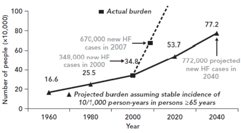
Nowadays, 5.7 million people in the US have HF, but the projections are worrisome since it is expected that by 2030 more than 8 million people will have this condition, accounting for a 46 % increase in prevalence (see Figure 1)[3].
Figure 1: Incidence of HF in the US and the future perspectives
In Europe, the Epidemiologia da Insuficiencia Cardiaca e Aprendizagem (Epidemiology of Heart Failure and Learning – EPICA) study performed in the late 1990s in Portugal reported HF prevalence
9
of 1.36 % in the 25–49-year-old group, 2.93 % in the 50–59-yearold group, 7.63 % in the 60–69-year-old group, 12.67 % in the 70–79-60–69-year-old group, and 16.14 % in patients >80 years [4]. Another analysis in Spain showed HF prevalence steadily increasing from 895 per 100,000 population per year in 2000 to 2,126 cases in 2007, with higher rates in men than women. The prevalence of HFpEF was higher than that of HFrEF; in the former rates were higher in women, while in the latter they were higher in men. The overall HF prevalence significantly increased with ageing, particularly among patients >64 years and with HFpEF [5]. In Germany in 2006 the prevalence of HF was 1.6 % in women and 1.8 % in men, with numbers increasing considerably with advancing age[6]In Sweden in 2010 the crude prevalence of HF was 1.8 % and was similar in men and women, but after adjustment for demographic composition the estimated rate was 2.2 %, with a weak decrease in temporal trend in women but not men between 2006 and 2010 [7]. A recent survey reported HF prevalence of 1.44 % in Italy, with rates increasing with the ageing of the population. HF is also an important health problem in Asia, and its prevalence seems to be even higher compared to Western countries, ranging between 1.3 % and 6.7 % Currently in China there are 4.2 million people with HF, with an estimated prevalence of 1.3 % (see Figure 2). [8].
Figure 2: Prevalence and incidence of HF worldwide [9]
HF outcomes have been extensively investigated in the US. The Organized Program to Initiate Lifesaving Treatment in Hospitalized Patients With Heart Failure (OPTIMIZE-HF) study enrolling 20,118 patients with HFrEF and 21,149 with HFpEF (EF ≥40 %) reported no differences between HFrEF and HFpEF in 60–90-day mortality (9.8 % versus 9.5 %) and re-hospitalisation (29.9 % versus 29.2 %), but higher in-hospital mortality in those with HFrEF (3.9 %) versus HFpEF (2.9 %). When the comparison between HFpEF (EF >50 %) and HFmrEF (EF 40–50 %) was performed, no differences in outcomes were observed.36 Similarly, the Get With The Guidelines (GWTG) registry that enrolled 15,716 patients with HFrEF, 5,626 with HFmrEF and 18,897 with HFpEF observed 37.5 %, 35.1 % and 35.6 % mortality at 1 yearm respectively, with no differences in risk after several adjustments. The 1-year HF hospital readmission rates were 30.9 %, 28.4 % and 24.3 % in HFrEF, HFmrEF and HFpEF, respectively, but there was a higher risk in HFrEF and HFmrEF compared with
HFpEF [10]. The Management Predischarge Process for Assessment of Carvedilol Therapy for Heart Failure (IMPACT-HF) study reported that >50 % of patients were discharged with unresolved symptoms, and within 60 days half had worsening symptoms, a quarter were re-hospitalised and >10 % died [11]. The Canadian Enhanced Feedback for Effective Cardiac Treatment (EFFECT) study enrolling 1,570 patients with HFrEF and 880 with HFpEF reported no differences in mortality at 30 days (7.1 % and 5.3 %, respectively) and 1 year (25.5 % and 22.2 %, respectively). Similarly, for HFrEF and HFpEF there were no differences between HF readmissions at 30 days (4.9 % and 4.5 %, respectively) and at 1-year (16.1 % and 13.5 %, respectively) [12].In Europe, the EuroHeart Failure Survey compared prognosis in 3,148 patients with HFpEF and 3,658 with HFrEF, reporting higher 90-day mortality in those with HFrEF (12 %) compared with HFpEF (10 %), but similar readmission rates (21 % versus 22 %, respectively).In the EuroHeart Failure Survey II, which enrolled 3,580 patients hospitalised for HF, overall in-hospital mortality was 6.4 %.
Figure 3: Stages of HF [13]
Recently, in the European Society of Cardiology Heart Failure Long-Term (ESC-HF-LT) registry that enrolled 12,440 patients with acute and chronic HF from 21 European and/or Mediterranean countries, the 1-year mortality rate was estimated to be 23.6 % for acute HF and 6.4 for chronic HF; whereas the rates for the combined endpoint of mortality or HF hospitalisation within 1 year were 36 % for acute HF and 14.5 % for chronic HF. Mortality rates ranged across the different regions from 21.6 % to 36.5 % for acute HF and from 6.9 % to 15.6 % for chronic HF [14]. The HF could
11
be classified in four stages (see Figure 3) [13], with worsening of the hemodynamic and clinical status of the patients from the Stage A to the Stage D.
The end-stage, Stage D, could be defined as the presence of progressive and/or persistent severe signs and symptoms of heart failure despite optimized medical, surgical, and device.
1.2 The Heart Transplantation
1.2.1 Definition
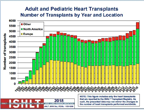
The heart transplantation (Htx) is the gold standard for the unsuccessful medical and surgical therapy in end-stage heart disease (Level of Evidence I C). In the last three years, the number of heart transplantation increased more and more (see Figure 4) [15] [16] [17] [2].
Figure 4: International Society Heart and Lung Transplantation Report of the 2018. The number of HTx procedure since the last 2018 in Europe, North America and other countries
13
1.2.2 Indications and contraindications of Htx
The main indications and contraindications of the heart transplantation are in the Table 2. The indications include [2, 15]:
✓ end stage of heart failure with severe symptoms with poor prognosis; ✓ end stage of heart failure with no evidence of pulmonary hypertension;
✓ refractory cardiogenic shock requiring continuous intravenous inotropic therapy; ✓ Peak VO2 (VO2 max) less than 10 ml/Kg per min;
✓ NYHA III and IV heart failure symptoms;
✓ recurrent life-threatening left ventricular arrhythmias despite an implantable cardiac defibrillator, antiarrhythmic therapy, or catheter-based ablation;
✓ refractory angina without potential medical or surgical therapeutic choice.
The Contraindications include [17, 2, 15]: ✓ relative contraindications:
▪ patients with HIV, ▪ hepatitis,
▪ Chagas disease, ▪ tuberculosis,
▪ active infection, excluded LVAD-related infection, ▪ severe peripherical vascular disease,
▪ severe osteoporosis, ▪ BMI> 35 Kg/m2,
▪ advanced age (more than 65 years old), ▪ phycological instability
▪ active or recent substance abuse
✓ absolute contraindications:
▪ severe cerebrovascular arterial disease;
▪ pharmacologically irreversible pulmonary hypertension,
▪ history of solid organ or hematologic malignancy within the last 5 years due to probability of recurrence
▪ irreversible renal dysfunction,
▪ advanced irreversible liver dysfunction,
▪ advanced irreversible pulmonary parenchymal disease or FEV1 < 1 L/min ▪ systemic disease with multi-organ involvement;
15
1.2.3 Surgical Techniques
The Heart transplantation could be performed by different techniques: ▪ Orthotopic technique:
o Shumway technique (BIATRIAL technique): During implantation, perfusate temperature is generally28°C, with intermittent topical cooling using 4°C saline ice slush. No additional cardioplegic solution is infused. The left atrial anastomosis is constructed first using continuous 3-0 polypropylene suture When constructing it, he first few stitches are placed “at a distance” before lowering the donor heart into the pericardial space. The remainder of the entire left atrial anastomosis is constructed in an everting fashion to provide endothelium-to-endothelium apposition, thereby reducing the chance of thrombus formation along the suture line. Construction of the far-leftward portion of the anastomosis along the left pulmonary veins is often facilitated by retracting the donor ascending aorta inferiorly with a traction suture. The right atrial anastomosis is also constructed with continuous 3-0 polypropylene suture. In the area over the interatrial septum, the suture lines are partially overlapping. Each chamber is filled with cold saline before securing the suture lines. The aortic anastomosis is constructed with continuous 4-0 polypropylene suture after the donor and recipient aortas are cut to appropriate length. A cardioplegia catheter to be used as a “needle vent” for aspirating air is placed in the donor ascending aorta. Air is evacuated from the heart through the aortic suture line, and the suture line secured. The aortic clamp is removed with strong suction on the needle vent. When a gentle sinus rhythm is established, preparations are made for the pulmonary artery anastomosis. (Some surgeons prefer to complete this anastomosis before removing the aortic clamp.) The pulmonary artery segments are cut to an appropriate length and the anastomosis constructed, usually with 4-0 or 5-0 polypropylene suture. The remainder of the operation is conducted as usual during rewarming, and CPB is gradually discontinued after thoroughly de-airing the heart through the aortic needle vent while examining it for residual air with TEE.
o Shumway procedure (BICAVAL technique): Orthotopic cardiac transplantation, bicaval technique. Right atrium is divided to create superior and inferior vena caval cuffs. Great vessels are divided as in biatrial method. Commencement of left atrial anastomosis. Completion of bicaval transplant technique, showing inferior vena caval, superior vena caval, aortic, and pulmonary trunk anastomoses
▪ Heterotopic technique: Heterotopic transplantation. Donor superior vena cava is anastomosed end to side to recipient superior vena cava. Anastomosis may be facilitated by transient removal of superior vena caval cannula. Aortic anastomosis completed. Pulmonary artery connection requires interposition of a polyester graft.
17
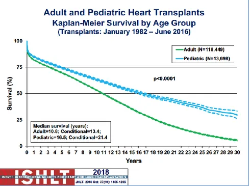
1.2.4 Survival
According to The International Society for Heart and Lung Transplantation estimates that more than 5,000 heart transplants are performed each year worldwide. The long-time survival of heart transplanted patients is 87.8%, 78.5% and 71.7% at 1, 3 and 5 years after surgery respectively ( see
Figure 5-6) [18, 19, 20, 21].
Figure 5: Adult and pediatric heart transplant Kaplan-Meyer cumulative survival curve
1.2.5 Complications
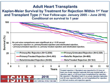
There are a lot of studies documented the management of heart transplant recipient in the postoperative period, short- and long-term. According to ISHLT registry and the most important articles in scientific [22] [23] [24] [9] [25, 26] , the main short-term complications are:
➢ early graft failure and primary graft dysfunction: they are the most common cause of short-term mortality after heart transplantation. Early graft failure (EGF), defined as a composite of death and/or re-transplantation associated with graft failure during the first 30 days after transplant, is the most severe form of primary graft dysfunction (PGD) and constitutes the most feared complication. The incidence of EGF reported for transplants performed between 2005 and 2013 was 3.8%, with a 96.3% mortality rate and 3.6% requiring re-transplantation. ➢ rejection: Hyperacute rejection is mediated by preexisting antibodies to allogeneic antigens and occurs immediately after transplantation with rapid graft failure. It is uncommon because of the current blood- and antigen-typing techniques. Acute rejection could be cellular or antibody-mediated rejection, with an incidence of deaths of 8% after heart transplantation. The figure 7 showed the reduction of rejection and treatment in the last years according to ISHLT registry, it documented the decreasing of its incidence due to early diagnosis and treatment.
Figure 7: Kaplan-Meyer Survival Curves in Adult Heart transplantation by treatment for rejection within 1st year.
➢ neurological complications: the rate of cerebrovascular accident (CVA) after heart transplant is reported up to 13%, and is associated with increased mortality post-transplant. CVAs can
19
be defined as either ischemic or hemorrhagic, with ischemic CVAs being twice as common as hemorrhagic CVAs after heart trans- plantation
➢ respiratory complications: the most of patients with advanced heart failure have considerable changes in pulmonary function, including abnormal pulmonary diffusion, evident by decreased diffusing capacity of the lungs for carbon mon- oxide. Gas exchange impairment persists in 67% of patients after transplantation, independent of smoking status, prior drug use, chest radiographic changes, hemodynamic findings, or duration of heart failure
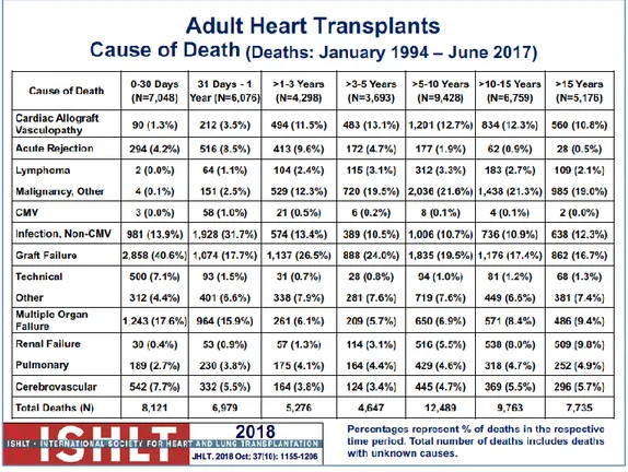
The long-term complications are reported by ISHLT 2018 report in the Figure 8.
Figure 8: Causes of death after heart transplantation during the post-transplantation
▪ Infections are common. The kind of infection in cardiac transplant recipient is vary, depending on time from transplantation [17]
▪ Chronic kidney disease, the calcineurin inhibitors can induce nephrotoxicity by a decrease in glomerular filtration rate (GFR), afferent arteriolopathy and striped tubulointerstitial fibrosis [27].
▪ Endocrine disease: While diabetes mellitus remains a common comorbidity in patients with advanced heart failure undergoing heart transplantation, hyperglycemia due to chronic steroid use may result in a new diagnosis of diabetes post-transplantation in up to 23 e 39% of patients in the first 2 years
▪ Malignancy is one of the major causes of long-term mortality in heart transplant recipient; the cutaneous ones are the most common but the post-transplantation lymphoproliferative disorder (PTDL) is a frequent fatal complication with high association to Ebstein-Barr virus. ▪ Cardiac allograft vasculopathy (CAV) is the largest long-term complication in the heart
transplant recipient, which has and had focused the research and the new digital technologies to improve the early detection in order to increase the patient and graft survival.
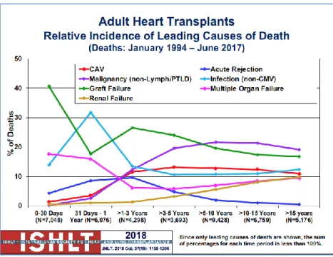
The heart transplant recipient causes of death are several, and according to the last ISHLT 2018 report the most relevant are (see Figure 9):
Figure 9: The causes of death after heart transplantation according to ISHLT registry and report 2018.
▪ at 1 year: Infection non-CMV, graft failure, multiple organ failure ▪ at 1-3 years: CAV, malignancy, graft failure, infection non-CMV ▪ at 3-5 years: CAV, malignancy, graft failure
▪ at 5-10 years: CAV, malignancy, graft failure ▪ at 10-15 years: CAV, malignancy, graft failure ▪ more than 15 years: CAV, malignancy, graft failure
Considering the long-term outcome (see Figure 9-10), the most frequent complications are coronary allograft vasculopathy (CAV) and neoplasm. CAV occurs in approximately 12,7% of patients by 5 years and 12,3% by 10 years, it is one of the major causes of graft loss and death.
21
2.1 Definition
Coronary heart disease of the transplanted heart (CAV) is characterized by characterized by intimal proliferation, develops early after trans- plant, is progressive, and accounts for major morbidity and mortality late in the transplant natural history [16, 28].
Also CAV was defined by ISHLT as:
a). A “Primary Vessel” denotes the proximal and Middle 33% of the left anterior descending artery, the left circumflex, the ramus and the dominant or co-dominant right coronary artery with the
posterior descending and posterolateral branches.
b). A “Secondary Branch Vessel” includes the distal 33% of the primary vessels or any segment within a large septal perforator, diagonals and obtuse marginal branches or any portion of a
non-dominant right coronary artery.
c). Restrictive cardiac allograft physiology is defined as symptomatic heart failure with echocardiographic E to A velocity ratio > 2 (>1.5 in children), shortened isovolumetric relaxation time (<60 msec), shortened deceleration time (<150 msec), or restrictive hemodynamic values (Right Atrial Pressure >12mmHg, Pulmonary Capillary Wedge Pressure >25 mmHg, Cardiac Index <2 l/min/m2)
2.2 Classification
Initially, it was described by Gao [28] and coded anatomic abnormalities into type A, B1, B2, and C lesions (see Figure 11).
Figure 10: The Classification of CAV according to Gao [28]
➢ Type A was discrete or tubular stenosis and multiple stenoses in the proximal, middle, or distal segment branches;
23
➢ type B1 was a proximal vessel maintaining nor- mal diameter with abrupt onset of distal concentric narrowing and obliteration;
➢ type B2 was a gradual transition from the normal proximal vessel with tapering, concentric narrowing progressively increasing in severity distally; and type C was a diseased vessel, diffusely irregular that lost small branches with terminations often non-tapered, squared off, and ending abruptly.
Recently, the ISHLT published a new and complete classification of the CAV according to the
2.3 Pathogenesis
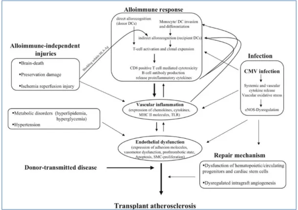
CAV is an accelerated fibroproliferative disease affecting the vasculature of the transplanted heart. Pathologically, smooth muscle proliferation, accumulation of inflammatory cells, and lipid deposition cause circumferential intimal thickening. In contrast to the focal, eccentric, proximal epicardial lesions in atherosclerosis, CAV is diffuse and affects epicardial and intramural vessels (see Figure 12) [29].
Intravascular imaging has shown disease occurs within the first year of trans- plant, and has a biphasic response, involving initial intimal thickening with expansion of the external elastic membrane and relative preservation of luminal area, followed by constrictive remodeling and luminal narrowing. Plaque composition changes from early fibrous and fibrofatty tissue to late atheromatous necrotic core and calcification [29].
Figure 12: Collaboration and interaction of alloimmune-dependent and -independent factors influencing the pathogenesis of transplant vasculopathy. Ag indicates antigen; CD, cluster of differentiation; eNOS, endothelial NO synthase; and SMC, smooth
25
2.4 Histopathological presentation
Coronary vasculopathy of the transplanted heart (CAV) has typical anatomic-pathological features that significantly differentiate it from coronary atherosclerosis affecting the general population, but that unite it to the lesions detectable in chronic rejection that affects the other transplanted organs. As for the type of vessels involved, CAV occurs at the level of the whole coronary tree, affecting both epicardial arteries and intramural vessels; it can also occur at the level of the coronary veins, while the vessels without smooth muscles are spared.
The lesions occur uniformly over the entire length of the vessels involved and it has been shown, thanks to a series of autopsy studies, how the severity of the same is comparable between the proximal and distal portions of the epicardial arteries, both as regards the percentage of surface concerned that due to the extent of intimal thickening. (see Figure 13).
Atherosclerosis, on the other hand, is characterized by focal and eccentric lesions that almost exclusively affect the proximal portion of the epicardial arteries, saving the intramyocardial circulation and coronary veins (see Figure 14).
The times of appearance of these two processes differ significantly; atherosclerotic lesions at the level of the native heart develop slowly starting from puberty and in most cases they occur clinically only after some decades.
Figure 13: Histological view of the cardiac allograft vasculopathy. It shows the typical concentric lesion versus the eccentric one of atheromatous disease.
In CAV, on the other hand, the first changes in the underwear take place already in the first weeks after the transplant; these initial lesions are characterized by a slight diffuse and concentric thickening of the intima, given by the presence of an inflammatory subendothelial infiltrate of lymphocytes and macrophages, by the proliferation of vascular smooth muscle cells (vascular smooth muscle cells, VSMCs) migrated into the intimate and from the presence of mild fibrosis and increased extracellular matrix proteins.
As the months progress, there is the appearance of intermediate lesions, developed following the accumulation of foam cells (foamy macrophages) and lipids in the intimate area and the accelerated intimal proliferation of modified VSMCs and fibroblast, and of atheromatous plaques, with a core well-formed lipid consisting of cholesterol and lipid residues.
In the long term these intimate fibrous and fibrolipid lesions lead to a picture of concentric and diffuse fibrous intimal thickening and to possible fibrous and fibroadipose plaques (atherosclerotic plaques mixed with a diffuse intimal thickening are late).
The histopathological presentation of these plaques is similar to that found in coronary vasculopathy, but the incidence of complications, such as plaque ulceration and thrombus formation, is very rare, as are the calcification phenomena.
Another difference compared to normal atherosclerosis, which affects all layers of the vascular wall with destruction of the internal elastic lamina, is the fact that CAV is a pathological process that mainly involves the intimate tunic of the vessels; the internal elastic lamina remains, in fact, relatively intact, while the medium tunic and the adventitia, not affected by aggressive intimal proliferation, are progressively replaced by fibrous tissue. The magnitude of this fibrotic process increases as the severity of the pathological process affecting the intima increases.
Figure 14: atherosclerotic disease of coronary artery.
27
2.5 Epidemiology and Aetiology
Both the immunologic and non-immunological factors are thought to contribute to the pathogenesis of CAV, still not fully understood; it seems that the former play a fundamental role in the onset of the disease, while the latter favor its progression and spread along the vascular tree (see Figure 15) [30].
Figure 15: Costimulatory molecules play crucial roles in this T cell activation. Many costimulatory pathways have been described, and some are involved in the pathogenesis of CAV, atherogenesis, and subsequent plaque formation. In this review, we summarize the present knowledge of the role of these pathways in CAV development and the possibility of manipulating these pathways as a means to treat heart allograft vascular disease and atherosclerosis [30]
Regarding the immunological risk factors, the degree of HLA incompatibility between donor and recipient and the number and duration of acute rejection episodes are important. In particular, a study published in 2004 identified a high Rejection Score (RS) for severe rejections (grade ≥3A) as an independent predictive factor for the onset of CAV.
Non-immunological risk factors include the donor's mode of death, ischemia and reperfusion injury, cytomegalovirus infection, age, sex and high donor and recipient weight, as well as common risk factors such as atherosclerosis, dyslipidemia, hyperhomocysteinemia, arterial hypertension, diabetes mellitus and cigarette smoking.
All these risk factors cause or contribute to the maintenance and perpetuation of a coronary endothelial dysfunction, which constitutes the primum movens and is fundamental in the pathogenesis of CAV.
This pathological process affecting the graft begins even before explantation, since it has been shown that a sudden brain death causes an increase in the circulating levels of catecholamines, inflammatory cytokines, chemokines and adhesion molecules at the level of the organ vessels to be transplanted; this cascade of events causes an inflammatory response in the heart, resulting in vascular damage. In the perioperative phase, ischemia and reperfusion damage play an important role in the development of endothelial dysfunction.
The extent of the damage caused depends not only on the time of ischemia, on the quality of conservation of the organ during transport, on the hemodynamic state of the donor and on the possible need for inotropic support with catecholamines, but also, paradoxically, from the same reperfusion. In the initial stages of this process, oxygen free radicals are formed which compromise the endothelium's ability to release nitric oxide, altering the coronary vascular tone.
The same free radicals also activate the leukocytes and macrophages of the host which, in turn, give rise to a vicious circle, through the production of additional free radicals, proinflammatory cytokines and chemokines. Ischemia and reperfusion damage also causes activation of endothelial cells, with an increase in the expression of adhesion molecules, stimulates platelet adhesion, complement activation and proliferation of vascular smooth muscle cells.
All of these processes lead to endothelial dysfunction resulting from ischemia and reperfusion injury. However, the main determinant in the pathogenesis of CAV appears to be the recipient's immune response to the transplanted organ, and in particular the graft endothelium. These endothelial cells therefore play a key role in the development of this atypical coronary vasculopathy, as they act as both antigen-presenting cells (APCs) and as a target for the immune response that they themselves helped to trigger.
The increased expression of class I alloantigens of the major histocompatibility complex (MHC) expressed by graft endothelial cells is directly recognized by CD8 + T lymphocytes, resulting in cytokine secretion and further activation of endothelial cells. Activated endothelial cells express increased levels of MHC class II antigens that late activate CD4 + T lymphocytes.
This may explain the predominance of CD8 + T lymphocytes in early vascular lesions and the increased proportion of CD4 + T lymphocytes in the advanced stages of the disease. The thus activated T cells release several cytokines, including IL-2, IL-4, IL-5, IL-6, INF-γ, α and
TNF-29
cytokines and adhesion molecules (VCAM-1, ELAM-1). Thanks to the chemotactic action of cytokines and adhesion molecules, it follows the recruitment and accumulation of macrophages and lymphocytes activated at the level of the vascular wall.
These cells secrete various growth factors, such as Platelet derived Growth Factor (PDGF), Insulin-like Growth Factor 1 (IGF-1), Fibroblast Growth Factor (FGF), Epidermal Growth Factor (EGF) and Transforming Growth Factor-β (TGF-β), which cause an intimal migration and an uncontrolled proliferation of modified vascular smooth muscle cells, associated with an increased production of extracellular matrix (see Figure 16) [31].
Figure 16: Immunological events involve in the CAV pathogenesis. Environmental and genetic factors contribute to the etiology of SSc. The pathogenesis of SSc involves an interplay between vascular, immunological, and fibrotic processes. Vascular injury and endothelial damage are the earliest events in the pathogenesis of SSc. Activated endothelial cells upregulate the expression of adhesion molecules and secrete chemokines, leading to inflammation and autoimmunity. Macrophages and T-cells are the predominant inflammatory cell types of the inflammatory infiltrates and produce cytokines and growth factors that drive the synthesis of extracellular matrix proteins by fibroblasts, resulting in progressive fibrosis. T-cells have also been implicated in autoantibodies production. [31].
CHAPTER 3:
31
3.1 Detection of CAV
In the early era of cardiac transplantation, the diagnosis of CAV was made pathologically. Angiographic diagnosis emerged rapidly and remained the most important diagnostic tool.
The development of IVUS allowed for detection of early stage CAV not identified by invasive coronary angiography.
In later years, circulating immunehistologic markers as well as gene-based and protein-based biomarkers have been studied to see if they can contribute to grading or detecting CAV.
Routine surveillance is important because HTX patients frequently are asymptomatic, particularly in the early stages of the disease.
Surveillance includes both evaluation of graft function and visualization of the coronary arteries. Echocardiography is the first-line imaging modality to assess graft function and is part of all serial evaluations during post-transplant follow-up.
With echocardiography, CAV is detected in a late stage when reduced coronary blood flow has resulted in allograft dysfunction.
Dysfunction first manifests as diastolic dysfunction with restrictive physiology, then as systolic dysfunction with reduced ejection fraction.
To detect the presence of CAV and identify potential significant stenosis eligible for intervention, annual or biannual screening with ICA is the current standard of care.
A number of other non-invasive and invasive imaging modalities are used for CAV evaluation (see Figure 17).
33
3.2 Non Invasive stress testing
Detection of CAV is challenging with non-invasive techniques, especially in the early stages [32, 33, 34, 35]. Various non-invasive techniques are used for CAV evaluation.
Stress Echocardiography
Dobutamine stress echocardiography (DSE) is commonly used for CAV screening [36, 37] [38]. The surveillance recommendations of ISHLT considered the DSE of Class IIa. DSE gives the information of:
- cardiac structure and function - regional wall motion
- myocardial deformation - coronary flow reserve
DSE has an important limitation: it is directly dependent on acustic window. Myocardial Perfusion Imaging
The myocardial perfusion imaging (MPI) has shown prognostic value and a moderate diagnostic accuracy in the investigation of CAV [39, 40]. Promising results have been demonstrated for MPI with single-photon emission computed tomography (SPECT, Surveillance Recommendation: Class IIa). The SPECT:
- identifies the myocardial perfusion, ventricular function - its limitation: radiation exposure
Positron emission tomography (PET, Surveillance Recommendation: not included) is the MPI with the most diagnostic accuracy due to its prognostic value and flow quantification (FQ). The FQ is better able to detect the microvascular or diffuse disease. The PET:
- documents myocardial perfusion, myocardial flow quantification and ventricular function - its strengths: quantify global/regional myocardial blood flow, quantify global/regional
myocardial flow reserve
- its limitations: limited availability, radiation exposure
both positron emission tomography and in magnetic resonance imaging in small studies. Cardiac Magnetic Resonance
The above-mentioned techniques evaluate myocardial structure, function, and/or perfusion by gadolinium enhancement [41, 42]. Cardiac MRI is:
- with limitations: high resting rates post-transplant, cardiac device contraindicated, challenging perfusion quantification software, nephrogenic systemic fibrosis in renal failure.
Cardiac Computed Tomography Angiography (CCTA)
CCTA is the only non-invasive technique assessing the coronary arteries, their lumen and wall. In the most recent International Society for Heart and Lung Transplantation (ISHLT) guidelines for the care of heart transplant recipients, coronary CT angiography (CCTA) is given a class IIb
recommendation (usefulness/efficacy is less well established by evidence/opinion) with a C level of evidence [2] [43, 44, 45].
CCTA shows promise in the evaluation of CAV in HTX recipients, although higher resting heart rates in these patients limit the technical image quality. It has got some limitations:
- high resting rates post-transplant - radiation exposure
- contrast-induced nephropathy
- limited ability to assess smaller vessels
Cardiac CT imaging includes coronary artery calcium (CAC) CT scanning and CCTA. The following technical considerations are predominantly concerned with CCTA but are also relevant for understanding CAC CT scanning. Imaging the coronary arteries with CT is technically demanding.
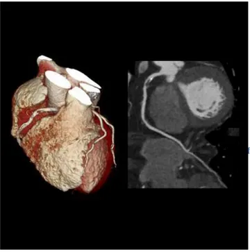
Figure 18: Example of the Cardiac Computed Tomography of the last generation.
35
Spatial and temporal resolution is challenged by the small, torturous vessels moving synchronously with the beating heart.
Electron beam CT was the first non-invasive imaging modality with cross-sectional visualization of the heart. It has a high temporal resolution of 100 ms, but the spatial resolution is limited by a slice thickness of 3 mm. With the introduction of multidetector technology, cardiac imaging with mechanical helical CT systems became possible. Starting out with cardiac imaging using 4-slice MDCT, a 64-MDCT is considered the minimum prerequisite for adequate scanning of the heart today
(see Figure 19).
Figure 19: Images of the new generations scanner in cardiac computed tomography devices.
Spatial resolution
To be visualized adequately, the coronary arteries require isotropic submillimeter spatial resolution. Spatial resolution with contemporary 64-MDCT is 300–400 μm and is 230–240 μm on the newest high-end scanners (vendor website information; GE Revolution CT, Siemens Somatom Force). Coarse coronary calcifications are still a challenge to reliable visualization of the lumen because of blooming artifacts and reduce the specificity of CCTA.
Temporal resolution
High temporal resolution is a prerequisite for imaging the coronary arteries to avoid cardiac motion artifacts. The data acquisition time per image is referred to as temporal resolution. In cardiac imaging,
a half gantry rotation is sufficient for reconstruction of one image; therefore, temporal resolution is half the gantry rotation time. Dual-source (DS) systems with two x-ray tubes and corresponding detectors operating simultaneously provide temporal resolution close to a quarter of a rotation time, which presently is 66 ms with the fastest scanner (vendor website information; Siemens Somatom Force). Shorter rotation time enables adequate imaging of higher heart rates. Medication to lower the heart rate to 60–65 beats per minutes (bpm) is currently recommended by European guideline/American guideline. Another important temporal aspect is to minimize the time needed to cover the heart in the z-axis (the long axis of the patient). The optimum is to cover the heart in only one heartbeat to avoid misalignment artifacts related to the heart being differently positioned in consecutive heartbeats, which is especially noticeable in arrhythmia. One-heartbeat coverage is achieved with wide detector technology or with DSCT high-pitch technology. The widest detectors are 16 cm wide and cover the whole heart in one rotation. In high-pitch technology, the high pitch facilitates data acquisition of the whole length of the heart within the diastole of a heartbeat, and the dual detector system enables gapless volume coverage despite the high pitch by doing two helical acquisitions almost simultaneously.
Figure 20: Multi-sector scanning and ECG
Scan modes
When imaging a beating heart, the images need to be reconstructed in consistency with a cardiac phase, i.e., systole or diastole. This reconstruction is facilitated with ECG-synchronized data acquisition. There are two types of ECG-synchronized scanning modes: retrospective ECG-gated helical scanning and prospective ECG-triggered axial (sequential) scanning. A variant of the prospective ECG-triggered method is used in high-pitch DS scanning where a helical data acquisition in the diastole of one heartbeat is prospectively triggered by the patient’s ECG. In retrospective ECG-gating, the data are acquired in a continuous, helical scan and a continuous movement of the table
37
with simultaneous recording of the patient’s ECG. The ECG recording guides data selection to ensure phase-consistent image reconstruction of data taken from several cardiac cycles. Sets of data can be reconstructed from any phase of the cardiac cycle, and the availability of both systolic and diastolic reconstructions makes the technique quite robust and can be essential in patients with high heart rates. In high heart rates, the optimal phase for reconstructing the left part of the coronary tree is most often the diastole while the right part is often best reconstructed in the late systole.
In prospective ECG-triggered sequential scanning, the data are acquired at a predefined phase of the cardiac cycle in an axial scan with a stationary table. Data acquisition is initiated by the patient’s ECG signal using the R peak as a reference. Depending on the detector width, one or more sequential axial scans are needed to cover the entire heart volume. If more than one scan is needed, the table has to be moved to the next scan position between each data acquisition; hence, the term
“step-and-shoot.”
Figure 21: The ECG phases in CARDIAC CT scan
The prospectively ECG-triggered scan mode effectively reduces the time of radiation exposure because only a short part of the cardiac cycle is scanned. The short exposure time does, however, restrict the possibility of multiple reconstructions throughout the cardiac cycle. Prospective triggering is limited to patients with low heart rates (<70–75 bpm in systems with gantry rotation time 250–280 ms) and with stable sinus rhythm.
Radiation exposure
Dose-saving strategies: In all procedures involving ionizing radiation, the small stochastic risk of malignancy induction should be taken into consideration, and radiation exposure should always be kept as low as reasonably achievable. There has been much focus on radiation dose in cardiac imaging, and great efforts have been put into the development of dose-saving strategies by the vendors. The most important dose saving strategies/techniques are choice of scan mode, ECG-synchronized tube current modulation, tube voltage reduction, and iterative CT data reconstruction. Choice of scan mode is probably the single most important factor influencing radiation exposure. Prospectively ECG-triggered axial scanning significantly reduces the radiation dose compared to retrospective ECG-gated helical scanning; reductions of up to around 70–80% have been reported. In the latest generation of high-end scanners, submillisievert dose levels have been demonstrated with the combined use of ECG-triggered scan mode, lower tube voltage, automated exposure control, and iterative reconstruction algorithms with both high-pitch and wide-volume scanners.
Patient-related dose factors
Patient-related factors are important predictors of radiation dose. Heart rate and heart rate regularity are important determinants of radiation dose because most of the dose-reducing alternatives depend on a low and steady heart rate. Depending on gantry rotation speed, there is an upper limit for prospective ECG-triggered scanning of 60–65 bpm in earlier systems and 70–75 bpm in high-end scanners. Body weight is another factor with a profound effect on radiation dose Heavier patients require higher tube voltage and current to achieve acceptable image noise levels, which consequently increases radiation exposure.
Radiation dose parameters
The radiation dose parameters used for CT are volume CT dose index (CTDIvol), expressed in units of mGy, and dose-length product (DLP), expressed in units of mGy*cm. Simplified, CTDIvol is an estimate of the average radiation dose for a specific scan protocol for one tomographic image with pitch incorporated. DLP is the product of the CTDIvol and the scan length. The CTDIvol is recommended for optimizing CT protocols whereas DLP should be used for comparing radiation doses and characterizing radiation dose from CT studies. To estimate an effective dose for adult patients, the DLP is multiplied by an organ-weighting factor (k). In cardiovascular imaging, the k value for chest examination is used, which currently is 0.014 mSv per mGy*cm
39
3.3 Invasive methods
Invasive methods for coronary evaluation enclose: visualizing vessel lumen (coronary angiography), evaluation of vessel wall dimensions and wall components (IVUS, IVUS virtual histology, and optical coherence tomography [OCT]), and evaluation of coronary flow parameters (fractional flow reserve [FFR], and index of microcirculatory resistance (IMR)) [46, 47, 48, 49, 50].
Coronary Angiography (CA)
Although a relatively insensitive method for diagnosing CAV, CA remains the accepted standard of care serving as a screening tool to grossly detect the presence of CAV and is typically performed at an annual or biannual routine basis. The method is clinically available and has documented prognostic significance. The ISHLT recommendations for CAV nomenclature is based on angiographically depicted lesions of CAV and the surveillance recommendation is Class I [15, 2, 16, 51, 52]. It is prognostic for its accuracy in the detection of coronary stenosis and myocardial blush. Its limitations:
- evaluation limited to epicardial vessels - insensitive for detection of early CAV - insensitive for the detection of diffuse disease - radiation exposure
- contrast-induced nephropathy.
The limitations are related to the nature of CAV lesions (concentric, longitudinal and diffuse disease) as well as expansive vascular remodeling.
Intra vascular Ultrasound (IVUS)
IVUS is superior to CA in detecting CAV. IVUS has been documented to detect CAV in apparently normal angiograms and to predict development of cardiac events even in the presence of a normal coronary angiogram [53, 15, 16, 45, 54]. A coronary artery intimal thickness ≥0.5 mm is defined as abnormal by ISHLT guidelines. A rapid progression of maximal intimal thickness (MIT) ≥0.5 mm during the first year after transplantation is a predictor of all-cause mortality and adverse cardiac events. On the other side, it has been demonstrated that IVUS-detected intimal hyperplasia does not correlate well with small-artery disease by histologic or immunohistochemical analysis.
Although IVUS is very sensitive for defining CAV, the ISHLT guidelines consider it to be an investigational tool and do not recommended IVUS for routine surveillance of CAV (Class IIa) [15] [16].
According to the same guidelines, IVUS is optional at baseline (5–6 weeks) and at 1 year after HTX to exclude donor CAD and detect rapidly progressive CAV, respectively, thus providing prognostic information. Being very sensitive for defining CAV, IVUS is an important research tool helping investigators to explore surrogate markers for CAV and evaluate the outcome of various therapeutic conditions.
Virtual histology (VH) is a relatively new IVUS-based technique providing information about plaque components. Four basic tissue components can be identified: fibrous, fibrofatty, calcified, and necrotic core. Although VH and IVUS are not a part of the routine surveillance of CAV, the added information on prevalence, morphologic patterns, and distribution from studies using these methods.
AUTHOR JOURNAL PARAMETERS Park et al [55, 56] JHLT 2017; 36: 185-192
EHJ 2016; 17: 272-279
•Vessel volume (mm3)
•Minimal vessel diameter (mm) •Maximal vessel diameter (mm) •Lumen volume (mm3)
•Minimal lumen diameter (mm) •Plaque Volume (mm3) •Percent Plaque volume
Clemmensen et al [57, 58] JHLT 2016, 35 (4S): s98 JACC 2017, 10 (7): 773-784
• Plaque type
• Mean Lumen/intima ratio • Maximal intima/media ratio • Percent plaque volume
Tomai et al [59] JHLT 2016, 35 (1): 74-79 • EEL area (mm2) • IEL area (mm2) • Lumen area (mm2) • Plaque thickness (mm) • Media/EEL area (%) • Intima /media ratio • Intimal thickness (mm) • Type of plaque
o eccentric o calcified o lipid pool
41 • Intimal thickness (mm) • Media thickness (μm) • Intimal volume (mm3) • Media volume (mm3) • Plaque volume (mm3) • Plaque index % • I/M ratio
Table 3: the most important literature experiences of IVUS in the detection of CAV [58] [59] [56] [55] [60].
According to the scientific literature and experience (see Table 3), the IVUS most interesting data to analyze the CAV are:
• Vessel volume (mm3)
• Minimal vessel diameter (mm) • Maximal vessel diameter (mm) • Lumen volume (mm3)
• Minimal lumen diameter (mm) • Plaque Volume (mm3)
• Percent Plaque volume (%, following for the results)
In particularly in the 2001, the American College of Cardiology Clinical Expert Consensus Document on Standards for Acquisition, Measurement and Reporting of Intravascular Ultrasound Studies [61] was published, which recommended a threshold for transplant vasculopathy as an intimal thickness of >0.5 mm measured at a target segment of a vessel. This is a widely accepted definition of CAV by IVUS today (see Table 4) [61].
Optical Coherence Tomography (OCT)
OCT provides high resolution (10-20 µm) in the detection of microvascular disease and macrovascular epicardial disease. It is not included in the surveillance recommendation of the ISHLT. OCT gives many important CAV details such as arterial wall description, arterial lumen quality, plaque volume, plaque characterization. Its strengths are high spatial resolution and high accuracy in the description of the plaque and arterial lumen [62]. Its limitations are:
- evaluation is limited to epicardial vessels - reduced tissue penetration than IVUS - high cost
- limited availability
Invasive Coronary Flow Studies
The CAV causes complex changes in coronary physiology. Invasive coronary sensor pressure and flow wires allow independent assessment of the epicardial arteries and microvasculature by measuring the fractional flow reserve (FFR), coronary flow reserve (CFR), and index of microcirculatory resistance (IMR). At this time, it is not included in the ISHLT Surveillance Recommendation in the detection of CAV [16] [15] [58, 63]. There are some limitations:
- Cost
- Limited availability
43
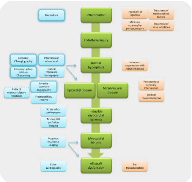
The management of CAV is focused on primary prevention, early detection, imaging surveillance and early treatment as reported by Chih and other author in his recent review (see Figure 22,23) [35, 64, 65, 66]
Figure 22: Algorithm for CAV surveillance and management.
In particularly the medications have a great role in the therapeutic choice.
➢ Aspirin: antiplatelet therapy; it reduces the formation of microthrombi at the sites of immune injury in the coronary endothelium [67];
➢ Statins: inhibit inflammatory and immune responses including the inhibition of natural killer cell cytotoxicity [68];
45
➢ Immunosuppression: mycophenolic acid reduces progression of intimal thickening, the mTORs sirolimus and everolimus inhibit vascular smooth muscle and fibroblast proliferation [69, 70, 71].
The Revascularization is limited to diffuse CAV and high mortality for surgical procedure.
First Author N^ of pts Procedural Success Rate Adverse clinical outcome Restenosis Bader et al [72] 40 91% 20% 6 death 2 repeat OHT BMS 31% DES 15% Simpson et al [73] 33 99% 39,3% 6 mts: 31% 12 mts: 46% 5 years: 66% Benza et al [74] 62 97% 34% 6 mts: 57% Wellnhofer et al [75] 160 97% / 38% Lee et al [76] 82 100% 20% DES 12% BMS 30% Zakliczynski et al [77] 37 / 18% DES 7% BMS 58% Lee et al [78] 140 98% 25% BMS 23% DES 10,4% Tremmel et al [79] 34 / 12% BMS 33% DES 12,5%
Table 5: The most important articles in scientific literature about the use of PCI in the treatment of CAV
The retransplantation is recommended to selected patients with advanced CAV but it is controversial because of organ storage, lower survival and re-presented CAV in de novo transplantation.
47
5.1 INTRODUCTION OF THE STUDY DESIGN
Firstly, I evaluate the capability of the 64-slice dual-source Coronary Computed Tomographic Angiography (CCTA) in the detection of Cardiac Allograft Vasculopathy (CAV) in the population of heart transplant recipients (FIRST STEP). In particularly I analyse the sensibility and specificity of CCTA versus the CA.
Then the CCTA is compared to intravascular ultrasound detection of CAV (IVUS, SECOND STEP). Finally, I compared the most important scientific articles regarding the early diagnosis of CAV with clinical, CCTA, CCA and IVUS and I create the “CAV Early Diagnosis score (CAVeD score)”
5.2 FIRST STEP
5.2.1 Material and Methods
Between January 2001 and December 2016, 84 patients undergoing heart transplantation at Heart Transplantation Center, Department of Heart and Vessels and followed by Heart Transplantation Ambulatory were screened for this retrospective observational study. Patients undergoing heart transplant in other Institution and subjects with renal failure were excluded from the analysis. Data collection included patient demographics (age, sex, height, and weight), donor age, CAD risk factors (hypertension, diabetes mellitus, dyslipidemia, and current smoking history), dates of CCTA and CCA procedures, and current medications. Blood glucose, glomerular filtration rate according to MDRD (Modification of Diet in Renal Disease), and creatinine levels were also recorded. All data were prospectively collected and recorded onto computerized database registries that remained consistent over the study period. The study was approved by the Ethics Committee of our Institution. Human rights statements and informed consent: All procedures followed were in accordance with the ethical standards of the responsible committee on human experimentation (institutional and national) and with the Helsinki Declaration of 1964 and later revisions. Informed consent was obtained from all patients for being included in the study.
Conventional coronary arteriography: Based on the ISHLT guidelines, CAV was classified by CCA
as follows: CAV0 (not significant) indicates no detectable angiographic lesion; CAV1 (mild) indicates angiographic left main <50%, primary vessel with a maximum lesion of <70%, or any branch stenosis <70% (including diffuse narrowing) without allograft dysfunction; CAV2 (moderate) indicates angiographic left main <50%, a single primary vessel >70%, or isolated branch stenosis
![Table 2: HTx indications and contraindications according to ESC 2016 HF Guidelines [2]](https://thumb-eu.123doks.com/thumbv2/123dokorg/2890344.11208/20.892.362.655.133.444/table-htx-indications-contraindications-according-esc-hf-guidelines.webp)