UNIVERSITY OF CATANIA
DEPARTMENT OF CHEMICAL SCIENCESINTERNATIONAL PhD IN CHEMICAL SCIENCES – XXXI CYCLE
Ivana Di Bari
Photoresponsive nanosystems
for therapeutic applications
PhD Thesis
Tutor: Prof. S. Sortino
Dr. G.M.L. Consoli
PhD Coordinator:
Prof. Salvatore Sortino
Abstract
Oncological and bacterial diseases are certainly the most troubling illnesses of the twenty-first century, and the data in this regard are staggering. Indeed, anticancer chemotherapy is affected by low specificity/selectivity and undesired side effects, and drug resistance phenomena are responsible for the failure of many conventional treatments. Multidrug-resistant-bacteria (MDRB) infection is become another burden to modern healthcare. For such a reason, there is an increasing demand for the development of new strategies for anticancer and antibacterial treatments without antibiotics to save millions of lives every year.1
In this frame, the light-controlled generation of cytotoxic species, such as singlet oxygen (1O
2) and nitric oxide (NO), by using appropriate photochemical
precursors represents a fascinating and unconventional approach for the treatment of cancerous and microbial diseases in non-invasive manner. However, many precursors have a hydrophobic nature, which favors their aggregation in aqueous medium strongly precluding their photochemical behavior.2 The entrapment of drugs and pro-drugs in nanocarriers permits to
overcome all these drawbacks and ensures protection from degradation, site-specific delivery, enhanced bioavailability, increased local concentration.
Here in are reported studies of two different types of nanocarriers carrying different photoactivable guests. The first part of this dissertation focuses on the achievement of novel photoresponsive calix[4]arene-based nanoconstructs which vehicle, by not covalent or covalent approach, different kinds of chromophores. The second part concerns the design, synthesis, antitumoral and antibacterial activity of polymersomes containing a nitric oxide photoprecursor by covalent and non-covalent approach.
Table of contents
Abstract ... I General aims of the project ... IX
1. Introduction and background ... 1
1.1. Antineoplastic Resistance ... 1
1.2. Antimicrobial Resistance ... 3
1.3. Nanomedicine ... 6
1.4. Light stimulated therapy ... 7
1.4.1. Photodynamic Therapy (PDT) ... 7
1.4.2. Nitric oxide based therapy ... 8
1.5. Calix[n]arenes (CAs) ...10
1.6. Polymersomes ...11
Part I ...13
Calixarene-based photoresponsive nanosystems ...13
2. Calixarene-based photoresponsive nanosystems entrapping different guests by non-covalent approach for antibacterial applications ...15
2.1. Introduction ...15
2.2. Results and discussion ...16
2.2.1. Synthesis of compound 1 ...16
2.2.2. Synthesis of compound 2 ...17
2.2.3. Characteristics of the components ...17
2.2.3.1. Compound 1 – Calix[4]arene derivative ...17
2.2.3.2. Compound 2 – NO photoprecursor ...18
2.2.3.3. Compound 4 – Foscan® ...19
2.2.3.4. Compound 5 – ZnPcS4 ...19
2.2.4. Characterization of the calix[4]arene 1-based nanoassemblies 20 2.2.5. Nanoassembly 1·2: spectroscopic and dimensional characterization ...21
2.2.6. NO photorelease from nanoassembly 1·2 ... 22
2.2.7. Antibacterial activity of nanoassembly 1·2 ... 23
2.2.8. Nanoassembly 1·4: spectroscopic and dimensional characterization ... 24
2.2.9. Nanoassembly 1·5: spectroscopic and dimensional characterization ... 25
2.2.10. 1O2 photogeneration from 1·4 and 1·5 nanoassemblies ... 27
2.2.11. Antibacterial activity of the nanoassemblies 1·4 and 1·5... 28
2.3. Experimental section ... 29
2.3.1. Materials ... 29
2.3.2. Synthesis ... 30
2.3.3. Preparation of the nanoassemblies ... 31
2.3.4. Instrumentation ... 32
2.3.5. Determination of fluorescence and 1O2 quantum yield ... 34
2.3.6. Antibacterial experiments ... 34
2.3.7. Phototoxicity assay ... 35
3. Simultaneous supramolecular activation of NO photodonor/photosensitizer ensembles by a calix[4]arene nanoreactor .. 37
3.1. Introduction ... 37
3.2. Results and discussion ... 38
3.2.1. Synthesis and characteristics of the components ... 38
3.2.2. Spectroscopic and dimensional characterization of the nanoassemblies 1·2·4 and 1·2·5 ... 38
3.2.3. NO photorelease from ternary 1·2·4 and 1·2·5 nanoassemblies ... 40
3.2.4. 1O2 photogeneration from ternary 1·2·4 and 1·2·5 nanoassemblies ... 41
3.3. Experimental Section ... 42
3.3.1. Materials ... 42
3.3.2. Preparation of the ternary nanoassemblies ... 42
3.3.4. Determination of fluorescence and 1O2 quantum yields ...44
4. Photophysical and photochemical properties preservation of Iodurate Bodipies PSs in aqueous environment – Preliminary studies ...45
4.1. Introduction ...45
4.2. Results and discussion ...46
4.2.1. Characteristics of the components ...46
4.2.2. Spectroscopic and dimensional characterization of the nanoassemblies 1·12 and 1·13 ...46
4.2.3. Photobleaching ...47
4.2.4. 1O2 Photogeneration from the nanoassemblies 1·12 and 1·13 49 4.2.5. Relative Efficency (Erel) of the two PSs ...50
4.3. Experimental section ...51
4.3.1. Preparation of the nanoassemblies ...51
4.3.2. Instrumentations ...51
4.3.3. Determination of the 1O2 quantum yields ...52
5. Design, synthesis and antibacterial activity of a multivalent polycationic calix[4]arene-NO photodonor conjugate ...53
5.1. Introduction ...53
5.2. Results and discussion ...54
5.2.1. Synthesis of compound 17 ...54
5.2.2. Characteristics of 17 ...54
5.2.3. Nanoassembly of 17: spectroscopic and dimensional characterization ...55
5.2.4. NO photorelease from 17 ...55
5.2.5. Antibacterial activity of 17 ...57
5.3.2. Synthesis ...59
5.3.3. Preparation of the hydro-alcoholic solution of compound 1761 5.3.4. Instrumentations ...61
Part II ... 63
Polymersomes-based nanoconstructs for anticancer and antibacterial photoactivated treatments ... 63
6. Design, synthesis, antitumoral and antibacterial activity of polymersomes containing a nitric oxide photoprecursor by covalent and non-covalent approach ... 65
6.1. Introduction ... 65
6.2. Results and discussion ... 66
6.2.1. Synthesis of polymers ... 66
6.2.2. Synthesis of compound 16 ... 68
6.2.3. Characterization of polymersome 21 ... 68
6.2.4. Nanoassembly 21·29: spectroscopic and dimensional characterization ... 69
6.2.5. Cellular uptake of nanoassembly 21·29 on HeLa cells ... 70
6.2.6. Cellular uptake of polymersomes 25 ... 71
6.2.7. Nanoassembly 21·28: spectroscopical and dimensional characterization ... 72
6.2.8. Comparison of EE (%) between polymersomes and micelles obtained by polymer 21 ... 73
6.2.9. NO detection ... 74
6.2.10. In vitro study: antitumoral activity of 21·28 on cervical (HeLa) and pancreatic (BxPc3) cancer cells ... 75
6.2.11. Characterization of polymersome 26 ... 77
6.2.12. NO release by photobleaching monitoring of polymersome 26 78 6.2.13. In vitro study: antitumoral activity of polymersome 26 on HeLa cells ... 78
6.2.14. Polymer 27 ... 79
6.2.15. Nanoassembly 27·28: spectroscopical and dimensional characterization ... 80
6.2.16. Antibacterial activity of nanoassemblies 21·28 and 27·28 .... 81
6.3.1. Synthesis ...87
6.3.2. Polymers Synthesis general procedure ...90
6.3.3. Preparation of polymerosomes entrapping non-polar guest .91 6.3.4. Preparation of micelles ...91 6.3.5. Instrumentations ...91 6.3.6. Antibacterial experiments ...92 6.3.7. Biological assay ...93 General conclusions ...95 Publications ...97 Acknowledgements ...99 References ...101
General aims of the project
The final goal of this research project is the design, synthesis and characterization of photoresponsive nanosystems as therapeutic devices able to release the transient cytotoxic species NOand 1O
2 upon light activation for
anticancer and antibacterial treatments. Specific attention is paid on water-soluble molecular systems, based on calixarenes and polymersomens, functionalized with suitable ligands able to recognize receptors overexpressed on the surface of cancer cells and/or promote the penetration in bacteria, which are typically characterized by an impermeable outer cell membrane. These systems are devised in order to exhibit either the single or simultaneous photo-stimulated release of the two cytotoxic agents.
Fabrication of these photoresponsive nanoconstructs implies collective cross-disciplinary efforts because of synthetic methodologies and physical characterization techniques. However, photochemistry plays a dominant role. In most cases, the systems are designed in order to avoid interchromophoric communication upon light excitation. It is important to note that, unlike non photoresponsive-molecules, the preservation of photophysical and photochemical properties of the chromophores in a confined region of the space is not a trivial result, but the logical consequence of the appropriate design of any specific component. The efficiency and the nature of any involved process, in fact, can be drastically affected by the energy and/or electron transfer mechanisms that preclude the ultimate goal.
1. Introduction and background
1.1. Antineoplastic Resistance
One of the main causes for the failure of an anti-cancer treatment is the development of drug resistance by the cancer cells.3 This is a very serious
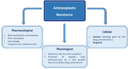
problem that can lead to the reappearance of the disease and even death. While it is quite common to think that the majority of cancers arise from a single precursor cell, it would be a mistake to think that a tumor is made up of all genetically identical cells. One of the characteristics of cancer is the increase in genetic instability and speed of mutation. These changes mean that the cells that are dividing, acquire genetic changes very quickly. Practically, this means that the cells of a tumor are similar, but they are not identical. When exposed to an anti-tumor drug, the sensitive cells die, while the resistant ones survive and multiply. The result is the re-growth of a tumor that is not sensitive to the drug used in origin. There are several reasons that can explain the existence of a first-resistant cell in the original tumor (Fig. 1).
Fig. 1 Schematic representation of the main causes of resistance to antineoplastics.
The resistance in fact is established on the basis of anatomical and physiological factors. As an example when the cancer cells are confined into organs hardly accessible by the drugs, the so-called “sanctuaries” (for example, the central nervous system and testicle, due to the presence of the blood-brain barrier and the blood-testis barrier, respectively), the drug is not
able to achieve or maintain cytotoxic concentrations for an appropriate period of time. In this case, the standard therapeutic scheme could result ineffective.
The drug resistance can also be induced by causes related to the peculiar physiology of the tumor mass. An excessive volume of the tumor determines in proximity of the same formation extensive ischemic areas, which make difficult both the drug release and the presence of oxygen in the proximity of the cancerous cells. Some antineoplastics cause cell death by the production of reactive oxygen species. Therefore, oxygen deficiency necessarily entails a reduction in activity.
There are also other factors typical of neoplastic cell, which oppose the action of antineoplastic drugs. These factors are responsible for intrinsic resistance, i.e. the resistance that develops during the first administration of the drug (a typical example of this is the Multidrug Resistance), or of secondary or acquired resistance, which occurs in tumor sensitive to a given molecule in the course of progression or recurrence of the disease. There are many types of intrinsic resistance mechanisms:
Decrease of intracellular transport: the intracellular concentration of a
particular antineoplastic agent may be reduced due to an altered binding with a transport protein;
Increased extracellular drug transport: this mechanism has been described for
many natural medicines, also called xenobiotics, such as vinca alkaloids, anthracyclines and epipodophyllotoxins. The increased outflow is due to the expression of specific proteins, which have the function of extruding xenobiotics from the inside of the cell. This mechanism is the basis of the phenomenon of multidrug resistance (MDR), i.e. of contemporary resistance to antineoplastic drugs with different structure and different mode of action;
Decreased activation of the drug: this mechanism interest drugs such as
ARA-C (cytosine arabinoside) which for their operation require enzymatic activation. The cells are resistant to ARA-C because they are poor in kinase and phosphoribosyl-transferase (enzymes necessary for intracellular activation of the drug);
Increased inactivation of the drug.
For all these mechanisms and for many others, chemotherapy drugs come generally administered in combination. It is rather unlikely that a particular tumor cell will launch a mechanism of resistance to several drugs simultaneously, especially if the different administered drugs are active
against different cellular processes, however, the large amount of cells present in a tumor mass makes the effect really possible.
1.2. Antimicrobial Resistance
After the development of penicillin in the 1940s, antibiotics have been used as “miraculous drugs” in clinical practice.4 However, due to their
extensive use in human, veterinary, and agricultural medicine, several bacterial strains have become resistant to numerous antibiotics. For this reason, the development of Antimicrobial Resistance (AMR) and MDRB (Multi drug resistance bacteria) have become critical issues not only in the battle against cancer, where antibiotics are crucial in helping chemotherapy patients to avoid and fight infections. Antimicrobial drugs in fact are medicines that are active against a range of infections, such as those caused by bacteria (antibiotics), viruses (antivirals), fungi (antifungals) and parasites (including antimalarials).
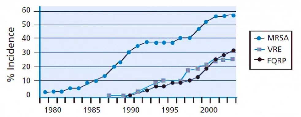
AMR arises when the micro-organisms which cause infection (e.g. bacteria) survive exposure to a medicine that would normally kill them or stop their growth. This allows those strains that are capable of surviving exposure to a particular drug to grow and spread, due to a lack of competition from other strains. This has led to the emergence of ‘superbugs’ such as Methicillin‑resistant Staphylococcus aureus (MRSA) and extremely drug‑resistant tuberculosis, bacteria which aredifficult or impossible to treat with existing medicines. Fig.2 shows the trend of antibiotic-resistance for three common bacteria.
Resistance to antimicrobials is a natural process that has been observed since the first antibiotics were discovered and, indeed, the genes that confer drug resistance upon some strains of bacteria pre-date antibiotics by millions of years.
Fig. 2 The graph shows the rapid increase of the resistance of three species of bacteria:
Staphylococcus aureus resistant to Methicillin (MRSA), Vancomycin-resistant enterococci
(VRE), and Pseudomonas aeruginosa resistant to Fluoroquinolones (FQRP). - SOURCE: Center for Disease Control and Prevention. (IMAGE ADAPTED FROM5)
However, AMR has increasingly become a problem in recent times because overuse of antimicrobials has increased the rate at which resistance is developing and spreading, but we lack new drugs to challenge these new superbugs. This results in us facing a growing enemy with a largely depleted armory.
In the past, resistant infections were predominantly associated with hospitals and care settings, but over the last decade resistant infections have been seen in the wider community too. With resistance on the rise, we stand to lose the immense ground we have gained in the last century. This includes: 1) the fight against life threatening infectious diseases such as pneumonia, TB, HIV and malaria; 2) the battle against conditions such as cancer; and 3) huge advances in surgical procedures like organ transplants and caesarean sections, which have now become routine and relatively low risk, thanks to our ability to effectively stave off or treat acute infections with antibiotics. It was estimated that in total about 700,000 people die every year from drug resistant strains of common bacterial infections, HIV, TB and malaria.5 This
number is likely to be an underestimate due to poor reporting and surveillance.
Based on scenarios of rising drug resistance for six pathogens to 2050, it was estimated that unless action is taken, the burden of deaths from AMR could balloon to 10 million lives each year by 2050, at a cumulative cost to global economic output of 100 trillion USD. On this basis, by 2050, the death
toll could be a staggering one person every three seconds and each person in the world today will be more than 10,000 USD worse off.
Fig. 3 Causes and number of deaths estimated to 2050. (IMAGE ADAPTED FROM5) It is impossible to predict the path of emerging drug resistance, but it is a trend that has largely run only in one direction so far. We can be certain that, in the absence of interventions to slow the emergence of resistance and increase the supply of new antibiotics, the impacts will be felt not just in isolated areas but at a far more fundamental level, across our societies and healthcare systems.
As the antibiotics available to us become less effective, so the risks of many treatments which rely upon antibiotics becomes higher. This will progressively undermine the viability of interventions that many may not directly associate with antibiotics. Cancer chemotherapy or organ transplantation are just two examples of medical treatments that leave the patient highly vulnerable to bacterial infections. Most invasive surgery (particularly ‘dirty’ procedures, such as those involving the gut) is today routinely and dependably ‘de-risked’ by effective antibiotic prophylaxis and by the availability of reliable therapy for infections that do occur despite best practices. Intubated patients in intensive care facilities already experience very high rates of infection, including drug-resistant ones, as a result of the ventilation that they receive and the mortality risk associated with this will
rise further if treatment options for such infections deplete. These secondary impacts are difficult to quantify but they threaten to dramatically change healthcare as we know it today.6
1.3. Nanomedicine
In the early 1900s Paul Ehrlich thought to an ideal therapy a “drug precisely targeted to an invader, which if linked to a toxic chemical would act like a missile, carrying a destructive payload directly to the disease” but only in 1959 Richard Feynman invited all the scientific community to arrange the atoms as we want, looking beyond what our eyes can see. So only when we became able to manipulate atoms, the era of Nanotechnology was born. Nanometric scale materials showed different electrical, optical and magnetic properties respect to the bulk, giving the possibility to find new applications for these new materials.
Application of nanotechnology to medical science has been emerging as a new field of interdisciplinary research among medicine, biology, toxicology, pharmacology, chemistry, material science, engineering, and mathematics, and is expected to bring a major breakthrough to address several unsolved medical issues. Nanomedicine, namely the use of nanotechnology for medicine, is starting to make an impact in areas like disease imaging and diagnosis, drug delivery and as reporters of therapeutic efficacy and of disease pathogenesis. Many multifunctional nanostructures, capable of performing one or more of the above duties, are now in various stages of preclinical and clinical development.7,8,9,10 Nanomedicine, represents an
alternative approach11 to oppose the development of resistance to antibiotics
and anticancer drugs. Various systems able to assemble in nanosized structures, as biopolymers, macrocycles, liposomes, et al., are used to vehicle various molecules in cellular environment by exploiting supramolecular interactions for their transport and release. The entrapment of activatable agents in nanocarriers in fact can ensure their (i) solubilization in aqueous medium, (ii) protection from premature degradation, (iii) transport in the blood stream where dilution and interfering biomolecules are present, (iv) delivery to pathological cells by enhanced permeability and retention (EPR) effect and/or homing ligands exposed on the vehicle surface, (v) enhanced biocompatibility and bioavailability, (vi) increased local concentration, and (vii) sustained release. The selectivity and specificity in disease treatment can be enhanced by using externally activatable agents to produce localized
cytotoxicity with minimized collateral damages. The ability to control the drug dosage in terms of quantity, location and time is a key goal for drug delivery science, allowing to maximize therapeutic effects. Systems responsive to stimuli such as temperature, pH, electric or magnetic applied field, ultrasound, light, or enzymatic action have been proposed as triggered delivery systems.12
1.4. Light stimulated therapy
In virtue of its simple manipulation in terms of wavelength, intensity and duration, low cost, low environmental impact, fast rate of photochemical processes, light represents the ideal external stimulus for the introduction of therapeutic agents in a desired bio-environment in a non-invasive way, mimicking an “optical microsyringe” with high spatio-temporal resolution. Moreover, it has the ability to not affect the values of physiological parameters such as temperature, pH and ionic strength, which is a prerequisite for biomedical application. For all these reasons activation of an appropriate precursor able to produce cytotoxic species under light stimulation represents an appealing strategy in biomedical field.
1.4.1. Photodynamic Therapy (PDT)
Photodynamic Therapy (PDT)13 is a minimally invasive therapeutic
approach in the treatment of cancer and infection diseases that involves the combination of visible light and a photosensitizer (PS). Each factor of this therapy (photosensitizer, light) is harmless by itself, but when combined with oxygen, can produce cytotoxic agents that can destroy cancer cells and bacteria.
PDT is based on the administration by intravenous, intraperitoneal or topical route, of a photosensitizing agent (PS) generally belonging to the class of porphyrins and phthalocyanines.14 These species localize preferentially in
diseased tissues, compared with normal cells, as only those cells that are simultaneously exposed to the photosensitizer, light and oxygen are exposed to the cytotoxic effect allowing to leave normal tissue intact.
After irradiation with opportune dose of appropriate wavelength light, the PS is excited and two different mechanisms can take place, Type I and II as reported in Fig. 4. In the Type II, which is the mechanism that takes place in PDT, the return to the ground state, starting from the lowest excited triplet
state, occurs mainly by energy transfer to molecular oxygen generating highly reactive 1O
2, which induces damage and cell death.
This process competes with the radiative (fluorescence and phosphorescence) and not radiative (internal conversion) deactivation processes of the PS.
Fig. 4 Type I and Type II reactions in PDT (photodynamic therapy). Schematic Jablonski’s
diagram showing PDT’s mechanism of action. Following light absorption, the PS reaches an excited singlet state. After an intersystem crossing, the PS, now in a triplet excited state, can react in two ways. It can react with biomolecules through a hydrogen atom (electron) transfer to form radicals, which react with molecular oxygen to generate ROS (type I reaction); or, it can react directly with oxygen through energy transfer, generating singlet oxygen (Type II reaction).
(IMAGE ADAPTED FROM15)
1O
2 is one of the major cytotoxic agents produced by light excitation for
treatment of many infectious and oncological diseases. This species is able to attack biological substrates of different nature like the plasma membrane, the mitochondria and the cell nucleus, and does not suffer Multidrug Resistance problems, typical for many anti-cancer and antibacterial drugs. Further, due to its short half-life in blood (µs), lack of charge and small sizes, 1O
2 diffuses
in cellular environment over short distances (up to 150 nm), confining its region of action to the irradiated area and minimizing systemic side effects.16
1.4.2. Nitric oxide based therapy
NO, a small free radical, is involved in many physiological and pathological17 processes such as stimulation of the immune system, smooth
muscle relaxation18, inhibition of platelet aggregation19, inflammatory
disorders20, iron metabolism, neurotransmission and neurotoxicity21 and
dysfunctions has generated numerous debates in recent years and NO has been labelled as the causative agent of several pathophysiological mechanisms. However, it seems that it also protects against many chemical species, such as those generated by oxidation, and seems to be related to the tumoricidal and antibacterial activity of the immune system. The multifaceted role of NO is complex and its effects in many biological processes and in cancer biology23 has attracted considerable interest. Several studies have
associated NO production by immune cells, endothelial cells and others able to release NO to various aspects of anti-cancer therapy24, such as the
inhibition of cancer cells proliferation, angiogenesis and metastasis. Difficulties in the delivery of gaseous NO to selected targets have inspired the development of a range of NO releasing molecular and macromolecular scaffolds25 as potential therapeutics that exploit NO’s multifaceted biological
roles, with the ambitious prospect to tackle important pathologies, including, of course, oncological and bacterial diseases. The biologic effects of NO are, however, strictly dictated by its concentration, delivery site and dosage, creating a complex role for the molecule in opposing beneficial and deleterious events.26
NO, as well as 1O
2, shows more or less the same properties respect to
conventional drugs, but has a half-life in tissues of ca. 5 s and diffuses in the range of 40-200 µm, allowing to act on a wider range of action.
Furthermore, NO photorelease is independent from O2 availability it can
potentially very well complement the 1O
2 effects at the onset of hypoxic
conditions typical of some tumors and infections by anaerobic bacteria. The possibility to obtain photo-activatable systems that are capable of releasing NO, conjugating the cytotoxic activity of the latter with the advantages offered by light stimulation, opens new perspectives for applications in bio-medical field.
1.5. Calix[n]arenes (CAs)
Calix[n]arenes (CAs) (Fig. 5) have established their own status as the third generation of macrocyclic molecules. They are listed as one of the three major class of macrocyclic molecules together with cyclodextrins and crown ethers.
Typically, CAs are produced by chemical condensation between para-substituted phenols and formaldehyde. The phenolic units are linked
by methylene bridges in orto-position to the OH groups. Because CAs with even numbers (n = 4, 6, 8) can be synthesized by one-step reaction and easily purified, they have been widely investigated. The distinctive feature of CAs is their cavity dimension according to the number of incorporated phenolic units.27 Structurally, they contain two well defined rims (lower and upper rim)
and an hydrophobic core (central annulus). Due to the presence of a hydrophobic cavity able to encapsulate many types of guests (small organic molecules, ions, sugars, etc), CAs are interesting host molecules in supramolecular chemistry.28 The inclusion complexation is driven by various
forces such as hydrophobic effect, ion-dipole interaction, and hydrogen-bonding interaction.
In the last decade, the favourable characteristics of CAs have been explored for diverse biomedical and pharmaceutical applications29 as well as
molecular recognition, sensing and self-assembly, catalysis and nanotechnology.30 CAs have shown to have a great potential as drugs or drug
delivery systems.31 CA derivatives having activities such as viral,
anti-bacterial, enzyme activators/blocker, anti-coagulant, anti-cancer32 etc. have
been described. A few studies concern the application of CAs in anticancer PDT. A PEGylated bodipycalix(4)arene was proposed as a near infrared amphiphilic photosensitizer33 and p-sulfonato-calix[6,8]arenes were found
possess antitumor activity in a K562 myelogenous leukemia cell line irradiated with mercury light at 430 nm.34 The photodynamic activity of a hydrophilic
PEGylated calix[4]arene and a calix[4]arene‒porphyrin forming supramolecular assemblies by host–guest interactions with chlorine6,35 and
biviologen derivatives,36 respectively, was investigated on tumor cells in vitro
experiments, but no photochemical and photophysical characterization of the nanostructured systems was carried out. To the best of our knowledge,
calixarene‒NO photodonor nanoassembly has never been investigated, some papers focused on calix[4]arene derivatives for storage of NO in the form of entrapped nitrosonium ions.37 However, no reports on photoactivatable
antibacterial systems based on calixarenes are known to date.
1.6. Polymersomes
Polymersomes (Fig. 6) are self-assembled polymer shells composed of block copolymer amphiphiles. The polymers used to make polymersomes are similar to lipids in that they are amphiphiles: at least one fraction or “block” of each molecule is hydrophilic, whereas the other fraction is hydrophobic. Either type of amphiphile, if made with suitable amphiphilic proportions, can self-assemble into vesicles when hydrated. The hydrophobic blocks of each molecule tend to associate with each other to minimize direct exposure to water, whereas the more
hydrophilic blocks face inner and outer hydrating solutions and thereby delimit the two interfaces of a typical bilayer membrane. 1The
structure of the macromolecule is composed of three distinct regions (Fig. 6):
- Hydrophilic shell - Hydrophobic bilayer - Aqueous core.
This structure permits to load hydrophobic, hydrophilic or amphiphilic guests making these nanoconstructs an attractive supramolecular host to vehicle in contemporary different drugs and/or bio-active molecules with the aim to exploit additive and/or synergistic effects or multimodal therapeutic approach.38,39
Polymerosomes can be prepared from different polymeric building blocks from whose nature will depend the physical-chemical and biological properties of the resulting polymersomes, like dimensions,40 encapsulation
efficiency, pharmacokinetics including release rates,41 degradability and
cellular uptake.42 Therefore, unlike the liposomes, polymersomes show a
highest chemical versatility and tend to have a much ticker outer membrane owing to the higher molecular weight of the co-polymers used. The larger
Fig. 6 Schematic representation of a polymersome structure.
thickness of the membrane results in a greater stability and mechanical strength (Fig. 7).
Fig. 7 Schematic representation of the advantages of polymersome respect to liposome.
Martin et al. have developed a charge neutral polymersome, using an amphiphilic PEGylated random copolymer, with an encapsulation efficiency of Fitc-CM-Dextran (Fluorescein isothiocyanate–Carboxymethyl–Dextran) of 70%, and the uptake of this guest by Hela cells was increased four-fold when encapsulated in the polymersomal system.43 In literature, is well known that
the use of PEG in nanoparticle preparations permits to enhance the circulation time in blood stream, reduce the aggregation in serum and prevents the elimination from reticuloendothelial system due to its immune-protection capabilities.44,45,46
However, it is also known that the PEGylation reduces cellular transfection and uptake due to interference with intracellular trafficking. A strategy to overcome these problems is the contemporary incorporation of PEG and lipid molecules such us cholesterol, since the latter is an essential structural component of cell membranes. Micelles and liposome formulated with cholesterol have shown increased cellular internalization, enhanced colloidal stability, higher encapsulation volume and slower release of payload compared to the ones without cholesterol.47 For all these reasons FDA have
only approved PEGylated liposomal drug delivery systems which incorporate cholesterol as a key component.48 At this regard, recently Martin et al. have
demonstrated that a polymersome containing an appropriate percentage of cholesteryl polymer showed a ten-fold increase in cellular uptake of Fitc-CM-Dextran compared to un-encapsulated drug.38
Part I
Calixarene-based
2. Calixarene-based
photoresponsive
nanosystems
entrapping different guests by non-covalent approach
for antibacterial applications
2.1. Introduction
The good biocompatibility and low immunogenicity exhibited by water soluble calixarene derivatives, has paved the way for a variety of applications of these molecules in pharmaceutical and biomedical fields as drugs and/or drug delivery systems. With this in mind, we planned to synthesize the amphiphilic calix[4]arene scaffold 1 able to spontaneously self-assemblies in nano-sized aggregates, and to investigate the capability of the nanoconstruct to be a versatile supramolecular host and act as a nanocarrier of different types of photostimulable precursors.
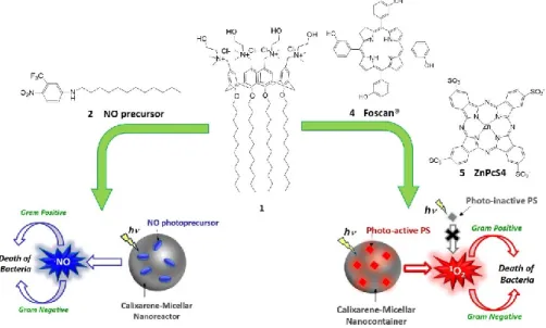
In the first part of this study, the hydrophobic NO precursor 2 and two porphyrinoid PSs 4 and 5 were entrapped separately in the nanoaggregates of 1. The calixarene-photostimulable supramolecular nanoassemblies represent novel potential antibacterial agents that under exclusive light-stimulation can produce cytotoxic species as singlet oxygen and nitric oxide radical useful in photodynamic therapy and NO-photostimulated therapy (Fig. 8).
2.2. Results and discussion
2.2.1. Synthesis of compound 1
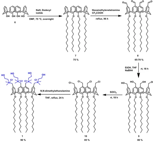
The amphiphilic calix[4]arene 1 was synthetized by following the procedure reported in Fig. 9. In particular, commercial p-H-Calix[4]arene 6 was alkylated at the lower rim and chloromethylated at the upper rim in order to obtain derivative 10, by passing through the formyl (8) and the alcohol (9) derivatives according the procedure reported by Eggers et al.49 The treatment
of compound 10 with N,N-dimethylethanolamine in THF as solvent gave compound 1.50 All the calix[4]arene derivatives were characterized by 1
H-NMR which agreed with the data reported in the literature.
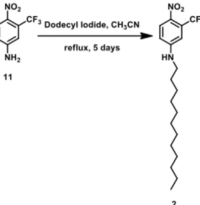
2.2.2. Synthesis of compound 2
The hydrophobic NO precursor 2 was synthetized starting from the commercial 4-Nitro-3-(trifluoromethyl)aniline treated with dodecyl iodide in CH3CN (Fig.10). Compound 2 was characterized by 1H-NMR and 13C-NMR.
Fig. 10 Schematic representation of the synthesis of compound 2.
2.2.3. Characteristics of the components
2.2.3.1.
Compound 1 – Calix[4]arene derivative
Calix[4]arene 1 (Fig. 11) is characterized by the presence of four polar choline groups at the upper rim which confer solubility in aqueous environment and could favour cell membrane penetration by electrostatic interactions; and dodecyl chains at the lower rim which ensure an amphiphilic behaviour. Due to its structure this molecule has the capability to self-assemble in nano-sized systems and serve as supramolecular host for different guests.
2.2.3.2.
Compound 2 – NO photoprecursor
Compound 2 (Fig. 12A), a 4-nitro-3-(trifluoromethyl)aniline derivative, presents a dodecyl alkyl chain at the amino group useful to encourage the assembly with calix[4]arene 1 by hydrophobic interactions, in addition to the pie-pie interactions between the aromatic rings of the two partners.
Compound 2 as well as other derivatives is an eligible NO precursor because it is able to release NO under exclusive light stimulation.51 The NO
release mechanism (Fig. 12B) is based on a nitro-nitrite rearrangement with consequent formation of a phenoxy radical intermediate and a phenol derivative as a stable reaction product. The reason resides in the no-coplanarity of the nitro group respect to the aromatic ring due to the presence of the -CF3 substituent which brings an orbital overlap between the oxygen of the nitro group and one of the carbons of the aromatic ring.
Fig. 12 (A) Structure of compound 2. (B) Schematic representation of the NO-release
mechanism.
B
A
2.2.3.3.
Compound 4 – Foscan®
Foscan® (Fig. 13) is the brand name with which has been labeled in the European Union the Temoporfirin (INN), which is the common name of 3’,3’’,3’’’(2,3-dihydroporphyrin-5,10,15,20-tetrayl)tetraphenol or more simply 5,10,15,20-Tetra(m-hydroxyphenyl)chlorin (m-THPC) a photosensitizer (based on chlorin) used in PDT for the treatment of squamous cell carcinoma of the head and neck. The EU approved its use in pharmacology in June 2001. Due to the poor solubility m-THPC can only be used with a formulation process prior to administration. Initial applications used formulations in PEG and ethanol or propylene glycol. Considerable effort has been spent on the preparation of nanocarrier formulations for m-THPC in fact in addition to the standard formulation (Foscan®) were prepared no-PEGylated and no-PEGylated liposomal formulations, Foslip® and Fospeg®.
Fig. 13 Structure of compound 4.
2.2.3.4.
Compound 5 – ZnPcS4
Zinc phtalocyanine tetrasulfonate (Fig. 14) is a water soluble PS and allows its intravenous administration without the need for alternative delivery vehicles. The Zinc as the central metal ion confers short triplet lifetime, high triplet quantum yields and high singlet oxygen quantum yield. All these characteristics which make ZnPcS4 a suitable PS are precluded by aggregation phenomena in aqueous environment.
2.2.4. Characterization
of
the
calix[4]arene
1-based
nanoassemblies
Compound 1 due to its amphiphilic nature self-aggregates in 10 mM PBS solution, pH =7.4, at concentrations above ca. 8 μM, as demonstrated by the calculation of cac by pyrene method. The measurements were performed on aqueous solutions of 1 (from 0.5 × 10⁻⁶ M to 100 × 10-6 M) containing pyrene
(0.3 μM) (Fig. 15A). Dynamic Light Scattering (DLS) measurements indicated an average hydrodynamic diameter of ca. 42 nm with a polydispersity index (PI) < 0.2 and a zeta potential ζ = +24.7 mV. TEM (Transmission Electron Microscopy) image confirmed approximately the dimensions acquired by DLS measurements and suggested that the calixarenes are aggregated in a micellar-like nanoassembly (Fig. 15B).
Fig. 15 (A) Intensity ratio I3/I1 of pyrene (0.3 μM) vs. the concentration of 1 in aqueous solution.
The arrow indicates the critical aggregation concentration (cac). (B) Intensity weighted hydrodynamic diameter distribution of the micellar aggregates of 1 (300 μM) obtained by DLS. The inset shows a representative Transmission Electron Microscopy image of 1.
300 400 500 0,0 0,2 0,4 0,6 0,8 1,0 A (nm) 1 2 3
2.2.5. Nanoassembly 1·2: spectroscopic and dimensional
characterization
The hydrophobic structure of compound 2 makes this molecule totally insoluble in aqueous environment. We used the assembled compound 1 as a suitable nanocarrier to introduce compound 2 in PBS aqueous solution obtaining a light yellow colloidal dispersion.
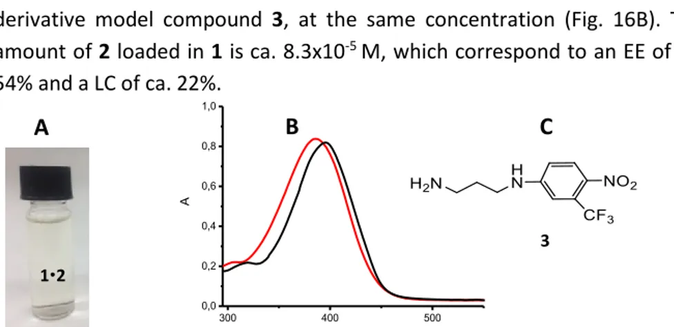
In order to verify the efficient encapsulation of 2 in 1 we characterized the system by Uv-vis spectroscopy. The absorption spectrum of 2 entrapped in the calixarene aggregate shows the typical profile centered at 396 nm ca. which is approximately the same observed for the water soluble nitro-aniline derivative model compound 3, at the same concentration (Fig. 16B). The amount of 2 loaded in 1 is ca. 8.3x10⁻⁵ M, which correspond to an EE of ca.
54% and a LC of ca. 22%.
3
Fig. 16 (A) Picture of the 12 colloidal dispersion. (B) Absorption spectrum of 2 (83 µM) in
phosphate buffer (pH 7.4, 10 mM) in the presence of 1 (300 µM) (black line) compared to absorption spectrum of the water soluble model compound 3 in PBS at the same concentration of 2. (C) Structure of the model compound 3.
DLS measurements (Fig. 17) have shown that the dimensions of the nanoassembly 1·2 are approximately the same of the empty NPs, distribution centered around 46 nm with a PDI = 0,24 and ζ = +23.7 mV, suggesting that the arrangement of the calixarene
aggregates is not disrupted by the NO precursor loading.
DLS measurements have been repeated after 30 days and the data are about equal to those previously obtained, demonstrating the long-time stability of the system in PBS solution.
Fig. 17 Size distribution of the nanoassembly 1·2.
12
B
A
C
40 45 50 55 60 0 5 10 15 Freq ue nc y (%) diameter (nm)0 200 400 600 800 0,0 0,2 0,4 0,6 0,8 1,0 OFF OFF ON ON OFF ON OFF OFF ON OFF ON [NO] M time (sec)
2.2.6. NO photorelease from nanoassembly 1·2
One of the most appropriate experimental methodologies to test NO photorelease capability of a system is to measure the concentration of NO species by an ultrasensitive electrode which directly detects NO with nM concentration. Irradiating with a 405 nm laser was monitored NO release by the samples, subjecting the photoactive component at regular intervals of light and darkness. The results illustrated in Fig. 18A provide evidence that the solution 1·2 and a PBS aqueous solution of 3 are stable in the dark and supply NO exclusively upon illumination with visible light. By comparison of the two NO release profiles is clear that the efficiency of NO photogeneration in the case of 1·2 is ca. 15 times higher than solution of 3. It is possible to assert that the reason of this behaviour is due to the presence of calixarene scaffold which behaves as a nanoreactor probably producing a favourable low polarity environment and facilitating the extraction of close hydrogens to the phenoxy-radical intermediate involved in the NO photogeneration as
previously described in section 4.3.2.
NO release issue of the solutions was further monitored by a photobleaching (Fig. 18B). Solution 1·2 was irradiated with a laser at 405 nm at regular intervals of 5 minutes. The absorbance profiles obtained show a clear reduction of the main signal relative to the formation of the phenol derivative as the only product obtained. This shows that there are not collateral or additional reactions which compete with the NO photorelease.
Fig. 18 (A) NO release profile observed for the nanoconstruct 1·2 (blue line, [1] = 300 µM; [2] =
83 µM) and an optically matched solution of the model compound 3 (black line) in water medium. exc = 405 nm. (B) Absorption spectral changes observed upon 405 nm light irradiation of an aqueous solution of the nanoconstruct 1·2 ([1] = 300 µM; [2] = 83 µM) for regular irradiation intervals of 5 min.
A
B
300 400 500 600 0,2 0,4 0,6 0,8 A (nm) 0 min2.2.7. Antibacterial activity of nanoassembly 1·2
Antibacterial activity tests were performed at the SIFI S.p.A. by Dr. Blanco and Dr. Picciotto. Two different bacterial strains were chosen, Staphylococcus aureus ATCC 6538 and Pseudomonas aeruginosa ATCC 9027, as Gram positive and Gram negative bacteria, respectively. These two species have acquired a high antibiotic resistance towards traditional antibiotics and, in some forms, are responsible for serious nosocomial infections. The bacterial cultures were kept in the dark or irradiated at different times with a 470 W Xenon lamp equipped with a cut-off filter at 380 nm. In Fig. 19 are reported the time-inhibition curves obtained testing the antibacterial activity of 1 and 1·2 on both bacterial strains. It is evident that calixarene 1 did not have any effect, while the nanoassembly 1·2 shows an evident action dependent on the irradiation time. In fact, the colloidal dispersion containing compound 2 is able to induce an inhibition of the growth of approximately 100% for both bacteria after 30 minutes of irradiation. Control experiments carried out with normal human skin fibroblast cells showed a negligible (<15%) antiproliferative effect of 1·2 with concentrations identical to those in Fig. 19 and up to 30 min irradiation.
Fig. 19 Comparison of the time-inhibition curves of Staphylococcus aureus and Pseudomonas
2.2.8. Nanoassembly 1·4: spectroscopic and dimensional
characterization
In Fig. 20B is reported the absorption spectrum of compound 4 in methanol solution which shows the typical porphyrin profile characterized by an intense peak centered at 416 nm (Soret band or B-band) to which followers of lesser intensity at higher wavelengths (Q-Band signals). Compound 4 which is totally insoluble in phosphate-buffered aqueous solution becomes fairly soluble in the presence of aqueous dispersion of 1 giving a colloidal solution as confirmed by the typical spectrum profile shown in Fig. 20B. The amount of 4 loaded was 3 µM, corresponding to an EE of 100% ca.
Fig. 20 (A) Picture of the colloidal solution 1·4 (B) Absorption spectra of 4 (3 µM) in methanol
(dashed line) and in phosphate buffer (pH 7.4, 10 mM) in the presence of 1 (300 µM) (red line).
The efficient encapsulation is confirmed by the emission spectra acquired exciting at 590 nm that corresponds to third Q-Band of Foscan. As shown in Fig. 21, the emission profile of 4 in 1 shows a similar trend to that observed in methanol confirming the effective encapsulation of 4 within the calixarene assembly mainly under its monomeric form with a fluorescence quantum yield of ɸf = 0.053.
Fig. 21 Fluorescence emission spectra of 4 (3 µM) in methanol (dashed line) and in phosphate
buffer (pH 7.4, 10 mM) in the presence of 1 (300 µM) (red line). exc = 590 nm.
14
The dimensions of the system 1·4 obtained by DLS measurements (Fig. 22) gave a distribution centered around 90 nm with a PDI = 0.28 and a substantially unvaried value of the zeta-potential respect to the empty calixarene nanoparticles. The measurements performed again after 20 and 77 days from preparation, gave approximately the same values showing the long-time stability of the system.
Fig. 22 Size distributions of the nanoassembly 1·4.
2.2.9. Nanoassembly 1·5: spectroscopic and dimensional
characterization
As shown in Fig. 23B the hydrophilic phthalocyanine PS 5 is soluble in PBS medium (10 mM pH=7.4) but in aggregated form. The aggregate shows an absorption spectrum centered at about 602 nm, while the profile in DMSO is characterized by an intense peak centered at 680 nm, relative to the monomeric species, preceded by a lower intensity peak relative to the aggregate one, as reported in literature.52 Absorption spectrum obtained for
5 loaded in the calixarene nanoassembly 1 shows that the phtalocyanine is present almost totally in its monomeric form.
Fig. 23 (A) Picture of the colloidal solution 1·5. (B)Absorption spectra of 5 (15 µM) in phosphate
buffer (pH 7.4, 10 mM) in the absence (blue) and in the presence (red) of 1 (300 µM). Dashed line is absorption spectra of 4 in DMSO, where it is present as monomeric species.
15
As shown in Fig. 24, the fluorescence emission spectra of 5 in PBS solution is totally quenched due to the aggregation phenomena, while emission profile of 5 in 1, though is lower in intensity, shows a similar trend to that observed in DMSO confirming the effective encapsulation of 5 within 1, mainly under its monomeric form with a fluorescence quantum yield of ɸf = 0.031.
Fig. 24 Fluorescence emission spectra of 5 (15 µM) in phosphate buffer (pH 7.4, 10 mM) in the
absence (blue) and in the presence (red) of 1 (300 µM). Dashed line is the spectrum of 5 in DMSO. exc = 540 nm.
Dynamic Light Scattering measurements (Fig. 25) gave hydrodynamic diameters of ca. 100 nm for the system 1·5 with a PDI = 0.3. The size increase compared to the empty nanoparticles could be ascribed to a rearrangement of the calixarene assembly, and the presence of surface electrostatic interactions between the positive charges of the choline moieties and the negative charge of the phtalocyanine. This explanation agrees with a slight decrease of the zeta potential to a value ζ= + 20.8 mV. The measurements performed again after 20 and 77 days gave approximately the same values showing the long time stability of the system.
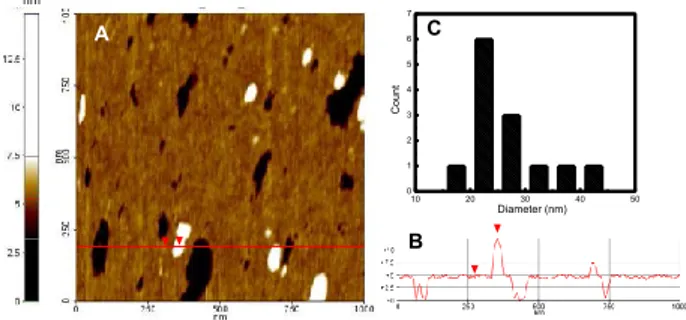
Thanks to a collaboration with Dr. Conoci and Dr. Petralia of ST Microelectronics-Catania the supramolecular nanoassembly was also investigated by Atomic Force Microscopy (AFM). In Fig. 26 is reported the two-dimension topography (A) obtained by compound 1·5 and its height profile (B). AFM measurements show a smaller dimension for the aggregate 1·5 respect to DLS measurements probably due to the different type of environment in which the measurements are recorded.53 In Fig. 26 C is
reported the distribution which is centered about 25 nm.
Fig. 26 A) Contact mode AFM of the supramolecular nanoassembly 1·5 deposited on cleaned
silicon surface. B) Height line profile of line shown in A. C) Diameter distribution of the nanoassembly 1·5.
2.2.10.
1O
2
photogeneration from 1·4 and 1·5 nanoassemblies
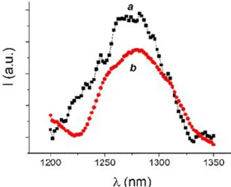
The generation of singlet oxygen, 1O
2, was detected by time resolving
near infrared luminescence with sub-microsecond time resolution. We have examined the singlet oxygen produced by energy transfer from 4 and 5 to molecular O2, in totally deuterated buffer solution of
calix[4]arene 1. The presence of the calixarene 1 permits to ensure the photochemical behaviour of the two PSs, otherwise precluded by low solubility and aggregation phenomena, in aqueous environment. Fig. 27 shows the typical phosphorescence signal at 1270 nm54 observed for all
samples investigated. The quantum yield for 1O
2 photogeneration, ,
were 0.89 and 0.57 respectively. These values are significantly higher than those observed for 4 in methanol ( = 0.43)55 and the 5 in CTAC
micelles ( = 0.31)56 and in DMSO ( = 0.46).57
nm A B 10 20 30 40 50 0 1 2 3 4 5 6 7 Cou nt Diameter (nm) C
Fig. 27 1O2 luminescence detected upon excitation of the nanoassembly 1·4 at exc = 405 nm
(a) and the nanoassembly 1·5 at exc = 680 nm (b) in deuterated phosphate buffer (pH 7.4, 10
mM). The 1O
2 luminescence spectrum observed for 4 in methanol is also shown, for sake of
clarity (c).
2.2.11. Antibacterial activity of the nanoassemblies 1·4 and 1·5
Bactericidal action of 1·4 and 1·5 systems were performed by SIFI S.p.A. always on Staphylococcus aureus ATCC 6538 and Pseudomonas aeruginosa ATCC 9027, at the same conditions reported in the section relative to the antibacterial activity of 1·2. The supramolecular nanoassemblies 1·4 and 1·5 did not show any significant action in the dark, but they exhibited a meaningful antibacterial activity under irradiation. No antibacterial activity was instead observed for the calix[4]arene nanoassembly alone (Fig. 28). The time-kill curves depicted in Fig. 28A, show that nanoassembly 1·5 has a faster antibacterial activity than 1·4 with total bactericidal effect in only 10 minutes of irradiation on both the bacteria strains. The reason of a greater slowness of 1·4 is could be due to a smaller fraction of absorbed light by the PS owing to the chemical filter used during the test which has a little absorption region in correspondence to that of the porphyrin 4. In Fig. 28B and C we report two pictures relative to the plates containing Staphylococcus aureus ATCC 6538 and Pseudomonas aeruginosa ATCC 9027, respectively, treated with calixarene 1 alone and nanosystem 1·4 at different time of irradiation. It is evident that after five minutes, the system 1·4 shows obvious bactericidal effect on both bacteria, even if, as expected, Pseudomonas shows a greater resistance.Fig. 28 (A) Time-kill curvesofthe nanoassembly 1·4 (squares), 1·5 (circles) and, for comparison,
1 alone (triangles) observed for Staphylococcus aureus ATCC 6538 (open symbols) and
Pseudomonas aeruginosa ATCC 9027 (filled symbols). Representative images for the antibacterial effect observed on Staphylococcus aureus ATCC 6538 (B) and Pseudomonas aeruginosa ATCC 9027 (C) at different irradiation times of (a) control, (b) 1 and (c) 1·5. [1] = 300 µM, [4] = 3 µM, [5] = 15 µM.
2.3. Experimental section
2.3.1. Materials
All reagents were of high commercial grade and were used without further purification. All solvents used were analytical grade. The PS 4 and 5 were purchased from Frontier Scientific Inc. (FSI) and Porphyrin Pdts, respectively and were used as received. Staphylococcus aureus (ATCC 6538) and Pseudomonas aeruginosa (ATCC 9027) were purchased from LGC Standards. Human skin fibroblast cells were obtained from biopsy after the informed consent of the subjects, according to the laws of the Italian Government and the Declaration of Helsinki Principles.
0 5 10 15 20 25 30 0 1 2 3 4 5 6 Log 10 CFU m L -1
Irradiation time (min)
t5 t10 t20 t0 B C a b c a b c A
2.3.2. Synthesis
25,26,27,28-Tetradodecyloxycalix[4]arene (7)
p-H-Calix[4]arene (6) (5.72 g, 13.5 mmol) and 5.4 g NaH (60% dispersion in oil, 135 mmol) were stirred in 140 mL of dry DMF for 15 min under argon. Dodecyl iodide (33.2 mL, 135 mmol) was then added and the mixture was stirred at 75 °C overnight. DMF was removed under vacuum and 0.5 N HCl (200 mL) was added. After stirring overnight, the solid was recovered by filtration and washed several times with MeOH and small amount of acetone. This operation can be repeated to reach the desired purity of compound 7 (75 %).
5,11,17,23-tetra-formyl-25,26,27,28-tetra-dodecyloxy-calix[4]arene (8) Under an inert gas atmosphere a mixture of compound 7 (3.3 g, 3 mmol) and hexamethylenetetramine (16.2 g, 120 mmol) in CF3COOH (100 mL) was
stirred for 96 h under reflux. The mixture was cooled to room temperature and then poured into a stirring solution of 2 M HCl (200 mL) and CH2Cl2 (200
mL), and vigorously stirred for 1 h. The mixture was extracted with CH2Cl2 (2
x 100 mL) and the combined organic layers were washed with saturated aqueous Na2CO3 (2 x 100 mL) and brine (2 x 100 mL), dried over sodium sulfate
and the solvent was then removed under reduced pressure. The raw product was further purified by column chromatography (silica gel 60; hexane: ethyl acetate: CH2Cl2, 7:3:2) to give a white solid (2.73 g, 65-70%).
5,11,17,23-tetra-hydroxymethyl-25,26,27,28-tetra-dodecyloxy-calix[4]arene (9) Under an inert gas atmosphere ethanol (50 mL) was added to a stirring solution of 8 (2.73 g, 2 mmol) in THF (10 mL). NaBH4 (2.6 g, 70
mmol) was then added and the mixture stirred for 18 h at room temperature. The mixture was then concentrated under vacuum and the resulting solid dissolved in CH2Cl2 (100 mL). 2 M HCl (100 mL) was added slowly and the
solution was stirred for 1 h. The reaction mixture was then extracted with CH2Cl2. The combined organic fractions were washed with 2 M HCl (3 x 100
mL) and dried over Na2SO4. The solvent was removed in vacuo to obtain a
white solid (1.5 g, 80%).
5,11,17,23-tetra-methylchloride-25,26,27,28-tetra-dodecyloxy-calix[4]arene (10) Under an inert gas atmosphere thionylchloride (2 mL) was added to calix[4]arene derivative 9 (0.5 g, 0.4 mmol) and then stirred at room
temperature for 18 h. The mixture was concentrated under vacuum and the resulting solid was dissolved in CH2Cl2 (50 mL), washed with saturated
aqueous Na2CO3 (3 x 50 mL) and dried over Na2SO4. The solvent was removed
under reduced pressure to obtain an off-white solid (0.436 g, 85%).
5,11,17,23-Tetra(N,N-dimethyl-N-hydroxyethylammonium)-methylene-25,26,27,28-tetradodecyloxycalix[4]arene tetrachloride (1)
A solution of N,N-dimethylethanolamine (1.33 g, 1.5 mL, 14.9 mmol) in THF (15 mL) was added to a stirring solution of 10 (4.1 g, 3.2 mmol) in THF (60 mL). The reaction mixture was refluxed for 24 h. After cooling, the suspension was centrifuged at 4000 rpm, 5 min. The precipitate was washed with THF (40 mL) and with acetonitrile (4 x 20 mL) by repeated centrifugation (4000 rpm, 5 min) and removal of the solvent. The solid precipitate was recovered and dried under vacuum to give a white compound (4.7 g, yield: 90%). 1H-NMR (MeOD): δ 0,91 (t, 12 H, J = 6.8 Hz, 4 x CH3), 1,31 (br m, 64 H, 32 x CH2), 1,47
(br t, 8 H, 4 x CH2), 2,01 (t, 8 H, J = 7.0 Hz, 4 x CH2), 2,98 (s, 24 H, 8 x CH3), 3,39
(br t, 8 H, 4 x CH2N), 3,41 and 4,52 (AX system, 4 H each, J = 13,2 Hz, 4 x CH2),
3,97-4,03 (overlapped, 16 H, 4 x OCH2 and 4 x CH2OH), 4,50 (s, 8 H, 4 x ArCH2),
7,04 (s, 8 H, 8 x ArH).
N-dodecyl-4-nitro-3-(trifluoromethyl)aniline (2)
4-Nitro-3-(trifluoromethyl)aniline (11) (200 mg, 0.96 mmol) was dissolved in acetonitrile (20 mL) and dodecyl iodide (0.92 mL, 3.72 mmol) was added. The reaction mixture was stirred at reflux for 5 days. The solvent was removed under vacuum and the solid was purified by preparative TLC (CH2Cl2/hexane
7:3) to give compound 2 as a yellow solid. 1H NMR (CDCl
3): δ 0.88 (t, 3 H, J = 7.0 Hz, CH3), 1.26 (br s, 16 H, CH2), 1.42 (m, 2 H, CH2), 1.66 (m, J = 7.0 Hz, 2 H, CH2), 3.20 (m, J = 7.0 Hz, 2 H, CH2), 4.56 (br t, 1H, NH), 6.63 (dd, J = 9.0 and 2.6 Hz, 1 H, ArH), 6.86 (d, J = 2.6 Hz, 1 H, ArH), 8.02 (d, J = 9.0 Hz, 2 H, CH2). 13 C-NMR (CDCl3): δ 13.9 (q), 22.6, 26.8, 28.7, 29.1, 29.2, 29.3, 29.4, 29.5, 31.8, 43.4 (t), 110.9, 111.0, 112.2 (d), 120.8, 129.0, 136.4, 151.7 (s).
2.3.3. Preparation of the nanoassemblies
Nanoassembly 12. Calixarene 1 (5 mg, 3 × 10⁻⁴ M) and compound 2 (0.58 mg, 1.55 × 10⁻⁴ M) were dissolved in 10 mL of 10 mM PBS, pH 7.4. The mixture
was vortex, sonicated for 15 min and stirred for 48 h, at rt. Then, the colloidal solution was filter through a 0.2 µm filter (GHP, Acrodisc) to remove
unentrapped compound 2. The amount of 2 entrapped in the nanoassembly of 1 after filtration was determined spectrophotometrically at 396 nm (ε= 10.000 M-1 cm-1). Encapsulation efficiency (EE %) and drug loading capacity
(LC %) were calculated using the formulae below: EE % = (WIN/Wi) ×100
LC% = WIN / (WIN + WNP) ×100
where WIN is the amount of drug in the nanoassembly, Wi is the total
amount of drug added initially during preparation, and WNP is the amount of
calixarene in the nanoassembly.
Nanoassembly 1·4. For the preparation of the nanoassembly 1·4, compound 4 was firstly dissolved in methanol and slowly evaporated to form a thin film. The film was then hydrated with a phosphate buffer solution of 1 (5 mg, 300 µM). The mixture was stirred for 10 days at room temperature and then filtered with 0.2 µm filter to remove any unentrapped compound 4.
Nanoassembly 1·5. The supramolecular nanoassembly 1·5 was prepared by mixing the calixarene 1 (5 mg, 300 µM) and compound 5 (15 µM) in 10 mL of 10 mM phosphate buffer, pH 7.4. The mixture was stirred for 15 days at room temperature and then filtered with 0.2 µm filter (GHP, Acrodisc) to remove any residual aggregate form.
2.3.4. Instrumentation
Spectra 1H and 13C NMR were recorded on a Bruker Avance 400.13 and
100.61 MHz, respectively.
The unstained specimens for the electron microscopy were prepared by placing a drop of calixarene 1 (1 mg/mL) on copper TEM grids coated with a thin amorphous carbon film. The grids were dried in air and the dried specimens were examined by a JEOL transmission electron microscope, using an accelerating voltage of 200 kV, at room temperature.
UV-Vis spectra absorption and fluorescence emission spectra were recorded with a JascoV-560 spectrophotometer and a Spex Fluorolog-2 (mod. F-111) spectrofluorimeter, respectively, in air-equilibrated solutions, using either quartz cells with a path length of 1 cm.
Irradiation of the samples in solution was performed in a thermostated quartz cell (1 cm pathlength, 3 mL capacity) under gentle stirring, by using a continuum laser with exc = 405 nm (ca. 100 mW) having a beam diameter of ca. 1.5 mm.
1O
2 emission was registered with the same spectrofluorimeter equipped
with a NIR-sensitive liquid nitrogen cooled photomultiplier, exciting the air-equilibrated samples of the nanoassemblies 1·4 and 1·5 at 405 nm with a 200 mW continuum laser or at 680 nm with the fluorimeter lamp, respectively.
DLS measurements of 1·2 were performed on Horiba LS 550 apparatus equipped with a diode laser with a wavelength of 650 nm, whereas DLS measurements of assemblies 1, 1·4 and 1·5 and zeta potential measurements of all systems were performed with a ZetaSizer NanoZS90 Malvern Instrument (UK), equipped with a 633 nm laser. (Scattering angle = 90°, T = 25 °C).
AFM images of 1·5 were acquired in air by using a Digital 3100 in tapping mode. Commercially available tapping etched silicon probes (Digital) with a pyramidal shape tip having a nominal curvature of 10 nm and a nominal internal angle of 35◦ were used. The samples for AFM analysis were prepared as a follows: an aliquot (10 µl) of the nanoassembly solution a was deposited on freshly cleaned Silicon flat substrate (silicon substrate was cleaned by Plasma-O2 process for 10 min at 100W). Afterwards, the residue liquid on the
surface was removed. The sample was dried in air before the measurement. NO release for samples in solution was measured with a World Precision Instrument, ISO-NO meter, equipped with a data acquisition system, and based on direct amperometric detection of NO with short response time (< 5 s) and sensitivity range 1 nM – 20 µM. The analog signal was digitalized with a four-channel recording system and transferred to a PC. The sensor was accurately calibrated by mixing standard solutions of NaNO2 with 0.1 M H2SO4
and 0.1 M KI according to the reaction: 4H+ + 2I- + 2NO
2- 2H2O + 2NO + I2
Irradiation was performed in a thermostated quartz cell (1 cm pathlength, 3 mL capacity) using the above continuum laser with exc = 405 nm. NO
measurements were carried out under stirring with the electrode positioned outside the light path in order to avoid NO signal artefacts due to photoelectric interference on the ISO-NO electrode.