Journal of Physiology (1995), 489.1, pp. 263-273
Distribution of I a effects onto human hand muscle
motoneurones as
revealed using an H
reflex technique
R.
Mazzocchio,
J.
C.
Rothwell * and
A.
Rossi
Laboratorio di
Neurofisiologia, Istituto di Scienze Neurologiche, Universita di Siena,
Viale
Bracci, I-53100 Siena, Italy
and
*MRC
Human
Movement
& Balance Unit,
The
Institute of Neurology, Queen Square, London WC1N
3BG, UK
1. The possibility of eliciting H reflexes in relaxed hand muscles using a collision between theorthodromic impulses generated by magnetic cortical stimulation and the antidromic motor volley due to a supramaximal (SM) peripheral nerve stimulus was investigated in seven
subjects.
2. Magnetic stimuli, applied through a circular coil (outer diameter, 13 cm) centred at the vertex, evokingEMG responses of 3-5 mV amplitudeinthe relaxedabductor digiti minimi (ADM) muscle, and SM test stimuli to the ulnar nerve at the wrist producing a direct maximal motor response
(Mmax)
intheADM muscle, were given either alone or combined. 3. In all subjects, combined cortical andSM ulnarstimulation produced a response after theMmax
with thelatency ofanHreflex evoked by the ulnar stimulus. This response occurredonlywithin interstimulus intervals (1-20 ms)compatible with collision in the motor axons. The response behaved like an H reflex being time-locked to the SM ulnar stimulus,
facilitated by voluntaryactivation of ADMmuscle, depressed byvibration(4s, 100Hz)of ADM tendon and by a submotor-threshold ulnar nerve stimulus applied 50 and 80ms
beforethecombinedstimulation, respectively.
4. In some subjects, it was also possible to distinguish an earlier response preceding the H reflexby3 ms.Evidence isgiventhat this response isprobablyof cortical origin.
5. Varying the intensity of magnetic stimulation resulted in a non-linear relationship between
theH reflex size and the size of the cortical response. When the latterwasbetween 5-25 % of
Mmax,
H reflexes were small (2-5-7-5% ofMmax);
with cortical responses between25-50%
ofMmax,
there was a steep increase in Hreflex amplitude(10-30%ofMmax).Wesuggest that this behaviour is due to an uneven distribution of Ia effects within the motoneuronepool.
Hreflexes may be recorded in several muscles of the leg and arm but only exceptionally can they be evoked in the resting intrinsicmusclesof the human hand (see Schieppati, 1987). This difficulty may arise for the following reasons.
(a) Motor axons may have similar or lower thresholds to stimuli than Ia fibres. Consequently, H reflex discharges elicitedby sensory inputs would be prevented, by collision with antidromicimpulses,from reaching muscles and being detected.(b)I aconnections tothesemotoneurones may be weak, as inferred from studies in cat (Fritz, Illert, de la
Motte, Reeh&Saggau,1989) andin man(Marsden, Merton & Morton, 1976). The excitatory postsynaptic potentials
(EPSPs) induced by the Ia fibre inputs would not be
sufficiently largeto depolarize the motoneurone membrane to the firing level in the resting state, but a discharge
may be evoked if transmission in the reflex pathway is increased, as during post-tetanic potentiation (Hagbarth,
1962), ortheexcitability of themotoneuronepoolisraised,
asduringavoluntarycontraction(Upton,McComas &
Sica,
1971; Stanley, 1978; Buller, Garnett &Stephens,
1980).
(c) Concomitant inhibitory processes set up by a mixed
afferent input may be present, which would cancel outthe excitatory effects of primary muscle spindle afferents, as
suggestedby Bulleret al.(1980). (d)Theremay beaskewed distribution of Ia effects in the pool (excitation being prevalentamonghigh-threshold motorunits), which would not be detected by classic H reflex testing, i.e. using
stimuli of subthreshold intensity for motor nerves
insufficienttoactivateall Iafibres.
R. Alazzocchio, J C. Rothwell and A. Rossi In thepresentstudy, wehave investigated the possibilityof
eliciting a short-latency reflex response in intrinsic hand musclesusing acollision between the orthodromicimpulses
generated by magnetic cortical stimulation and the
anti-dromic motor volley dueto a supramaximal(SM)peripheral
nerve stimulus. The axons in which collision has occurred arefreed from the effects of the antidromic volley and can let pass a reflex response produced by the discharge of the respective motoneurones activated by the large I a volley concomitantly set upby the SM peripheral nerve stimulus. We showhere that Hreflexescan indeed be revealed using this approach and that the gain of these reflexes changes according to whether low- or high-threshold motor units arerecruited.
A preliminary accountof these findings has been presented toThePhysiological Society (Mazzocchio, Scarpini & Rossi, 1994).
METHODS
The experiments were performed on a total of seven normal volunteers who gave their informed consent; they were five males and two females with ages ranging from 22 to 38years. The procedures were approved by the University Hospital Ethics Committee. Subjects wereseated comfortablyin anarmchair with their lefthand andforearmfirmlysecuredon awoodenboard. All recordings weremade fromthe left abductor digitiminimi (ADM) muscle using silver-silver chloride cup electrodes of 07cm diameter taped ovei the belly and tendon of the ADM muscle. The
EMIG recordings were amplified, filtered (3 Hz to 2 kHz) and stored on floppy disk for later analysis using a commercial EMG/evokedpotential machine (Medelec 'Sapphire';Old Woking, Surrey, UK). Relaxation of the leftADM muscle wasmonitored acoustically by surface EMG recording(0 1 mVD-').
Transcranial magneticstimuli were delivered with aMagstim200
(Mlagstim Co., W;\hitland, Dyfed, UK) using a 13 cm external diameter coil centred at the vertex with the inducing currents clockwise when viewed from above. At maximum output of the device, thefield generated at the centre of the coilwzas 1 5 T. The lowest intensity that gaveareproducible response wasdefined as threshold for excitation of the totally relaxed ADAI muscle. Infive
subjects, the threshold fell between 40 and 65%of the maximum
outputof the stimulator, while in two it was about 80%.The ulnar nerve was stimulated supramaximally at the wsNrist through a bipolar electrode (0 7 ms rectangular pulses). Initially, brain stimuli 20-25% above threshold were delivered in the resting condition about oncepereight seconds.Suchintensities and rate of stimulationyielded motor-evokedpotentials (AIEP)of2-5-4-5mV
amplitude. Inrandom trials, cortical and SMi ulnar nerve stimuli were delivered either alone or combined with various time intervals between them. If the SM ulnar shock preceded the magnetic stimulus, time intervals were defined as negative. At eachinterstimulus interval, eight to twelve trialswNrere performed with the muscle relaxed in all the subjects. In three subjects, further trials were also performed with the muscle slightly
contracted(10%of maximum).In allsubjects, blocks of trialswere conductedat restwithv-aryingintensitiesofmagneticstimulation
using a fixed interstimulus interval (5 ms) between the cortical shockandtheSM ulnar stimulus. The datafrom thetwosubjects whose threshold value was very high were not included because
they were not representative ofacomparable range of magnetic stimulusintensities.
In three subjects, a long-lasting vibration (4s, 100Hz) was applied to the relaxed left ADMI muscle proximal to the metacarpophalangeal joint by an electromagnetic mechanical stimulator (Bruel and Kjaer model 4809, Naerum, Denmark) driven by monophasic rectangular pulses of2 ms duration. The amplitude of vibration was such as not to produce a muscle contraction of the ADM muscle. Transcranial shocks were given 50 ms after the beginning of the vibration stimulus and at intersvalsof 5, 10 and 15 ms before theSMulnar shock. Trials were collected before,during andafterthevibration stimulus. Inthree subjects, a conditioningstimulus was applied to the ulnar nerve and was delivered through the sameelectrodes used for the SM stimulus. The intensityof such stimuluswasjustbelowthemotor
threshold. The intervals between the conditioning ulnar stimulus and theSMulnar stimuluswvere5and 80 ms,respectively. In one subject, a large H reflex from the ADM muscle could be exceptionally obtainedby stimulating the ulnar nerve at the wrist with anintensity below the threshold for activation of the motor axons. A conditioning electrical stimulus was applied to the median nerve at the wrist through a bipolar electrode (05ms
rectangular pulses). The intensity ofsuchstimulus wasbelow the
motor threshold and produced no current spread to the ulnar nerve. Also,tworingelectrodeswereattachedtothe index finger
fordigitalnerxve stimulation, the cathodeproximal and the anode distal to theproximalinterphalangealjoint. Thestimulusstrength was twice the perception threshold. The threshold of the direct motorresponseand of sensation wasdeterminedatthebeginning of each experiment and checked regularly throughout the experiment. Control ADMHreflexeswererandomly alternated to
H reflexes conditioned by either the median nerve stimulus or
digital stimulation using different time intervals between conditioningand teststimuli.
RESULTS
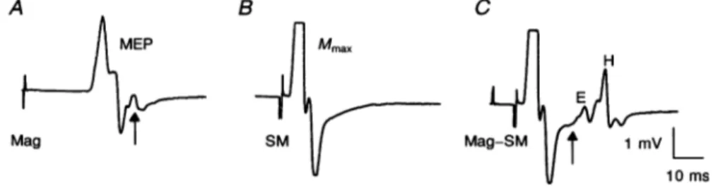
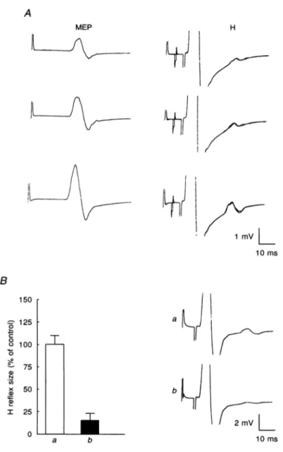
Figure 1A showvsaMEP of about4mV in theADM muscle
produced by a magnetic stimulus 25% above threshold
appliedatthevertex inarelaxedhealthy subject. Figure1B shows adirect motorresponse of maximum size produced by a SM stimulus applied to the ulnar nerve at the wrist.
Figure 1Cshows the resultofcombiningthe two typesof
stimulation, the brain shockprecedingtheSM ulnarshock
by 8 ms. Under these conditions, the antidromic response set up in everymotor fibre stimulatedby the SM stimulus should collide with the corticospinal volley due to the
magnetic stimulus. At thesametime, part of the SM
anti-dromic volleyshould be eliminatedby themagnetic volley, clearing the a-fibres for unblocked EMIG activity. Indeed, the MEP disappeared and extra EAIG activity remained
followingthe directmotor response. Theactivityconsisted of two responses. (a) The first was an early and small response(seearrowinFig.1C),theonsetandpeakofwhich J Physiol.489.1 264
H
reflexes
in human hand muscleswere at about 18 and 23ms, respectively, from the SM stimulus artifact or 25 and 30 5ms, respectively, from the magnetic stimulusartifact. Theshapeofthisresponse was very similar to the last part of the MEP before it returned to baseline (see arrow in Fig.1A); it will be henceforth referred to as the E (early) response. (b) The second response was a later and larger response, which, if measured from theSM stimulus artifact, started at about 28 ms, peaked at 30msand ended at about 35-36 ms;ifmeasured fromthe brain stimulus artifact, the onset was at about 35 ms and the peak at 37 ms. This response was called H for reasons which will be made clearer below.
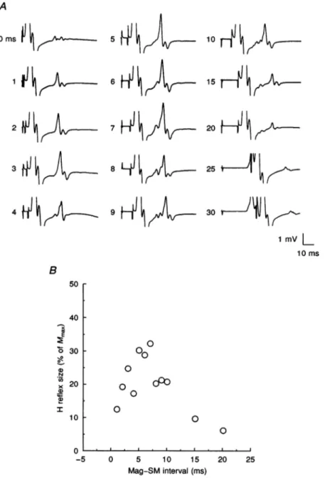
Figure 2A shows the variations of both responses when the
SM stimulus relative to the brain stimulus was shifted in the same subject at rest. The E response was not evident at all theintervals examined.It became apparent with a 4 ms interval, was very clear between 7 and 9ms, and became difficult to recognize between 10 and 15 ms because a substantial part of it fell in the direct motor response from theulnar shock. Nevertheless, it was clearly time-locked to the brain stimulus rather than to the SM stimulus and occurred 4-5 ms later than the MEP shown in Fig. 1A. It could be clearly distinguished in the relaxed muscle in three out of seven subjects. In contrast, the H response was time-locked to the SM ulnar stimulus; it was also much more stable and evident at almost all the time intervals used. The latency values of the H response were very similar to those of the H reflex that could be obtained at rest in one of the subjects (not illustrated). Figure 2B illustrates the amplitude variations of the H response when thetimeinterval separating the magnetic stimulus and the
SM ulnar stimulus was increased. The H response was always observed in relaxed muscles when this interval was changed from 1 to 20 ms. This is the range within which collision between the antidromic volley set up by the SM
A B
ulnar shock and the orthodromic volley set up by the cortical shock isexpectedto occur along thea-motorfibres. Inall subjects, the size of the H responserosesteeplyto its maximum between 5-7 ms and progressively declined thereafter. Above 20 ms,a MEPbegantoappearbeforethe directmotorresponsefrom the SMstimulus,whichimplied that the MEPwaspassing under the electrodeonits way to the muscle when the SM shock wasdelivered. Witha0 ms interval, no clearresponse could be discernedinthissubject. Althoughthe brainstimulus wasgiven simultaneouslywith theSM stimulus, thecorticalvolleyshould reach thecervical cord some 3-4ms earlier than the peripheral volley from the ulnar stimulus. This was the difference between the latencies of the MEP and the H reflex evoked in relaxed musclesinthis subject. Therefore, with a0msdelaythere should still betimefor collisionto occur in the motor axons. These, however, having passed the corticospinal volley,are in a relative refractory state for about 2 ms (see Kimura, Yamada & Rodnitzky, 1978). During such period, further impulseswouldnot be transmitteddistally. This conduction failure may explain the low-amplitude response obtained with a 0ms delay. Similarly, when the SM stimulus preceded the brain stimulus by 1-3ms(not illustrated), no clear H response could be distinguished. Longer negative intervals (from -5 to -10ms), at which the SM ulnar stimulus reached the motoneurones before the brain stimulus, gaverise to asingle, large response whichhad the same latency as the MEP. Thiswaspresumably produced
by reactivationof thea-motorfibresby the brainstimulus after collision between the orthodromic and antidromic volleysof theSM stimulus.
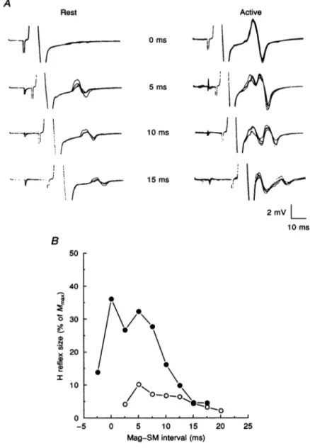
Figure 3A shows an example of the EMG responses obtained using different time intervals between the magnetic cortical stimulus and the SM stimulus under relaxed conditions and during slight tonic voluntary
C
MEP Mmax
H
A
~~~~~~~~~~~~E
Mag ISM Mag-SM 1 mVL
10Ms
Figure 1. EMG responsesfrom motoneurones of the relaxedADM muscle in arepresentative subject
A, isolated magnetic (Mag) stimulation of the contralateral motor cortex at 80% maximum output producingamotor-evokedpotential (MEP). B, supramaximal(SM) electrical stimulation ofmotorfibres of the ulnarnerveatthe wrist causing maximalmotor(Mmax)response. C, SMstimulation of the ulnarnerve 8msafter the cortical stimulus.Under thiscondition, the MEPdisappearsand thereisadditional EMG activityafter theMmax.Thelatency and shape (see arrows) of the early (E) responsearecompatiblewitha
corticalorigin, while thesubsequentresponse(H) has the latency ofanHreflexevokedbytheSM ulnar nervestimulus. Eachtrace isthe averageof three trials.
266 R.
Mazzocchio, J
C.contraction of the ADM muscle (10% of maximum voluntary effort). In this subject, the H response was not preceded by the E response at any interval at rest. However, during voluntary activity, the E response could beprogressively distinguished as detaching itself from the H response and showing a clear peak of its own with longer interstimulus intervals. At 0 ms, the E response was
Rothwell and A.
Rossi
J Physiol. 489.1presumably present, its peak coinciding with that of the Hresponse. Theonsetofthe E response was3-4mslater than the onset of the MEP in the active muscle and was
time-locked to the brain stimulus. Compared with relaxed conditions, therewas aconsistentincrease in the size of the H response at any time interval during voluntary effort;
however, the hump-shaped curve was preserved, though
A 0ms|¼ 1 5 1tOFI10
tJ
]1 <H7H
6'
/H
15 2 ; 1 > 7 202 f--4 @ 111 { 9 HJ11 g a 30 t l 1 mv L 10ms B 50 r 40 F E 0 U1) N C,1) x a) a) I 30 F 20 F 10F
O L -5 000
0 0 0 0O0C
0 0 0 5 10 15 Mag-SM interval (ms) 20 25Figure 2. Variations ofHreflex from therelaxedADM muscle with changesin the Mag-SM interval
SamesubjectasinFig. 1. A, SM ulnar stimulusprecededby Mag stimulus. TheMag-SM intervalwas progressively increased from 0to30 ms(value indicated atleft of eachtrace); each traceisthe average of threetrials. Notethat,inthissubject,when theMag-SM interval reached4ms,the E responsebeganto appearbefore theH reflex.B, timecourse of the changesinHreflexsize. Thiswasmeasuredusing the
falling phaseof the H response andexpressedas a percentageof Mmax. Each pointisthe meanof five measurements;S.D.valuesranged from about 5-15%of mean values.
(,-H
reflexes
in humanhand muscles
shifted to shorter timeintervals, as shown in Fig. 3B. In fact, maximum H amplitudes werereached between0 and 5 ms; additionally, an H response could still be obtained usinga-2-5 msinterval(SM ulnar stimulus before cortical stimulus), whereas no H responsecould be observed abovea 17-5 msinterval, since by that time theMEP had reached the stimulating electrode when the SM stimulus occurred. This is due to the fact that shorter conduction times from the cortex to the spinal cord are obtained in activated muscles compared with resting conditions (see Rothwell,
A
Rest
71
\
-n
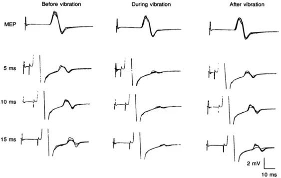
11Thompson, Day, Boyd & Marsden, 1991). In this subject, the difference in time between the two conditions was 3f6ms. Similar results were obtained in two othersubjects. Figure 4 illustrates theeffects of a long vibration stimulus (4 s) applied to the tendon of the relaxedADMmuscle on
the MEP (upper traces) and on the H responses (lower traces). The latter are shown at three different time intervals between the cortical stimulus and the SM stimulus. The resultswere obtained from thesame subject
Active 0ms b I j 5ms 10
ms
kArW
-iS
t)
..'I
a 2mVL
10ms B 50 40 30 N x 20-a) 20 I 10 _ 0 --5 0 5 10 15 Mag-SMinterval(ms) 20 25Figure 3. Comparison of ADM H reflex variations with different Mag-SM intervals under restingand active conditions
Data from another subject. A, SM ulnar stimulus preceded by Magstimulus. Theintensityof theMag
stimuluswasthe maximumpossible.TheMag-SMintervalisindicatedinmilliseconds.Notethat,inthis
subject, the E responsewas notpresentat restbutappeared onlyduringatonicvoluntarycontractionof
ADM muscle (10%of maximum). Each record shows four superimposed traces. B, timecourse of the
changes inH reflex size under relaxed(0) and active(0) conditions. Seelegend toFig.2Bfor further
details.
267
J Physiol.489.1
15Ms -Ij
R.
Mazzocchio,
JC. Rothwell
and A. Rossiinwhom therewasnoevidence of anEresponse at rest(see Fig. 3A). Cortical stimulus intensity corresponded to the maximum output of the magnetic stimulator. Vibration preceded the magnetic stimulus by 50ms. In this case, no significant variation in the size of the MEP was observedas previously reported by Claus, Mills & Murray (1988). In contrast, the amplitude of the H response was almost completely depressed by the vibratory stimulus. This was observed at all the time intervals explored. Recovery of the size of the H response occurred about 1 min after the end of vibration. Similar results were seen in two other subjects. Figure 5 shows the effect of a weak conditioning stimulus, just below the threshold for the direct motor response, applied to the ulnar nerve 5 ms (A) and 80 ms (B) before the SM stimulus, on the H response. The former (A), by evoking EPSPs in the motoneurones via I a pathways, should increase the excitability of the a-motoneurones during the arrival of the SM volley; the latter (B), presumably via Ia afferents, should produce inhibition of Hreflexes during the classic excitability cycle (see Rossi, Mazzocchio & Schieppati, 1988). The sensitivity of H reflexes to facilitation was tested on H responses of small size
(1-7% of
Mmax)
obtained using MEPs of relatively low amplitude (10-25% ofMmax).
There was no significant change between control and conditioned H responses at each MEP amplitude used (Fig. 5A). On the other hand, when the same conditioning stimulus was applied 80 msBefore vibration
MEP
earlier, the size of the H response was significantly
depressed (Fig. 5B). The size of the MEP did not show significant changes between control and conditioned trials. Similar resultswereobtained intwoothersubjects.
Since the size of the MEP determined theoccurrenceof the H response,- we studied the relationship between the H response and the MEP with increasing intensities of transcranial magnetic stimulation. Discharge of a
moto-neurone to aphasic excitatory input, particularlyatrest,is probabilistic in nature (see Bawa & Lemon, 1993). As a
result, the magnitudes of the MEP to the same magnetic
stimulation intensitycanbe quitevariable (see alsoDavey,
Ellaway, Maskill, Anissimova, Rawlinson & Thomas,1994).
We thereforeexpressed the variations of Hresponsesizeas
a function of MEPs of increasing amplitude. Figure 6A shows the data from five subjects (see Methods). The interval between the magnetic shock and the SM stimulus was 5 ms.ArangeofMEP amplitudes between 5and20% of
Mmax
producedsmall changesinthe size of the H reflex (2x5-7x5% of Mmax). In contrast, MEPs of larger size (25-50% of Mmax) produced a steep increase in H reflex size(10-30% ofMmax).
Itisknown that the strength of Iaeffects varies with motoneurone size so that small motoneurones with a lowconduction velocity exhibitlarge I a EPSPs, whereas large motoneurones with a high conduction velocity show small Ia EPSPs (see Awiszus & Feistner, 1993, for references). It has also been shown in
During vibration After vibration
5ms ft
i
10 ms LI 15ms 2mVL
10 msFigure4.Effectofvibrationof the ADMtendonon MEPand H reflexunderrestingconditions
Same subject as in Fig. 3. The vibratory stimulus (100Hz, 4s) was delivered 50ms before the Mag
stimuluswhichtriggered the trace. In each panel: upper trace, isolated Mag stimulus (maximum output of the stimulator); lower traces, combined Mag and SM ulnar stimuli atdifferent intervals. Each record showsfoursuperimposedtraces.
268 JPhysiol.489.1
I
.k-J I
-j
Hreflexes in human hand muscles
the monkey that, among motoneurones of the ulnarnerve,
those with large Ia EPSPs tend to receive larger cortico-motoneuronal EPSPs thando cells in which the IaEPSPs are small (Clough, Kernell & Phillips, 1968). Accordingly, the H response would be expected to reach the maximum
A MEP B 150 125 a 0 o 100 0 - 75 N x 50 a) I 25 0
I
a bsizewithlow-amplitude MEPs, since thesearepresumably produced by the discharge of the small, low-threshold
motoneurones receiving a larger amount of corticospinal EPSPs. This did not occur, the H response reaching the maximalamplitudewiththelargestpossible MEPs.
H
ILt~
tfI
ut-wi
1 mv 10ms 10msb4
Figure 5. Effect ofa conditioning ulnar nerve stimulus on the H reflex ofthe relaxedADM muscle
Same subject as in Fig. 3. The intensity ofthe conditioning stimuluswasjust below the threshold for activationofmotorfibres. A, left panel,isolatedMagstimulusofincreasing intensity(between 60-80%
ofmaximumoutput). Eachtraceistheaverageof eightresponses.Right panel,theconditioning stimulus precedes the SM ulnar stimulus by 5ms. The Mag-SM interval was 10 ms. Each record shows the averagesofeight superimposed control andeightconditioned Hreflexes.B, averagesof twelve trials(the
size ofthe Hreflex being expressedasapercentageofitscontrolvalue) andsample records(eachtrace
wastriggeredbytheMag stimulus; the Mag-SM intervalwas7 5ms) show thesizeof the H reflexinthe controlsituation(a)andwhen theconditioningstimulusprecededtheSM ulnar stimulusby80ms(b).The difference between the two conditions isstatistically significant (P<005; Student's two-tailed t test).
Verticalbars indicate1S.D.
R. Mazzocchio, J C. Rothwell and A. Rossi A B 140 r AA A A I
IE
_ I~~A
:;;2
iL~
0 10 20 30 40 50 60MEPsize (%ofMmax)
_ 120 0ioo 0 8 100 N °- 80 a) a, 60 a) I AN _ 40 r 20 -4 -2 0 2 4 6 8 10 12 14 Conditioning-test interval (ms)
Figure6.Variations of ADM H reflex withMEPs of increasing size under resting conditions(A) and effect of median anddigitalnervestimulationonADM H reflex(B)
A,datafrom fivesubjects. Theamplitude ofthe Hreflex (Magpreceded SMulnar stimulusby5ms)is plotted against that ofthe MEP, both expressed as apercentage ofMmax. Each point is the mean of
6-8 measurements; S.D. values ranged from about 5-15% of the mean values. B, this H reflex was
exceptionallyobtained inonesubjectat restusing submotor-thresholdstimulation of the ulnarnerveat
thewrist. Theconditioning median(0)nervestimuluswasappliedatthewristand its intensitywasjust
belowmotorthreshold. Theconditioningdigital (0)nervestimulation wasappliedtotheindexfingerand itsstrengthwastwicetheperceptionthreshold. ThesizeofthetestH reflex, expressedas apercentageof
itscontrol value(dashed line), isplotted against the conditioning-test interval. Thisisnegativewhenthe
test stimulus precedes the conditioning one. Each symbol represents the mean of 20 measurements.
Vertical bars indicate1S.D.)
One possible reason for this could be the presence of an
inhibitoryprocess, induced by the SM stimulation, affecting
the ADM motoneurones at very short latencies. This was
investigated intheonly subject inwhom itwaspossibleto
elicit an H reflex from the relaxed ADM muscle with a
stimulus intensity below the threshold for activating the ulnarmotoraxonsatthewrist(see Methods). The effect of
a conditioning submotor-threshold stimulation of the
mediannerveat the wristonthe size ofthis H reflexwas
then studied (Fig. 6B). Inhibition ofthe H reflex (m) was
evidentataconditioning-testintervalasearlyas0msand
wasoverby 8ms. Incontrast, nosignificantchange in the
size of the H reflex was observed when the conditioning
stimulus was replaced by digital nerve stimulation (0). In
thiscase,theconditioning-test intervalswerecorrectedby
subtracting thelatency of the finger-wrist tract.Thus, at
conditioning-test intervals of0ms, the volleys elicited by bothstimulishouldarriveatthespinal cordsimultaneously, given the similar conduction velocities of low-threshold muscleand cutaneous afferents(see Macefield, Gandevia & Burke, 1989).
DISCUSSION
Combinedmagneticstimulation of the scalpand SM ulnar
nervestimulation elicited additional EMGactivityafter the
direct motor response in the relaxed ADM muscle. This
consisted ofa constant response (H), the onset of which
was occasionally masked by the occurrence of other EMG
activity (E response) preceding the H response by about
4ms.Bothresponseswererecordedonlywhentherewasa
corticospinal volley capable of freeing the motor axons,
through collision, from the antidromic motor volley set up
bytheSMnervestimulation.
It has been suggested that one possible wayEMG activity can appear under the above conditions is for the cortical
shock to produce more than a single discharge of spinal
motoneurones(Day et al. 1987, 1989; Hess, Mills &Murray, 1987). Assuming that a single cortical shock in man can
give rise, asin monkey(Kernell &Wu, 1967), toseries of
descending volleys lasting up to 6-8ms after the first
volley(seeRothwell et al. 1991), the Hresponsecannot be
the expression of such activity since it occurred about
13 mslaterthantheMEPunderrestingconditions. On the
40 35 30 25 20 15 10 5 0 N 1-x ao I JPhy8iol.489.1 270 L
H
reflexes
in human hand musclesother hand, the E response, observed during voluntary contraction and in some cases at rest (see Figs 2 and 3), may well be due t-, thedischarge of the same motoneurones following the arrival of succeeding corticospinal volleys not affected by the collision. Indeed, this E responseoccurred 4-5 mslater than theMEP elicited under either relaxed or active conditions. This value compares favourably withthe estimated interval between double discharges observed in human hand muscles (see Rothwell et al. 1991).
The H response must then have a different origin and may result from the discharge of those motoneurones which, havingfired in the MEP, have had their axons cleared from the effects of the antidromic motor volley by means of collision and are thus available for activation by the la afferent volley elicited by the SM stimulus. Several lines of evidence indicate that such a response is of reflex origin: (a) its latency was remarkably stable and very similar to that of the H reflex obtained using the classic technique in one of the subjects studied; (b) it was facilitated by active contraction of the ADM muscle; and (c) it was strongly inhibited by long-lasting vibration of the ADM tendon or by a conditioning group I stimulation delivered 80 ms earlier (seeRossi et al. 1988, for references).
The time course of the H reflex effect when changing cortical-wrist intervals showed a hump-shaped curve (see Figs 2 and 3). This may reflect a combination of two factors: first, recovery from an absolute refractory period within the motoneurones(postspike after-hyperpolarization), andsecond, a decliningpostsynaptic facilitation. Concerning the origin of the latter, it should be noted that with the Hreflex obtained using collision in the motor axons only that fraction of the ADM motoneurone pool that has already discharged in response to the corticospinal volleys istested. That is to say, the subliminal fringe created by the samecorticospinal volleys will not be recruited by the
SMstimulus. Therefore, the rise in the excitability of this Hreflex will depend on events which follow the first corticospinal discharge, such as EPSPs elicited either by multiple descending volleys after the same cortical shock
(see Rothwell et al. 1991; Mazzocchio, Rothwell, Day & Thompson, 1994), or by peripheral volleys after the SM stimulus itself producing central effects at very short latencies (see below and also Macefield et al. 1989).
The occurence of the H response at rest very much depended on the amplitude of the MEP. The relationship between theirsizeswith increasing intensities of magnetic stimulation was non-linear so that the recruitment gain, i.e.the number of motoneurones participating in the Hresponse (see Kernell & Hultborn, 1990), was initially low and then
becamne
markedly increased along with the activationof new, high-threshold motor units inthe MEP. There may bevariousreasonsfor such behaviour.(a) Achangein the depth of the after-hyperpolarization of
discharge. Despite the complex and phasic nature of the descending corticospinal volley generated by transcranial magnetic stimulation, recruitment of a-motoneurones
underrestingconditionsappearstooccurinanorderly
size-related fashion (Bawa & Lemon, 1993). Therefore, with increasing magneticstimulusintensity, motoneuroneswith shorterafter-hyperpolarization might be recruited.
(b) Changes in the 'setting' of the recurrent Renshaw inhibition. Although transcranial magneticstimulation has been shown recently to depress the activity of Renshaw cells in humans(Mazzocchio, Rossi & Rothwell, 1994) and therefore, in theory, could be one of the factors which
makes the recruitment gain steeper, there is evidence suggestingalack ofrecurrentinhibition in themotornuclei of the more distal muscles of both limbs (Rossi &
Mazzocchio, 1991; Katz, Mazzocchio, Penicaud & Rossi, 1993).
(c) A disproportionate increase in the total amount of descending excitatory input to the motoneurone pool. When asmall cortical stimulus is given, there is relatively
little continuing excitatory activity in the corticospinal
tractwhich couldsummatewith the H reflex; witha very
strong cortical shock, there would be a large amount of
persisting corticospinalfacilitation of themotoneuronepool sustained bythemanyvolleyscoming down after theones
responsible for the first motoneurone discharge. The time interval between the cortical shock and the SM stimulus used to study the relationship between MEPs and Hreflexes (Fig. 6A)and the estimated central time during which the size of the H reflexcanbe influencedby synaptic
events(see Burke,Gandevia &McKeon, 1984)wouldgivea
total time of about 10ms, which should allow for
cortico-spinal EPSPs summation with increasing magnetic stimulus intensities. Thesteeprise in the H reflex sizecould
be theexpressionof suchamechanism.
(d) Small MEPs havelongerlatenciesatrest(see Day et al. 1987). Then, the motoneurones tested with the H reflex
might suffer a somewhat stronger 'relative refractoriness'
(earlier during their after-hyperpolarization) than with
larger shorter-latency MEPs. Thismightbeanexplanation
for the 'unproportionally' small H reflexes with small
MEPs.
(e) A distribution of synaptic input activated by the SM
stimulation favouring the recruitment of large
moto-neurones.If theIainputwas moreeffectiveindischarging
large motoneurones than small motoneurones, this would steepen the input-output curve for the H reflex. This
possibilityis discussed inmoredetail below.
It is not possible to say whether one or all five of these
reasons contributes to the steep rise in the input-output curve for H reflexes in the present experiments.
Never-theless, whatever the explanation, the findings may also
haveadirectbearingonthe problemofwhyH reflexesare
the motoneurones that have fired in the corticospinal
272 R.
Mazzocchio,
JC.
Rothwell andA. Rossi
J Physiol.489.1sodifficultto obtain in hand muscles at rest. In the past, it hasoften been tacitly assumed that H reflexes are absentin relaxed hand muscles because the monosynapticI a input to motoneurones is relatively weak (see introduction). The present experiments show that this is not the case, and that when tested appropriately, Iainputcan be very powerful. We propose that the steep input-output curve is responsible for the lack of the H reflexes at rest. Effectively, the steep curve means that the Ia input is ineffective on small, low-threshold motoneurones.Whythisshould bethe case is notclear, but of theexplanations given above forthe steep input-output curveonly the last(e)canapply to the simple situation in which H reflexesare elicited inrelaxed muscle since only that doesnotdepend upon the effects ofa prior cortical shock. We shall therefore explore this explanation in a little more detail.
There are several possible reasons why the effectiveness of I ainput could be skewed in favour of recruitmentof large motoneurones. (a) The Ia terminals themselves might be distributed preferentially to large motoneurones. This would beindirectcontrast toresultsreportedinmostother situations in which the I a input is distributed evenly to motoneurones of differentsize (see Calancie &Bawa, 1990). (b) The anatomical distribution of Ia connections amongst the motoneurone pool may resemble that seen in other systems but the effectiveness of these connections onto motoneurones of different size could be changed by
presynapticfactors or synaptic differences (Collins, Honig
&Mendell, 1984;Davis, Collins &Mendell, 1985). (c)Other inputs, activated at the same time, could mask the distribution of I a effects. Group I stimulation of median nerve fibres at the wrist elicited ashort-latency inhibition of ADM motoneurones(see Fig. 6B). This input, by tending tocancel out I aeffects, may well be responsible for the slow rise in the excitability of ADM motoneurones, possibly explaining theirlowsensitivitytofacilitation(see Fig.5A). Presumably, a similar effect might be produced, through activation of homonymous inhibitory pathways, when testingroutinely for H reflexesinhand muscles. Cutaneous input could also be important.Cutaneous fibres at the wrist have the same diameter and threshold as large diameter muscleafferents (seeMacefield et al. 1989)andcanproduce a predominantly inhibitory effect on small, low-threshold motor units and an excitatory effect on high-threshold,
large motor units (Garnett & Stephens, 1981; Kanda &
Desmedt, 1983; Masakado, Kamen & De Luca, 1991). While a cutaneous contribution to the inhibition of the small, low-threshold motoneurones is possible, though improbable (see Fig.6B), cutaneous facilitatory input would be the most likely candidate for explaining the prevalence of Ia excitation amongst large, high-threshold motoneurones.
We conclude that H reflexes are difficult to obtain in
relaxed hand muscles because of the steepness of the
input-output curve. This is dueto a skewed distributionof I a excitation to the motoneurone pool, which favours the discharge of large motoneurones over small motoneurones. Interestingly, in a theoretical study of the input-output relationships of a motoneurone pool model (Kernell & Hultborn, 1990), a change in output gain, as caused by synaptic input systems with an uneven distribution, is represented by a non-linear curve very similar to that obtainedinthepresentstudy.
The fact that the distribution of I a effects does not reflect the stereotyped pattern of I a connections may perhaps point to a specific synaptic organization favouring motoneurones with fast axons. Such a differential distribution of excitation could increase the accessibility of some motor units of the hand muscles to cortical control throughfast corticospinal fibres as seen during fractionated movementsof the fingers involving precision grip and fine manipulation (see Johansson, Lemon & Westli-g, 1993; Lemon, Werner, Bennett & Flament, 1993; Maier, Bennett, Hepp-Reymond & Lemon, 1993).
Awisus, F. & FEISTNER, H. (1993). The relationship between estimates ofIa-EPSPamplitude and conduction velocity in human soleus motoneurones.Experimental BrainResearch 95,365-370. BAWA, P. & LEMON, R. N. (1993). Recruitment of motor units in
response to transcranial magnetic stimulation in man. Journal of Physiology471, 445-464.
BULLER, N. P., GARNETT, R. & STEPHENS, J. A. (1980).The reflex responses of single motor units in human hand muscles following muscle afferent stimulation. Journal ofPhysiology303,337-349. BURKE, D.,GANDEVIA, S. C. & MCKEON,B.(1984). Monosynapticand
oligosynaptic contributions to human ankle jerk and H reflex. Journalof Neurophysiology 52,435-448.
CALANCIE,B.& BAwA, P. (1990). Motor unit recruitment in humans. In The Segmental Motor System, ed. BINDER, M. D. & MENDELL, L.M., pp. 75-95.Oxford University Press,Oxford.
CLAUS,D.,MILLS,K.R.&MURRAY,N. M. F. (1988). The influence of vibration on the excitability of alpha motoneurones. Electroencephalogra.phyand ClinicalNeurophysiology 69, 431-436. CLOUGH, J. F. M., KERNELL, D. & PHILLIPS, C. G. (1968). The
distribution of monosynaptic excitation from the pyramidal tract
and fromprimaryspindleafferents to motoneurones of the baboon's hand andforearm. Journal ofPhysiology 198,145-166.
COLLINS, W. F. III, HONIG, M. G. & MENDELL, L. M. (1984). Heterogeneity of group Ia synapses on homonymous a-motoneurons
as revealed by high-frequency stimulation of Ia afferent fibres. JournalofNeurophysiology 52,980-993.
DAVEY, N. J., ELLAWAY, P. H., MASKILL, D.W., ANISSIMOVA, N. P., RAWLINSON, S. R. & THOMAS, H. S. (1994). Variability in the amplitude of skeletal muscle responses to bilateral transcranial magneticstimulationin man.JournalofPhysiology 476.P, 33P. DAVIS, B. M., COLLINS, V. F. III & MENDELL, L. M. (1985).
Potentiationof transmission atla-motoneuronconnectionsinduced by repeated short bursts of afferent activity. Journal of Neurophysiology 54, 1541-1552.
J. Physiol. 489.1 H
reflexes
in
human hand muscles 273DAY, B. L.,DRESSLER, D., MAERTENS DE NOORDHOUT, A., MARSDEN, C. D.,NAKASHIMA, K., ROTHWELL, J. C. & THOMPSON, P. D. (1989). Electricand magneticstimulation of humanmotor cortex: surface EMG and single motor unit responses. Journal of Physiology 412, 449-473.
DAY, B. L., ROTHWELL, J. C., THOMPSON, P. D., DICK, J. P. R., COWAN, J. M. A., BERARDELLI, A. & MARSDEN, C. D. (1987). Motor
cortex stimulation in intact man. 2. Multiple descending volleys.
Brain 110,1191-1209.
FRITZ, N., ILLERT, M., DE LA MOTTE, S., REEH, P. & SAGGAU, P.
(1989). Pattern of monosynaptic Iaconnections inthecatforelimb. Journal of Physiology 419, 321-351.
GARNETT, R. &STEPHENS, J. A. (1981). Changes in the recruitment threshold ofmotorunitsproduced bycutaneousstimulationinman.
Journalof Physiology 311, 463-473.
HAGBARTH, K.-E. (1962). Post-tetanic potentiation of myotatic reflexes in man. Journal of Neurology, Neurosurgery and Psychiatry 25,1-10.
HESS, C. W., MILLS, K. R. & MURRAY, N. M. F. (1987). Responsesin
small hand muscles from magnetic stimulation of the human brain. JournalofPhysiology 388, 397-419.
JOHANSSON, R. S., LEMON, R. N. & WESTLING, G. (1993). Cortical influence overprecision grip in manis strongly modulated during
differentphases of the task. Journal of Physiology 459, 469P. KANDA, K. & DESMEDT, J. E. (1983). Cutaneous facilitation of large
motorunitsandmotorcontrol of humanfingersinprecision grip.In Advances in Neurology. Motor Control Mechanisms in Health and Disease, vol. 39, ed. DESMEDT, J. E., pp. 253-261. Raven Press,
NewYork.
KATZ, R., MAZZOCCHIO, R., PENICAUD, A. & RossI, A. (1993). Distribution ofrecurrentinhibition inthe humanupperlimb. Acta
Physiologica Scandinavica 149, 183-198.
KERNELL, D. & HULTBORN, H. (1990). Synaptic effects on
recruitment gain: amechanism ofimportancefor theinput-output
relations of motoneurone pools? Brain Research 507, 176-179. KERNELL,D.&Wu,C.-P.(1967). Responses of thepyramidaltractto
stimulation of the baboon's motor cortex. Journal of Physiology 191,653-672.
KIMURA, J., YAMADA, T. & RODNITZKY, R. L. (1978). Refractory period of human motor nerve fibres. Journal of Neurology, Neurosurgery and Psychiatry 41, 784-790.
LEMON, R. N., WERNER, W., BENNETT, K. M. B. & FLAMENT, D. A. (1993). The proportion of slow and fast pyramidal tract neurones
producing post-spike facilitation of hand muscles inthe conscious
monkey. Journal ofPhysiology 459, 166P
MACEFIELD, G., GANDEVIA, S. C. & BURKE, D. (1989). Conduction velocities of muscle andcutaneousafferentsintheupperand lower
limbs of humansubjects. Brain 112,1519-1532.
MAIER,M.A., BENNETT, K. M. B.,HEPP-REYMOND,M.-C.&LEMON, R. N. (1993). Contribution of the monkey corticomotoneuronal
system to the control of force in precision grip. Journal of Neurophysiology 69,772-785.
MARSDEN, C. D., MERTON, P. A. & MORTON, H. B. (1976). Stretch reflex andservoaction inavariety of human muscles. Journal of
Physiology 259, 531-560.
MASAKADO, Y., KAMEN, G. & DE LUcA, C. J. (1991). Effects of
percutaneous stimulation on motor unit firing behavior in man.
ExperimentalBraini Research 86, 426-432.
MAZZOCCHIO, R., RossI, A. &ROTHWELL,J. C. (1994). Depressionof
Renshawrecurrent inhibition by activation of corticospinal fibresin
human upper andlowerlimb. Journal of Physiology 481, 487-498.
MAZZOCCHIO, R., ROTHWELL, J. C., DAY, B. L. & THOMPSON, P. D. (1994). Effect of tonic voluntary activity on the excitability of human motorcortex. JournalofPhysiology 474, 261-267. MAZZOCCHIO,R.,SCARPINI, C. & Rossi, A. (1994). H-reflexes in small
hand muscles can be elicited routinely by a means of a collision technique. Journalof Physiology 480.P, 43P.
Rossi, A. & MAZZOCCHIO, R. (1991). Presence of homonymous recurrentinhibition inmotoneurones supplying different lower limb muscles inhumans. Experimental Brain Research 84, 367-373. Rossi, A., MAZZOCCHIO, R. & SCHIEPPATI, M. (1988). The H-reflex
recovery curve reinvestigated: low-intensity conditioning stimulation and nerve compression disclose differential effects of presumed group Ia fibres in man. Human Neurobiology 6, 281-288. ROTHWELL, J.C., THOMPSON, P. D., DAY, B. L., BOYD, S. & MARSDEN,
C. D. (1991). Stimulation of the human motor cortex through the scalp.Experimental Physiology 76, 159-200.
SCHIEPPATI, M. (1987). The Hoffmann reflex: a means of assessing spinalreflex excitability and its descending control in man. Progress
inNeurobiology 28, 345-376.
STANLEY, E. F. (1978). Reflexes evoked in human thenar muscles during voluntary activity and their conduction pathways. Journal ofNeurology, Neurosurgery and Psychiatry 41, 1016-1023. UPTON, A. R. M., MCCOMAS, A. J.& SIcA, R. E. P. (1971). Potentiation
of 'late' responses evoked in muscles during effort. Journal of Neurology, Neurosurgery and Psychiatry 34, 699-711.