The Journal of Infectious Diseases
M A J O R A R T I C L E
Live Attenuated In
fluenza Vaccine in Children Induces
B-Cell Responses in Tonsils
Kristin Greve-Isdahl Mohn,1Karl Albert Brokstad,2Rishi D. Pathirana,1Geir Bredholt,1Åsne Jul-Larsen,1Mai Chi Trieu,1Sarah Larteley Lartey,1
Emanuele Montemoli,8,9Camilla Tøndel,3,4Hans Jørgen Aarstad,3,5and Rebecca Jane Cox1,6,7 1
The Influenza Center,2
Broegelmann Research Laboratory, Department of Clinical Science,3
Department of Clinical Medicine, University of Bergen,4
Department of Pediatrics,5
Department of Otolaryngology/Head and Neck Surgery,6
Department of Research & Development, Haukeland University Hospital, Bergen,7
K.G. Jebsen Center for Influenza Vaccines, University of Bergen, Norway;
8
Department of Molecular and Developmental Medicine, University of Siena, and9
VisMederi, Siena, Italy
Background. Tonsils play a key role in eliciting immune responses against respiratory pathogens. Little is known about how tonsils contribute to the local immune response after intranasal vaccination. Here, we uniquely report the mucosal humoral respons-es in tonsils and saliva after intranasal live attenuated influenza vaccine (LAIV) vaccination in children.
Methods. Blood, saliva, and tonsils samples were collected from 39 children before and after LAIV vaccination and from 16 age-matched, nonvaccinated controls. Serum antibody responses were determined by a hemagglutination inhibition (HI) assay. The sali-vary immunoglobulin A (IgA) level was measured by an enzyme-linked immunosorbent assay. Antibody-secreting cell (ASC) and memory B-cell (MBC) responses were enumerated in tonsils and blood.
Results. Significant increases were observed in levels of serum antibodies and salivary IgA to influenza A(H3N2) and influenza B virus strains as early as 14 days after vaccination but not to influenza A(H1N1). Influenza virus–specific salivary IgA levels correlated with serum HI responses, making this a new possible indicator of vaccine immunogenicity in children. LAIV augmented influenza virus–specific B-cell responses in tonsils and blood. Tonsillar MBC responses correlated with systemic MBC and serological respons-es. Naive children showed significant increases in MBC counts after LAIV vaccination.
Conclusions. This is the first study to demonstrate that LAIV elicits humoral B-cell responses in tonsils of young children. Fur-thermore, salivary IgA analysis represents an easy method for measuring immunogenicity after vaccination.
Keywords. pediatric; influenza; LAIV; lymphoid tissue; tonsils; mucosa; saliva immune response; humoral; memory B cell; antibody-secreting cell; longevity.
Influenza continues to be an important infectious disease, with annual epidemics claiming up to half a million lives and causing a significant economic burden [1]. Annual seasonal immuniza-tion with inactivated trivalent influenza vaccine (TIV) is the most widely used and cost-effective measure for limiting the impact of influenza. An alternative vaccination strategy is to use live attenuated influenza vaccine (LAIV), which was li-censed in Europe for children (2–17 years old) in 2012 [2]. LAIV is genetically stable and attenuated to have limited repli-cation in the upper respiratory tract. Meta-analysis of LAIV ef-ficacy studies have demonstrated up to 80% efef-ficacy to matched
strains in children <6 years old and 40% efficacy in adults [3–5]. However, the immunological mechanisms and correlates of protection of LAIV are not yet clearly understood.
Serum antibody levels are known to underestimate the pro-tection achieved by LAIV [6]. Other immunological mecha-nisms are thought to be involved in conferring protection after intranasal immunization, and mucosal responses warrant further investigation. Tonsils are local lymph nodes serving the upper respiratory tract and are a collection of mucosa-associated lymphoid tissues. They consist of a pharyngeal (adenoid) and lingual tonsil and 2 tubal and palatine tonsils (referred to as ton-sils). Tonsils play a key role in eliciting mucosal immune respons-es against rrespons-espiratory pathogens [7], but their role in eliciting immune response against antigens delivered by intranasal vacci-nation is not widely reported.
Delivery of LAIV via the intranasal route is perhaps the most efficient way of boosting mucosal immunity at the site of viral entry and induces a weaker systemic response as compared to that of TIV [8]. The tonsil’s location at the site of entry into the upper respiratory tract suggests a major role in anti-influenza immunity. The tonsillar epithelium is composed of deep crypts to maximize the surface area exposed to antigens, with Langer-hans and M cells transporting luminal antigens into the tonsillar
Received 14 January 2016; accepted 23 May 2016; published online 30 May 2016. Presented in part: Options for the Control of Influenza VIII Conference, Cape Town, South Africa, September 2013; Fifth European Scientific Working Group on Influenza Conference, Riga, Latvia, 14–17 September 2014.
Correspondence: K. G.-I. Mohn, The Influenza Center, University of Bergen, Bergen, Norway ([email protected]).
The Journal of Infectious Diseases®
2016;214:722–31
© The Author 2016. Published by Oxford University Press for the Infectious Diseases Society of America. This is an Open Access article distributed under the terms of the Creative Commons Attribution-NonCommercial-NoDerivs licence (http://creativecommons.org/licenses/by-nc-nd/ 4.0/), which permits non-commercial reproduction and distribution of the work, in any medium, provided the original work is not altered or transformed in any way, and that the work is properly cited. For commercial re-use, contact [email protected]. DOI: 10.1093/infdis/jiw230
tissue [9,10]. Evidence suggests that tonsils have functional T cells and can provide B cells for mucosal effector sites, including upper airway mucosa and lacrimal and salivary glands [11,12]. In this unique study, we have vaccinated young children with LAIV at specific time points prior to elective tonsillectomy. We aimed to characterize the early local immune responses after LAIV vaccination, using the blood, saliva, and tonsils obtained from these children. We have previously reported that the sys-temic B- and T-cell responses persisted for 1 year after LAIV vaccination in some children [13]. Here we show that the LAIV induces early salivary antibody and B-cell responses in the tonsils, which may play a significant role in mediating pro-tection against influenza.
MATERIALS AND METHODS Study Design
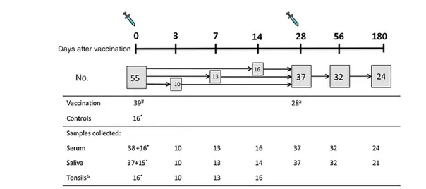
Fifty-five healthy children (3–17 years old) scheduled for tonsil-lectomy were recruited from outpatients at the ear, nose, and throat (ENT) clinic at Haukeland University Hospital (Figure1). Thirty-nine children were vaccinated with trivalent LAIV (Flu-enz, Astra Zeneca, United Kingdom) during the influenza season from October 2012 to February 2013. The study had ethical and regulatory approval (clinical trials registration NCT01866540). Exclusion criteria have been published previously [13].
To study the early immunological responses after LAIV, we chose the earliest time point that was considered safe by the anesthesiologist (day 3), in addition to 1 and 2 weeks after
vaccination. The sampling days were based around the distribu-tion of antibody-secreting cells (ASCs) in peripheral blood, the levels of which peak around day 7 after vaccination, but they have been observed as early as day 2 after TIV vaccination [14–16]. The children were randomized into 3 subgroups, de-pending on scheduled tonsillectomy date: 3–5 days (n = 10), 7 days (range, 6–9 days; n = 13), and 14 days (range, 11–20 days; n = 16) after vaccination. The number of children was obtained from asking eligible children set up for elective tonsillectomy during the vaccination period. A nonvaccinated control group consisted of 16 age-matched children as a prevaccination com-parator for the tonsillar responses in the vaccinated children; the controls were recruited in parallel to the study subjects. Samples were collected at a single time point during the opera-tion (Table1). Blood and saliva specimens were only used to show the suitability of the controls, as prevaccination (day 0) blood and saliva samples were collected from the vaccinated children for comparison with samples obtained at subsequent time points up to 180 days after vaccination.
Vaccine
LAIV was administered intranasally as a 0.1-mL spray dose into each nostril. LAIV contained 107fluorescent focus units of A/Cal-ifornia/7/2009(H1N1)pdm09, A/Victoria/361/2011(H3N2), and B/Wisconsin/1/2010. Children <10 years of age (n = 28) were given 2 doses of LAIV as per the manufacturer’s recommendations. None of the children had earlier received LAIV, as it was not licensed in Norway in 2012–2013. Most children were born after
Figure 1. Study design and sample collection. Children scheduled for tonsillectomy were recruited from outpatients at the ear, nose, and throat clinic at Haukeland Uni-versity Hospital during the influenza season of October 2012–February 2013. Control children were recruited in parallel throughout the study from the same patient population (ie, if the parents were willing for their child to join the study but did not wish for their child to receive the live attenuated influenza vaccine). Asterisks denote nonvaccinated controls. One control did not provide a sufficient saliva sample owing to dry mouth prior to operation. The hash indicates that 1 vaccinated child provided samples on the day of tonsillectomy but no sample on day 0.aOnly children aged <10 years old required 2 doses of LAIV. Two children aged <10 years did not receive a second dose, 1 child was sick on the day of the second vaccination, and another child withdrew from the study due to postoperative discomfort.bThe patients had both of their tonsils removed in 1 operation, and therefore tonsils were only sampled at a single time point. Nonvaccinated controls were used as a prevaccination (day 0) comparator for tonsillar samples. Tonsils were collected from vaccinated children at 3–5 days, 7 days (range, 6–9 days), or 14 days (range, 11–20 days) after vaccination. Serum and saliva samples were collected at multiple time points from each vaccinated subject and at only a single time point, at the time of tonsillectomy, from the nonvaccinated controls. The exclusion criteria and study details for this clinical trial have been published earlier [13].
the pandemic, and the only influenza vaccine the older children had received was the monovalent, adjuvanted pandemic influen-za A(H1N1) vaccine in 2009 (6 controls and 21 vaccinees). Samples
Peripheral blood samples (8 mL) were collected at day 0 and after vaccination, using CPT tubes (BD), and peripheral blood mono-nuclear cells (PBMCs) and plasma were separated [17]. Plasma samples were stored at−80°C. Immediately following tonsillecto-my, whole tonsils were collected to isolate the tonsillar mononu-clear cells (TMCs) by Lymphoprep (Stemcell tech, United Kingdom). Saliva samples were absorbed from the lower buccal mucosa for 2 minutes, using a swab (Salimetrics). The swabs were placed in a tube and kept on ice until centrifuged (at 600 × g for 10 minutes at 4°C) before storage at−80°C. Serological Assays
Hemagglutination Inhibition (HI) Assay
Plasma samples from each subject were tested at the same time, in duplicate. In the HI assay, 8 hemagglutinin units of the homologous influenza A(H1N1) and influenza A(H3N2) virus strains or ether-treated influenza B virus vaccine strains (50 µL/well) were used, and 0.7% turkey red blood cells, with receptor destroying enzyme–treated serum at a starting dilution of 1:10 [18]. The influenza virus antigens were either provided by the WHO Influenza Reagent Resources or were grown in eggs in our laboratory.
B-Cell Assays
The influenza virus–specific immunoglobulin G (IgG), immu-noglobulin A (IgA), and immuimmu-noglobulin M (IgM) ASC [19] and memory B-cell (MBC) [20] responses were determined by an enzyme-linked immunospot (ELISPOT) assay using fresh lymphocytes from blood and tonsils. Results are presented as influenza virus–specific ASCs or MBCs per 1 × 10⁶PBMCs/ TMCs.
Salivary IgA
The concentration of influenza virus–specific IgA antibodies in the saliva was measured in ELISA plates coated with 2 µg/mL of split influenza virus antigens (A(H1N1), A(H3N2), or B strains) as previously described [14].
Statistical Analysis
Statistical analysis was performed using GraphPad Prism, ver-sion 6, for Mac OS X. Differences between prevaccination and postvaccination ASC and MBC responses were analyzed by matched-paired signed rank t test, (Wilcoxon), and the P value was adjusted accordingly (by the Bonferroni method). The comparison of HI and saliva IgA responses over time were evaluated by analysis of variance, (nonparametric, Kruskal–Wallis) with the Dunn multiple comparisons test. Cor-relation analysis was performed by nonparametric Spearman correlation. A P value of < .05 was considered statistically significant.
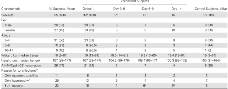
Table 1. Demographic and Clinical Characteristics of the Study Children
Characteristic All Subjects, Value
Vaccinated Subjects
Control Subjects, Value Overall Day 3–5 Day 6–9 Day 14
Subjects 55 (100) 39a (100) 9a 13 16 16 (100) Sex Male 28 (51) 20 (51) 6 7 6 8 (50) Female 27 (49) 19 (49) 3 6 10 8 (50) Age, y 3–4 31 (56) 23 (59) 6 8 8 8 (50) 5–9 15 (27) 8 (20.5) 2 3 3 7 (44) 10–17 9 (16) 8 (20.5) 1 2 5 1 (6) Weight, kg, median (range) 19 (9–81) 19 (13–81) 18.5 (14–81) 19.3 (13–60) 19.4 (13–61) 19 (9–64) Height, cm, median (range) 107 (88–177) 107 (88–177) 104.2 (99–176) 108.4 (95–171) 105.9 (88–172) 100 (91–164)b
A(H1N1)pdm09c
vaccination 26 (47) 21 (54) 3 7 4 6 (38)d
Reason for tonsillectomye
Only recurrent tonsillitis 11 8 3 2 2 3 Only hypertrophyf
20 13 5 4 4 7
Both reasons 22 16 1 6g
9g
6
Data are no. (%) of children, unless otherwise indicated. Thirty-nine subjects received 1 dose of live attenuated influenza vaccine, and 28 children received 2 doses. Ten subjects did not receive the second dose owing to age (8 subjects were >10 years old), illness on the day of the second dose (1 subject), and postoperative discomfort and later withdrawal from study (1 subject).
a
One child had the operation delayed, therefore no samples were collected at the day of tonsillectomy, but the rest of the time points were collected.
b
Data for 4 subjects are missing.
c
Vaccination with 2009 pandemic influenza A(H1N1) vaccine (Pandemrix) in 2009.
d
Data for 5 subjects are missing.
e
Data for 2 subjects on reason for tonsillectomy are missing.
f
Defined as hypertrophy-related problems such as sleep apnea/snoring, speech impairment, and recurrent ear infections.
g
RESULTS Study Subjects
Fifty-five healthy children were enrolled in the study during the influenza season from October 2012–January 2013. Of these, 39 were vaccinated with LAIV, and 16 were nonvaccinated controls. The vast majority (32 of 39) were ethnic Norwegian caucasian in-dividuals. Among the vaccinated children, there were 20 boys and 19 girls, with a median age of 4 years (range, 3–17 years). The chil-dren were vaccinated at 3 days (range, 3–5 days; n = 10), 7 days (range, 6–9 days; n = 13), or 14 days (range, 11–20 days; n = 16) prior to tonsillectomy, to allow evaluation of early tonsillar B-cell responses after LAIV vaccination. The demographic characteris-tics and vaccination history were similar in the 3 subgroups and controls (Table1). Sequential blood samples were collected before vaccination, on the day of tonsillectomy, and 28, 56 and 180 days after vaccination (Figure1) [13]. The median sampling time point was close to the target sampling day. For comparison of differenc-es in kinetics in blood and tonsils, the early time points (days 3, 7, and 14) were used, while the later time points were used to study the duration of the systemic and salivary responses after LAIV vaccination. For the comparison of background prevaccination tonsillar responses and the responses in vaccinated children, 16 matched, nonvaccinated controls were used.
Among the 39 vaccinated children, 21 (54%) had received the inactivated, monovalent influenza A(H1N1) pandemic vaccine in 2009. Two vaccinees (5%) were born to mothers who had been immunized with the pandemic vaccine during pregnancy. Apart from 1 child, none had earlier received seasonal TIV or LAIV, as routine influenza vaccination of children is not recom-mended in Norway.
Serological Responses
An HI titer of≥40 is considered protective against seasonal in-fluenza [21]. No significant changes were observed in the post-vaccination response against influenza A(H1N1) virus, with
45%–82% having titers of ≥40 after LAIV vaccination (Fig-ure2A). Significant increases in influenza B and influenza A
(H3N2) virus antibody responses were observed from 14 and 28 days after vaccination, respectively, and maintained until day 180 (Figure2B and2C). Overall, the percentage of subjects
with HI titers of≥40 against influenza A(H3N2) virus increased from 49% at day 0 (geometric mean titer [GMT], 36) to 94% in the group that underwent tonsillectomy 14 days after vaccina-tion (GMT, 137; Figure2B) and was maintained until 180 days.
The majority (89%) of children had no detectable antibodies to the influenza B virus strain before vaccination. As early as 14 days after vaccination, 69% had protective antibody titers, in-creasing to 76% and 84% at days 28 and 56 (P = .0001; Fig-ure 2C). The nonvaccinated controls had similar antibody
titers to the prevaccination (day 0) titers, justifying their use as a day 0 comparator for tonsillar responses (Figure2; con-trols/day 0). When studying the individual responses, no in-crease in titers was observed on day 3, but inin-creases were observed in 2 children on day 7 (influenza A(H3N2) and B virus strains), and in 10 children at day 14 (71%) for the in flu-enza B virus strain (Supplementary Figure 2). A boost after the second dose was observed in 1 child (for influenza A(H1N1) virus), 7 children (for influenza A(H3N2) virus), and 11 chil-dren (for the influenza B virus strain), with the strongest re-sponses in the unprimed children. There were no significant differences in responses over time in the 3 groups, except at the time of tonsillectomy (Supplementary Figure 2).
IgA Response in Saliva
Figure3A–3C shows the influenza virus–specific IgA response
in saliva after LAIV vaccination. Significant increases (P < .001) in saliva IgA response were detected against influenza B virus and influenza A(H3N2) virus strains from 0 to 14 days after vaccination and also at days 56 and 180 for the influenza B virus strain. The IgA response was maintained to day 180
Figure 2. Serological response after live attenuated influenza vaccine (LAIV) vaccination. Plasma was collected from nonvaccinated controls (open circles) and LAIV recip-ients (closed circles), and the serological antibody response was investigated by a hemagglutination inhibition (HI) assay. The data indicate influenza A(H1N1) virus–specific (A), influenza A(H3N2) virus–specific (B), and influenza B virus–specific titers (C). Each symbol represents an individual subject, and the horizontal lines represent the geometric mean titers ± 95% confidence intervals. The dotted lines represents an HI titer of 40, considered indicative of a protective level. Statistical significance between prevaccination and postvaccination responses was measured by the nonparametric Kruskal–Wallis multiple comparisons test. **P ≤ .01, ***P ≤ .001, and ****P ≤ .0001.
above prevaccination levels for the influenza A(H3N2) and B virus strains. However, no significant increase in IgA responses was observed against the influenza A(H1N1) virus strain at any time point after vaccination. Furthermore, there was a signi fi-cant positive correlation between the postvaccination (day 3– 14), salivary IgA titers and the serum HI responses for all 3 strains (r = 0.37–0.48; P < .05). The controls had titers that matched the prevaccination titers of the vaccinated children. ASC Responses in Tonsils and Blood
As tonsils could only be collected at a single time point, nonvac-cinated control children were included to show background pre-vaccination tonsillar B-cell responses (Figure1and Table1). Serological and salivary IgA titers (Figures 2and3) observed in controls were similar to the prevaccination (day 0) samples from the vaccinated children, making them suitable for compar-ison in the ASC and MBC assays. Influenza virus–specific ASC responses in blood and tonsils were analyzed by ELISPOT to a mixture of the 3 vaccine strain antigens (influenza A(H1N1), A(H3N2), and B virus strains; Figure4A–4F). The
antigen-specific ASC response in TMCs was dominated by IgM and in-creased significantly 7 days after vaccination (Figure4C). There
were low numbers of IgG and IgA ASCs detected in the tonsils, with the highest responses on day 14 after vaccination, com-pared with control responses (Figure4A and4B).
Figure4D–4F shows the influenza virus–specific ASC
re-sponse in PBMCs, with very low numbers detected before and 3 days after vaccination but with significant increases in IgG and IgA ASCs on days 7 and 14 (Figures 4D and 4E ).
IgM also increased although not significantly. At day 28, the IgA and IgM frequencies were similar to prevaccination levels
(mean, 3 and 4 ASCs/1 × 106PBMCs, respectively). We found a significant positive correlation between influenza virus– specific IgG ASC frequencies detected in tonsils and blood after LAIV vaccination (r = 0.51; P = .007), suggesting that pe-ripheral ASCs reflect the local tonsillar response.
MBC Responses in Tonsils and Blood
Influenza virus–specific MBC responses were detected by ELI-SPOT in blood and tonsils. No significant increases were ob-served in short-term MBC responses in peripheral blood or tonsils up to day 14 (Supplementary Figure 1). In general, much higher frequencies of IgG and IgM MBCs were detected, compared with IgA, in both tonsils and blood.
We observed a significant positive correlation between the IgG MBC responses in the TMCs and PBMCs to the 3 LAIV strains (r = 0.82–0.59; P < .05). A significant positive correlation was also detected between the IgG MBC responses in the TMCs and the HI responses at the corresponding time points to the influenza A(H1N1) virus (r = 0.68; P = .0004) and influenza A(H3N2) virus (r = 0.47; P = .0189) strains but not to the influenza B virus strain.
To see whether previous infection ( priming status) of the subjects influenced their short-term MBC response after LAIV vaccination, we stratified each individual on the basis of their prevaccination serological titer as primed (HI titer of≥40) or naive (unprimed; HI titer of <40; Figure5). The primed sub-jects had significantly higher IgG MBC frequencies than the naive subjects against influenza A(H1N1) virus in both tonsils (mean, 469 and 51 MBCs/106TMCs, respectively) and blood (mean, 1100 and 130 MBCs/106 PBMCs, respectively) and against the influenza A(H3N2) virus strain in tonsils (mean, Figure 3. The immunoglobulin A (IgA) response in saliva after live attenuated influenza vaccine (LAIV) vaccination. Saliva samples were collected from nonvaccinated controls (open circles) and LAIV recipients (closed circles) on the day of tonsillectomy (day 3, 7, or 14) and 28–180 days after vaccination. The IgA antibody levels in saliva were determined by enzyme-linked immunosorbent assay for each strainA (H1N1), B (H3N2), and C (B strain). Each symbol represents the IgA response of 1 subject, with means and standard errors of the mean indicated. Statistical significance between prevaccination and postvaccination responses was measured by the nonparametric Kruskal– Wallis multiple comparisons test. *P < .05 and **P ≤ .01.
710 and 182 MBCs/106TMCs, respectively). Low frequencies of MBCs were observed for the influenza B virus strain in the un-primed children, and generally a higher response was observed in the primed child (only 1 of the 4 primed children had results for the B strain). No significant differences in influenza virus–
specific IgA and IgM MBC responses were observed between primed and naive individuals (data not shown).
We have earlier shown that LAIV significantly increases MBC responses in these children, which persist for up to 6– 12 months [13]. When we analyzed these long-term IgG Figure 4. Antibody-secreting cell (ASC) response in tonsils and peripheral blood after live attenuated influenza vaccine (LAIV) vaccination. Children were vaccinated with 2012–2013 seasonal LAIV, and the immunoglobulin G (IgG), immunoglobulin A (IgA), and immunoglobulin M (IgM) ASC responses in tonsillar mononuclear cells (TMCs) and peripheral blood mononuclear cells (PBMCs) were measured by enzyme-linked immunospot assay. The influenza virus–specific IgG (A), IgA (B), and IgM (C) ASC responses against a combination of the 3 viruses (influenza A(H1N1), A(H3N2), and B viruses) were determined in TMCs isolated from nonvaccinated controls (open circles) and LAIV recipients (closed circles) 3, 7, and 14 days after vaccination. The IgG (D), IgA (E), and IgM (F) ASC responses against a combination of 3 influenza viruses (influenza A(H1N1), A (H3N2), and B viruses) were measured in PBMCs isolated before vaccination (day 0) and at 3, 7, 14, and 28 days after vaccination.A–F, Each symbol represents influenza virus– specific ASCs per 1 × 10⁶cells with mean ± standard error of the mean indicated. Statistical differences between vaccinated and nonvaccinated subjects were determined by
MBC responses (up to day 180) according to the priming status, we found a significant increase in MBCs in the unprimed children after LAIV vaccination and not in the primed children (Figure6A–6C). This indicates that the increase in the MBC
re-sponse after LAIV vaccination was largely influenced by the priming status of the child.
DISCUSSION
Tonsils represent both an induction and maintenance site for mucosal immune responses in the nasopharynx against respira-tory pathogens (ie, influenza virus) encountered through natu-ral infection [22]. However, limited data are available on the tonsillar role and contribution to the local immune response Figure 5. The short-term effect of priming on memory B-cell (MBC) response in tonsils and blood after live attenuated influenza vaccine (LAIV) vaccination. The children were classified as“primed” if they had a hemagglutination inhibition (HI) antibody titer of ≥40 and as “naive” if the HI titer was <40 prior to vaccination. The figure shows the influenza A(H1N1) virus–specific (A), influenza A(H3N2) virus–specific (B), and influenza B virus–specific (C) immunoglobulin G (IgG) MBC response in tonsillar mononuclear cells (TMCs). The IgG MBC results for the 3 strains in peripheral blood mononuclear cells (PBMCs) are shown in panelsD–F from primed and naive subjects. Each symbol represents the MBC response of 1 subject, and the horizontal lines represent the means ± standard errors of the mean indicated. Statistical differences between the primed and naive groups were measured by the Mann–Whitney nonparametric test. **P ≤ .01 and ***P ≤ .001.
after intranasal vaccination. In this unique study, we were able to collect tonsils, saliva, and blood samples from young children (median age, 4 years) who were intranasally vaccinated with a LAIV prior to elective tonsillectomy. These pediatric samples
enabled us to assess the local lymphoid and saliva responses, as well as the systemic immune responses.
The immune responses elicited by LAIV are multifaceted, similar to those after natural infection. The induction of ade-quate immune responses to LAIV is dependent on local replica-tion of the virus, and hence preexisting mucosal antibodies may reduce viral replication. Protection after LAIV is thought to be associated with induction of mucosal antibodies, but challenges in sampling and assaying these antibodies have hampered de-velopment of mucosal antibodies as a correlate of protection [2]. IgA is the predominant secreted antibody at mucosal surfac-es [23,24]. We detected elevated IgA levels in saliva 14 days after vaccination, and the response persisted in some subjects for 180 days, which is similar in duration to the nasal IgA response ob-served after LAIV vaccination in children [8,25]. This indicates that the mucosal immune response is well developed in young children and that the LAIV provides local protection in the nasal and oral cavities. Importantly, a significant positive asso-ciation was observed between the influenza virus–specific sali-vary IgA and serum antibody responses. This implies that salivary IgA levels could be a possible noninvasive biomarker to predict the immunogenicity of LAIV and could be particular-ly useful when assessing the effectiveness of the vaccine in children.
The LAIV enhanced the systemic antibody responses toward the influenza A(H3N2) and B virus strains in most subjects, but no boost was observed against the influenza A(H1N1) virus strain. A lack of measurable effectiveness against the influenza A(H1N1) virus strain in the 2013 LAIV has been reported in the United States, and the LAIV is no longer the sole recommended vaccine for children [26–28]. The LAIV influenza A(H1N1) virus strain had reduced viralfitness due to temperature insta-bility, and the vaccine manufacturer has developed a new influ-enza A(H1N1) virus strain to overcome these problems [29]. However, the preexisting antibodies to influenza A(H1N1) virus may have also contributed to the low effectiveness, since most children had previously been infected or vaccinated with this strain during the 2009 pandemic.
The humoral immune response was further characterized by analyzing the ASC responses in the tonsils and blood. Our ear-lier work in adults has shown that after intramuscular TIV vac-cination, antigen-specific ASC responses appear transiently in the blood. These ASCs peak 1 week after vaccination, correspond-ing to the peak plasmablast (CD19+CD20−CD27highCD38high) response [14,19]. The kinetics of the circulating ASC response was slower after LAIV vaccination as compared to TIV vaccina-tion, with the highest response observed 2 weeks after vaccination. The extended ASC response after intranasal immunization could be due to longer persistence of the vaccine viruses. Data from re-spiratory syncytial virus–infected subjects show that ASCs are pro-duced for as long as the virus is shed [30]. An extended ASC response was also detected in the tonsils; however, we cannot Figure 6. The long-term effect of priming on memory B cells (MBCs) in blood
after live attenuated influenza vaccine (LAIV) vaccination. The children were classi-fied as“primed” if they had a hemagglutination inhibition (HI) antibody titer of ≥40 and as“naive” if the HI titer was <40 prior to vaccination. The immunoglobulin G (IgG) MBC results for the 3 strains were measured in peripheral blood mononuclear cells (PBMCs) isolated before vaccination (day 0) and 28, 56, and 180 days after vac-cination (A–C). Each symbol represents influenza virus–specific MBCs per 1 × 10⁶
cells with mean ± standard error of the mean indicated. Statistical differences be-tween the different time points and day 0 were determined by the nonparametric Kruskal–Wallis multiple comparisons test. *P < .05, **P ≤ .01, and ***P ≤ .001, respectively.
rule out a peak later than our last sampling point (day 14). In the tonsils, the predominant influenza virus–specific responses were unswitched IgM ASCs, which indicates activation of naive B-cell responses to novel epitopes on the viral surface glycoproteins. Rel-atively low IgA responses were observed in tonsils after LAIV vac-cination, in agreement with our previous observation in children after TIV vaccination [15]. Although the number of influenza
virus–specific IgG and IgA ASCs remained relatively low in tonsils, compared with blood, it still represents a significant immunolog-ical response, as >109cells were isolated from some tonsils.
Recall antibody responses produced by MBCs are crucial for influenza vaccine–induced protective immunity. We observed influenza virus–specific MBCs in both tonsils and blood before vaccination in most children. The presence of tonsillar MBCs could be a significant factor in providing long-term protection at the site of initial virus infection in the upper respiratory tract, as murine studies have shown that long-lived antibody responses against viruses originate from existing MBCs [31]. The impor-tance of mucosal MBCs in protective immunity is highlighted by the fact that at baseline, primed subjects had significantly high-er influenza virus–specific IgG MBCs in their tonsils than un-primed subjects. The MBCs measured at day 0 or in the controls reflected prior infection history. The observation of high levels of influenza A(H1N1) virus– and A(H3N2) virus–spe-cific MBCs in some of the control children in both tonsils and blood (Supplementary Figure 1) is probably a result of earlier infection by the circulating influenza A(H1N1) or A(H3N2) virus strains, not previous vaccination, as influenza vaccination is only recommended for children with high-risk conditions in Norway. We observed interstrain differences in the immune response after LAIV vaccination, with the influenza B virus strain inducing the highest antibody responses both locally (saliva and tonsils) and systemically (blood). Most of the children were naïve to the influenza B virus strain; nonetheless, the majority reached protec-tive HI titers 14 days after vaccination, indicating a rapid induc-tion of protective antibody. Although individual variance will affect the kinetics of the immune response, the results from our study may represent different types of immune responses: an experienced population (with the majority previously exposed to influenza A(H1N1) virus) and a naïve population (with the majority without prior influenza A(H3N2) and B virus strain exposure).
Importantly, there was a significant positive correlation be-tween influenza virus–specific tonsillar IgG MBC responses and serum HI titers, which is consistent with findings that IgG MBCs are more likely to become plasmablasts that appear in the circulation, capable of producing influenza virus–specific antibody responses [32–34]. Although the early influenza virus– specific MBC responses were not boosted by vaccination within the 2-week period (Supplementary Figure 1), the MBC respons-es increased in blood by 28–56 days after vaccination, lasting up to 12 months [13]. Importantly, our results show that the
increased MBC responses after LAIV occur mainly in unprimed (naïve) children (Figure6).
MBC responses are rapid, producing more high-affinity anti-bodies than naïve B cells. The primed children had higher MBC levels, which were not boosted further after LAIV vaccination, possibly because a threshold level was reached (Figure6), which has been observed by Sasaki et al [16]. LAIV may however help maintenance and maturation of the MBC response, producing antibodies with a broader repertoire to influenza virus [35]. The finding that primed children had a greater IgG MBC response in both tonsils and blood toward the 2 influenza A virus strains (Figure5) could therefore suggest that a more mature immune response is achieved in these children. This is supported by ear-lier studies showing that serum IgG levels induced after in fluen-za virus infection or vaccination are correlated with resistance to infection [26], with higher antibody levels with increased avid-ity. The lack of correlation between influenza B virus strain se-rological titers and IgG MBCs could indicate that the influenza B virus strain kinetics are different or that the response arises at a later time point, as the vast majority were naïve to this strain. Only the naïve children showed a significantly increased long-term MBC response in blood beyond the time points we sam-pled the tonsils (Figure6). The weaker influenza A(H1N1) virus
strain [29] did not boost the MBC response, but the influenza A(H3N2) and B virus strains elicited boosted MBCs responses in naïve children, and the majority seroconverted. Ourfindings support European recommendations of LAIV vaccination by children only and can explain the lower effectiveness of LAIV found in adults as compared to children. Our study is limited by the small number of subjects and because we asked the par-ents about influenza illnes in their children but did not test the children during the trial.
Future studies may elucidate the ability of LAIVs to induce cross-reactive antibodies and cellular immunity. In the present study, we are thefirst to show that vaccination with a mucosal influenza vaccine enhances antibody and B-cell immune re-sponses in palatine tonsils. Better understanding of the tonsillar immune responses will aid the development of vaccination strategies aimed at enhancing local immunity against influenza.
Supplementary Data
Supplementary materialsare available athttp://jid.oxfordjournals.org. Consisting of data provided by the author to benefit the reader, the posted materials are not copyedited and are the sole responsibility of the author, so questions or comments should be addressed to the author.
Notes
Acknowledgments. We thank the children and their parents who par-ticipated altruistically in this study; all of the staff at the Ear, Nose, and Throat Department; Dr Lorentz Sandvik, Dr Ole Gamlemshaug, Dr Per An-ders Hunderi, Hildegunn Grimstad, Wendela Mathisen, Nina Haugland, and the Children’s trial unit, with Hildur Grindheim, Renate Håpoldøy, Anne Marthe Østerbø, Marianne Heradstveit, and all colleagues at the In-fluenza Center, for assistance with the clinical trial; Steinar Sørnes, Emilia Lohndal, and Jane Kristin Nøstbakken, for invaluable technical and
logistical assistance; and Glaxo SmithKline, for kindly providing the split virus antigen.
Financial support. The work was supported by the University of Bergen Influenza Center (funded by the Ministry of Health and Care Ser-vices, Norway; the Norwegian Research Council Globvac program [220670/ H10], the European Union [Univax 601738 and EU IMI115672 FLUCOP], Helse Vest, and the K. G. Jebsen Center for Influenza Vaccines); and the Bergen Clinical Vaccine Consortium.
Potential conflicts of interest. All authors: No reported conflicts. All authors have submitted the ICMJE Form for Disclosure of Potential Con-flicts of Interest. ConCon-flicts that the editors consider relevant to the content of the manuscript have been disclosed.
References
1. Molinari NA, Ortega-Sanchez IR, Messonnier ML, et al. The annual impact of seasonal influenza in the US: measuring disease burden and costs. Vaccine 2007; 25:5086–96.
2. Sridhar S, Brokstad KA, Cox RJ. Influenza vaccination strategies: comparing inactivated and live attenuated influenza vaccines. Vaccines (Basel) 2015; 3:373–89.
3. Jefferson T, Rivetti A, Di Pietrantonj C, Demicheli V, Ferroni E. Vaccines for pre-venting influenza in healthy children. Cochrane Database Syst Rev 2012; 8: CD004879.
4. Jefferson T, Di Pietrantonj C, Rivetti A, Bawazeer GA, Al-Ansary LA, Ferroni E. Vaccines for preventing influenza in healthy adults. Cochrane Database Syst Rev 2010; doi:10.1002/14651858.CD001269.pub4.
5. Cha TA, Kao K, Zhao J, Fast PE, Mendelman PM, Arvin A. Genotypic stability of cold-adapted influenza virus vaccine in an efficacy clinical trial. J Clin Microbiol 2000; 38:839–45.
6. Bandell A, Woo J, Coelingh K. Protective efficacy of live-attenuated influenza vac-cine (multivalent, Ann Arbor strain): a literature review addressing interference. Expert Rev Vaccines 2011; 10:1131–41.
7. Brandtzaeg P. Immune functions of nasopharyngeal lymphoid tissue. Adv Otorhi-nolaryngol 2011; 72:20–4.
8. Clements ML, Murphy BR. Development and persistence of local and systemic antibody responses in adults given live attenuated or inactivated influenza A virus vaccine. J Clin Microbiol 1986; 23:66–72.
9. Perry ME. The specialised structure of crypt epithelium in the human palatine tonsil and its functional significance. J Anat 1994; 185 (Pt 1):111–27. 10. Howie AJ. Scanning and transmission electron microscopy on the epithelium of
human palatine tonsils. J Pathol 1980; 130:91–8.
11. Brandtzaeg P. Immunology of tonsils and adenoids: everything the ENT surgeon needs to know. Int J Pediatr Otorhinolaryngol 2003; 67(suppl 1):S69–76. 12. Sada-Ovalle I, Talayero A, Chavez-Galan L, et al. Functionality of CD4+ and
CD8+ T cells from tonsillar tissue. Clin Exp Immunol 2012; 168:200–6. 13. Mohn KG, Bredholt G, Brokstad KA, et al. Longevity of B-Cell and T-Cell
Re-sponses After Live Attenuated Influenza Vaccination in Children. J Infect Dis 2014; 211:1541–9.
14. Brokstad KA, Cox RJ, Olofsson J, Jonsson R, Haaheim LR. Parenteral influenza vaccination induces a rapid systemic and local immune response. J Infect Dis 1995; 171:198–203.
15. el-Madhun AS, Cox RJ, Soreide A, Olofsson J, Haaheim LR. Systemic and mucosal immune responses in young children and adults after parenteral influenza vacci-nation. J Infect Dis 1998; 178:933–9.
16. Sasaki S, He XS, Holmes TH, et al. Influence of prior influenza vaccination on an-tibody and B-cell responses. PLoS One 2008; 3:e2975.
17. Cox RJ, Madhun AS, Hauge S, et al. A phase I clinical trial of a PER.C6 cell grown influenza H7 virus vaccine. Vaccine 2009; 27:1889–97.
18. Madhun AS, Akselsen PE, Sjursen H, et al. An adjuvanted pandemic influenza H1N1 vaccine provides early and long term protection in health care workers. Vaccine 2010; 29:266–73.
19. Cox RJ, Brokstad KA, Zuckerman MA, Wood JM, Haaheim LR, Oxford JS. An early humoral immune response in peripheral blood following parenteral inacti-vated influenza vaccination. Vaccine 1994; 12:993–9.
20. Crotty S, Aubert RD, Glidewell J, Ahmed R. Tracking human antigen-specific memory B cells: a sensitive and generalized ELISPOT system. J Immunol Methods 2004; 286:111–22.
21. Committee for proprietary medicinal products CPMP EMa. Note for guidance on harmonisation of requriements for influenza vaccines. 1997.
22. Brandtzaeg P, Pabst R. Let’s go mucosal: communication on slippery ground. Trends Immunol 2004; 25:570–7.
23. Renegar KB, Small PA Jr, Boykins LG, Wright PF. Role of IgA versus IgG in the control of influenza viral infection in the murine respiratory tract. J Immunol 2004; 173:1978–86.
24. van Riet E, Ainai A, Suzuki T, Hasegawa H. Mucosal IgA responses in influenza virus infections; thoughts for vaccine design. Vaccine 2012; 30:5893–900. 25. Johnson PR Jr, Feldman S, Thompson JM, Mahoney JD, Wright PF.
Compari-son of long-term systemic and secretory antibody responses in children given live, attenuated, or inactivated influenza A vaccine. J Med Virol 1985; 17: 325–35.
26. Clements ML, Betts RF, Tierney EL, Murphy BR. Serum and nasal wash antibodies associated with resistance to experimental challenge with influenza A wild-type virus. J Clin Microbiol 1986; 24:157–60.
27. Chung JR, Flannery B, Thompson MG, et al. Seasonal Effectiveness of live atten-uated and inactivated influenza vaccine. Pediatrics 2016; 137:1–10.
28. Potter CW. A history of influenza. J Appl Microbiol 2001; 91:572–9.
29. de Vinuesa CG, Cook MC, Ball J, et al. Germinal centers without T cells. J Exp Med 2000; 191:485–94.
30. Lee FE, Falsey AR, Halliley JL, Sanz I, Walsh EE. Circulating antibody-secreting cells during acute respiratory syncytial virus infection in adults. J Infect Dis 2010; 202:1659–66.
31. Purtha WE, Tedder TF, Johnson S, Bhattacharya D, Diamond MS. Memory B cells, but not long-lived plasma cells, possess antigen specificities for viral escape mu-tants. J Exp Med 2011; 208:2599–606.
32. Dogan I, Bertocci B, Vilmont V, et al. Multiple layers of B cell memory with dif-ferent effector functions. Nat Immunol 2009; 10:1292–9.
33. Pape KA, Taylor JJ, Maul RW, Gearhart PJ, Jenkins MK. Different B cell popula-tions mediate early and late memory during an endogenous immune response. Science 2011; 331:1203–7.
34. Wrammert J, Koutsonanos D, Li GM, et al. Broadly cross-reactive antibodies dom-inate the human B cell response against 2009 pandemic H1N1 influenza virus in-fection. J Exp Med 2011; 208:181–93.
35. Toellner KM, Jenkinson WE, Taylor DR, et al. Low-level hypermutation in T cell-independent germinal centers compared with high mutation rates associated with T cell-dependent germinal centers. J Exp Med 2002; 195:383–9.