A Propensity Matched Comparison for Open and Endovascular Treatment of
Post-carotid Endarterectomy Restenosis
Walter Dorigoa,*, Aaron Fargiona, Elena Giacomellia, Raffaele Pulli b, Fabrizio Mascielloa, Sara Spezialia, Giovanni Pratesic, Carlo Pratesia
aDepartment of Vascular Surgery, University of Florence, Italy bDepartment of Vascular Surgery, University of Bari, Italy
cDepartment of Vascular Surgery, University of Rome Tor Vergata, Italy
WHAT THIS PAPER ADDS
This study showed that CAS in post-CEA restenosis is as effective as redo CEA in equivalent subgroups of patients from the clinical and anatomical point of view. Moreover, CAS may be preferred over CEA in patients with respect to secondary restenosis and re-interventions. This suggests a primary role for an endovascular strategy in such conditions.
Objectives:To compare results of open and endovascular management of post-carotid endarterectomy (CEA) restenosis.
Methods:This was a retrospective single centre matched case control study. From 2005 to 2015, 148 consecutive interventions for post-CEA restenosis were performed: 80 cases received carotid artery stenting (CAS) and 68 cases received redo CEA. Propensity score based matching was performed in a 1:1 ratio to compare outcomes. Coronary artery disease, degree of the carotid restenosis, timing of the re-intervention with respect to the primary intervention (greater or less than 24 months) and the presence of ipsilateral brain lesions were the covariates included in the matching. Peri-operative outcomes were analysed with
c
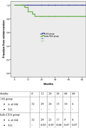
2tests, while late results were estimated by KaplaneMeier methods.Results:After propensity matching, 32 CAS interventions were matched with 32 redo CEAs. There were no peri-operative deaths or strokes. Cranial nerve palsy occurred in seven patients in the redo CEA group. Median duration of follow-up was 36 months (interquartile range 24e60; range 6e120). The estimated 5 year survival rate was 94% in the CAS group and 72% in the redo CEA group (p¼.1, log rank 2.4). There were no significant differences between the groups in terms of stroke free survival. In the CAS group, no severe restenosis were found, while in the redo CEA group eight patients had severe restenosis or occlusion of the operated carotid artery. Freedom from secondary restenosis at 4 years was 100% in the CAS group and 72.5% in the redo CEA group (p¼.005, log rank 7.9). The corresponding figures in terms of freedom from secondary re-intervention were 100% and 83%, respectively (p¼.02, log rank 4.8).
Conclusions:CAS and redo CEA in patients with post-CEA restenosis provided similar peri-operative results in a sample of equivalent patients. CAS patients had better follow-up results in terms of secondary restenosis and re-interventions. Further analysis is required with a larger number of patients and a longer follow-up time. ! 2017 European Society for Vascular Surgery. Published by Elsevier Ltd. All rights reserved.
Article history: Received 21 July 2017, Accepted 13 November 2017, Available online 26 December 2017 Keywords:Post-CEA restenosis, Redo CEA, CAS, Outcomes
INTRODUCTION
The optimal management of post-carotid endarterectomy (CEA) restenosis is still controversial. There are no rando-mised studies comparing the outcomes of open and endo-vascular treatment in this field. The current evidence is derived from retrospective non-randomised registries and
studies, and contemporary practice guidelines do not pro-vide specific advice on how these patients should be managed. While best medical treatment (BMT) seems to be reserved for stable severe asymptomatic and for mild symptomatic lesions, the most recent guidelines suggest that in patients needing re-intervention both open and endovascular interventions are effective, provided that a multidisciplinary decision including surgeon preference and patient choice is taken.1 Carotid artery stenting (CAS) is considered by several authors to be the treatment of choice because of its reduced invasiveness in comparison with the challenges associated with repeat CEA (redo CEA)
* Corresponding author.
E-mail addresses: dorigow@unifi.it; [email protected] (Walter Dorigo).
1078-5884/! 2017 European Society for Vascular Surgery. Published by Elsevier Ltd. All rights reserved.
procedures.2On the other hand, several studies and meta-analyses have shown similar rates of peri-operative com-plications with these techniques,3,4 while the risk of neurological complications and development of secondary restenosis during follow-up varies widely among the pub-lished data.5,6One possible explanation for these inconsis-tent results is that most published studies include heterogeneous patients in terms of demographic, clinical, and anatomical characteristics. Moreover, the duration of follow-up often varies between CAS and redo CEA. Finally, there is a general trend to treat late de novo atherosclerotic plaques with open surgery, while patients with early (<24 post-operative months) hyperplastic restenosis are more frequently treated by CAS.7
The aim of the present paper was to retrospectively compare open and endovascular treatment of post-CEA restenosis, in terms of peri-operative and late outcomes, among a homogenous population. To this end, a single centre matched case control study design was employed. MATERIALS AND METHODS
Patient population, pre-operative workup, indications for surgery, and surgical strategy
From January 2005 to December 2015, 3112 consecutive CEAs were performed at the study institution. In the same period of time, 140 consecutive CAS procedures were per-formed. Data on these interventions were prospectively collected in a dedicated database, the characteristics of which have been reported previously.8
A retrospective analysis of this database was performed, and 155 interventions carried out for post-CEA restenosis were identified. CAS was performed in 80 cases and redo CEA in 68. Seven patients underwent a common to internal ca-rotid artery bypass and were excluded from the study. Informed consent for the treatment of personal data was obtained from each patient before the insertion of their data in the prospective registry. The retrospective analysis of the data did not require approval of the institutional review board.The present study included all the patients from a prior study,7plus those who were operated on between January 2012 and December 2015, namely 49 new interventions.
All of the patients underwent both duplex scanning of the extracranial vessels and computed tomography angi-ography (CT) scans of the intra- and extra-cranial vessels and the cerebral parenchyma. The degree of carotid ste-nosis was measured using the North American Symptomatic Carotid Endarterectomy Trial (NASCET) criteria. The duplex criteria employed for the definition of post CEA carotid restenosis were those indicated in the national guidelines: namely, a peak systolic velocity (PSV) greater than 180 cm/s indicated the presence of a >70% restenosis, while a PSV greater than 225 cm/s was used to define a >80% reste-nosis.9 During the pre-operative workup, a phoniatrist conducted an otolaryngological evaluation of the motility of the vocal cords and damage to the cranial nerves.
The indications for re-intervention were the presence of symptomatic stenosis >50% or asymptomatic stenosis
>80% in progression despite optimal medical management (antiplatelets drugs, statins). Patients were considered to be symptomatic in the presence of ipsilateral neurological events (transient ischemic attack [TIA], or stroke) in the previous 6 months.
The selection of either open or endovascular treatment was not randomised; it was made at the surgeon’s discre-tion on the basis of clinical and anatomical consideradiscre-tions, including the characteristics of the neck and the previous surgical wound, the site of the lesion (distal lesions beyond the angle of the jaw), the presence of past cranial nerve injuries, and the duplex appearance of the lesion. In addi-tion, the time from the primary interventions was taken into account: in patients with early (<24 months) restenosis an endovascular approach was preferred, while in patients with late recurrent disease a new open surgical intervention was generally carried out. Technical details of open and endovascular interventions were as described previously.7 In CAS patients, a double antiplatelet regimen (aspirin and clopidogrel) was adopted in all patients for at least 6 months and, after that, ASA was administered indefinitely. In all redo CEA patients, post-operative medical treatment consisted of single antiplatelet therapy. Patients in both groups were treated with statins indefinitely.
Peri-operative and follow-up evaluation
A neurological evaluation was independently performed by an experienced neurologist at discharge and within 30 post-operative days, to determine the presence of minor or ma-jor strokes. A minor stroke was defined as any post-operative neurological event of more than 24 h duration followed by a recovery in the subsequent weeks or months either without impairment or with minimal residual functional impairment. A major stroke was defined as any post-operative neurolog-ical event of more than 24 h duration with residual functional impairment. An otolaryngological evaluation was also per-formed within the first peri-operative month. Patients with cranial nerve injuries at the first post-operative evaluation were further followed up every 6 months.
Follow-up was performed at 1, 6, and 12 months, and yearly thereafter, with a clinical examination and a duplex scan. All the follow-up visits were performed by a vascular surgeon; the patients were asked to report their clinical history during the index period, with particular interest paid to the occurrence of neurological events and their time of appearance, as well as major cardiovascular events. The collaboration of a neurologist was sought in cases where the clinical history suggested the occurrence of a new neurological symptom. DUS studies were performed by two vascular surgeons (W.D., R. P.), certified as institutional national tutors by the Italian Society for Vascular Investi-gation, using an Acuson Sequoia 512 Ultrasound System (Acuson Corporation, Mountain View, Ca, USA). During DUS examination, the patency of the operated vessel and the status of the contralateral internal carotid artery were assessed. Criteria for secondary restenosis were the same as primary in the redo CEA group, while in CAS patients
starting from 2008 the criteria as suggested by Lal et al. were used: namely, a PSV between 220 and 339 cm/s indicated the presence of a 50e79% restenosis, while a PSV higher than 340 cm/s was used to define >80% reste-nosis.10Additional data regarding long-term survival, major cardiovascular events and later hospitalisations were ob-tained from the Regional Health Care database. Indications for secondary re-interventions were the same as for primary re-interventions. The analysis of follow-up results ceased in December 2016.
Statistics
Patients in the CAS group were matched to patients in the redo CEA group on the basis of a propensity score, which is defined as the probability of treatment assignment condi-tional on observed baseline characteristics.11 For its calcu-lation, all the baseline characteristics that were reliably measured and were potentially relevant in the decision making process were included as confounders in a logistic regression model (forward conditional; 0.05 for entry, 0.10 for removal); they included demographic data, cardiovas-cular risk factors and comorbidities, clinical status (presence or absence of neurological symptoms), time between the re-intervention and the primary re-intervention, and anatomical characteristics of the lesions and of the cerebral parenchyma (Table 1). Logistic regression analysis revealed four baseline key factors that were significantly different between the two groups: the time from primary intervention, the degree of restenosis on the operated side, the presence of ipsilateral brain lesions in the pre-operative CT scan and the presence of coronary artery disease. These factors were then used to perform a propensity matching analysis and to generate a score and a score based matched control group. The two matched groups (CAS group, 32 patients; CEA group, 32 patients) were compared in terms of peri-operative (<30 days) results of interventions (TIA, stroke and death rates, cranial nerve injuries) with
c
2tests and Fisher’s exact tests, when necessary. Follow-up data were analysed with KaplaneMeier curves in terms of survival, stroke free and neurological symptom free survival, freedom from severe restenosis and occlusion, and freedom from re-intervention. The results in the two groups were compared by means of the log-rank test. The follow-up index (FUI) for late survival in the study group was assessed; follow-up index was defined as the ratio between the investigated follow-up period and the theoretically possible follow-up period, up to December 2016. Statistical analysis was performed with dedicated Windows software (Statistical Package for the Social Sciences (SSPS) 23; SPSS Inc., Chicago, IL, USA). Sta-tistical significance was defined as a p value less than .05. RESULTSDemographic data, risk factors, comorbidities, clinical and anatomical status
Of the 155 patients who had re-intervention during the study period, 38 had been operated on for primary CEA
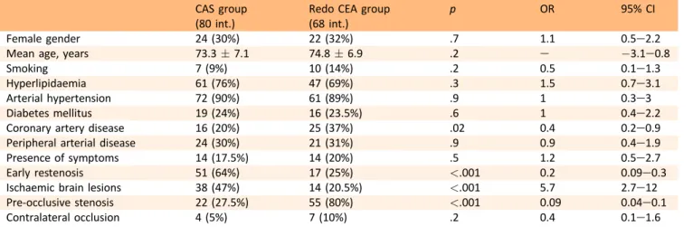
before the beginning of the study period and five had been operated on at other institutions. As a consequence, the crude rate of restenosis requiring re-intervention in the overall study group of 3112 CEAs was 112/3112 (3.5%). Baseline characteristics of the CAS and CEA patients in the original data set are reported inTable 2; the median time from primary intervention was 24 months (range 3e240, interquartile range [IQR] 14e36) in the CAS group and 60 months (range 12e254, IQR 25e254) in the redo CEA group (p<.001). Before the propensity matching, patients in the redo CEA group were more frequently affected by coronary artery disease and had a higher rate of pre-occlusive restenosis, while patients in the CAS group had a higher rate of silent ipsilateral ischemic brain lesions, and were more frequently operated on for an early restenosis. After propensity matching, there were 32 patients in both the CAS and redo CEA groups with correction of all differences in baseline characteristics (Table 3). In two patients with early restenosis in the redo CEA group, CAS had been excluded for significant tortuosity of the vessels, while for one patient with late restenosis in the CAS group, redo CEA had been excluded because of extensive scarring at the site of primary CEA. Most patients were asymptomatic at the time of the intervention, without differences between the two groups. The three symptomatic patients in the CAS group were operated on for a TIA, while in the redo CEA group, three patients had a TIA and two patients had a minor stroke. There were no differences between the two matched groups in terms of degree of carotid stenosis on the operated side and most patients were operated on for a >90% carotid restenosis; the status of the contralateral internal carotid artery was also similar. The median time from primary intervention was 36 months (range 12e140, IQR 21e81) in the CAS group and 48 months (range 12e 180, IQR 16e120) in the redo CEA group (p¼.04); however, the number of interventions performed for early or late Table 1. Definitions of baseline characteristics.
Covariate Definition
Gender Male or female patients
Diabetes mellitus Need for specific anti-diabetic drugs History of ischemic
heart disease
Prior myocardial infarction or surgical or percutaneous revascularisation Hyperlipidaemia Hyperlipidaemia on medical treatment Arterial hypertension Hypertension on medical treatment Smoking Patient still smoking following the
primary intervention Peripheral arterial
disease
ABI <0.9 or prior surgical or percutaneous revascularisation Clinical status Presence or absence of pre-operative
symptoms related to the operated internal carotid artery
Early restenosis Restenosis occurring within 24 months from the primary intervention Status of cerebral
parenchyma
Presence or absence of ipsilateral ischemic lesion at the pre-operative brain CT scan
Pre-occlusive stenosis Degree of stenosis greater than 90% at pre-operative evaluation
restenosis was equivalent between the groups. In three patients in the CAS group, the primary intervention con-sisted of endarterectomy and primary closure, while the remaining patients had been operated on with a patch closure. In the redo CEA group, the primary intervention consisted of endarterectomy and primary closure in four cases, in eversion endarterectomy in one case and in end-arterectomy with patch closure in the remaining 27. In the pre-operative evaluation, neither alterations of the motility of the vocal cords nor damage to the cranial nerves related to the primary intervention were detected in either group. The characteristics of patients excluded by matching in both the CAS and redo CEA groups are shown inTable 4. Intra-operative details
In the CAS group, a protective filter (Epifilter EZ 6.5, 0.01800
over a 0.01400guidewire, Boston Scientific, Natick, MA, USA)
was placed in the distal internal carotid artery in all but four patients, who had proximal protection devices (NPS system, W.L. Gore & Associates Inc, Flagstaff, AZ, USA). In 26 pa-tients an elgiloy stent was inserted, and in the remaining five a nitinol stent was used. The mean diameter of the stent was 8.5 mm (range 7e9 mm); the mean length of the covered segment of the carotid artery was 37.5 mm (range
30e40 mm). In one patient, concomitant angioplasty and stenting of a tight stenosis of right common iliac artery was performed.
In the redo CEA group, locoregional or cooperating pa-tient general anaesthesia with clinical monitoring of neurological status was used in 17 patients (47%), while the remaining patients had general anaesthesia with somato-sensory evoked potential monitoring. The cumulative shunt insertion rate was 18.5% (six cases); in four cases, a shunt was placed in patients undergoing general anaesthesia. For arterial reconstruction, polyurethane patch closure was used in all cases.
Peri-operative and early (<30 days) results
Technical success was achieved in all the CAS group pa-tients. There were no peri-operative deaths or strokes; one patient in the redo CEA group, operated on for a late (7 years) asymptomatic restenosis, suffered from a hemi-spheric TIA (contralateral upper limb weakness) in the first post-operative day. Both duplex scanning and CT angiogram confirmed the patency of the internal carotid artery and of the intracranial vessels, without signs of acute/subacute ischaemic lesion, and the patient was discharged asymp-tomatic on the fourth post-operative day. Another patient Table 3. Demographic data, comorbidities, risk factors in the two matched groups.
CAS group (32 int.)
Redo CEA group (32 int.)
p OR 95% CI
Female gender 7 (22%) 9 (28%) .5 1.9 0.5e7.8
Mean age, years 71.5 " 7.5 75.1 " 6.9 .4 e #7.2e0.04
Smoking 3 (9%) 4 (12%) .6 0.6 0.08e4.1
Hyperlipidaemia 26 (81%) 21 (66%) .2 1.6 0.7e4.8
Arterial hypertension 27 (84%) 28 (87%) .7 0.5 0.09e3.6
Diabetes mellitus 4 (12%) 4 (12%) 1 0.8 0.1e4.9
Coronary artery disease 6 (18%) 11 (34%) .1 0.2 0.06e1.1
Peripheral arterial disease 10 (31%) 6 (18%) .2 1.9 0.6e6.2
Presence of symptoms 3 (9%) 5 (15%) .4 2.1 0.3e13.2
Early restenosis 12 (37.5%) 13 (41%) .8 2.1 0.4e10.7
Ischaemic brain lesions 7 (22%) 7 (22%) 1 1.1 0.2e6
Pre-occlusive stenosis 21 (65%) 22 (68%) .8 0.9 0.1e5.3
Contralateral occlusion 4 (12%) 2 (6%) 0.4 1.3 0.3e1.9
Table 2. Demographic data, comorbidities, risk factors in the whole study group. CAS group
(80 int.)
Redo CEA group (68 int.)
p OR 95% CI
Female gender 24 (30%) 22 (32%) .7 1.1 0.5e2.2
Mean age, years 73.3 " 7.1 74.8 " 6.9 .2 e #3.1e0.8
Smoking 7 (9%) 10 (14%) .2 0.5 0.1e1.3
Hyperlipidaemia 61 (76%) 47 (69%) .3 1.5 0.7e3.1
Arterial hypertension 72 (90%) 61 (89%) .9 1 0.3e3
Diabetes mellitus 19 (24%) 16 (23.5%) .6 1 0.4e2.2
Coronary artery disease 16 (20%) 25 (37%) .02 0.4 0.2e0.9
Peripheral arterial disease 24 (30%) 21 (31%) .9 0.9 0.4e1.9
Presence of symptoms 14 (17.5%) 14 (20%) .5 1.2 0.5e2.7
Early restenosis 51 (64%) 17 (25%) <.001 0.2 0.09e0.3
Ischaemic brain lesions 38 (47%) 14 (20.5%) <.001 5.7 2.7e12
Pre-occlusive stenosis 22 (27.5%) 55 (80%) <.001 0.09 0.04e0.1
in the redo CEA group, operated on for a late (4 years) asymptomatic, pre-occlusive restenosis, developed typical chest pain with a significant increase in levels of circulating troponin I, and electrocardiographic signs of a myocardial infarction on the first post-operative day. The patient un-derwent a coronary angiogram with successful stent placement. Finally, a third patient in the redo CEA group underwent surgical revision on the first post-operative day for neck haematoma inducing dyspnoea. The cumulative rate of complications in the redo CEA group was 9.3%. Neither access related nor systemic complications were recorded in the CAS group (p¼.03 compared with the redo CEA group).
At the 30 day neurological evaluation no new events were recorded.
Peri-operative cranial nerve palsy occurred in seven pa-tients (21.8%) in the redo CEA group; in four cases, a deficit in the inferior laryngeal nerve was present, while the remaining three patients had an isolated deficit of the hy-poglossal nerve. One month otolaryngological evaluation showed complete regression of the deficit in six patients, while one of the patients with injured laryngeal nerve had persistent paralysis of the vocal cord, in the absence of dysphagia and with only a mild dysphonia.
Follow-up results
In the whole study group, 144 patients (97%) had available follow-up data, with a median duration of 36 months (range 3e120, IQR 24e65). With regard to matched groups, all patients had follow-up data available, with a median dura-tion of 36 months (range 6e120, IQR 24e60); follow-up duration was similar in the two groups (33.3 months in the CAS group and 35.6 months in the redo CEA group, p¼.1). The mean follow-up index was 0.72 (range 0.1e1) and it was similar between the two groups (0.71 and 0.73, respectively, p¼.5).
During follow-up there were seven deaths, one in the CAS group and six in the redo CEA group. The cause of death in the CAS group was pulmonary cancer, whereas in the redo CEA group, two cases were caused by cancer, one by acute myocardial infarction, one by a brain abscess and unknown causes in the remaining two patients. The esti-mated 5 year survival rate was 94% (standard error [SE] 0.06) in the CAS group and 89% (SE 0.1) in the redo CEA group (p¼.1, log rank 2.4). The corresponding figures in non-matched patients were 89% (SE 0.05) in the CAS group and 84% (SE 0.07) in the redo CEA group (p¼.6, log rank 0.2).
One major stroke was recorded at 5 years in a redo CEA patient who had a mild secondary restenosis. Cumulative stroke free survival at 5 years was 94% (SE 0.06) in the CAS group and 63% (SE 0.1) in the redo CEA group (p¼.07, log rank 3.2). No adjunctive ipsi- or contralateral neurological events were recorded during follow-up. In non-matched patients, the stroke free survival rate at 5 years was 84% (SE 0.07) in the redo CEA group and 88% in the CAS group (SE 0.05; p¼.3, log rank 0.7).
Significant (>80%) secondary restenosis (seven cases) or occlusion (one case) of the operated artery occurred in the redo CEA group; this was asymptomatic in all the patients. Secondary disease occurred at a mean time of 18.1 " 8.4 months from the re-intervention. One adjunctive moderate secondary restenosis was detected in this group. No sig-nificant restenosis was found in the CAS group. Freedom from secondary restenosis at 5 years was 100% in the CAS group and 72.5% (SE 0.08) in the redo CEA group (p¼.005, log rank 7.9;Fig. 1). Similar values were found among non matched patients (91% in the CAS group and 81% in the redo CEA group, p¼.05, log rank 2.7). Five of the seven patients who had significant restenosis underwent sec-ondary re-intervention, while the remaining two patients declined. In four cases the re-intervention consisted of a Table 4. Characteristics and outcome of patients excluded from propensity score matching compared with the matched groups.
CAS REDO CEA
Matched Not matched p Matched Not matched p
Patients 32 48 32 36
Female gender 7 (22%) 17 (35%) .2 9 (28%) 13 (36%) .4
Mean age, years 71.5 " 7.5 74.4 " 6.7 .4 75.1 " 6.9 74.4 " 7 .7
Smoking 3 (9%) 4 (8%) .7 4 (12%) 7 (19%) .4
Hyperlipidaemia 26 (81%) 35 (73%) .5 21 (66%) 26 (72%) .6
Arterial hypertension 27 (84%) 45 (93%) .2 28 (87%) 33 (91%) .7
Diabetes mellitus 4 (12%) 15 (31%) .09 4 (12%) 12 (33%) .04
Coronary artery disease 6 (18%) 10 (21%) .8 11 (34%) 14 (39%) .7
Peripheral arterial disease 10 (31%) 14 (29%) .6 6 (18%) 15 (42%) .04
Presence of symptoms 3 (9%) 11 (23%) .1 5 (15%) 9 (25%) .3
Early restenosis 12 (37.5%) 39 (81%) <.001 13 (41%) 4 (11%) .005
Ischaemic brain lesions 7 (22%) 31 (64%) <.001 7 (22%) 7 (19%) .6
Pre-occlusive stenosis 21 (65%) 1 (2%) <.001 22 (68%) 33 (91%) .01 Contralateral occlusion 4 (12%) 0 .01 2 (6%) 5 (14%) .3 Peri-operative results Death 0 0 e 0 0 e Stroke 0 0 e 0 1 (3%) .2 Myocardial infarction 0 0 1 (3%) 0 .2
new CAS, while one patient with mild chronic renal failure underwent autologous saphenous vein common to internal carotid bypass. At further controls, none of these patients showed disease recurrence following secondary re-intervention. Estimated 5 year freedom from secondary re-intervention in the redo CEA group was 83.5% (SE 0.07, p¼.02, log rank 4.8 in comparison with the CAS group;
Fig. 2).Table 5reports the follow-up results in the overall population and in the matched and non-matched groups.
DISCUSSION
Several studies have addressed the peri-operative and follow-up outcomes of open and endovascular treatment of post-CEA restenosis, showing satisfactory early and late results with both techniques. However, there is a limited number of studies directly comparing the two treatment options,5e7,12e19and in the majority of these studies CAS was reserved for early hyperplastic lesions, while redo CEA was preferred in patients with late de novo atherosclerotic plaques. In the present study, results of a previous comparative series7were updated by adding data on the interventions performed in the years following its publica-tion, and attempts were made to make the comparison reliable by adjusting for baseline confounding factors.
Cases of moderate and severe symptomatic restenosis and severe asymptomatic restenosis continue to be oper-ated on; although the effectiveness of treating asymptom-atic patients is debatable,20 there is a well established relationship between restenosis and late ipsilateral stroke,21 particularly when progression occurs.1,22 Also the recent guidelines of the European Society for Vascular Surgery Figure 2. KaplaneMeier curves for 5 year freedom from secondary re-intervention in both groups with number of patients at risk at different time intervals (p¼.02, log rank 4.8).
Table 5. Results at 5 years in the whole study group and in matched and non-matched patients.
Survival Stroke free survival Freedom from secondary restenosis Freedom from secondary re-intervention Whole study group
CAS 90% 90% 94% 96% redo CEA 85% 77% 78% 86% p .1 .08 .006 .03 Matched groups CAS 94% 94% 100% 100% redo CEA 89% 63% 72.5% 83.5% p .1 .07 .005 .02
Non matched groups
CAS 89% 88% 91% 94.5%
redo CEA 84% 84% 81% 89%
p .6 .3 .05 .2
Figure 1. KaplaneMeier curves for 5 year freedom from severe secondary restenosis in both groups with number of patients at risk at different time intervals (p¼.005, log rank 7.9).
(ESVS) recommend, with a B level of evidence, intervention in patients with severe asymptomatic restenosis, provided that an accurate multidisciplinary evaluation is performed.1 This is typical of daily practice, in which surgeons, neurol-ogists, and otolaryngologists are involved in the evaluation of patients. In the present study, the main criterion for the choice of the treatment was the type of lesion and conse-quently the time from the primary intervention; the ma-jority of studies in the extant literature include patients with different lesions treated with different techniques. This can cause a significant bias, considering that early restenosis is usually a fibrous and hyperplastic stable lesion, with low potential for microembolisation, while late atherosclerotic recurrent plaques are often complex and complicated le-sions with high embolic risk. Moreover, CAS patients more frequently have a less tight stenosis than redo CEA patients; the reduced invasiveness of CAS, along with the higher percentage of ipsilateral silent brain lesions observed on the pre-operative CT scan, could probably have pushed treat-ment of less severe stenosis with CAS rather than with redo CEA. A similar finding was reported in a recent meta-analysis, in which redo CEA patients had a severe ipsilat-eral stenosis >70% in 95% of the cases, compared with some 85% among CAS patients.2Finally, redo CEA patients had a significantly higher percentage of coronary artery disease than CAS patients, probably reflecting the more advanced state of atherosclerotic disease in those patients. After propensity matching, it was possible to compare early and late outcomes of CAS and redo CEA in clinically and anatomically equivalent populations. Recently, Arhuidese et al.5performed a similar analysis in a very large cohort of 1862 patients; however, in that study the time between primary and secondary intervention was not reported and the authors were unable to stratify the results for that parameter.
In the present study, peri-operative results were good in both groups; no deaths or strokes were recorded. This was achieved in spite of the presence of a high rate of late restenosis in both groups, placing the patients at potentially high risk of intra-operative brain embolisation, particularly among CAS patients. Considering that, on the basis of recent guidelines,1 low rates of peri-operative stroke and death are crucial to confer long-term benefit mainly in patients with asymptomatic restenosis, the present results further encourage continued treatment of severe asymp-tomatic restenosis.
As expected, the rate of peri-operative systemic and local complications was higher in redo CEA patients, and the same findings were evident when cranial nerve injuries were examined. There was a significant number of post-operative cranial nerve injuries; this is higher than re-ported in the literature (approximately 5e7%) and closer to the reported rate of 14% in AbuRahma’s series.4,12 A possible explanation for this finding is that the present study used a very accurate otolaryngological surveillance program, while in the majority of the published studies, the diagnosis of cranial nerve injuries was surgeon driven; this could have contributed to the diagnosis of minimal
peripheral lesions in the current study, which might be under reported in other series.5Furthermore, in the present study the majority of cranial nerve injuries were reversible, and when the rate of permanent lesions only was analysed, the percentage of cranial nerve injuries fell to 3%, which is in line with the data in the literature.
Follow-up data were available in all the patients, with a robust follow-up index and a median duration of 36 months; this is higher than that reported in a recent meta-analysis,3where the mean duration of follow-up of the main comparative studies was approximately 13 months. Most importantly, the duration of follow-up was similar between the two groups, while previous studies have observed a significantly shorter follow-up time in CAS patients.5 Follow-up results were fairly good, with satisfactory survival rates and a very limited number of neurological events at 5 years. In their meta-analysis, Fokkema et al.3reported only seven strokes and seven deaths during follow-up among 453 pa-tients treated by CAS and 731 papa-tients treated by redo CEA, after a mean follow-up of 13 months. Similar satisfactory results were also reported after 1 year of follow-up by Arhuidese et al.,5thus confirming the effectiveness of both approaches for stroke prevention during follow-up.
As far as secondary restenosis and re-interventions are concerned, the present results confirmed results from a previous study, with a significantly increased risk among redo CEA patients.
This again is a surprising finding, considering that in the majority of the published studies, the risk of secondary restenosis is higher or at least equivalent in CAS patients compared with redo CEA patients. Only Bettendorf et al.16 reported results similar to those in the present study, with a 14% rate of secondary recurrences among patients treated by redo CEA compared with 6% in patients treated by CAS; they found that the failure to take
b
blockers during follow-up was an independent predictor of multiple re-currences. Moreover, a recent study from De Marino et al.6 reported a trend towards a higher severe recurrent reste-nosis rate in redo CEA patients at 3 years; however, this was not statistically significant. As previously reported, possible explanations for these findings include differences in post-operative medical treatment and a possible “literature driven” stricter control of risk factors in patients treated with CAS than in patients treated with redo CEA, thus optimising the post-operative strategy primarily in the former patients. Moreover, a possible overestimation of secondary restenosis in the literature, because of different criteria for CAS duplex surveillance between groups, must be taken into account. The present authors adopted the criteria suggested by Lal9as soon as they were published, and this may further contribute to the explanation for the low rate of secondary restenosis among CAS patients. However, once recurrent restenosis has occurred, it can be safely treated again by both open and endovascular surgery, as demonstrated by the present study. However, consid-ering the relatively low potential for late neurological events in patients with post-CEA restenosis, it is likely that in the future, there will be an increasing role for aggressivecontrol of risk factors and optimal medical management, which will probably reduce the need for invasive interventions.
Based on these data, the present authors are now changing strategy for dealing with patients with post-CEA restenosis. As such, the possibility of CAS is evaluated for all patients who are candidates for re-intervention, inde-pendently from the kind of lesion and from the time be-tween primary and secondary intervention. Redo CEA is reserved for patients with anatomically complex lesions for which CAS is contraindicated.
This study has several limitations that should be consid-ered. First, it is a retrospective, non-randomised study, with a limited number of patients and events. Moreover, while peri-operative results were assessed by an independent neurologist, follow-up outcomes were evaluated by a vascular surgeon and only selected patients were evaluated by a neurologist. On the other hand, the propensity matching allowed reduction of the selection bias in the choice of treatment. Moreover, the follow-up was accurate and robust and its duration was similar between CAS and redo CEA patients. Considering that it is probably not feasible to perform a randomised controlled trial, in an achievable time period,3in this small group of patients with a low event rate in the early and long-term, these kind of studies can contribute to building knowledge and adding a good level of evidence to the paucity of data in the contemporary literature.
CONCLUSIONS
CAS and redo CEA in patients with post-CEA restenosis provided similar peri-operative results in equivalent groups of patients. CAS patients had better follow-up results in terms of secondary restenosis and re-interventions; this, together with the non-negligible rate of peri-operative cranial nerve palsy in redo CEA patients, suggests a pri-mary role for CAS in post-CEA restenosis.
ACKNOWLEDGEMENTS
Special acknowledgements to Dr. A. Santucci for statistical revision. CONFLICTS OF INTEREST None. FUNDING None. REFERENCES
1 Naylor AR, Ricco JB, de Borst GJ, Debus S, deHaro J, Halliday A,
et al. Management of atherosclerotic carotid and vertebral artery disease: 2017 clinical practice guidelines of the Euro-pean Society for Vascular Surgery (ESVS). Eur J Vasc Endovasc Surg 2018;55:3e81.
2 Schermerhorn ML, Fokkema M, Goodney P, Dillavou ED, Jim J,
Kenwood CT, et al. The impact of Centers for Medicare and Medicaid Services high-risk criteria on outcome after carotid
endarterectomy and carotid artery stenting in the SVS Vascular Registry. J Vasc Surg 2013;57:1318e24.
3 Fokkema M, Vrijenhoek JE, Den Ruijter HM, Groenwold RH,
Schermerhorn ML, Bots ML, et al. Stenting versus endarter-ectomy for restenosis following prior ipsilateral carotid end-arterectomy: an individual patient data meta-analysis. Ann Surg 2015;261:598e604.
4 Tu J, Wang S, Huo Z, Wu R, Yao C, Wang S. Repeated carotid
endarterectomy versus carotid artery stenting for patients with carotid restenosis after carotid endarterectomy: systematic review and meta-analysis. Surgery 2015;157:1166e73.
5 Arhuidese I, Obeid T, Nejim B, Locham S, Hicks CW, Malas MB.
Stenting versus endarterectomy after prior ipsilateral carotid endarterectomy. J Vasc Surg 2017;65:1e11.
6 Marques de Marino P, Martinez Lopez I, Hernandez
Mateo MM, Cernuda Artero I, Cabrero Fernandez M, Reina Gutierrez MT, et al. Open versus endovascular treatment for patients with post-carotid endarterectomy restenosis: early and long-term results. Ann Vasc Surg 2016;36:159e65.
7 Dorigo W, Pulli R, Fargion A, Pratesi G, Angiletta D, Aletto I,
et al. Comparison of open and endovascular treatments of post-carotid endarterectomy restenosis. Eur J Vasc Endovasc Surg 2013;45:437e52.
8 Dorigo W, Pulli R, Pratesi G, Fargion A, Marek J, Alessi
Innocenti A, et al. Early and long-term results of carotid endarterectomy in diabetic patients. J Vasc Surg 2011;53: 44e52.
9 Antignani PL, Benedetti-Valentini F, Aluigi L, Baroncelli TA,
Camporese G, Failla G, et al. Diagnosis of vascular diseases. Ultrasound investigations e guidelines of the Italian Society for Vascular Investigation. Int Angiol 2012;31:1e7.
10 Lal BK, Hobson 2nd RW, Tofighi B, Kapadia I, Cuadra S, Jamil Z.
Duplex ultrasound velocity criteria for the stented carotid ar-tery. J Vasc Surg 2008;47:63e73.
11 Austin PC. An introduction to propensity score methods for
reducing the effects of confounding in observational studies. Multivariate Behav Res 2011;46:399e424.
12 AbuRahma AF, Abu-Halimah S, Hass SM, Nanjundappa A,
Stone PA, Mousa A, et al. Carotid artery stenting outcomes are equivalent to carotid endarterectomy outcomes for patients with post-carotid endarterectomy stenosis. J Vasc Surg 2010;52:1180e7.
13 Antonello M, Deriu GP, Frigatti P, Amistà P, Lepidi S,
Stramanà R, et al. Does the type of carotid artery closure in-fluence the management of recurrent carotid artery stenosis? Results of a 6-year prospective comparative study. Surgery 2008;143:51e7.
14 Fokkema M, de Borst GJ, Nolan BW, Lo RC, Cambria RA,
Powell RJ, et al. Carotid stenting versus endarterectomy in patients undergoing reintervention after prior carotid endar-terectomy. J Vasc Surg 2014;59:8e15.
15 Attigah N, Kulkens S, Deyle C, Ringleb P, Hartmann M,
Geisbusch P, et al. Redo surgery or carotid stenting for reste-nosis after carotid endarterectomy: results of two different treatment strategies. Ann Vasc Surg 2010;24:190e5.
16 Bettendorf MJ, Mansour MA, Davis AT, Sugiyama GT, Cali RF,
Gorsuch JM, et al. Carotid angioplasty and stenting versus redo endarterectomy for recurrent stenosis. Am J Surg 2007;193: 356e9.
17 Bowser AN, Bandyk DF, Evans A, Novotney M, Leo F, Back MR,
et al. Outcome of carotid stent-assisted angioplasty versus open surgical repair of recurrent carotid stenosis. J Vasc Surg 2003;38:432e8.
18 Hobson 2nd RW, Goldstein JE, Jamil Z, Lee BC, Padberg Jr FT, Hanna AK, et al. Carotid restenosis: operative and endovascular management. J Vasc Surg 1999;29:228e38.
19 Rockman CB, Bajakian D, Jacobowitz GR, Maldonado T,
Greenwald U, Nalbandian MM, et al. Impact of carotid artery angioplasty and stenting on management of recurrent carotid artery stenosis. Ann Vasc Surg 2004;18:151e7.
20 Brott TG, Halperin JL, Abbara S, Bacharach JM, Barr JD, Bush RL,
et al. 2011 ASA/ACCF/AHA/AANN/AANS/ACR/ASNR/CNS/SAIP/ SCAI/SIR/SNIS/SVM/SVS guideline on the management of
pa-tients with extracranial carotid and vertebral artery disease. J Am Coll Cardiol 2011:1002e44.
21 Kumar R, Batchelder A, Saratzis A, AbuRahma AF, Ringleb P,
Lal BK, et al. Restenosis after carotid interventions and its relationship with recurrent ipsilateral stroke: a Systematic re-view and meta-analysis. Eur J Vasc Endovasc Surg 2017;53: 766e75.
22 Ouriel K, Green RM. Appropriate frequency of carotid duplex
testing following carotid endarterectomy. Am J Surg 1995;170: 144e7.
Eur J Vasc Endovasc Surg (2018) 55, 161
COUP D’OEIL
Complex Open Surgical Revascularisation for Successful Resolution of
Bilateral Lower Limb Ischaemia
Alexandros Malliosa,*, William C. Jenningsb
aDepartment of Vascular Surgery, Institut Mutualiste Montsouris, Paris, France bUniversity of Oklahoma College of Medicine, Tulsa, OK, USA
A 68 year old male patient with previous aorto-iliac stenting, occlusion of the right iliac and both common femoral arteries after endarterectomies, occluded right ilio-femoral and left femoro-popliteal bypasses, occluded left superficial femoral and popliteal arteries, and right below knee amputation presented with left sided rest pain. Left axillo-profunda (Dacron; (A,B) yellow arrows) and profunda-posterior tibial (left wrist to shoulder cephalic vein; (B) red arrows) bypasses were performed. Six months later the patient presented with right stump pain and a non-healing ulcer. A left to right crossover bypass (graft to profunda; (A) green arrow) was performed. One year later all grafts remain patent; the patient is asymptomatic and ambulating.
* Corresponding author. Department of Vascular Surgery, Institut Mutualiste Montsouris, 42 Boulevard Jourdan, 75014, Paris, France. E-mail address:[email protected](Alexandros Mallios).
1078-5884/! 2017 Published by Elsevier Ltd on behalf of European Society for Vascular Surgery.