A model of Helicobacter pylori persistence
in a case of gastric cancer
Rossella Grande, Mara Di Giulio, Emanuela Di Campli, Soraya Di Bartolomeo, Luigina Cellini Department of Drug Sciences, “G. d’Annunzio” University, Chieti-Pescara, Italy
INTRODUCTION
Gastric cancer represents the second most com-mon cancer in the world and its association with
Helicobacter pylori infection was confirmed on the
basis of several epidemiological studies (Ito et al., 2009). H. pylori virulence genes and genomic plas-ticity play a key role in the onset of the infection (Cellini and Donelli 2000; Cellini et al., 2006; Sgouras et al., 2009). The vacA gene contains three variable parts corresponding to the s-region, the i-region and the m-i-region which encode the signal, intermediate and the middle peptides, respective-ly. For the signal region, two distinct isotypes, s1 and s2, were recognized, whereas, for the middle
Corresponding author
Prof. Luigina Cellini
Department of Drug Sciences University “G. d’Annunzio”
Via dei Vestini, 31 - 66100 Chieti, Italy E-mail: [email protected]
region, m1 or m2 alleles were categorized. The
vacA genotype is associated with in vitro
cytotox-in activity: s1 and m1 have greater cytotoxcytotox-in ac-tivity than s2 and m2, respectively (van Doorn et
al., 1998; Chisholm et al., 2002; Sgouras et al.,
2009). Recently, a new polymorphism in the in-termediate (i) region, located between the s and m regions, was identified and associated with gas-tric cancer in Iran (Rhead et al., 2007). With regard to the cagA gene, H. pylori strains, possessing the
cag pathogenicity island (PAI), are more
associat-ed with disease development than those lacking this region (Argent et al., 2005). The existence of EPIYA motifs, repeated within the variable region of the protein, induces the phosphorylation of CagA protein by the Src kinases of the host, there-by producing a morphological modification of the epithelial cells (Argent et al., 2005). Helicobacter
pylori strains which encode CagA proteins
con-taining a greater number of EPIYA motifs are mainly associated with gastric cancer develop-ment (Argent et al., 2005).
The aim of this work was to analyze several clones of Helicobacter pylori isolated from a patient with gastric cancer, to evaluate i) genetic variability ii) virulence factors profile and iii) antimicrobial susceptibility against the drugs com-monly used in the H. pylori therapy. A total of 32 H. pylori clones isolated from a biopsy sample coming from a pa-tient with gastric cancer previously treated for H. pylori infection, were analyzed for: the genetic variability by ampli-fied fragment polymorphism analysis; the vacA, cagA virulence status by PCR; the antimicrobial susceptibility by min-imum inhibitory concentrations with the agar dilution method towards amoxicillin, clarithromycin, levofloxacin and tinidazole. The patient showed a mixed infection with the presence of at least 3 different strains. The clones isolated possessed the vacA, cagA virulence factors with a different allelic combination (vacA s1/i1/m1; s1/i1i2/m1; s2/i2/m2; s2/i1i2/m2) together with repeated cagA EPIYA motif pattern P1P2P3P3P3. Moreover, a pattern of multi-drug resist-ance was disclosed in the different clones. The presence of multiple H. pylori strains colonizing the same patient, with the main virulence factors displaying a different allelic combination and a different multi-drug resistance among iso-lates, point out the role of genetic variability generating, in time, more virulent and adapted strains.
KEY WORDS: cagA status, Drug resistance, Helicobacter pylori, Mixed infection
SUMMARY
With regard to genetic variability, H. pylori strains, isolated from different patients, show a signifi-cantly high degree of variability either in size or in gene order because of the acquisition of new DNA sequences (Taylor et al., 1992; Suerbaum, 2000): mixed H. pylori infection in the same pa-tient is significantly related to strains more re-sistant to antibiotics with a more virulent geno-type than strains responsible for single infection (Cellini et al., 2006).
This study analyses several clones isolated from a patient with gastric cancer, previously treated for H. pylori infection, for the evaluation of: i) ge-netic variability, ii) virulence factors profile and iii) antimicrobial susceptibility against the drugs commonly used in the H. pylori therapy, to better understand the adaptation dynamic of the mi-croorganism to the host.
MATERIAL AND METHODS Patient and H. pylori culture
A 68-year-old female, selected from a previous study among a group of individuals subjected to upper gastrointestinal (GI) endoscopy for gas-trointestinal complaints, was considered in this study (Cellini et al., 2008b). Written informed con-sent was obtained from the patient.
The patient, previously treated for H. pylori in-fection with a 7-day standard treatment consist-ing of a proton pump inhibitor (PPI) (20 mg b.d.) combined with clarithromycin (500 mg b.d.) and amoxicillin (1 g b.d.) underwent an endoscopy because of persistent GI disturbances. A biopsy sample was collected from the antrum for cul-ture in Portagerm-pylori (Bio-Merieux, Marcy L’Etoile, France) and processed microbiological-ly within 24 h as previousmicrobiological-ly described (Cellini et
al., 2008b). Helicobacter pylori colonies were
iden-tified for colony morphology, Gram staining and positive reaction with urease, catalase and oxi-dase. From the isolated strain, named H. pylori 9L, 32 clones were picked up randomly from the primary culture, transferred on CA and incubat-ed in a microaerophylic atmosphere for 3 days at 37°C.
Isolated clones were collected and stored until use at -80°C by Drumm and Sherman method (1989).
Amplified fragment length polymorphism (AFLP)
The chromosomal DNA was extracted from each clone by using Qiamp Tissue DNA isolation minikit (QIAGEN S.p.a, Milan, Italy) and the AFLP analysis was carried out following the methodology reported by Gibson et al., (1998). The DNA fingerprintings were analyzed with GEL COMPAR Software, Windows version 4.1 (Applied Math, Kortrijk, Belgium). The similari-ty coefficient indicating the relationship between the strains was calculated by using band posi-tions (Gerner-Smidt et al., 1998) by the GEL COMPAR program as previously reported (Cellini
et al., 2006). A similarity coefficient <70% was
considered significant for mixed infection. The experiments were performed in triplicate.
Virulence factors genotyping
PCR reactions of vacA s/m, cagA EPIYA motifs of the H. pylori isolated clones were carried out as described before (Chisholm et al., 2002; Cellini et
al., 2008a), while, the amplification of vacA
i-Region was performed by using the methodolo-gy reported by Rhead et al. (2007). The oligonu-cleotide primers used were listed in Table 1. The amplification was performed in a 2700 Thermocycler (PE-Applied Biosystems) and con-sisted of 5 min of denaturation followed by 30-35 cycles consisting of: 40 s at 94°C of denatura-tion, 1 min of annealing at 52°C and 1 min and 30 s of extension at 72°C for the analysis of vacA s/i/m regions; 1 min at 94°C, 1 min at 55°C and 1 min at 72°C for the amplification of cagA 3’ vari-able region and EPIYA phosphorylation motifs. After the last cycle, the extension was continued for 5 min (Cellini et al., 2008a). The PCR prod-ucts were examined by electrophoresis in 2% (w/v) agarose gel at 100 V for 30 min. The exper-iments were performed in triplicate.
Analysis of a new cagA-P3 amplified product
The cagA-P3 PCR products were analyzed by agarose gel electrophoresis. A new band of about 300 bp was detected in all 32 samples. The band was cut from the gel and the amplified DNA frag-ment was eluited by using QIAquick Spin GEN), purified by spin column QIAQuick (QIA-GEN) and cyclesequenced (on both strands) us-ing the ABI PRISM Big Dye Terminator Cycle Sequencing kit (Applied Biosystems). DNA
quences were analyzed on an automated se-quencer, ABI PRISM 310, version 3.4.1 (Applied Biosystems). The band was isolated and se-quenced in all samples in which was detected. The resulting nucleotide sequences of about 300 bp were aligned using the Sequence Navigator Software package (Applied Biosystems). Sequence comparisons were subsequently car-ried out using BLAST Search in the National Center of Biotechnology Information (NCBI). All detections were done in triplicate.
Antibiotic susceptibility tests
Amoxicillin (Sigma Chemical Co., St Louis, MO, USA), clarithromycin (Abbott Laboratories, Abbott Park, Ill., USA), levofloxacin (FLUKA-Biochemika, Buchs, Switzerland) and tinidazole (Sigma), commonly used in H. pylori therapy, were tested against the isolated clones. The Minimum Inhibitory Concentrations (MICs) were evaluated using the standard agar dilution method accord-ing to the Clinical and Laboratory Standards Institute (CLSI) (2005) guidelines, using Mueller-Hinton agar (Oxoid) with 7% of defibrinated horse blood. Two-fold dilutions of the antibiotics were
added to melted agar in order to obtain the fol-lowing final concentrations: from 2 to 0.06 µg/ml for amoxicillin and clarithromycin, from 8 to 0.25 µg/ml for levofloxacin and from 20 to 0.6 µg/ml for tinidazole. Agar plates were inoculated using a Steers replicator delivering a bacterial suspen-sion of approximately of 5x10⁴colony-forming
units (CFUs)/spot. Test plates were incubated as mentioned before (Cellini et al., 2008b). MIC was defined as the lowest concentration of the antibi-otics inhibiting the visible growth.
Bacteria were considered resistant when MIC was greater than 0.5 µg/ml for amoxicillin, 1 µg/ml for clarithromycin, 5 µg/ml for levofloxacin and tinidazole (Cellini et al., 2008b). The reference strain H. pylori ATCC 43629 was inserted in the experiments as the control.
The experiments were performed in triplicate.
RESULTS AFLP analysis
A total of 32 clones were isolated from a patient with gastric cancer which was previously treated
TABLE 1 - Oligonucleotides used for PCR-based typing.
Primer designation Gene Sequence (5’-3’) Expected size
of PCR product (bp)
VA1-F vacA ATGGAAATACAACAAACACAC 259 (s1)
VA1-R CTGCTTGAATGCGCCAAAC 286 (s2)
VAG-F vacA CAATCTGTCCAATCAAGCGAG 567 (m1)
VAG-R GCGTCAAAATAATTCCAAGG 642 (m2)
VacF1 vacA GTTGGGATTGGGGGAATGCCG
C1R TTAATTTAACGCTTGTTTGAAG 426 (i1)
C2R GATCAACGCTCTGATTTGA 432 (i2)
cag2a cagA GGAACCCTAGTCGGTAATG 550-800
cag4a ATCTTTGAGCTTGTCTATCG
cagA28F cagA TCTCAAAGGAGCAATTGGC 264-291 (P1) cagA-P1C GTCCTGCTTTCTTTTTATTAACTTKAGC
cagA-P2CG cagA TTTAGCAACTTGAGCGTAAATGGG 309-336 (P2) cagA-P2TA cagA TTTAGCAACTTGAGTATAAATGGG 309-336 (P2) cagA-P3E cagA ATCAATTGTAGCGTAAATGGG 465-498/672 (P3)
ADH1* ACGGTATGCGACAG
ADH2* AGCTCTGTCGCATACCGTGAG
HI-A GGTATGCGACAGAGCTTA
for H. pylori infection. The genetic analysis of the isolates, performed by AFLP technique, showed different DNA fingerprintings associated to the presence of a mixed infection in the host as shown in Figure 1. At least 3 different strains were detected, as confirmed by the Jaccard co-efficients whose values were <70%. In particular, the comparison of the clone 9/4 with the clones 9/6 and 9/5 displayed a Jaccard coefficient of 29% and 46% respectively, while the Jaccard coeffi-cient derived by the comparison of 9/5 with 9/6 was equal to 62%.
Virulence factors detection
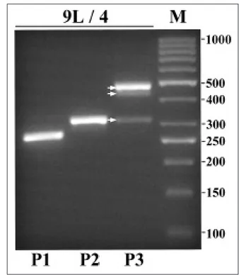
The analysis of the main virulence factors in each clone showed a genetic plasticity associated to a different allelic combinations detected in the dif-ferent analyzed colonies. All the isolates studied were cagA positive. A new amplified fragment size of about 300 bp was detected and sequenced in 32 clones. The 32 colonies displayed the cagA EPIYA motifs pattern corresponding to P1P2P3 P3P3 as shown in Figure 2. In particular, the cagA EPIYA motifs pattern P1P2P3P3P3, displays a new fragment size of about 300 bp, which was sequenced and aligned by using BLAST Search with H. pylori J99 genomic sequence present in
DATA BASE (NCBI), confirming that the se-quences of 300 bp belong to cagA gene with an identity of about 83-85%. The sequence data, ob-tained by the 32 clones, had an identity of 93% when compared each other and 2 representative sequences of the 32 colonies were deposited in GenBank (accession numbers GQ855282 and GQ855283). With regard to vacA gene, the allel-ic combination s1/i1/m1 was detected in 25 out of 32, with a frequency of 78.13%; s1/i1i2/m1 in 2 out of 32 with a frequency of 6.25%; s2/i1i2/m2 in 4 out of 32 with a frequency of 12.5% and s2/i2/m2 in 1 out of 32 isolates with a frequency of 3.12%. A comparative analysis between the
vacA isotypes and the cagA EPIYA combination
pattern detected in the 32 colonies studied, showed a prevalence of isolates (71.87%) pos-sessing both vacA s1/i1/m1 and cagA EPIYA P1P2P3P3P3.
Antibiotic susceptibility test
The antibiotic susceptibility test, performed on 32 clones displayed a different pattern of multi-drug resistances. A resistance against
clar-FIGURE 1 - Representative image of Amplified Fragment
Length Polymorphism (AFLP) profiles of 13 Helicobacter pylori 9L clones isolated from a biopsy obtained from a
patient with gastric cancer. FIGURE 2 - Representative image of PCR amplification
of the cagA EPIYA motifs pattern P1P2P3P3P3 from a clone isolated from a patient with gastric cancer. M, size marker in base pairs.
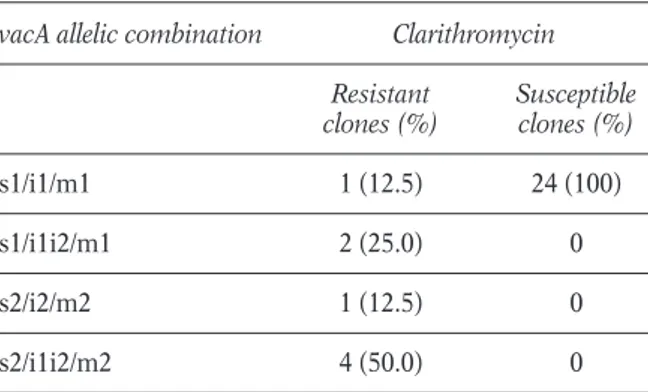
ithromycin and levofloxacin was found in dif-ferent coinfectant clones. In particular, the re-sistance to clarithromycin was detected in 8 out of 32 (25%) analyzed clones, while the resistance to levofloxacin was detected in all isolated colonies. On the contrary, the amoxicillin and tinidazole were efficacious against all detected clones. Interestingly, no association was detect between the DNA profile or virulence marker pattern and the clarithromycin resistance. In fact, among the clones resistant to clar-ithromycin 4 out of 8 were s2/i1i2/m2; 2 out of 8 were s1/i1i2/m1; 1 out of 8 was s1/i1/m1 and 1 out of 8 was s2/i2/m2 (Table 2).
DISCUSSION
In the present work we describe a case of a pa-tient colonized by H. pylori and affected by gas-tric cancer. Multiple H. pylori strains, possessing the main virulence factors and displaying a dif-ferent antimicrobial susceptibility pattern were harboured in the patient studied. Several studies demonstrate that recombination events occurred frequently during chronic infection, producing multiple H. pylori mosaic genotypes (Kersulyte et
al., 1999; Falush et al., 2001; Kraft et al., 2006).
The macroevolution of the strains, detected in the present study, might be associated either to a col-onization of the host by multiple strains acquired in time or to an adaptation of a single strain to the host. The first hypothesis might be explained with the existence of a mixed infection, in which, one or more strains were present in a minor fraction or at a different site in the gastric mucosa so that
they escaped identification in the first isolation (Wang et al., 1998).
The second hypothesis could be associated with a microbe adaptation to the stressing stimuli such as an ineffective antimicrobial therapy. Failure of the H. pylori treatment or sub-hibitory concentrations of antibiotics might in-duce the evolution of more resistant strains, dif-ficult to eradicate (Hung et al., 2009; Wueppenhorst et al., 2009). In the present work, the clones isolated from the same patient, dis-played differences in the DNA fingerprintings, the antimicrobial susceptibility patterns and the allelic status of virulence genes. Interestingly, contrary to Miehlke et al. (1999), who demon-strated that patients with gastric cancer were col-onized by a single predominant strain, we found multiple strains in the same host. These differ-ences might be explained by an increased con-sumption of antibiotics in patients during the last few years that presumably generated strains which developed a high genetic variability as well as a retention of the virulent factors associated with more severe gastric diseases. On the other hand (Suerbaum and Josenhans 2007) the per-sistence of H. pylori in the host is due to the con-tinuous improvement of its genome, predomi-nantly by inter-strain recombination, in the same. Therefore, the genetic variability of the mi-croorganism contributes to the host adaptation also by evading the natural immune response fa-voring the bacterial survival and enabling the on-set of the most dangerous complication of H.
py-lori infection: the gastric carcinoma. The
analy-sis of the status of virulence markers in all stud-ied clones confirmed the correlation between the severity of the disease and the presence of a greater number of the cagA EPIYA motifs (Basso
et al., 2008; Jones et al., 2009) as well as the
prevalence of the allelic combination s1i1m1 of the vacA gene. The wide genetic variability de-tected in the virulence marker status confirmed the genomic plasticity recorded in the DNA pro-files, although no association was found between the same DNA fingerprintings and the same al-lelic combinations of the virulence markers. Sgouras et al. (2009) have recently demonstrat-ed, in a study performed on 98 children, the pres-ence of microevolving strains having identical RAPD profiles and producing CagA proteins pos-sessing a variable number of EPIYA motifs. The
TABLE 2 - Comparison between vacA allelic
combination and clarithromycin susceptibility of 32 Helicobacter pylori clones. vacA allelic combination Clarithromycin
Resistant Susceptible clones (%) clones (%) s1/i1/m1 1 (12.5) 24 (100) s1/i1i2/m1 2 (25.0) 0 s2/i2/m2 1 (12.5) 0 s2/i1i2/m2 4 (50.0) 0
authors hypothesize that children may be colo-nized by multiple H. pylori variants which over time, under a selective pressure due to the host genetics and bacterial factors, generate different
H. pylori genotypes that predominate in the
adult-hood (Sgouras et al., 2009).
With regard to the antimicrobial susceptibility test, a resistance to clarithromycin and lev-ofloxacin was detected in different co-infectant colonies. In particular, H. pylori cells were found to be either susceptible or resistant to clar-ithromycin suggesting that the selection of a sin-gle colony may not be representative of the H.
py-lori population to define the susceptibility profile
in one host (Schwarz et al., 2008). On the basis of the comments outlined above an antimicrobial susceptibility test of at least three different H.
py-lori colonies should be recommended to obtain
a realistic situation of the colonizing strains, al-so considering that H. pylori heteroresistance has already been demonstrated (Toracchio et al., 2005; Kim et al., 2003). On the other hand, H.
py-lori resistance to antimicrobials is the leading
cause of failure in eradication therapy (Toracchio
et al., 2005; Kim et al., 2003). Moreover, our data
emphasize the widespread variability of vacA al-lelic combination only within clarithromycin re-sistant clones.
In conclusion, the presence of multiple H. pylori strains colonizing the same patient, with the main virulence factors and different multi-drug resist-ance among isolates, disclose the role of genetic variability generating, in time, more virulent and adapted strains (Schwarz et al., 2008; Wen and Moss 2008).
The intragastric distribution of H. pylori and severity of the chronic inflammatory process in-volves complex mechanisms such as characteris-tics of the colonizing strain, host genecharacteris-tics and im-mune response, diet, and the level of acid pro-duction (Figueiredo et al., 2002; Lee et al., 2003; Jarosz et al., 2009). This suggests that other fac-tors must play a role in disease pathogenesis. In fact, the bacterium-host interaction involves com-plex mechanisms that can balance or emphasize the effect of virulence factors together with envi-ronmental and dietary factors. Our data underline the wide genetic variability of H. pylori aimed at the survival and persistence in the host and em-phasize the need for careful H. pylori surveillance to improve management of the infection.
ACKNOWLEDGMENTS
The Authors thank Stefania d’Aloisio, Nunzio Di Paolo and Lucinda Bessa for their technical assi-stance. This study was supported by a grant awar-ded by the “Ministero Università e Ricerca”, PRIN 2007, Rome, Italy.
REFERENCES
ARGENTR.H., ZHANGY., ATHERTONJ.C. (2005). Simple method for determination of the number of
Helicobacter pylori CagA variable-region EPIYA
ty-rosine phosphorylation motifs by PCR. J. Clin.
Microbiol. 2, 791-795.
BASSO D., ZAMBON C.F., LETLEY D.P., STRANGES A., MARCHETA., RHEADJ.L., SCHIAVONS., GUARISOG., CEROTI M., NITTI D., RUGGE M., PLEBANI M., ATHERTON J.C. (2008). Clinical relevance of
Helicobacter pylori cagA and vacA gene
polymor-phisms. Gastroenterology. 135, 91-99.
CELLINIL., DONELLIG. (2000). Virulence factors of
Helicobacter pylori. Microb. Ecol. Health Dis. 12,
259-262.
CELLINIL., GRANDER., DICAMPLIE., DIBARTOLOMEOS., CAPODICASAS., MARZIOL. (2006). Analysis of genet-ic variability, antimgenet-icrobial susceptibility and vir-ulence markers in Helicobacter pylori identified in Central Italy. Scand. J. Gastroenterol.13, 280-287. CELLINIL., GRANDER., DICAMPLIE., DIBARTOLOMEOS., DI GIULIO M., TRAINI T., TRUBIANI O. (2008a). Characterization of an Helicobacter pylori environ-mental strain. J. Appl. Microbiol.3, 761-769. CELLINIL., GRANDER., DICAMPLIE., TRAINIT., DIGIULIO
M., LANNUTTIS.N., LATTANZIOR. (2008b). Dynamic colonization of Helicobacter pylori in human gas-tric mucosa Scand. J. Gastroenterol. 43, 178-185. CHISHOLMS.A., TEAREE.L., PATELB., OWENR.J. (2002).
Determination of Helicobacter pylori vacA allelic types by single-step multiplex PCR. Lett. Appl.
Microbiol. 35, 42-46.
CLINICAL AND LABORATORY STANDARDS INSTITUTE. Performance Standards for Antimicrobial Susceptibility Testing: Fifteenth Informational Supplement. (2005). Villanova, PA: CLSI; Publication No. M100-S15.
DRUMMB., SHERMANP. (1989). Long-term storage of
Campylobacter pylori. J. Clin. Microbiol. 27,
1655-1656.
FALUSHD., KRAFTC., TAYLORN.S., CORREAP., FOXJ.G., ACHTMANM., SUERBAUMS. (2001). Recombination and mutation during long-term gastric coloniza-tion by Helicobacter pylori: estimates of clock rates, recombination size, and minimal age. Proc. Natl.
Acad. Sci. USA. 98, 15056-15061.
FIGUEIREDOC., MACHADOJ.C., PHAROAHP., SERUCAR., SOUSAS., CARVALHOR., CAPELINHAA.F., QUINTW.,
CALDASC., VANDOORNL.J., CARNEIROF., SOBRINHO -SIMÕESM. (2002). Helicobacter pylori and inter-leukin 1 genotyping: an opportunity to identify high-risk individuals for gastric carcinoma. J. Natl.
Cancer Inst. 94, 1680-1687.
GIBSONJ.R., SLATERE., XERRYJ., TOMPKINSD.S., OWEN R.J. (1998). Use of an Amplified Fragment Length Polymorphism technique to fingerprint and differ-entiate isolates of Helicobacter pylori. J. Clin.
Microbiol. 36, 2580-2585.
GERNER-SMIDT P., GRAVES L.M., HUNTER S., SWAMINATHANB. (1998). Computerized analysis of restriction fragment length polymorphism patterns: comparative evaluation of two commercial soft-ware packages. J. Clin. Microbiol.36, 1318-1323. HUNGK.H., SHEUB.S., CHANGW.L., WUH.M., LIUC.C.,
WU J.J. (2009). Prevalence of primary fluoro-quinolone resistance among clinical isolates of
Helicobacter pylori at a University Hospital in
Southern Taiwan. Helicobacter. 14, 61-65.
ITOM., TAKATAS., TATSUGAMIM., WADAY., IMAGAWAS., MATSUMOTOY., TANAKAS., YOSHIHARAM., CHAYAMA K. (2009). Clinical prevention of gastric cancer by
Helicobacter pylori eradication therapy: a
system-atic review. J. Gastroenterol.44, 365-371.
JAROSZM., RYCHLIKE., SIUBAM., RESPONDEKW., RYZKO -SKIBAM., SAJÓRI., GUGABAS., BBAZEJCZYKT., CIOK J. (2009). Dietary and socio-economic factors in re-lation to Helicobacter pylori re-infection. World J.
Gastroenterol. 15, 1119-1125.
JONES K.R., JOOY.M., JANG S., YOO Y.J., LEE H.S., CHUNGI.S., OLSENC.H., WHITMIREJ.M., MERRELL D.S., CHAJ.H. (2009). Polymorphism in the CagA EPIYA motif impacts development of gastric cancer.
J. Clin. Microbiol. 47, 959-968.
KERSULYTE D., CHALKAUSKAS H., BERG D.E. (1999). Emergence of recombinant strains of Helicobacter
pylori during human infection. Mol. Microbiol. 31,
31-43.
KIMJ.J., KIMJG, KWOND.H. (2003). Mixed-infection of antibiotic susceptible and resistant Helicobacter
py-lori isolates in a single patient and underestimation
of antimicrobial susceptibility testing. Helicobacter. 8, 202-206.
KRAFTC., STACKA., JOSENHANSC., NIEHUSE., DIETRICHG., CORREAP., FOXJ.G., FALUSHD., SUERBAUMS. (2006). Genomic changes during chronic Helicobacter pylori infection. J. Bacteriol.188, 249-254.
LEES.A., KANGD., SHIMK.N., CHOEJ.W., HONGW.S., CHOIH. (2003). Effect of diet and Helicobacter
py-lori infection to the risk of early gastric cancer. J. Epidemiol. 13, 162-168.
MIEHLKES., THOMASR., GUITERREZO., GRAHAMD.Y., GO M.F. (1999). DNA fingerprinting of single colonies of Helicobacter pylori from gastric cancer patients suggests infection with a single predomi-nant strain. J. Clin. Microbiol.37, 245-247. RHEADJ.L., LETLEYD.P., MOHAMMADIM., HUSSEINN.,
MOHAGHEGHIM.A., ESHAGHHOSSEINIM., ATHERTON J.C. (2007). A new Helicobacter pylori vacuolating cytotoxin determinant, the intermediate region, is associated with gastric cancer. Gastroenterology. 133, 926-936.
SCHWARZ S., MORELLI G., KUSECEK B., MANICA A., BALLOUX F., OWEN R.J., GRAHAM D.Y., VAN DER MERWE S., ACHTMAN M., SUERBAUM S. (2008). Horizontal versus familial transmission of
Helicobacter pylori. PLos Pathog. 4, e1000180.
SGOURASD.N., PANAYOTOPOULOUE.G., PAPADAKOSK., MARTINEZ-GONZALEZB., ROUMBANIA., PANAYIOTOU J., VANVLIET-CONSTANTINIDOUC., MENTISA.F., ROMA -GIANNIKOU E. (2009) CagA and VacA polymor-phisms do not correlate with severity of histopatho-logical lesions in Helicobacter pylori-infected Greek children. J. Clin. Microbiol. 47, 2426-2434. SUERBAUM S. (2000). Genetic variability within
Helicobacter pylori. Int. J. Med. Microbiol. 290,
175-181.
SUERBAUMS., JOSENHANSC. Helicobacter pylori evolu-tion and phenotypic diversificaevolu-tion in a changing host. (2007). Nat. Rev. Microbiol. 5, 441-452. TAYLORD.E., EATONM., CHANGN., SALAMAS.M. (1992).
Construction of a Helicobacter pylori genome map and demonstration of diversity at the genome lev-el. J. Bacteriol.174, 6800-6806.
TORACCHIOS., CAPODICASAS., SORAJAD.B., CELLINIL., MARZIOL. (2005). Rifabutin based triple therapy for eradication of H. pylori primary and secondary resistant to tinidazole and clarithromycin. Dig. Liver
Dis. 37, 33-38.
VANDOORNL.J., FIGUEIREDOC., SANNAR., PLAISIERA., SCHNEEBERGERNP., DEBOERW., QUINTW. (1998). Clinical relevance of the cagA, vacA, and iceA status of Helicobacter pylori. Gastroenterology. 115, 58-66. WANGG., JIANGQ., TAYLORD.E. (1998). Genotypic char-acterization of clarithromycin- resistant and sus-ceptible Helicobacter pylori strains from the same patient demonstrates existence of two unrelated isolates. J. Clin. Microbiol.36, 2730-2731.
WUEPPENHORSTN., STUEGERH.P., KISTM., GLOCKERE. (2009). Identification and molecular characteriza-tion of triple and quadruple-resistant Helicobacter
pylori clinical isolates in Germany. J. Antimicrob. Chemother. 63, 648-653.