Early View
Original article
Outcomes of COVID-19 patients treated with
continuous positive airway pressure outside ICU
Rosanna Vaschetto, Francesco Barone-Adesi, Fabrizio Racca, Claudio Pissaia, Carlo Maestrone, Davide Colombo, Carlo Olivieri, Nello De Vita, Erminio Santangelo, Lorenza Scotti, Luigi Castello, Tiziana Cena, Martina Taverna, Luca Grillenzoni, Maria Adele Moschella, Gianluca Airoldi, Silvio Borrè, Francesco Mojoli, Francesco Della Corte, Paolo Navalesi, Gianmaria Cammarota, Marta Baggiani, Sara Baino, Piero Balbo, Simona Bazzano, Valeria Bonato, Sara Carbonati, Federico Crimaldi, Veronica Daffara, Luca De Col, Matteo Maestrone, Mario Malerba, Federica Moroni, Raffaella Perucca, Mario Pirisi, Valentina Rondi, Daniela Rosalba, Letizia Vanni, Francesca Vigone
Please cite this article as: Barone-Adesi F, Racca F, Pissaia C, et al. Outcomes of COVID-19 patients treated with continuous positive airway pressure outside ICU. ERJ Open Res 2020; in press (https://doi.org/10.1183/23120541.00541-2020).
This manuscript has recently been accepted for publication in the ERJ Open Research. It is published here in its accepted form prior to copyediting and typesetting by our production team. After these production processes are complete and the authors have approved the resulting proofs, the article will move to the latest issue of the ERJOR online.
Copyright ©ERS 2020. This article is open access and distributed under the terms of the Creative Commons Attribution Non-Commercial Licence 4.0.
Title: Outcomes of COVID-19 patients treated with continuous positive airway pressure outside ICU
Rosanna Vaschetto1,2, MD, PhD; Francesco Barone-Adesi2§, MD, PhD; Fabrizio Racca3, MD; Claudio Pissaia4, MD; Carlo Maestrone5, MD; Davide Colombo6, MD, PhD; Carlo Olivieri7, MD; Nello De Vita2, MD; Erminio Santangelo2, MD; Lorenza Scotti2, PhD; Luigi Castello2,8, MD; Tiziana Cena1, MD; Martina Taverna3, MD; Luca Grillenzoni9, MD; Maria Adele Moschella10, MD; Gianluca Airoldi11, MD; Silvio Borrè12, MD, Francesco Mojoli13, MD; Francesco Della Corte1,2, MD; Paolo Navalesi14*, MD, FERS; Gianmaria Cammarota, MD1, PhD.
COVID-19 Eastern Piedmont Network:
Marta Baggiani2, MD; Sara Baino2, MD;Piero Balbo15, MD; Simona Bazzano1, MD; Valeria Bonato3, MD; Sara Carbonati2, MD; Federico Crimaldi2, MD; Veronica Daffara2, MD; Luca De Col4, MD; Matteo Maestrone2, MD; Mario Malerba2,16, MD; Federica Moroni2, MD; Raffaella Perucca1, MD; Mario Pirisi2,17, MD; Valentina Rondi2, MD; Daniela Rosalba2, MD; Letizia Vanni2, MD; Francesca Vigone2.
1. Azienda Ospedaliero Universitaria “Maggiore della Carità”, Anestesia e Terapia Intensiva, Corso Mazzini, 18 - 28100 Novara, Italy.
2. Università del Piemonte Orientale, Dipartimento di Medicina Traslazionale, Via Solaroli, 17 - 28100 Novara, Italy.
3. Azienda Ospedaliera SS. Antonio e Biagio e Cesare Arrigo, Department of Anesthesia and Intensive Care, Via Venezia, 16 - 15121 Alessandria, Italy, EU.
4. Ospedale degli Infermi, Dipartimento di Anestesia e Terapia Intensiva, Via dei Ponderanesi, 2 - 13875 Ponderano, Biella, Italy.
5. Presidio Ospedaliero Domodossola e Verbania, Anestesia Rianimazione ASL VCO, Direzione dipartimento chirurgico, Largo Caduti Lager Nazisti, 1 - 28845 Domodossola, Verbania, Italy.
6. Ospedale Ss. Trinità, Department of Anesthesia and Critical Care, Viale Zoppis, 10 - 28021 Borgomanero, Italy.
7. Azienda Ospedaliera Sant’Andrea, Department of Anesthesia and Critical Care, Corso M. Abbiate, 21 - 13100 Vercelli, Italy.
8. Azienda Ospedaliero Universitaria “Maggiore della Carità”, Medicina d’Urgenza, Corso Mazzini 18 - 28100 Novara, Italy.
9. Ospedale degli Infermi, Medicina D’Urgenza, Via dei Ponderanesi, 2 - 13875 Ponderano, Biella, Italy.
10. Presidio Ospedaliero Domodossola, Medicina Interna ASL VCO, Largo Caduti Lager Nazisti, 1 - 28845 Domodossola, Verbania, Italy.
11. Ospedale Ss. Trinità, Medicina Interna, Viale Zoppis, 10 - 28021 Borgomanero, Italy.
12. Azienda Ospedaliera Sant’Andrea, Malattie Infettive, Corso M. Abbiate, 21 - 13100 Vercelli, Italy.
13. University of Pavia, Fondazione IRCCS Policlinico San Matteo, Anaesthesia and Intensive Care, Viale Camillo Golgi, 19 - 27100 Pavia, Italy.
14. Istituto di Anestesia e Rianimazione, Azienda Ospedale-Università di Padova, Dipartimento di Medicina - DIMED - Università di Padova, Via Gallucci, 13 - 35121 Padova.
15. Azienda Ospedaliero Universitaria “Maggiore della Carità”, Pneumologia, Corso Mazzini, 18 - 28100 Novara, Italy.
16. Azienda Ospedaliera Sant’Andrea, Pneumologia, Corso M. Abbiate, 21 - 13100 Vercelli Italy.
17. Azienda Ospedaliero Universitaria “Maggiore della Carità”, Clinica Medica, Corso Mazzini, 18 - 28100 Novara, Italy.
* PN and GC equally contributed
Corresponding author: Rosanna Vaschetto, Università del Piemonte Orientale, Dipartimento di
Medicina Traslazionale, via Solaroli 17, 28100, Novara, Italy. e-mail:
[email protected]; phone: +39 0321 3733380 fax: +39 0321 3733973 ORCID ID: 0000-0003-4625-4367
Take-home message: Our study describes characteristics and in-hospital mortality of the largest
population of COVID-19 patients treated with CPAP outside ICU. Treatment duration for patients failing CPAP prior to intubation represents a risk factor for mortality.
Abstract
Aim We aim at characterizing a large population of Coronavirus 19 (COVID-19) patients with
moderate-to-severe hypoxemic acute respiratory failure (ARF) receiving CPAP outside intensive care unit (ICU), and ascertaining whether the duration of CPAP application increased the risk of mortality for patients requiring intubation.
Methods In this retrospective, multicentre cohort study, we included COVID-19 adult patients, treated
with CPAP outside ICU for hypoxemic ARF from March 1st to April 15th, 2020. We collected
demographic and clinical data, including CPAP therapeutic goal, hospital length of stay (LOS), and 60-day in-hospital mortality.
Results The study includes 537 patients with a median age of 69 (IQR, 60-76) years. Males were 391
(73%). According to predefined CPAP therapeutic goal, 397 (74%) patients were included in full treatment subgroup, and 140 (26%) in the do-not intubate (DNI) subgroup. Median CPAP duration was 4 (IQR, 1-8) days, while hospital LOS 16 (IQR, 9-27) days. Sixty-day in-hospital mortality was overall 34% (95%CI, 0.304-0.384), and 21% (95%CI, 0.169-0.249) and 73% (95%CI, 0.648-0.787) for full treatment and DNI subgroups, respectively. In the full treatment subgroup, in-hospital mortality was 42% (95%CI, 0.345-0.488) for 180 (45%) CPAP failures requiring intubation, while 2% (95%CI, 0.008-0.035) for the remaining 217 (55%) patients who succeeded. Delaying intubation was associated with increased mortality [HR, 1.093 (95%CI, 1.010-1.184)].
Conclusions We described a large population of COVID-19 patients treated with CPAP outside ICU.
Intubation delay represents a risk factor for mortality. Further investigation is needed for early identification of CPAP failures.
Introduction
Noninvasive ventilation (NIV) administered as bi-level positive airway pressure (BiPAP) or continuous positive airway pressure (CPAP) is commonly used in various critical care settings across a variety of aetiologies of acute respiratory failure (ARF). For hypercapnic ARF, mainly consequent to chronic obstructive pulmonary disease exacerbation, BiPAP can be used at an early stage to prevent intubation, at a later stage as alternative to first-line endotracheal intubation, or as a mean to facilitate weaning [1]. For hypoxemic ARF, recommendations strongly support the use of both BiPAP and CPAP in patients with episodes of cardiogenic pulmonary edema [2,1], while fewer data suggest their use in
immunosuppressed [3,1] and in post-operative [4,1] patients. In patients with de novo hypoxemic ARF evidences and recommendations on the use of NIV are still to be determined [1]. Moreover, the
application of NIV in patients with acute respiratory distress syndrome (ARDS) complicating viral pneumonia is controversial [5].
During coronavirus disease 2019 (COVID-19) pandemic, Piedmont together with Lombardy, Emilia-Romagna and Veneto was one of the most affected Italian regions. Due to the exceptional demand on intensive care unit (ICU) resources, hospitals increased the number of ICU beds and converted many general wards in respiratory intermediate care units (RICU) to treat patients with severe pneumonia and ARDS-needing respiratory support and monitoring. Indeed, NIV in patients with different therapeutic indications i.e., full-treatment and do-not-intubate [6] has been shown to be successfully applicable also outside the ICU [7,8], when appropriate monitored setting and trained personnel are employed.
Data on NIV during COVID-19 pandemic, so far, consider predominantly patients admitted to the ICU [9,10,11,12,13]. The rate of patients receiving NIV at ICU admission ranges from 11%, as reported by an Italian multicentre investigation [10], to 56% according to a Chinese single centre study [11]. Exposure to noninvasive forms of respiratory support might have been even more diffuse outside ICU, though only data from two monocentre studies are presently available, accounting overall for 40 patients, 38 treated with CPAP [14] and two with NIV or high flow oxygen therapy [15].
We designed this retrospective multicentre study to describe the clinical characteristics of patients with laboratory-confirmed COVID-19 treated with CPAP outside ICU, to assess 60-day in-hospital mortality, and hospital length of stay (LOS), and to ascertain whether the duration CPAP application prior to CPAP failure affects outcome in patients requiring endotracheal intubation.
Methods
Study Design
This is a multicentre, retrospective observational study performed in six hospitals from the area of Eastern Piedmont in Northern Italy. All the participating centres obtained ethic committee approval. More details on study design and ethics approval are provided in the supplementary material.
Patient enrolment and data collection
All patients admitted to one of the participating hospitals from March 1st to April 15th 2020 with hypoxemic ARF secondary to confirmed SARS-CoV-2. Inclusion criteria were: 1) age ≥18 years, 2) respiratory distress and partial pressure of arterial oxygen to fraction of inspired oxygen ratio < 200 mmHg during Venturi mask oxygen therapy, 3) CPAP initiation outside ICU. Patients who received post-extubation CPAP were excluded.
Patients were classified according to predefined CPAP therapeutic goal applied by the medical team, in two subgroups [6]: 1) full treatment, i.e. patients scheduled to receive intubation in the case of CPAP failure; and 2) do-not-intubate (DNI), when CPAP was ceiling of treatment. In case patient changed the therapeutic goal during hospital stay, the last CPAP goal has been considered. The therapeutic goal of CPAP was collegially discussed within the multidisciplinary teams, with the patients and with the families, taking into account comorbidities [16], quality of life and patient wishes.Possible
discrepancies between patients and relatives were solved through additional discussions between patient, relatives, and the medical team.
Demographic characteristics, body mass index (BMI), blood sample exams performed at hospital entrance (white blood cell count, lymphocytes count, creatinine, alanine transaminase, aspartate transaminase, lactate dehydrogenase, C-reactive protein, D-dimer, ferritin), arterial blood gas (ABG) values obtained prior to CPAP initiation and 2 to 24 hours after; and coexisting comorbidities were also recorded. Charlson Comorbidity Index (CCI)[17] was also computed on the first day of hospital
admission. This index contemplates 17 categories of comorbidity recorded via anamnesis. Age is not included as comorbidity in the CCI version adopted. Finally, we collected data about the clinical
outcomes such as duration of CPAP use, hospital length of stay (LOS), intubation and hospital mortality. For patients still in the hospital on May 15th (n=32), the outcomes have been censored on that day.
CPAP and RICU organization
Details on CPAP setting, schedule, RICU organization and criteria for intubation are described in the supplementary material.
Statistical analysis
Descriptive statistics are used to summarize the main demographic characteristics and the results of laboratory findings of all patients included in the study. Categorical variables are reported as absolute frequencies and percentages, while numerical variables as median and interquartile range (IQR). The frequency and percentage of missing values for all variables is also reported. Mann Whitney U test is used to assess the difference between two independent samples, while Wilcoxon signed-rank test for repeated measurements. Chi-square statistic is used for testing relationships of categorical variables.
Curves of cumulative incidence of in-hospital mortality are drawn to describe mortality along 60 days, either overall and stratified for treatment goal, and in the full treatment subgroup separately for patients succeeding CPAP or receiving intubation.
In order to avoid immortal time bias, in the survival analysis of patients receiving intubation,
observation period started at the day of intubation. In the other analyses, observation period started at the day of CPAP initiation. Since discharge must be considered an informative censoring [18],cumulative incidence was calculated using methods accounting for competing risk. To evaluate the cumulative incidence of in-hospital mortality for patients not undergoing intubation, all full treatment subjects are considered, and intubation is treated as a competing event allowing to account for the contribution of the time spent by intubated patients on CPAP. The Gray’s test is used to assess the difference between cumulative incidence functions. Fine and Gray multivariate competing risk model is adopted to calculate the sub-distribution hazard ratios (sHR) and the corresponding 95% confidence intervals (95%CI) for the association between CPAP duration and in-hospital mortality risk in intubated patients, considering discharge as competing event. In the main analyses, missing data are managed by listwise deletion. We
also carried out a secondary analysis using multiple imputation to evaluate the impact of missing values on the association estimates. Missing imputation is performed using the Expectation-Maximization algorithm (500 imputations) and considering the “missing at random” mechanism. More details about the model are provided in the supplementary material.
All hypothesis tests are two-tailed and a significance level of 0.05 is considered. All statistical analysis was performed using STATA (Stata Statistical Software: Release 15. College Station, TX, USA: StataCorp LLC), SAS (version 9.4; SAS Institute Cary, NC, USA) and R (version 3.5.1).
Results
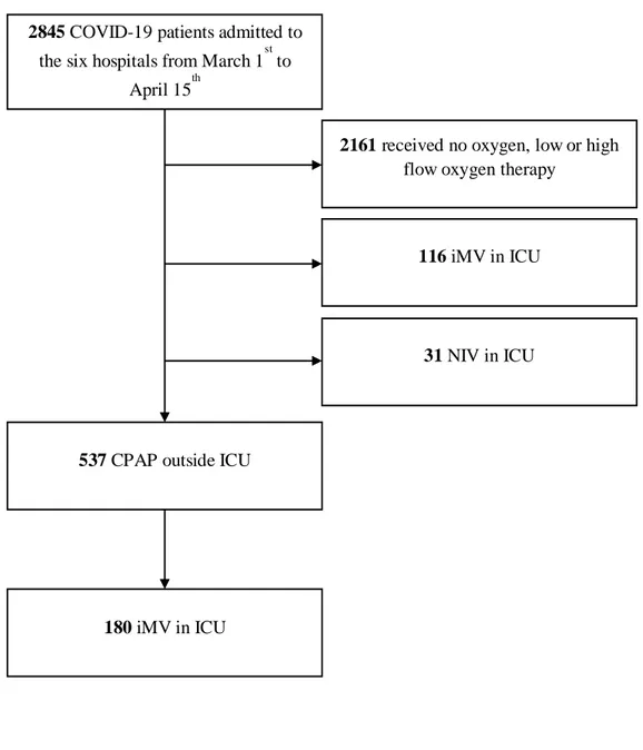
From March 1st to April 15th, a total of 2845 patients with confirmed COVID-19 were admitted to the six hospitals of the eastern Piedmont (Figure 1s). Of these, 326 (11%) patients were treated in ICU, 31 (1%) and 295 (10%) with noninvasive and invasive mechanical ventilation, respectively. CPAP was applied to 537 (22%) patients in RICU.
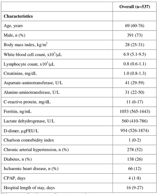
Table 1 shows the demographic and clinical patients’ characteristics. The number of observations for each variable is displayed in table 1s. The median age was 69 (IQR, 60-76) years, and BMI 28 (IQR, 25-31) kg/m2. Males were 391 (73%). Laboratory values at hospital entrance are also summarized in table 1. Median white blood cell count was 6.9 (IQR, 5.1-9.5) x 103/µL, with lymphopenia, i.e., 0.8 (0.6–1.1) x 103/µL. Median values of creatinine, aspartate-aminotransferase, alanine-aminotransferase were in the normal range, while C-reactive protein, ferritin, lactate dehydrogenase and D-dimer were all above the normal range. CCI median value was 1 (IQR, 0-2), chronic arterial hypertension was present in 278 (52%) patients, diabetes in 138 (26%) patients, and ischaemic heart disease in 66 (12%) patients.
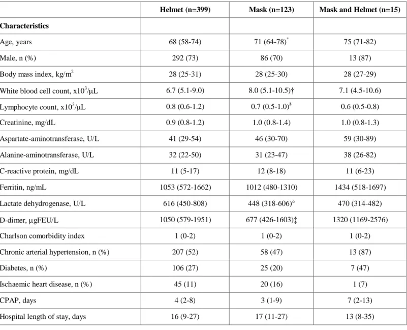
The most common interface was the helmet, in 399 (74%) patients, while face masks were used in 123 patients (23%); 15 patients (3%) alternated both interfaces. Median CPAP duration was 4 (IQR, 1 -8) days. Overall cumulative 60-day in-hospital mortality was 34% (cumulative incidence, 0.344, 95%CI, 0.304-0.384), as depicted in Figure 1A, while hospital LOS was 16 (IQR, 9-27) days.
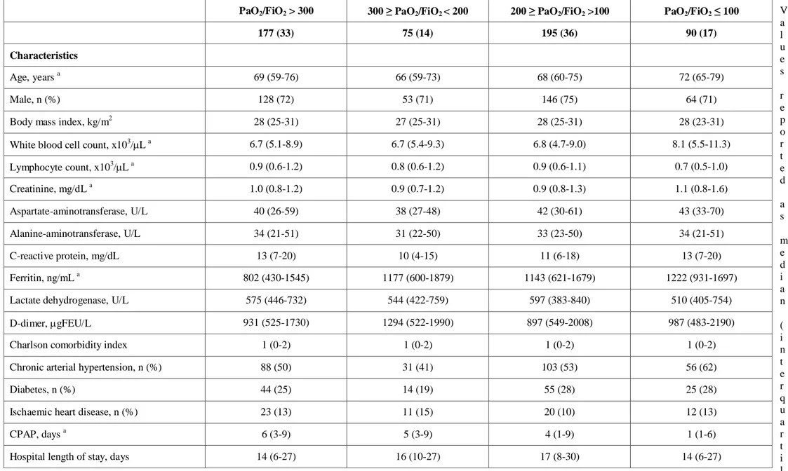
Demographic and clinical patients’ characteristics stratified by PaO2/FiO2 performed 2 to 24 hours after
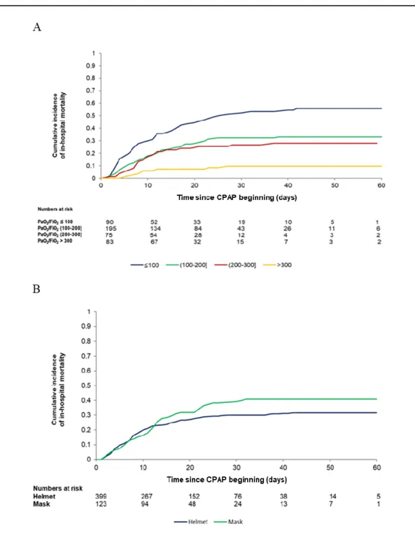
CPAP initiation and interface applied, are provided in the supplementary material in table 2s and 3s, respectively. Overall cumulative 60-day in-hospital mortality stratified according to PaO2/FiO2 and
interface applied is also depicted in Figure 2s A and B, in the supplementary material. As expected, mortality seemed to increase in patients with lower PaO2/FiO2,while was not different between patients
treated with helmet (cumulative incidence, 0.315, 95%CI, 0.270-0.361) or face mask (cumulative incidence, 0.407, 95%CI, 0.320-0.493), p= 0.094.
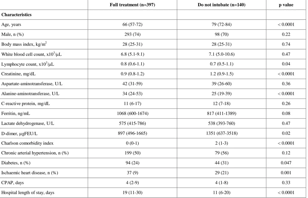
When dividing patients according to the therapeutic goal (table 2), patients in the full treatment subgroup were younger [median, 66 (IQR, 57-72) years] compared to DNI [median, 79 (IQR, 72-84) year]. DNI subgroup had higher D-dimer values than full treatment patients [median, 1351 (IQR, 637-3518)
µgFEU/L vs. 897 (IQR, 496-1665) µgFEU/L] (p=0.02), and greater CCI [median, 2 (IQR, 1-3) vs. median, 0 (IQR, 0-1)] (p<0.0001). The number of observations for each variable is displayed in table 4s.
Table 5s shows ABGs and table 6s the number of observations for each variable. While the first ABG indicated in table 5s was performed 1-day (IQR, 0-3 days) after hospital admission, the second was performed 2 to 24 hours after CPAP initiation.
Before starting CPAP, patients presented a PaO2/FiO2 of 108 (IQR, 71-157) mmHg and a respiratory
rate of 27 (IQR, 22-32) breaths/min. Soon after CPAP, PaO2/FiO2 raised to 157 (IQR, 109-255) mmHg
and respiratory rate decreased to 24 (IQR, 20-28) breaths/min with median CPAP of 10 (IQR, 10-12) cmH2O, and FiO2 of 50% (IQR, 50-60%). Median CPAP duration was 4 days for both subgroups, while
hospital LOS was 19 (IQR, 11-30) days for full treatment and 11 (IQR, 6-20) days for DNI subgroup.
Figure 1B depicts 60-day in-hospital mortality for full-treatment [21% (cumulative incidence 0.208, 95%CI, 0.169-0.249)] and DNI [73% (cumulative incidence 0.731, 95%CI, 0.648-0.787)], (p < 0.0001). Within the full treatment subgroup, 60-day in-hospital mortality was 42% (cumulative incidence 0.418, 95%CI, 0.345-0.488) for patients receiving intubation (Figure 2), and 2% (cumulative incidence 0.018, 95%CI, 0.008-0.035) for patients succeeding CPAP (Figure 3s).
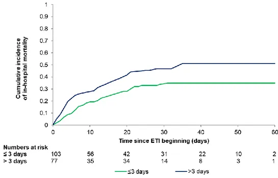
CPAP duration was 2 days (IQR, 1-3 days) in patients who survived and 3 days (IQR. 1-5 days) in patients who died (p=0.061). Table 3 shows that duration of CPAP application was an independent predictor of mortality for patients requiring intubation. The model, adjusting for age, gender, comorbidities, LDH, C-reactive protein values, and lymphocyte count, indicates a 9.3% [HR, 1.094 (95%CI, 1.010-1.184)] increase of the risk of death for each day of treatment. The association between duration of CPAP and mortality does not substantially change in the secondary analysis using multiple imputation [HR, 1.060 (95%CI, 1.001-1.121)], as presented in Table 7s. 60-day in-hospital mortality was significantly higher in patients subjected to CPAP for more than 3 days (cumulative incidence 0.510, 95%CI, 0.393-0.615) as compared to those receiving CPAP for 3 days or less (cumulative incidence 0.350, 95%CI, 0.259-0.441), as shown in Figure 4s.
Discussion
This multicentre retrospective observational study on 537 patients hypoxemic ARF secondary to laboratory-confirmed COVID-19 infection, shows that CPAP applied to different therapeutic goals i.e., candidate to intubation in the case of CPAP failure and do-not-intubate in which CPAP is considered the ceiling of treatment, is feasible outside ICU. Treatment duration for patients failing CPAP prior to intubation represents a risk factor for mortality.
CPAP can be delivered both in ICU and outside ICU. Grasselli et al. [10], found that 11% of the
patients entering ICU needed NIV, while early data from China revealed a higher percentage i.e., 41.7% [13], 43.3% [19], 56% [11], 62%[15]. In keeping with data by Grasselli et al. [10], as CPAP was
delivered to 31 out of 326 patients (9.5%) entering ICU.
Data on the use of CPAP in COVID-19 patients treated outside ICU are scarce. Two over 28 patients (7%) received NIV or HFNC outside ICU in a single centre study in Wuhan [15]. Oranger et al., treated 38 patients with CPAP in a respiratory ward [14]. Although the study included a limited patients’ number, CPAP resulted to be feasible and the authors also suggest a potential benefit for both full treatment and DNI patients, as opposed to those treated with oxygen only [14].
As far as mortality concerns, we showed an overall cumulative 60-day in-hospital mortality of 34% in patients with moderate to severe forms of ARF COVID-19-related needing CPAP. The rate of mortality observed in our study, is not divergent from those reported in several prior studies [9,10,15,12,13] for ICU patients, predominantly intubated, which varied from 17% [13] to 67% [9].
Lastly, our study includes 26% of DNI patients, for whom CPAP was considered ceiling of treatment. Rate of DNI patients reported in our study is similar to 15% observed in a small cohort of patients treated outside ICU during COVID-19 pandemic [14], i.e., as well as to 30% reported by a large Italian multicentre observational study in patients with pneumonia non-COVID-19 related, treated with NIV outside ICU [20]. In our study, 60-day in-hospital mortality for DNI patients was 73%.
A major concern when treating patients with hypoxemic ARF with NIV, is related to NIV-failure rate that might occur in up 50% of the cases with consequent recourse to intubation [21]. Undue prolongation
of NIV may worsen lung injury resulting in the so-called patient self-inflicted lung injury [22], while the direct consequence of NIV failure is delaying intubation and adequate treatment with invasive
ventilation [23,1]. Our data confirm that intubation delay for those requiring afterward invasive ventilation is associated with increased risk for mortality. In other pandemics, such as influenza, H1N1, and severe acute respiratory syndrome (SARS), NIV failure ranges from 10 to more than 70% [24], reaching 90% with middle east respiratory syndrome [25]. In our study, CPAP failure rate was 45%, which indicates that effective treatment occurred in more than half of patients, who avoided invasive ventilation through an endotracheal tube, which is a life-saving procedure, but it is also prone to several side-effects and complications [26].
Limitations
The study has several limitations. First, we were not able to compare our population with an historical control. Second, most of data have been retrospectively derived from the medical records. According to the retrospective nature of the study, formal criteria to start CPAP treatment were not defined a priori , and the time span between CPAP initiation and control ABG was relatively long. Third, definitions of full treatment and DNI patients, although internationally accepted [6], are influenced by patients,
families, and clinicians thinking and might be influenced by cultural, religious and geographical factors. Fourth, due to the diversity of interfaces and devices used in our study, the actual applied pressure could somewhat differ from the preset value [27]. Fifth, due to the number of missing data among many
important variables such as D-Dimer and respiratory rate, we were not able introduce them in the model that explore the correlation between CPAP duration and mortality. Lastly, because of the exceptionality of pandemic outbreak, our results are not generalizable to other conditions.
Conclusions
To the best of our knowledge, this is the largest retrospective cohort study on patients with COVID -19 treated with CPAP outside ICU. We show that CPAP is feasible outside ICU with overall in-hospital mortality similar to that reported in other studies treating critically ill ICU patients. In-hospital mortality is closely related to the therapeutic goal, patients having DNI order being affected by much higher mortality. Intubation delay is confirmed to be an independent risk factor for mortality. Further studies
are necessary to ascertain the potential infective risk related to CPAP treatment outside ICU among healthcare workers.
Table 1 General characteristics of patients with CPAP outside ICU
Values are median (interquartile range) or number (percentage).
CPAP, noninvasive continuous positive airway pressure; ICU, intensive care unit; FEU, fibrinogen-equivalent unit
Overall (n=537) Characteristics
Age, years 69 (60-76)
Male, n (%) 391 (73)
Body mass index, kg/m2 28 (25-31)
White blood cell count, x103/L 6.9 (5.1-9.5)
Lymphocyte count, x103/L 0.8 (0.6-1.1) Creatinine, mg/dL 1.0 (0.8-1.3) Aspartate-aminotransferase, U/L 41 (29-59) Alanine-aminotransferase, U/L 31 (22-50) C-reactive protein, mg/dL 11 (6-17) Ferritin, ng/mL 1053 (565-1643)
Lactate dehydrogenase, U/L 560 (410-786)
D-dimer, gFEU/L 954 (526-1874)
Charlson comorbidity index 1 (0-2)
Chronic arterial hypertension, n (%) 278 (52)
Diabetes, n (%) 138 (26)
Ischaemic heart disease, n (%) 66 (12)
CPAP, days 4 (1-8)
Table 2 General characteristics of patients stratified according to CPAP therapeutic goal
Values reported as median (interquartile range) or number (percentage). p values were calculated by Mann-Whitney U test or χ2 test, as appropriate
Full treatment (n=397) Do not intubate (n=140) p value
Characteristics
Age, years 66 (57-72) 79 (72-84) < 0.0001
Male, n (%) 293 (74) 98 (70) 0.22
Body mass index, kg/m2 28 (25-31) 28 (25-31) 0.74
White blood cell count, x103/L 6.8 (5.1-9.1) 7.1 (5.0-10.6) 0.47
Lymphocyte count, x103/L 0.8 (0.6-1.1) 0.7 (0.5-1.1) 0.04 Creatinine, mg/dL 0.9 (0.8-1.2) 1.2 (0.9-1.5) < 0.0001 Aspartate-aminotransferase, U/L 42 (31-59) 39 (26-60) 0.36 Alanine-aminotransferase, U/L 34 (24-53) 25 (19-39) < 0.0001 C-reactive protein, mg/dL 11 (6-17) 12 (7-18) 0.26 Ferritin, ng/mL 1068 (600-1674) 817 (411-1389) 0.08
Lactate dehydrogenase, U/L 575 (415-786) 538 (393-760) 0.47
D-dimer, gFEU/L 897 (496-1665) 1351 (637-3518) 0.02
Charlson comorbidity index 0 (0-1) 2 (1-3) < 0.0001
Chronic arterial hypertension, n (%) 199 (50) 79 (56) 0.12
Diabetes, n (%) 94 (24) 44 (31) 0.047
Ischaemic heart disease, n (%) 37 (9) 29 (21) 0.001
CPAP, days 4 (2-9) 4 (1-8) 0.33
Table 3 Fine and Gray model for the association between CPAP duration and mortality
sHR 95%CI CPAP, days 1.094 (1.010-1.184) Male 1.596 (0.814-3.129) Age 1.023 (0.985-1.064) Hypertension, yes vs. no 1.224 (0.729-2.056) Charlson index 1.392 (1.145-1.693)
Lactate dehydrogenase, U/L 1.001 (1.000-1.002) C-reactive protein, mg/dL 0.983 (0.947-1.020) Lymphocytes, x103/mL 1.269 (0.699-2.302) CPAP, noninvasive continuous positive airway pressure; sHR, sub-distribution hazard ratio; 95% CI, 95% confidence interval.
Sub-distribution hazard ratio and 95% corresponding confidence interval deriving from the multivariate Fine and Gray models. Estimates are further adjusted by center. N:146
Figure legends
Fig. 1 Cumulative incidence of in-hospital mortality in the overall patients (Panel A) and stratified
by treatment (Panel B) i.e., DNI and full treatment patients in grey and in black lines, respectively. Curves and corresponding 95% confidence intervals (dashed lines).
CPAP, noninvasive continuous positive airway pressure; DNI, do not intubate.
Fig. 2 Cumulative incidence of in-hospital mortality in patients requiring invasive mechanical
ventilation. Curve and corresponding 95% confidence interval (dashed lines).
ETI, endo-tracheal intubation.
Acknowledgements
We acknowledge all health-care workers involved in the diagnosis and treatment of patients in the eastern Piedmont region. We thank Dr. Davide Crimaldi for database support. The study received no funding. Preliminary data have been sent, in abstract form, to the European Society of Intensive Care Medicine Congress 2020.
Reference List
1. Rochwerg B, Brochard L, Elliott MW, Hess D, Hill NS, Nava S, Navalesi PMOT, Antonelli M, Brozek J, Conti G, Ferrer M, Guntupalli K, Jaber S, Keenan S, Mancebo J, Mehta S, Raoof S Members Of The Task Force. Official ERS/ATS clinical practice guidelines: noninvasive
ventilation for acute respiratory failure. Eur Respir J 2017; 50:
2. Masip J, Roque M, Sanchez B, Fernandez R, Subirana M, Exposito JA. Noninvasive ventilation in acute cardiogenic pulmonary edema: systematic review and meta -analysis. JAMA 2005; 294: 3124-3130.
3. Lemiale V, Mokart D, Resche-Rigon M, Pene F, Mayaux J, Faucher E, Nyunga M, Girault C, Perez P, Guitton C, Ekpe K, Kouatchet A, Theodose I, Benoit D, Canet E, Barbier F, Rabbat A, Bruneel F, Vincent F, Klouche K, Loay K, Mariotte E, Bouadma L, Moreau AS, Seguin A, Meert AP, Reignier J, Papazian L, Mehzari I, Cohen Y, Schenck M, Hamidfar R, Darmon M, Demoule A, Chevret S, Azoulay E. Effect of Noninvasive Ventilation vs Oxygen Therapy on Mortality Among Immunocompromised Patients With Acute Respiratory Failure: A Randomized Clinical Trial. JAMA 2015; 314: 1711-1719.
4. Jaber S, Lescot T, Futier E, Paugam-Burtz C, Seguin P, Ferrandiere M, Lasocki S, Mimoz O, Hengy B, Sannini A, Pottecher J, Abback PS, Riu B, Belafia F, Constantin JM, Ma sseret E, Beaussier M, Verzilli D, De JA, Chanques G, Brochard L, Molinari N. Effect of Noninvasive Ventilation on Tracheal Reintubation Among Patients With Hypoxemic Respiratory Failure Following Abdominal Surgery: A Randomized Clinical Trial. JAMA 2016; 315: 1345-1353.
5. Namendys-Silva SA, Hernandez-Garay M, Rivero-Sigarroa E. Non-invasive ventilation for critically ill patients with pandemic H1N1 2009 influenza A virus infection. Crit Care 2010; 14: 407-
6. Curtis JR, Cook DJ, Sinuff T, White DB, Hill N, Keenan SP, Benditt JO, Kacmarek R, Kirchhoff KT, Levy MM. Noninvasive positive pressure ventilation in critical and palliative care settings: understanding the goals of therapy. Crit Care Med 2007; 35: 932-939.
7. Cabrini L, Moizo E, Nicelli E, Licini G, Turi S, Landoni G, Silvani P, Zangrillo A.
Noninvasive ventilation outside the intensive care unit from the patient point of view: a pilot study. Respir Care 2012; 57: 704-709.
8. Cabrini L, Esquinas A, Pasin L, Nardelli P, Frati E, Pintaudi M, Matos P, Landoni G, Zangrillo A. An international survey on noninvasive ventilation use for acute respiratory failure in general non-monitored wards. Respir Care 2015; 60: 586-592.
9. Arentz M, Yim E, Klaff L, Lokhandwala S, Riedo FX, Chong M, Lee M. Characteristics and Outcomes of 21 Critically Ill Patients With COVID-19 in Washington State. JAMA 2020;
10. Grasselli G, Zangrillo A, Zanella A, Antonelli M, Cabrini L, Castelli A, Cereda D, Coluccello A, Foti G, Fumagalli R, Iotti G, Latronico N, Lorini L, Merler S, Natalini G, Piatti A, Ranieri MV, Scandroglio AM, Storti E, Cecconi M, Pesenti A, Nailescu A, Corona A, Zangrillo A, Protti A, Albertin A, Forastieri MA, Lombardo A, Pezzi A, Benini A, Scandroglio AM, Malara A, Castelli A, Coluccello A, Micucci A, Pesenti A, Sala A, Alborghetti A, Antonini B, Capra C, Troiano C, Roscitano C, Radrizzani D, Chiumello D, Coppini D, Guzzon D, Costantini E, Malpetti E, Zoia E, Catena E, Agosteo E, Barbara E, Beretta E, Boselli E, Storti E, Harizay F, Della MF, Lorini FL, Donato SF, Marino F, Mojoli F, Rasulo F, Grasselli G, Casella G, De FG, Castelli G, Aldegheri G, Gallioli G, Lotti G, Albano G, Landoni G, Marino G, Vitale G, Battista PG, Evasi G,
Citerio G, Foti G, Natalini G, Merli G, Sforzini I, Bianciardi L, Carnevale L, Grazioli L, Cabrini L, Guatteri L, Salvi L, Dei PM, Galletti M, Gemma M, Ranucci M, Riccio M, Borelli M, Zambon M, Subert M, Cecconi M, Mazzoni MG, Raimondi M, Panigada M, Belliato M, Bronzini N, Latronico N, Petrucci N, Belgiorno N, Tagliabue P, Cortellazzi P, Gnesin P, Grosso P, Gritti P, Perazzo P, Severgnini P, Ruggeri P, Sebastiano P, Covello RD, Fernandez-Olmos R, Fumagalli R, Keim R, Rona R, Valsecchi R, Cattaneo S, Colombo S, Cirri S, Bonazzi S, Greco S, Muttini S, Langer T, Alaimo V, Viola U. Baseline Characteristics and Outcomes of 1591 Patients Infected With SARS-CoV-2 Admitted to ICUs of the Lombardy Region, Italy. JAMA 2020;
11. Yang X, Yu Y, Xu J, Shu H, Xia J, Liu H, Wu Y, Zhang L, Yu Z, Fang M, Yu T, Wang Y, Pan S, Zou X, Yuan S, Shang Y. Clinical course and outcomes of critically ill patients with SARS-CoV-2 pneumonia in Wuhan, China: a single-centered, retrospective, observational study. Lancet Respir Med 2020;
12. Richardson S, Hirsch JS, Narasimhan M, Crawford JM, McGinn T, Davidson KW, Barnaby DP, Becker LB, Chelico JD, Cohen SL, Cookingham J, Coppa K, Diefenbach MA, Dominello AJ, Duer-Hefele J, Falzon L, Gitlin J, Hajizadeh N, Harvin TG, Hirschwerk DA, Kim EJ, Kozel ZM, Marrast LM, Mogavero JN, Osorio GA, Qiu M, Zanos TP. Presenting Characteristics,
Comorbidities, and Outcomes Among 5700 Patients Hospitalized With COVID -19 in the New York City Area. JAMA 2020;
13. Wang D, Hu B, Hu C, Zhu F, Liu X, Zhang J, Wang B, Xiang H, Cheng Z, Xiong Y, Zhao Y, Li Y, Wang X, Peng Z. Clinical Characteristics of 138 Hospitalized Patients With 2019 Novel Coronavirus-Infected Pneumonia in Wuhan, China. JAMA 2020;
14. Oranger M, Gonzalez-Bermejo J, Dacosta-Noble P, Llontop C, Guerder A, Trosini-Desert V, Faure M, Raux M, Decavele M, Demoule A, Morelot-Panzini C, Similowski T. Continuous
positive airway pressure to avoid intubation in SARS-CoV-2 pneumonia: a two-period retrospective case-control study. Eur Respir J 2020;
15. Huang C, Wang Y, Li X, Ren L, Zhao J, Hu Y, Zhang L, Fan G, Xu J, Gu X, Cheng Z, Yu T, Xia J, Wei Y, Wu W, Xie X, Yin W, Li H, Liu M, Xiao Y, Gao H, Guo L, Xie J, Wang G, Jiang R, Gao Z, Jin Q, Wang J, Cao B. Clinical features of patients infected with 2019 novel coronavirus in Wuhan, China. Lancet 2020; 395: 497-506.
16. Gristina GR, Orsi L, Carlucci A, Causarano IR, Formica M, Romano M. [Part I. End-stage chronic organ failures: a position paper on shared care planning. The Integrated Care Pathway]. Recenti Prog Med 2014; 105: 9-24.
17. Charlson ME, Pompei P, Ales KL, MacKenzie CR. A new method of classifying prognostic comorbidity in longitudinal studies: development and validation. J Chronic Dis 1987; 40: 373-383.
18. Resche-Rigon M, Azoulay E, Chevret S. Evaluating mortality in intensive care units: contribution of competing risks analyses. Crit Care 2006; 10: R5-
19. Guan WJ, Ni ZY, Hu Y, Liang WH, Ou CQ, He JX, Liu L, Shan H, Lei CL, Hui DSC, Du B, Li LJ, Zeng G, Yuen KY, Chen RC, Tang CL, Wang T, Chen PY, Xiang J, Li SY, Wang JL, Liang ZJ, Peng YX, Wei L, Liu Y, Hu YH, Peng P, Wang JM, Liu JY, Chen Z, Li G, Zheng ZJ, Qiu SQ, Luo J, Ye CJ, Zhu SY, Zhong NS. Clinical Characteristics of Coronavirus Disease 2019 in China. N Engl J Med 2020; 382: 1708-1720.
20. Brambilla AM, Prina E, Ferrari G, Bozzano V, Ferrari R, Groff P, Petrelli G, Scala R, Causin F, Noto P, Bresciani E, Voza A, Aliberti S, Cosentini R. Non-invasive positive pressure ventilation in pneumonia outside Intensive Care Unit: An Italian multicenter observational study. Eur J Intern Med 2019; 59: 21-26.
21. Frat JP, Thille AW, Mercat A, Girault C, Ragot S, Perbet S, Prat G, Boulain T, Morawiec E, Cottereau A, Devaquet J, Nseir S, Razazi K, Mira JP, Argaud L, Chakarian JC, Ricard JD,
Wittebole X, Chevalier S, Herbland A, Fartoukh M, Constantin JM, Tonnelier JM, Pierrot M, Mathonnet A, Beduneau G, Deletage-Metreau C, Richard JC, Brochard L, Robert R. High-flow oxygen through nasal cannula in acute hypoxemic respiratory failure. N Engl J Med 2015; 372: 2185-2196.
22. Brochard L, Slutsky A, Pesenti A. Mechanical Ventilation to Minimize Progression of Lung Injury in Acute Respiratory Failure. Am J Respir Crit Care Med 2017; 195: 438-442.
23. Brochard L, Lefebvre JC, Cordioli RL, Akoumianaki E, Richard JC. Noninvasive
ventilation for patients with hypoxemic acute respiratory failure. Semin Respir Crit Care Med 2014; 35: 492-500.
24. Esquinas AM, Egbert PS, Scala R, Gay P, Soroksky A, Girault C, Han F, Hui DS, Papadakos PJ, Ambrosino N. Noninvasive mechanical ventilation in high-risk pulmonary infections: a clinical review. Eur Respir Rev 2014; 23: 427-438.
25. Alraddadi BM, Qushmaq I, Hameed FM, Mandourah Y, Almekhlafi GA, Jose J, Omari A, Kharaba A, Almotairi A, Al KK, Shalhoub S, Abdulmomen A, Mady A, Solaiman O, Al-Aithan AM, Al-Raddadi R, Ragab A, Balkhy HH, Al HA, Sadat M, Tlayjeh H, Merson L, Hayden FG, Fowler RA, Arabi YM. Noninvasive ventilation in critically ill patients with the Middle East respiratory syndrome. Influenza Other Respir Viruses 2019; 13: 382-390.
26. Esteban A, Frutos-Vivar F, Muriel A, Ferguson ND, Penuelas O, Abraira V, Raymondos K, Rios F, Nin N, Apezteguia C, Violi DA, Thille AW, Brochard L, Gonzalez M, Villagomez AJ, Hurtado J, Davies AR, Du B, Maggiore SM, Pelosi P, Soto L, Tomicic V, D'Empaire G, Matamis D, Abroug F, Moreno RP, Soares MA, Arabi Y, Sandi F, Jibaja M, Amin P, Koh Y, Kuiper MA,
Bulow HH, Zeggwagh AA, Anzueto A. Evolution of mortality over time in patients receiving mechanical ventilation. Am J Respir Crit Care Med 2013; 188: 220-230.
27. Vargas F, Thille A, Lyazidi A, Campo FR, Brochard L. Helmet with specific settings versus facemask for noninvasive ventilation. Crit Care Med 2009; 37: 1921-1928.
FIGURE 1 B
Outcomes of COVID-19 patients treated with continuous positive airway pressure outside ICU
Supplementary material
Study Design
This was a multicentre, retrospective observational study performed in six hospitals of the eastern Piedmont region, Northern Italy i.e., “Maggiore della Carità” University hospital in Novara, “SS. Antonio Biagio e Cesare Arrigo”, hospital in Alessandria, “S. Andrea” hospital in Vercelli, “VCO ASL” in Domodossola, “Nuovo Ospedale degli Infermi” hospital in Biella. One centre i.e.,
“Maggiore della Carità” University hospital in Novara, enrolled patients prospectively n=138 patients, as the protocol for the data collection, has been approved on the 07th June 2019 (CE 64/19) for a study, monitoring patients treated with CPAP outside ICU.
All the participating centres obtained ethics committee approval for the present research project (CE 87/20; CE 112/20, CE 111/20, CE 110/20, ASO.RianGen.20.02, AslVC.RianGen.20.01). Informed consent was obtained for all the patients prospectively enrolled and for those still in the hospital after ethics committee protocol approval, while for the others, ethics committee waived the need for informed consent. Local investigators were responsible for ensuring data integrity and validity.
Criteria for intubation
Criteria for intubation were cardiac or respiratory arrest; inability to protect the airway; coma or psychomotor agitation; unmanageable secretions or uncontrolled vomiting; life threatening
arrhythmias or electrocardiographic signs of ischemia; hemodynamic instability defined as systolic arterial pressure < 90 mmHg despite adequate filling or use of vasoactive agents; intolerance to all interfaces; dyspnoea during noninvasive continuous positive airway pressure (CPAP), respiratory rate >30 breaths/min; peripheral oxygen saturation (SpO2) below 92% during CPAP, acidosis with a
CPAP
CPAP was delivered through helmets (Intersurgical, Mirandola (MO), Italy; Dimar, Medolla, MO, Italy) and face masks (Intersurgical, Mirandola, MO, Italy; Dimar, Medolla, MO, Italy;
Fisher&Paykel, Auckland, New Zealand; ResMed, San Diego, CA, USA; Philips Respironics, Murrysville, PA, USA) via Boussignac systems or via flow-meters (typically 30-50 l/min
depending on the interface chosen) with a scale that allowed the clinician to regulate oxygen and air flow separately to set inspiratory oxygen fraction (FiO2). Bacterial and viral filter was applied to the
expiratory port. CPAP was set between 10 and 12 cmH2O according to patient’s needs, tolerance
and any side-effects. CPAP pressure could be increased up to 15 cmH2O. CPAP was delivered on
an as-needed basis. When respiratory parameters improved, CPAP support was gradually reduced with a progressive increase of time off CPAP, until discontinuation.
Respiratory intermediate care unit organization
Nurse to patient ratios varied from a maximum of 1:6 both during day and night to a minimum of 1:8 and 1:12, respectively during days and nights. In three hospitals, medical staff treating CPAP COVID-19 patients was an ad-hoc mixed team, mainly internists, pneumologists, emergency physicians, cardiologists, anaesthesiologists/ICU physicians, while in the other three hospitals the medical team was the same as before COVID-19 pandemic. CPAP was prescribed mainly by anaesthesiologists actively working with the ad-hoc COVID-19 ward team, but also by pneumologists and emergency doctors or by consulting anaesthesiologists. Personnel was adequately trained for NIV; those who were not, received a short-organized training during pandemic. Ward monitoring included SpO2, non-invasive blood pressure, ECG applied
continuously or at a defined time point depending on the severity of the patient. Blood gas analysis was performed when clinically relevant. Patients received daily visit from the consulting physician who prescribed CPAP if not present in the ad-hoc ward team.
Statistical analysis
Fine and Gray model included as adjustment age, gender, comorbidities i.e., Charlson comorbidity index and hypertension, LDH, C-reactive protein levels and lymphocyte count. The model was further adjusted by centre. The adjustment variables were selected on the base of their clinical relevance. Multiple imputation procedures were applied to account for missing data.
Figure 1S Study flow chart
COVID-19, coronavirus disease 19; iMV, invasive mechanical ventilation; ICU, intensive care unit; NIV, noninvasive ventilation; CPAP, noninvasive continuous positive airway pressure.
2845 COVID-19 patients admitted to
the six hospitals from March 1st to April 15th
537 CPAP outside ICU
116 iMV in ICU
31 NIV in ICU
2161 received no oxygen, low or high
flow oxygen therapy
Figure 2S Cumulative incidence of in-hospital mortality stratified by PaO2/FiO2 levels (Panel A) and interface
applied (Panel B).
PaO2/FiO2
Cumulative incidence at 60 days – PaO2/FiO2≤100: 0.558 (0.448-0.654).
Cumulative incidence at 60 days – PaO2/FiO2 (100-200]: 0329 (0.264-0.395).
Cumulative incidence at 60 days – PaO2/FiO2 (200-300]: 0.280 (0.183-0.385).
Cumulative incidence at 60 days – PaO2/FiO2>300: 0.100 (0.045-0.172).
P-value Grey’s test for equality of CIF <0.0001
CPAP device
Cumulative incidence at 60 days – CPAP Helmet: 0.315 (0.270-0.361). Cumulative incidence at 60 days – CPAP Mask: 0.407 (0.320-0.493). P-value Grey’s test for equality of CIF 0.0938
B B B B B B
Figure 3S Cumulative incidence of in-hospital mortality in full treatment patients not requiring
invasive mechanical ventilation
Curves and corresponding 95% confidence intervals (dashed lines). Cumulative incidence at 60 days 0.018 (0.008-0.035).
Figure 4S Cumulative incidence of in-hospital mortality stratified by number of CPAP days among
patients undergoing endotracheal intubation (median)
Cumulative incidence at 60 days – CPAP days ≤3: 0.350 (0.259-0.441). Cumulative incidence at 60 days – CPAP days >3: 0.510 (0.393-0.615). P-value Grey’s test for equality of CIF 0.0256
B B
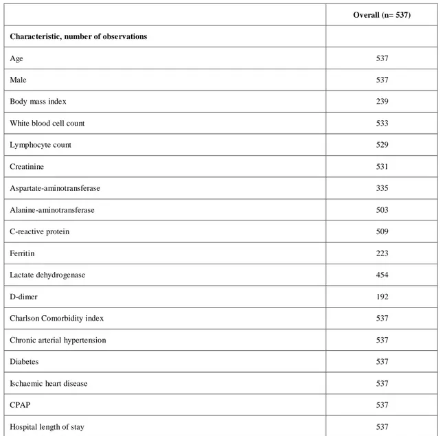
Table 1S Number of observations for each variable
CPAP, noninvasive continuous positive airway pressure
Overall (n= 537) Characteristic, number of observations
Age 537
Male 537
Body mass index 239
White blood cell count 533
Lymphocyte count 529 Creatinine 531 Aspartate-aminotransferase 335 Alanine-aminotransferase 503 C-reactive protein 509 Ferritin 223 Lactate dehydrogenase 454 D-dimer 192
Charlson Comorbidity index 537
Chronic arterial hypertension 537
Diabetes 537
Ischaemic heart disease 537
CPAP 537
Table 2S General characteristics of patients stratified according to PaO2/FiO2 value V a l u e s r e p o r t e d a s m e d i a n ( i n t e r q u a r t i l e range) or number (percentage). p values were calculated by Mann-Whitney U test or χ2 test, as appropriate
PaO2/FiO2 > 300 300 ≥ PaO2/FiO2 < 200 200 ≥ PaO2/FiO2 >100 PaO2/FiO2 ≤ 100
177 (33) 75 (14) 195 (36) 90 (17)
Characteristics
Age, years a 69 (59-76) 66 (59-73) 68 (60-75) 72 (65-79)
Male, n (%) 128 (72) 53 (71) 146 (75) 64 (71)
Body mass index, kg/m2 28 (25-31) 27 (25-31) 28 (25-31) 28 (23-31)
White blood cell count, x103/L a 6.7 (5.1-8.9) 6.7 (5.4-9.3) 6.8 (4.7-9.0) 8.1 (5.5-11.3)
Lymphocyte count, x103/L a 0.9 (0.6-1.2) 0.8 (0.6-1.2) 0.9 (0.6-1.1) 0.7 (0.5-1.0) Creatinine, mg/dL a 1.0 (0.8-1.2) 0.9 (0.7-1.2) 0.9 (0.8-1.3) 1.1 (0.8-1.6) Aspartate-aminotransferase, U/L 40 (26-59) 38 (27-48) 42 (30-61) 43 (33-70) Alanine-aminotransferase, U/L 34 (21-51) 31 (22-50) 33 (23-50) 34 (21-51) C-reactive protein, mg/dL 13 (7-20) 10 (4-15) 11 (6-18) 13 (7-20) Ferritin, ng/mL a 802 (430-1545) 1177 (600-1879) 1143 (621-1679) 1222 (931-1697)
Lactate dehydrogenase, U/L 575 (446-732) 544 (422-759) 597 (383-840) 510 (405-754)
D-dimer, gFEU/L 931 (525-1730) 1294 (522-1990) 897 (549-2008) 987 (483-2190)
Charlson comorbidity index 1 (0-2) 1 (0-2) 1 (0-2) 1 (0-2)
Chronic arterial hypertension, n (%) 88 (50) 31 (41) 103 (53) 56 (62)
Diabetes, n (%) 44 (25) 14 (19) 55 (28) 25 (28)
Ischaemic heart disease, n (%) 23 (13) 11 (15) 20 (10) 12 (13)
CPAP, days a 6 (3-9) 5 (3-9) 4 (1-9) 1 (1-6)
Table 3S General characteristics of patients according to CPAP interface applied
Values are median (interquartile range) or number (percentage).
CPAP, noninvasive continuous positive airway pressure; ICU, intensive care unit; FEU, fibrinogen-equivalent unit Helmet vs. mask: * p =0.030, † p=0.002, § p=0.001, °p<0.0001, ‡ p=0.032.
Helmet (n=399) Mask (n=123) Mask and Helmet (n=15)
Characteristics
Age, years 68 (58-74) 71 (64-78)* 75 (71-82)
Male, n (%) 292 (73) 86 (70) 13 (87)
Body mass index, kg/m2 28 (25-31) 28 (25-30) 28 (27-29)
White blood cell count, x103/L 6.7 (5.1-9.0) 8.0 (5.1-10.5)† 7.1 (4.5-10.6)
Lymphocyte count, x103/L 0.8 (0.6-1.2) 0.7 (0.5-1.0)§ 0.6 (0.5-0.8) Creatinine, mg/dL 0.9 (0.8-1.2) 1.0 (0.8-1.4) 1.0 (0.8-1.3) Aspartate-aminotransferase, U/L 41 (29-54) 46 (30-70) 59 (30-89) Alanine-aminotransferase, U/L 32 (22-50) 31 (23-47) 38 (26-82) C-reactive protein, mg/dL 11 (5-17) 12 (8-18) 11 (6-23) Ferritin, ng/mL 1053 (572-1662) 1012 (480-1310) 1434 (518-1697)
Lactate dehydrogenase, U/L 616 (450-808) 448 (318-606)° 470 (314-482)
D-dimer, gFEU/L 1050 (579-1951) 677 (426-1603)‡ 1320 (1169-2576)
Charlson comorbidity index 1 (0-2) 1 (0-2) 1 (0-2)
Chronic arterial hypertension, n (%) 207 (52) 58 (47) 13 (87)
Diabetes, n (%) 106 (27) 25 (20) 7 (47)
Ischaemic heart disease, n (%) 45 (11) 20 (16) 1 (7)
CPAP, days 4 (2-8) 3 (1-9) 7 (2-13)
Table 4S Number of observations for each variable
Full treatment (n=397) Do-not-intubate (n=140) Characteristic, number of observations
Age 397 140
Male 397 140
Body mass index 196 43
White blood cell count 395 138
Lymphocyte count 392 137 Creatinine 394 137 Aspartate-aminotransferase 248 87 Alanine-aminotransferase 371 132 C-reactive protein 377 132 Ferritin 180 43 Lactate dehydrogenase 345 109 D-dimer 150 42
Charlson Comorbidity index 397 140
Chronic arterial hypertension 397 140
Diabetes 397 140
Ischaemic heart disease 397 140
Cancer 397 140
CPAP 397 140
Hospital length of stay 397 140
Table 5S Arterial blood gas analysis at baseline and after CPAP outset
ABG, arterial blood gas analysis; CPAP, noninvasive continuous positive airway pressure; PaCO2, arterial partial
pressure of carbon dioxide; PaO2, arterial partial pressure of oxygen; SpO2, peripheral oxygen saturation; HCO3-,
bicarbonate; FiO2, inspired oxygen fraction; PaO2/FiO2, arterial partial pressure of oxygen to inspired oxygen fraction
ratio; SpO2/FiO2, peripheral oxygen saturation to inspired oxygen fraction ratio a
Arterial blood gas analysis performed before CPAP initiation
b
First arterial blood gas analysis performed 2-24 hours after CPAP outset
†
Mann Whitney U test p < 0.01 full treatment vs. do not intubate
*
Wilcoxon signed-rank test p < 0.01 ABG before CPAP vs. ABG after CPAP
Overall (n=537) Full treatment (n=397) Do not intubate (n=140) ABG before CPAPa
pH 7.47 (7.43-7.49) 7.47 (7.44-7.49)† 7.46 (7.42-7.48) PaCO2, mmHg 35 (31-38) 35 (31-38) 34 (31-37) PaO2, mmHg 60 (49-73) 60 (51-73)† 54 (46-68) SpO2, % 91 (86-95) 92 (87-95)† 90 (83-93) HCO3-, mmol/L 25 (23-27) 26 (23-28)† 24 (22-26) Lactate, mmol/L 1.2 (0.9-1.7) 1.2 (0.9-1.6) 1.2 (1.0-1.7) FiO2, % 50 (50-100) 50 (50-100) 50 (50-100) PaO2/FiO2, mmHg 108 (71-157) 115 (73-158) 94 (66-146) SpO2/FiO2, % 164 (95-194) 171 (96-194) 161 (94-190)
Respiratory Rate, breaths/min 27 (22-32) 26 (22-32) 28 (24-32)
ABG after CPAPb
pH 7.45 (7.42-7.48) 7.45 (7.42-7.48)†* 7.43 (7.40-7.46)* PaCO2, mmHg 36 (33-40) 36 (33-40)* 36 (32-41)* PaO2, mmHg 85 (60-132) 87 (62-135)* 75 (56-126)* SpO2, % 96 (91-98) 96 (92-98)†* 94 (90-98)* HCO3-, mmol/L 26 (23-28) 26 (24-28)† 25 (21-28) Lactate, mmol/L 1.2 (0.9-1.6) 1.1 (0.8-1.6)†* 1.4 (1.0-1.7) FiO2, % 50 (50-60) 50 (50-60)* 50 (50-60)* PaO2/FiO2, mmHg 157 (109-255) 162 (109-259)* 148 (105-232)* SpO2/FiO2, % 176 (153-196) 176 (152-196)* 180 (157-196)*
Respiratory Rate, breaths/min 24 (20-28) 24 (20-28)*
25 (21-28)*
T able 6S Num ber of obse rvati ons for each vari able
Abbreviations: ABG, arterial blood gas analysis; CPAP, noninvasive continuous positive airway pressure; PaCO2,
arterial partial pressure of carbon dioxide; PaO2, arterial partial pressure of oxygen; SpO2, peripheral oxygen
saturation; HCO3-, bicarbonate; FiO2, inspired oxygen fraction; PaO2/FiO2, arterial partial pressure of oxygen to
inspired oxygen fraction ratio; SpO2/FiO2, peripheral oxygen saturation to inspired oxygen fraction ratio a
Arterial blood gas analysis performed before CPAP initiation
b
First arterial blood gas analysis performed 2-24 hours after CPAP outset
Overall (n= 537) Full treatment (n=397) Do not intubate (n=140) ABG before CPAPa
pH 492 370 122 PaCO2 493 371 122 PaO2 497 374 123 SpO2 535 395 140 HCO3- 484 365 119 Lactate 434 329 105 FiO2 409 307 102 PaO2/FiO2 383 290 93 SpO2/FiO2 408 306 102 Respiratory Rate 351 258 93
ABG after CPAPb
pH 462 353 109 PaCO2 462 352 110 PaO2 466 356 110 SpO2 534 395 139 HCO3- 456 347 109 Lactate 392 299 93 FiO2 499 376 102 PaO2/FiO2 443 344 99 SpO2/FiO2 499 376 123 Respiratory Rate 348 257 91 CPAP 454 344 110
Table 7SFine and Gray model for the association between CPAP treatment duration and mortality after the application of multiple imputation procedure.
sHR 95%CI CPAP, days 1.060 (1.001-1.121) Male 1.229 (0.668-2.261) Age 1.037 (1.002-1.073) Hypertension, yes vs. no 1.075 (0.671-1.722) Charlson index 1.260 (1.049-1.515)
Lactate dehydrogenase, U/L 1.000 (0.999-1.001) C-reactive protein, mg/dL 0.996 (0.963-1.030) Lymphocytes, x103/mL 1.323 (0.854-2.049)
Abbreviations: CPAP, continuous positive airway pressure; sHR, sub-distribution hazard ratio; 95% CI; 95% confidence interval. Sub-distribution hazard ratio and 95% corresponding confidence interval deriving from the multivariate Fine and Gray models. Estimates are further adjusted by center. N:180