INDEX
SUMMARY ... I INTRODUCTION ... I
AIM OF THE STUDY ... III
MATERIALS AND METHODS ... IV
Entomological successions ... IV Sampling of necrophagous species ... V RESULTS ... VI
Seasonal successions ... VI Sampling of necrophagous species ... VII CONCLUSIONS ... IX
AIM OF THE STUDY ... 1
CHAPTER 1 ... 1
INTRODUCTION ... 1
THANATOLOGICAL METHODS TO EVALUATE TIME OF DEATH ... 2
Algor mortis ... 3
Rigor mortis ... 4
Livor mortis ... 5
Putrefaction ... 6
ENTOMOLOGICAL METHODS TO EVALUATE PMI ... 8
Entomological successions ... 10
Using animal models in bodies succession studies ... 20
Life cycle of necrophagous Diptera ... 21
MYIASIS ... 27
CHAPTER 2 ... 31
MATERIALS & METHODS ... 31
1. ENTOMOLOGICAL SUCCESSIONS ... 31
Animal models ... 31
Study area ... 33
Experimental procedure ... 36
2. SAMPLING OF NECROPHAGOUS SPECIES ... 40
Traps design ... 40 Study areas ... 42 Wild area ... 43 Rural area ... 45 Urban Area ... 46 Experimental procedure ... 47 Data analysis ... 49 CHAPTER 3 ... 51 RESULTS ... 51 ENTOMOLOGICAL SUCCESSIONS ... 51 Autumn investigation ... 51 Summer investigation ... 56 Winter investigation ... 62 Spring investigation... 68
SAMPLING OF NECROPHAGOUS SPECIES ... 75
Spatial variability ... 80
Temporal variability ... 85
Seasonal activity of sampled species ... 90
Influence of environmental temperature on the species activity ... 97
CHAPTER 4 ... 101
CASEWORK ... 101
PMI ESTIMATION ... 101
Case 1 ... 101
Case 2 ... 103
POST MORTEM ARTIFACTS ... 105
Case 3 ... 105 MYIASIS ... 107 Case 4 ... 107 Case 5 ... 109 Case 6 ... 110 CHAPTER 5 ... 111
CONCLUSIONS AND DISCUSSIONS ... 111
I
SUMMARY
I
NTRODUCTIONForensic entomology is a new field of applied entomology that studies the insects involved in human activities. This science is applied in three branches: urban entomology, which focuses on the insects that live in a human environment and infest private houses and public buildings. This branch also investigates the species that cause myiasis, diptera larvae infestation in living human beings and animals; stored products
entomology, which studies the insects breeding in badly preserved
foodstuff; medico-legal (or forensic) entomology, which focuses on the insects that thrive on human and animal corpses1.
The medico-legal entomology pays particular attentions to the insects that occur on corpses discovered in unknown circumstances, indoors and outdoors. At the point of death, a corpse becomes a trophic resource for several species of insect, that start to settle on it until decomposition is completed. The insect activity on a corpse is important to study in order to establish how much time has passed between death and the discovery, i.e.
II evaluate the PMI (post mortem interval)2, as each species infects the corpse at different stages of decomposition. Thus, since the body appears in the environment, several ecological categories start to colonize it, following a normal ecological succession3. Necrophagous species (above all Diptera Calliphoridae) start the insect succession; in fact they can reach a corpse from distances far away, through smelling the decomposition odor. This family begins by laying eggs in natural cavities of the body in order to have available food for the hatching larvae. In general Calliphoridae lay eggs immediately after death, so the age of their coincides with PMI. As the body continues the decomposition process, several other species appear and each one having a different ecological role. Following this necrophagous, saprophagous, predator and parasite species occur on the cadaver, and insect activity accelerates the decomposition process. When skeleton stage is reached, only a few coleopteran and mites species remain, using the corpse to feed on dry skin4,5,6.
Time of colonization, duration of settlement, species composition and duration of decomposition process are influenced by several factors, such as environmental temperature and humidity, season, geographic position,
2 Hall, 1990 in Byrd&Castner, 2010 3
Megnin, 1894
4 Megnin, 1894
5 Smith, 1986 in Amendt et al., 2004 6 Campobasso et al, 2001
III habitat type, corpse exposure (shade, sunlight, indoor, outdoor), presence of competitors species7.
A
IM OF THE STUDYIn Italy this science is still rarely applied, only little knowledge about necrophagous species is available. No data concerning South Italy is known, above all, for the Mediterranean areas. Moreover, the influence of environmental factors and species competition are unknown, thus there isn’t an official data base for necrophagous species in Italy.
The aim of this study is to discover which insect species play the role of necrophagous in Cosenza province, focusing on the Calliphoridae family as main indicators of early PMI. We investigated the ecological, seasonal and biological preferences of Calliphoridae species.
The first step undertaken was to study which species colonize a corpse in the Cosenza area, using animal models to simulate human corpses, over the course of several seasons. This study was carried out to understand the difference in species composition over different seasons and also to gain better knowledge concerning the influence of seasonal variation on decomposition time insects activity and also if there are other factors, such
IV as interspecific competition or predation, which could affect a normal succession process. Moreover this study looks to learn if insect activity can also cause thanatological changes on the bodies.
The second step was to study if Calliphoridae species have different ecological preferences, at which points of the seasons the species appear, trapping them in three different areas with the help of bait bottle traps over a period of two years.
The final goal of this study is to have a global knowledge about Calliphoridae habits that live in the Cosenza province, in order to create the first database for South Italy that can be applied to real cases.
M
ATERIALS AND METHODSEntomological successions
In order to study insect succession and time of colonization, four pig carcasses (Sus scrofa L.) were used as animal models, each presented for each season. They were taken from a pig farm near the study area and immediately after death placed in the Botanical Garden of University of Calabria. Carcasses have simulated human bodies dead in an outdoor environment. Since the death, daily inspections took place in each seasonal
V experiment. Environmental data (temperature, humidity), thanatological changes and insect activity were noted every two hours every day. Adults and larvae were collected in order to identify the species. When the skeleton stage was reached, the remains were moved and entomological finds were collected from surrounding area. The collected samples were identified and ecological categories were defined for each season.
Sampling of necrophagous species
For the sampling, three areas with different urbanization/naturalness level were chosen for this study. The wild area was represented by a wood of
Fagus sp, situated at about 900 meters above sea level; rural area,
represented by the Botanical Garden of University of Calabria, was a transition area because its naturalness is influenced by surrounding urbanization. The urban area, represented by the city of Cosenza- Rende, is a none-natural area. In each area, eight bait bottle traps were positioned and replaced each month. In each replacement, samples were separated from adults and larvae, and adult families were sorted. The Calliphoridae family was identified at species level. Data analysis for Calliphoridae species were performed with IBM SPSS Software and Past Software.
VI
R
ESULTSSeasonal successions
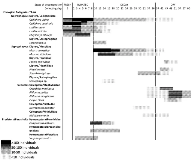
In the autumn, summer and spring experiments four stages of decomposition were identified (fresh, bloated, decay and dry) (figures 22, 26, 36). In the winter experiment five stages of decomposition were recognized (fresh, bloated, decay, adipocere and dry) (figure 31). In each season, the entire decomposition process had different durations, due to the influence of environmental conditions. Ecological categories occurred on the carcasses presenting different species composition according to the phenology of the species. For the necrophagous category, the Calliphoridae species were the dominant taxon, represented by different species for each season. According to Dominance Index, calculated for the sampled individuals (table 1), Calliphora vicina Robineau-Desvoidy 1830,
Calliphora vomitoria (L.) and Lucilia caesar (L.) were dominant in the
autumn experiment; in the summer investigation Chrysomya albiceps (Wiedeman 1819) was the most abundant species; in the spring investigation L. caesar and Lucilia sericata (Meigen 1826) were the dominant species; in the winter experiment a very low number of
VII temperature, thus the role of necrophagous in this investigation was played by Coleoptera Silphidae Thanatophilus rugosus L. and Thanatophilus
sinuatus (Fabricius 1775). Furthermore species composition of the other
ecological categories and their time of arrival were different in each investigation, as reported in the figures 25, 29, 35, 41. We observed the important role of Hymenoptera Formicidae that acted as predator towards the Calliphoridae larvae and also caused post mortem artifacts on the carcasses skin (figure 30).
Sampling of necrophagous species
In two years, several Diptera families were trapped in the three areas, as reported in table 2. The Calliphoridae family was the most abundant for
wild area (36,57%) and urban area (30,86%). In rural area Calliphoridae
represented the 18,99% of sampled families, and in this area the most abundant was the Platystomatidae family (27,63%).
Data analysis were performed for Calliphoridae because this family is the most important in PMI estimation. Calliphoridae species are identified as
Calliphora vicina Robineau-Desvoidy 1830, Calliphora vomitoria (L.), Chrysomya albiceps (Wiedeman 1819), Lucilia ampullacea Villeneuve
VIII From Correspondene Analysis (CA) the species-habitat associations can be observed in graphic 8. As shown, C.vicina and L.caesar are related to
urban area; L. ampullacea, Ch. albiceps and L.caesar are related to rural area; C.vomitoria is the most representative for wild area.
According to Synanthropyic Index (table 10), the most synanthropic species is L. sericata that was never trapped in the wild area, instead C.
vomitoria is not related to the urban area but is strongly related to the wild area.
Concerning the temporal variability, phenology of each species, it is different in each area (as shown in the graphs 9, 10 and 11), probably because the average temperatures for each area are different. In general
Calliphora spp are more active during the cooler months, but not in the
severe winters of wild area (winter average temperature 3,56 ± 1,08°C). In this area Calliphora species shift their activity in the spring and autumn months. L. caesar is the most abundant species for rural area for almost the entire sampling period except for winter months when the C. vicina becomes the dominant species. L. sericata is the most abundant in summer months in urban area, the rest of year, C. vicina is the dominant urban species. L. ampullacea is related to rural area during spring, Ch. albiceps represents the species with the lowest number of individuals, although it had colonized the carcass in the summer investigation. Also, Lucilia
IX species show a strong correlation with environmental temperatures, as reported by the Pearson Correlation in table 25. From this analysis, it appears that the abundances of Lucilia spp are positively related with temperatures, instead for the other species there isn’t a correlation. Maybe
Ch. albiceps is represented by a very low number of individuals and Calliphora spp are more influenced by the habitat then the temperature.
C
ONCLUSIONSThis research has collected important data for forensic entomology in Calabria. Now we are able to know which species we expect to find in human corpses and which ones are affected by the surrounding environment and possible competitors.
The important role of Calliphoridae was evaluated, along with the ecological and biological factors that affect species colonization were observed and analyzed. We also observed the important role of Hymenoptera Formicidae in post mortem artifact creation.
We studied the ecological habits of Calliphoridae of forensic importance, and have a general knowledge concerning the habitat preferences and the period of species activity.
X Future studies will focus on the life cycle of species, breeding dipteral colonies in laboratory conditions in order to time the development cycle duration at different temperatures, and complete the biological aspects useful in forensic investigations.
1
AIM OF THE STUDY
Medico-legal entomology (or forensic entomology), is a developing science in Italy, although in the rest of Europe and in America, it is already fully applied. This science investigates in both medical and forensic fields. The insects involved in forensic cases, above all Diptera Calliphoridae, are attracted to the decomposing organic matter, thus allowing to colonize human and/or animals corpses. Forensic entomology can be applied through several ways because with the presence of the insects on the corpses, it is also possible to understand if the body was displaced or if it had suffered traumas or mutilations ante mortem. Moreover, lots of the necrophagous species are involved in myiasis cases, infestations of diptera larvae on living individuals that are neglected in hygienic conditions. Entomological finds represent important investigative tools in cases of unknown death and in myiasis in vulnerable individuals (elderly, children and animals). Insect evidence is considered valid proof to present in court for the correct interpretation of a death scene or for negligence cases.
2 Necrophagous species activity is strongly affected by geographical position, due to the fact that species composition is different according to their spatial distribution.
In Italy this science is rather new, totally unknown for Southern Italy. The lack of information represents the starting point of this study. Nowadays there is not an official data base to use as reference, thus the aim of this research was to evaluate which insect species living in the Cosenza province are involved in corpses colonization and myiasis cases. This study was carried out following two steps:
1. Studying which species are involved in cadavers colonization in several season, using animal models;
2. Studying the environmental preferences of necrophagous species through a two-years sampling in three different areas characterized for a different level of urbanization.
The final aim of this study is to realize the first official data base facilitating the application of experimental data in real cases.
1
Chapter 1
INTRODUCTION
Forensic entomology is a rather new branch in the forensic and medico-legal field. The science that applies the entomological knowledge in this field is forensic entomology, which studies the species of insects and other arthropods involved in civil and criminal disputes and/or involved in human environment infestation. In turn, this science is divided into three branches: stored products entomology that investigates arthropods that infest badly preserved foodstuffs; urban entomology that studies the species infesting human environment. Myiasis cases also belong to this branch. Negligence on vulnerable individuals (elderly, children and animals) in public or private building, when hygienic conditions are unsatisfactory, is pursued by the law and myiasis gives proof of the time of larvae infestation, along with the time during which the individual was neglected8 along with information concerning the duration of neglection; medico-legal
entomology (or forensic entomology) that studies the insect species
involved in criminal investigation such as homicide, suicide, sudden death
2
etc., and in criminal offenses (physical abuse, smuggling, drugs)9. This
field mainly investigates the duration of time a corpse was exposed to the environment (post mortem interval or PMI) and the area in which the corpse was discovered, using insect evidence found on it and in the surrounding areas10.
Forensic entomology represents an important investigative tool in case of sudden death or discovery of corpses in advanced stages of decomposition, because since the moment of death several insect species colonize the corpse feed on the decomposing organic matter, putrefactive liquids or preying other species.
T
HANATOLOGICAL METHODS TO EVALUATE TIME OF DEATHThe decomposition of a body is a continuous process that starts with the death and ends when the corpse reached the skeleton stage. The time of death (post mortem interval or PMI) estimation is usually calculate according to thanatological changes occurred on the body after death11. The fisical-chemical changes that start the decomposing process appear immediately or shortly after death and progress in an orderly manner until
9 Byrd&Castner, 2010
10 Hall, 1990 in Byrd&Caster, 2010 11 Goff, 2009
3 complete disintegration. The duration of thanatological changes is strongly influenced by unpredictable endogenous or environmental factors12. PMI evaluation is an estimation based on putrefactive phenomena that occur on the body, not an absolute value, so its precision decreases with the passing of time13,14.
Thanatological changes which occur on the body after death are mainly
algor mortis (body cooling), rigor mortis (cadaverous rigidity), livor mortis
(lividity) and putrefaction.
Algor mortis
Body cooling is the more useful element to estimate time of death during the first post mortem 24 hours. With the end of metabolic activities that produce heat (thermogenesis), the body undergoes a progressive lowering of the temperature, which is dispersed in the form of heat through the surface of the body. The lowering of the body temperature continues until it reaches the same value as the environment. The PMI estimation can be made knowing the speed of lowering, namely the gradient. This parameter can be calculate if it is possible to make temperature measurements at
12 Pounder, 1995 13 Goff, 2009 14 Puonder, 1995
4 precise time intervals. The temperature gradient starts when the inner part of the body begins to cool, and this parameter can vary from an instantaneous time to several hours. The cooling speed of the body depends on several factors, such as the size of the body (larger bodies dissipate heat quickly because the dispersion surface is greater), the presence of clothes (which slow down the process because they retain heat), air humidity and the immersion of the body in water (which instead promote the cooling rate). After this step, the body temperature will increase again because of metabolic activity of bacteria and other organisms present inside the body15.
Rigor mortis
The cadaveric rigidity is due to the chemical reactions that occur in the muscles. After death, the muscles relax completely, and because the muscle tissues become anoxic, the cells work anaerobically to produce ATP. The final product of anaerobiosis is the lactic acid that decreases the cell pH level and causes the glycogen desegregation and ATP diminution. The ATP function in the muscles is to inhibit the link between actine and myosin. After anaerobiosis, the ATP diminution causes the actine-myosin link,
5 inducing the muscle rigidity. The rigor mortis appears 2-3 hours after death, the time to resolution vary from 24 to 84 hours, according to the factors which influence the duration. Generally, the rigidity occurs firstly in the face muscles and continues in a cranio-caudal sense extending first to the upper limbs and then to the lower limbs. The total rigidity of the body remains stationary for about 36-48 hours from death and finally the muscles return to relax following the same cranio-caudal order.
In general, rigor mortis duration is affected by environmental temperature and muscle activity before death16,17.
Livor mortis
The post mortem lividity consists of the brownish coloration of the body, it’s a physical event due to the interruption of blood circulation. The blood doesn't receive the heart's thrust, so it starts to migrate to the lower parts of the body according to the gravity, forming clots defined hypostasis. The hypostasis pass through an initial phase during which the blood is still semiliquid and they can migrate if the body is displaced. When the clot becomes permanent, the hypostasis become fixed and do not follow the
16 Goff, 2009 17 Pounder, 1995
6 body movements. The livor mortis appears from half an hour to two hours after death and ends about 9-12 hours after death18,19.
Putrefaction
The putrefaction is the most important process degrading the organic matter after death, due to the activity of bacteria and enzymes. The cellular autolysis leads to the gradual dissolution of tissues in gasses, liquids and mineral salts20,21,22. As reported by several authors, the decomposition process has several stages, which occur gradually. In outdoor environments, or in uncontrolled conditions, a decomposing body represents a source of food and shelter for many species of insects and arthropods, which contribute to the decay of organic matter, sometimes accelerating the process. When the corpse reach an advanced decomposition stage, the thanatological methods are not sufficient to estimate the PMI and insect evidence is essential in determining the time of death23. The body decomposes through several stages, each one attractive
18 Goff, 2009 19 Pounder, 1995 20 Campobasso et al., 2001 21 Gennard, 2007 22 Pounder, 1995 23 Byrd&Castner, 2010
7 for different insect and arthropod groups24,25. The stages of decomposition which occur on the corpse can be reassumed in:
Fresh stage: it starts at the moment of death and ends with the first signs of
bloating. This stage attracts necrophagous species which use natural cavities of the body as oviposition sites. In general the first colonizers are the Diptera Calliphoridae and Sarcophagidae26,27;
Bloated stage: it starts the putrefaction. Anaerobic bacteria living inside the
body start the tissues digestion. The gasses emitted by their metabolism confer the state of swelling of the body. Diptera Calliphoridae are mainly attracted by the putrefaction smell, and also Coleoptera occur in this stage preying diptera larvae28.
Active decay stage: it starts when the skin lacerates and gasses are released
outside. The decomposition process goes on leading to the formation of butirric and caseic acids that attract Diptera Piophilidae. During this stage the maggot mass is conspicuous on all the body and predator Coleoptera are more abundant (Staphylinidae, Histeridae, Silphidae)29,30.
24 Gennard, 2007 25 Goff, 2009 26 Gennard, 2007 27 Goff, 2009 28 Gennard, 2007 29 Gennard, 2007 30 Goff, 2009
8
Post-decay stage: the body reduces in skin, cartilage and bones. Diptera are
no more the dominant taxon, the majority of them have already completed their life cycle. During this stage the corpse is attractive for several species of Coleoptera which feed on the decomposing remains (Dermestidae, Cleridae)31,32.
Skeletal stage: only bones and hair remain on the body, in general this
stage is not colonized by specialist insects but by accidental species that seek refuge in the dry remains.
The time of decomposition vary strongly with the size of the body, with environmental factors and the conditions in which the body stay. PMI estimation becomes more difficult and less precise using only thanatological methods33.
E
NTOMOLOGICAL METHODS TO EVALUATEPMI
The time since death (post mortem interval or PMI) is the most important information to obtain in cases of homicides or in unknown death investigations34. Even in cases of natural death, this information is useful
31
Gennard,
32 Goff,
33 Byrd&Castner,
9 for inheritance and insurance reasons35. Often, forensic investigations concentrate on maximum PMI determination, or the time interval between the discovery of body and the moment that the deceased was seen alive for the last time. This method can be applied only if the deceased can be identified36. Legal medicine can only estimate the time of death accurately during the first 72 hours after death, because after this period the body undertakes drastic changes and thanatological inspections are not sufficient to determinate the PMI. When a corpse is discovered in the advanced stage of decomposition, it is not simple to establish the time of permanence of the body in the environment. Moreover, with traditional investigations, it is not always possible to know if the place of death coincides with the place of discovery37. The use of insect evidence turns out of fundamental importance in determination of time of death because, as observed through a lot of research, the necrofauna activity shows PMI-dependent processes38. Necrophagous insects are attracted to decomposing organic matter, not by living individuals, except in particular cases. From their presence on a body, it is possible to estimate minimum PMI, or the time elapsed between the moment of death and the moment of discovery of the body39.
35 Henssge et al., 1995 n Byrd&Castner, 2010 36
Henssge et al., 1995 in Byrd&Castner, 2010
37 Marchenko, 2001
38 Hall, 1990 in Byrd&Castner, 2010 39 Smith, 1986 in Byrd&Castner, 2010
10 Entomological method to evaluate PMI is based mainly on two approaches. The first one concerns the succession with which several ecological categories colonize the body, replacing themselves as the decomposition goes on. This method is especially utilized when the discovered corpse is in advanced stage of decomposition, considering the ecological category present at a precise stage. The succession method is used to estimate both minimum and maximum PMI40.
The second method concerns the estimation of larval age of immature that feed on the body tissues. The first colonizers occur almost immediately after death, laying the offspring in the natural cavities of the body or in the open wounds. The young feed on the tissues for the entire larval cycle, so the age of samples indicates the time of the body exposure in the environment. This method is more accurate and is very useful for estimation of minimum PMI41.
Entomological successions
In natural environments, the ecosystems modify as the time passes changing the species composition of communities that live in it. The same
40 Shoenly et al., 1992 in Byrd&Castner, 2010 41 Smith, 1996 in Byrd&Castner, 2010
11 happens with the cadaveric resource, that modify with the decomposition process, attracting gradually different groups of species. Since death, the corpse undergoes several physical, chemical and biological changes, modifying its thanatological conditions through several stages of decomposition until the complete skeletonization42. Each stage is attractive for different insect species which colonize the body at different times but always related to the stage of degradation of the trophic resource. The experiments carried out by Francesco Redi43, that refuted the aristotelian theory about spontaneous generation, focused the attention on the existence of necrophagous species attracted by decomposing organic matter. The relation between insects and corpses, although known since the ancient Egyptians, already applied in China during XIX century and observed during mass exhumations in France and Germany during XVIII and XIX centuries44, starts to be studied in detail by P. Mégnin at the end of 180045. In 1894, in fact, after the publication of the book La faune des cadavres, it is highlighted how different insect species are attracted to decomposing bodies and how they are replaced as the decomposition continues, due to the fact that each one is trophically related at a precise thanatological
42
Coe and Curran, 1980; Henssge et al., 1995; Van den Oever, 1976 in Byrd &Castner 2010
43 Redi, 1875 44 Benecke, 2001 45 Mégnin, 1894
12 condition. Mégnin observed which species occur on the corpses and which role they play, attributing them to several ecological categories grouped in eight teams of travailleurs de la mort46(figure 1).
Figure 1. The travailleurs de la mort described by Mégnin (from Benecke, 2002).
From the Mégnin observations, several studies about insect colonization were carried out, and still today the successions study represents the starting point for entomological investigations in forensic field47,48. The eight teams of travailleurs de la mort described by Mégnin, defined
46 Mégnin, 1894 47 Byrd&Castner, 2010 48 Gennard, 2007
13 successional waves49, are nowadays counted in four main ecological categories necrophagous, predators & parasites, omnivorous, accidental50 (figure 2). The first colonizers, pioneers in the cadaveric succession, are represented by necrophagous, in which belong Diptera Calliphoridae and Sarcophagidae families, which lay eggs or directly larvae in natural cavities of the body or in the wounds. Larvae hatch and feed on the decomposing tissues. As the decomposing process continues, other Diptera families occur in the body, such as Muscidae, Fannidae, Piophilidae, Phoridae, attracted by advanced decay stages. The last stages of decomposition are attractive for Coleoptera Dermestidae and Cleridae that feed on dry rests of skin and bones51.
Predators and parasites feed on other insect or arthropod species that are present on the corpse. The most important representatives are Coleoptera Staphylinidae, Silphidae, Nitidulidae, Histeridae, although some Silphidae species can act also as necrophagous in particular circumstances52,53. Also schyzophagous species belong to this category, which first feed on the body and when they reach mature larval stages feed on other larval species,
49 Tuschetto&Vanin, 2004 50
Smith, 1986 in Amendt et al., 2004
51 Campobasso et al., 2001 52 Bonacci et al., 2010 53 Campobasso et al., 2001
14 such as Chrysomya genus (Calliphoridae)54. Omnivorous species, which include Hymenoptera Formicidae, Vespidae and some Coleoptera species, feed on both the body and other colonizers.
Finally, accidental species, such as Springtails and Arachnids, use the body as extra resources or occasional shelter55.
Figure 2. Entomological succession on an animal carcass (from Klein, 2005).
Based on the presence of a certain ecological category on the body, it is possible dating the period of the exposure in the environment, this method is used above all in cases of discovery of bodies exposed for a long period
54 Campobasso et al., 2001
15 of time and/or in cases of death happened in unknown circumstances56. Although the modalities of replacement of categories are rather predictable, specie composition varies for each category because it affected by the environment in which the corpse is exposed and time of arrival of each species and time of stay depend on their ecological requirements57.
A correct identification of the species found on the body represents the starting point in a crime scene interpretation and PMI evaluation58. Several variables affect the process of corpse colonization, in particular the modalities of succession vary depending on geographical position, the body exposure (sunlight, shade) and the kind of habitat in which the body is placed59.
Geographical position is one of the most principal factors that affect specie composition of ecological categories in a succession. The biogeoclimatic zone defines a particular habitat, the vegetation, the soil type and meteorological conditions of the area. This affects both the presence and the phenology of the colonizer species. Moreover, thanatological processes are different according to the same factors, along with the time of arrivals of the involved species, which vary between regions60.
56 Byrd&Castner, 2010 57 Byrd&Castner, 2010 58 Campobasso et al., 2001 59 Byrd&Castner, 2010 60 Byrd&Castner, 2010
16 The season has a strong influence on weather conditions and on the typical flora and fauna characterizing a region, in other words from season depends phenology of the species. Many necrophagous species have different abundance depending on the season because their activity takes place in a determinated period of the year. Some species are tipically summer species due to being related to high temperature, others in contrast are active in cooler months61,62. The presence of a species or of its tracks on the rests depends on its phenology and on the ability to withstand seasonal weather conditions63.
The position of the corpse has a decisive effect on remains colonization. The main effect is the difference of heat between corpses directly exposed in sunlight and those in shade. The direct exposition to sunlight determinates an increase of temperature and this fact accelerates the decomposition process. The biomass reduces more rapidly than a corpse in shade, accelerating the passage from a decomposition stage to another. Heliophilous species are more attracted by sunlight exposed corpses and the increase of heat caused by sun radiation, which also accelerates their
61 Arnaldos et al., 2001 62 Arnaldos et al., 2004 63 Byrd&Castner, 2010
17 activity. Sciaphilous species are instead attracted by shade corpses, as in this case the decomposition process is slower64,65.
The kind of habitat in which a body is discovered has fundamental importance because necrophagous species have a spatial distribution affected by their ecological preferences. Some species have cosmopolitan distribution, because they are present worldwide and in every habitat, others are specialist and they can only be found in rural or woodland habitats. Synanthropic species live in urban environments, closely with humans. The synanthropy level of a particular species is important to know during a forensic investigation, for example when entomological finds belonging to a urban species on a body discovered in a woodland are found, it could be surmised that the body was displaced after death66.
The environment surrounding the body (city, countryside, indoor or outdoor), the conditions in which it is (buried, burnt, on a beach, in a woodland or at sunlight exposed), the microclimatic conditions (temperature, humidity, weather) represent elements to consider for the entomological research and an accurate PMI estimation67.
64 Joy et al., 2006 65 Byrd&Castner, 2010 66 Byrd&Castner, 2010 67 Byrd&Castner, 2010
18
Post mortem artifacts
The cadaveric resource is colonized by a variety of insect species, some of them occur feeding on the body tissues, others occur instead preying on other species attracted by the remains68,69. Many species are omnivorous, playing the double role of necrophagous and predators. The species that occur on the body leave traces of their activity, such as larval exuviae, empty puparia, peritrophic membranes, all elements useful in forensic investigations70. But, often, their activity can also alterate the thanatological conditions of the body, causing post mortem artifacts difficult to interpret with the only medico-legal surveys71.
In outdoor or uncontrolled environments the exposed corpse can be preyed by macrofauna (rodents, carnivores, other animals) and also by microfauna (insects in particular) that living the area72,73. The artifacts caused by macrofauna are quite distinguishable from ante mortem injuries because they don’t show bleeding or redness in the tissues adjacent the lesion margins and also have well-defined boundaries based on the shape of the
68 Rodriguez&Bass, 1983 in Campobasso et al., 2009 69 Smith, 1986 in Campobasso et al., 2009
70
Byrd&Castner, 2010
71 Byard, 2005 72 Tsokos, 2005 73 Byard et al., 2002
19 teeth74. The microfauna activity instead is more difficult to identify because
post mortem artifacts inflicted by insects are often confused with wounds or
lesions suffered ante mortem (figure 3).
Figure 3. Post mortem artifacts caused by insect activity (from Batalis & Cina, 2012).
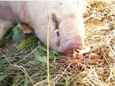
For example, Calliphoridae larvae or other necrophagous Diptera, pass through the skin of the corpse making the small circular holes often confused with gunshot wounds suffered before death. Other opportunistic species, such as cockroaches, produce abrasions on the skin surface that mimic skin deseases75. Hymenoptera Formicidae, as well as being predators of immature stages of Diptera, can also cause lesions on the
74 Tsokos, 2005 75 Tsokos, 2005
20 cadaver skin through corrosive secretions, easily confused as strangulation marks or excoriation suffered ante mortem76,77,78.
The Formicidae intervention on bodies is immediate and often impedes or hinders the access to other ecological categories. The lesions caused by Formicidae on a body represent their interference with Diptera Calliphoridae colonization, therefore the identification of post mortem artifacts contributes to a better PMI interpretation79,80. Sometimes, Hymenoptera Formicidae represents the numerically dominant taxon among the all colonizers and occasionally they can establish a colony on the colonized corpse. Goff (1997) reports a case of minimum PMI estimation calculated according to the period of establishment of an ant colony Anoplolepsis longipes, found on a human corpse remains in Oahu island (Haway)81.
Using animal models in bodies succession studies
The decomposition processes and the modalities of species colonization are strongly influenced by environmental and other factors inherent in both the 76 Tsokos, 2005 77 Bonacci et al., 2011 78 Byard, 2005 79 Bonacci et al., 2010 80 Bonacci et al., 2011 81 Goff, 1997
21 position and condition of exposed bodies. Therefore it is essential to study the necrophagous fauna in a determinated area in different experimental conditions. The studies of seasonal variations, habitat influence and the effect of bodies exposition in several environments have been carried out by several researcher worldwide, thanks to the use of animal models82,83,84. Nowadays, international works include several models used for forensic purposes85, but the model considered most valid in application of data to real cases is the domestic pig Sus scrofa (L.). The experimental model shows decomposition times and modes quite similar to those of a human corpse. The domestic pig, in fact, is an omnivorous species which has an intestinal bacterial flora similar to the human, has a very thick skin and modes of decomposition correspond approximately to the human body with similar size86,87.
Life cycle of necrophagous Diptera
Necrophagous Diptera, mainly represented by Calliphoridae and Sarcophagidae families, are the principal elements for PMI estimation 82 Byrd&Castner, 2010 83 Bonacci et al., 2010 84 Tomberlin et al., 2012 85 Tomberlin et al., 2012 86 Campobasso et al., 2001 87 Tomberlin et al., 2012
22 because they occur on a cadaver immediately after death88,89. The adults, attracted by decomposition smell even from long distances, colonize the corpse laying eggs (or directly larvae as in Sarcophagidae) in the natural cavities of the body or in the open wound, in order to ensure the trophic resource for the offspring. Immature feed on the decaying tissues completing the entire larval cycle on the body. Once the third larval stage is reached and completed, they migrate from remains and pupate in surrounding soil waiting for metamorphosis90,91 (figure 4).
Figure 4. Life cycle of a Diptera Calliphoridae (from Goff, 2000).
88
Byrd&Castner, 2010
89 Donovan et al., 2006 90 Byrd&Castner, 2010
23 The larval size, usually indicated by their length, is related to their age and (it, not needed) is expressed as a function of time and temperature. Taking into account the environmental temperature, the larval age estimation of an older sample found on or near the body, represents the minimum PMI92. Necrophagous flies don’t show the same time of development, because each species has optimal ranges of temperature dictated by their ecological needs. So, the larval development is a temperature-dependent process, each species has a thermal history defined as the amount of heat necessary to complete the cycle 93. In general an increase of temperature contributes to a decrease of development time, in contrast lower temperatures increase it, in any case the amount of heat necessary to complete the cycle is constant94. The identification of the species found on a corpse represents the first step during the entomological investigations95. For each species, every development stage needs a certain amount of heat, that is quantified as
accumulated degree/hours (ADH) or accumulated degree/days (ADD),
necessary to reach the pupa stadium96,97,98. The heat accumulation, in hours or days, for any temperature is calculated as99:
92
Donovan et al., 2006
93 Joseph et al., 2011
94 Wilson et al., 1993; Greenberg&Kunich, 2002 in Niederegger et al., 2010 95 Joseph et al., 2011 96 Donovan et al., 2006 97 Joseph et al., 2011 98 Ames&Turner, 2003 99 Niederegger et al., 2010
24 ADH = (T - TLDT) * hours to develop
ADD = (T - TLDT) * days to develop
When:
T = temperature (or means temperatures) gained on the discovery location
TLDT = Lower Development Treshold, low temperature at which
development stops
The difficulty in this method is to establish the minimum temperature of development, different for each species but also for the same species, this parameter can vary according to other factors. For example, the geographic position influences the phenology, whereby the same species shows a greater or lesser tolerance to extreme temperatures according to the environment in which lives. Several European authors report a LDT different for the same studied species100,101.
Another approach to estimate the larval age is by comparing the length of the older found samples with isomorphen and isomegalen diagramas102,103.
100 Ames&Turner, 2003 101 Donovan et al., 2006 102 Reiter, 1984 103 Grassberger&Reiter, 2001
25 From isomorphen diagrams, it is possible to predict how much time it takes a species to reach a particular stadium at a certain temperature (figure 5). This graph is useful above all when samples are found in post feeding or pupa stages, namely when the length is not more an useful criterion to estimate larval age104.
Figure 5. Isomorphen diagram for the species Lucilia sericata (Calliphoridae). All the immature stage are shown, from eggs to adults emergence at different temperatures. The spaces between the lines represent the same morphological stages at different temperatures. a: eggs; b: L1; c: L2: d: L3; e: postfeeding (prepupae); f: pupae; g: adults (from Grassberger & Reiter, 2001).
From isomegalen diagrams, it is possible to estimate the larval age based on the length of the samples, assuming that it developed at constant temperature (figure 6).
26
Figure 6. Isomegalen diagram for the species Lucilia sericata (Calliphoridae) from the hatching to third larval stage. The temperature is represented as a function of the time, each line in the graph represents the same measure of length in mm. In the upper right, the graph shows the duration of egg stage before the hatching at different temperatures (from Grassberger & Reiter, 2001).
However, the isomegalen diagrams are developed through constant temperatures and this fact can provide a loss of information, because in real cases it could be calculated as a means of environmental temperatures105.
27
M
YIASISIn some particular situations, necrophagous species which generally colonize corpses can also infest living individuals causing myiasis in natural cavities or in open wounds106. The correct definition of myiasis is given by Zumpt (1965) defining them as infestation of diptera larvae on living human and other vertebrates that at least for a certain period feed on living or dead tissues, corporal liquids or ingest food of the host107. The most important criterion of this definition is that infesting larvae complete, or continue at least for a certain period, their normal development on the host body108.
Myiasis agent Diptera belong to three main groups, divided according to their habits109,110:
1. Obligatory parasites: which develop exclusively on living tissues. To complete their cycle they must find a living host. To this group belong myiasis caused by Oestridae and species of Sarcophagidae families, which infest livestock (figure 7);
106 Byrd&Castner, 2010 107 Zumpt, 1965 108 Zumpt, 1965 109 Zumpt, 1965 110 Hall, 1991
28
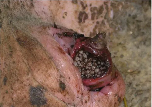
Figure 7. Urogenital myiasis in a sheep farmed caused by Wohlfahrtia magnifica (Sarcophagidae) (from Giangaspero et al., 2011).
2. Facultative parasites: which are usually free living, in general are represented by necrophagous species that feed on dead bodies and only occasionally infest living individuals. Diptera Calliphoridae, main PMI indicators, belong to this group. Facultative parasites can cause primary myiasis if they start the infestation; secondary myiasis if they occur only when the infestation was already started by other species; tertiary myiasis is the host is close to death111,112;
3. Accidental parasites: the infestations in this case are defined
pseudomyiasis because larvae don’t infest the host directly but they
are ingested accidentally, for example for food contamination, and
111 Hall, 1991 112 Cruz, 2004
29 pass through the digestive tract. In this case, the larvae ingestion is almost always passive and often the passage in the digestive tract causes their death. Larvae can be expelled through the feces or establish in the intestine causing enteric disorders113.
Myiasis can be classified also according to the site of infestation or the site of next development in the host. From the medical point, the infestations are defined as gastrointestinal, urogenital, nasopharyngeal, auricular and
cutaneous based on the anatomical area selected by infesting larvae114,115
(figure 8).
113 Amendt et al., 2010 114 Hall, 1991
30
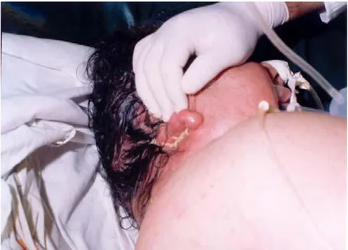
Figure 8. Auricolar myiasis in a 62 aged female caused by Lucilia sericata (Calliphoridae) (from Yaghoobi et al., 2005).
In the forensic field, myiasis are related more communally to facultative parasites of Calliphoridae, Sarcophagidae and Muscidae families, generally used for PMI estimation. In some particular cases, myiasis can represent an element of confusion because provide to a longer PMI estimation then the real time elapsed from death (such as can occur in tertiary myiasis cases). In other situations, for example in the case of infestation on living individuals, myiasis represent a valid help to solve negligence cases on vulnerable individuals116.
31
Chapter 2
MATERIALS & METHODS
1. E
NTOMOLOGICAL SUCCESSIONSThe study of entomological successions was carried out over four experiments, taking place in each season (autumn, summer, winter, spring). The autumn experiment was carried out from November; the summer one in July; the winter one from February; the spring experiment from May. Entomological researches were carried out in the same study area (in the Botanical Garden of University of Calabria) using specimens of domestic pigs, Sus scrofa (L.), as animal models.
Animal models
For the seasonal experiments, four specimens of Sus scrofa (figure 9) weighing 20-25 kg were used, one for each season. The models were taken from a farm located close to the study area and euthanised by lethal injection with the assistance of a veterinarian. Following their death, the
32 models were positioned on the soil covered by a wire netting fine mesh and enclosed in a fence, thus avoiding avoid a possible macrofauna attack. The experimental procedure's aim was to simulate situations of death in an outdoor environment and evaluate the seasonal influence (and accordingly the climatic factor influence) on both the presence and activity of colonizer species and on thanatological changes of exposed carcasses.
33 Study area
The carcasses were placed in an area of Botanical Garden of University of Calabria (figure 10), suitably selected respecting to hygiene and health standards established by law.
The Botanical Garden is developped on a hilly terrain at 180-230 meters a.s.l. and covers an area of about 8 hectares. Geographic coordinates are included between 39°18’ and 39°24’ of latitude N and 16°10’ and 16°20’ longitude E. The boundaries that trace the perimeter of the garden are determined by a concrete wall with a wire netting masked by creepers and bordered to the south and west sides by a row of cypress trees. The wall stops at five gates of which only one is used as an input to (the) visitors. The garden encloses semi-natural areas with a significant sample of flora Calabria: more than 4000 species of about 2500 total census for the whole of Calabria. The geological substratum of the area consists of sand and conglomerates and cement-crystalline metamorphic from brown to reddish clays with interbedded gray to blue-gray. The presence of these layers of clay permits the formation of a discrete water system at the surface.
34 The botanical garden in fact, besides being crossed from the west to east by a stream that goes in the direction of the river Crati includes four wells and two small perennial springs.
The vegetation in the area consists mainly of plant communities established on former cropland and fragments of training subspontaneous represented by an oak grove and a riparian poplars grove and willows along the creek.
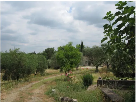
Figure 10. Entrance of the Botanical Garden of University of Calabria (da Greco S.).
The potential vegetation of the area is represented by deciduous oaks classifiable in Querceta ilicis (thermophilic elements species-rich evergreen) and Querceta robori-petraea (mesophilic elements). The dominant species is Quercus virgiliana, oak belongs to the cycle of Q.
35
pubescens (downy oak). It features a little disturbed forest formation,
where there are many characteristic species of thermophilous woods (Erica
arborea, Cornus sanguinea, Clinopodium vulgare, Brachypodium sylvaticum, Knautia calycina, Helleborus bocconei).
Part of the site is occupied by a thicket of willows (Salix caprea and S.
triandra), in which undergrowth is abundant Equisetum telmateja. Other
hygrophilous species are represented by Dorycnium rectum, Potentilla
reptans, Mentha suaevolens. This vegetation is related in this area to a
perennial source. The valley floor is characterized by a grove of trees in riparian Populus alba belonging to the order Populetalia albae. In the undergrowth there are abundant species typical for hygrophilous woods such as Equisetum telmateja, Lamium flexuosum, Lythrum junceum.
Part of the area is characterized by secondary vegetation abandoned cropland with scattered fruit trees and now converted into meadow hay (Avena barbata, Agrostis castellana, Chrysanthemum segetum, Echium
plantagineum) 117.
The experiments were carried out in an area characterized by Quercus
pubescens Willd. 1805, Pirus spp (L.) and Olea europea (L.) (figure 11).
36
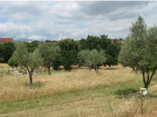
Figure 11. The area of the Botanical Garden surrounding the experiment site (from Greco S.).
Experimental procedure
The carcasses were transported to the study area immediately after death, placed on the soil inside a fence and covered with a metallic netting in absence of researcher (figure 12).
37
Figure 12. Animal model placement in the study area (from Bonacci T.).
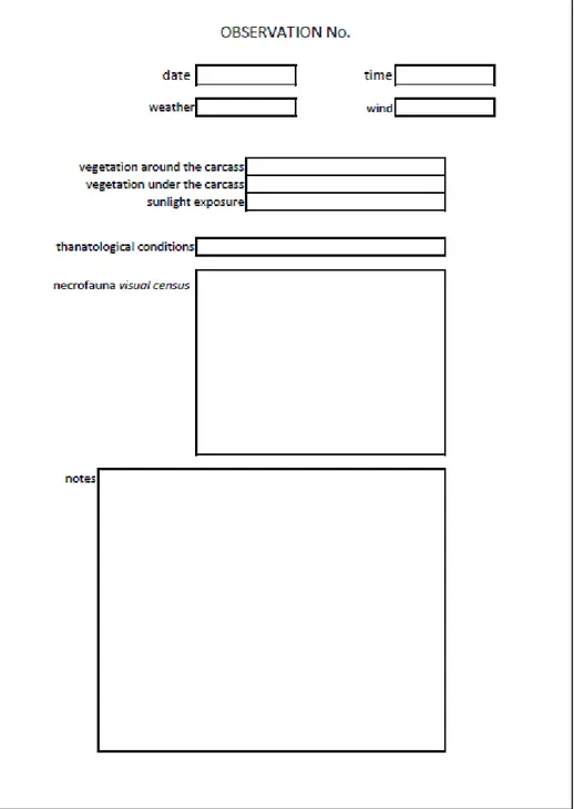
From the moment of placement, two digital data logger were activated, one with a probe inserted in the carcass the other outside, for continuous recording of the temperature and humidity. Data concerning rainfall related to the studied period were found on the official website of ARPACAL. Daily inspections are made, more times a day until the thanatological conditions were established. When the dry stage was reached, the inspections were less frequent, waiting for the complete body skeletonization. Daily, direct catches of necrofauna were made through an entomological net, also larvae were sampled with tweezers. Procedures for the collecting and preserving the samples followed the guidelines proposed by Amendt et al., 2007.
38 Thanatological changes of the body and the necrofauna activity related to them, were photographed, recorded and noted on appropriate experimental protocols (figure 13).
39 When carcasses were completely dry, the remains were removed and buried and entomological finds were collected in the soil under and around the carcasses site (figure 14).
Figure 14. Collecting of entomological finds after the carcass removal (from Greco S.).
The adults samples collected during the experiments were identified using international literature118,119,120, sorted by ecological category and stored in the Entomological Collection of Ecology Department (University of Calabria).
118 Chynary, 2004 119 Oosterboek, 2006 120 Rivosecchi, 2000
40
2. S
AMPLING OF NECROPHAGOUS SPECIESThe sampling of necrophagous species was carried out using bottle traps. Three areas with different degrees of nature were selected, in order to evaluate the influence of the habitat and climatic factors on the species distribution.
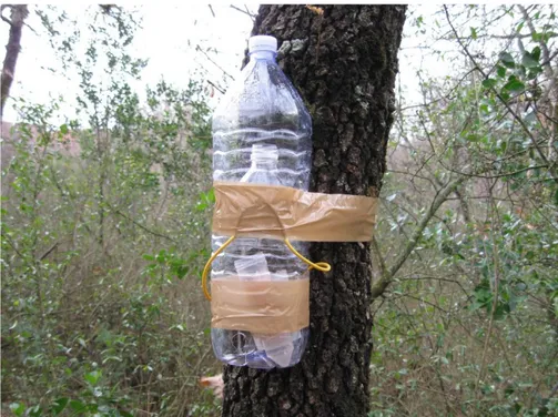
Traps design
The traps were made following the model proposed by Hwang & Turner (2005).
A trap is realized with the aid of two plastic bottles of 1,5 l, of the same shape and dimensions. The traps are positioned in order to create two chambers: the collecting chamber situated at the top of the trap and the bait
chamber represented by the lower part. The collecting chamber is made
with the two upper parts of both bottles, one pushed in the other (figure 15).
41
Figure 15. Design of the traps used for the sampling (from Greco S.).
Inside the bait chamber were positioned two plexiglas containers, one with mince and the other with NaCl solution in order to avoid the rapid dryness of the bait.
After the bait position, the two chambers are closed with sellotape and the traps are moved in the study areas (figure 16).
42
Figure 16. Bottle traps placement in one of the study areas (from Greco S.).
Study areas
The sampling of necrophagous species were carried out in three different areas of the Cosenza province, characterized by a different level of nature and defined wild area, rural area and urban area. Geographic localization of the areas is indicated in figure 17.
43
Figure 17. Geographical position of the study areas (from Google Earth).
Wild area
The natural site considered (figure 18) is located at about 15 Km from the University of Calabria, situated near San Fili village, at 985 m of elevation. Geographical coordinates are 39°19’12.37’’ latitude N and 16°6’45.86’’ longitude E.
The potential vegetation of the area is represented by a macrothermal beechwood linked to a humid tempered bioclimate markedly oceanic. These climatic conditions favor the spread of the beech on the Catena Costiera even at relatively low altitude at which the other Calabrian mountains are more thermophilous forest types (oak, chestnut and pine forests). The presence of beech at low altitudes is explained, in fact, by the
44 extreme nebulosity which also occurs in the summer period starting from about 650-700 m above sea level. This climatic peculiarity favoring the development of beech forests are characterized by a more complex structure with a rich shrub layer with mostly evergreen shrubs such as holly (Ilex aquifolium), butcher's broom (Ruscus aculeatus), Dafne laurella (Daphne laureola). This type of beech wood is Anemono
apenninae-Fagetum, association Fagetalia sylvaticae, endemic to the mountains of the
southern Apennines. The beech wood is generally governed by high forest, pure, in good vegetative conditions and with a full density.
45 The area is crossed by numerous rivers that create valleys characterized by forest vegetation in lindens and maples typical for moist ravines. These woods form a continuous band which characterizes the sides of the valley and come into contact with the hydric wood at the bottom and at the top of the beech wood. The undergrowth is rich in species nemoral such as
Hedera helix, Vinca major, Acanthus mollis, Helleborus bocconei, etc..
These formations are quite rare in the province. They are framed in Ostryon
carpinifoliae alliance, which brings together mainly mixed forests with
maples, lindens and elms linked to a rather wet and cool microlimate, located in ravines and valleys121.
Rural area
The rural site is represented by the Botanical Garden of the University of Calabria. The description of the area is already reported at page 33. The traps were positioned in four sub-habitats: two oak wood, broom wood, poplar wood (figure 19).
46
Figure 19. Bottle trap positioned in the rural area (from Greco S.).
Urban Area
The urban site of Rende-Cosenza (figure 20) is located in a hilly area at an elevation between 185-575 m a.s.l.; the geographical coordinates are 39°18’39’’60 latitude N and 16°15’3’’60 longitude E. the site is situated in a totally man-made environment in which the potential vegetation, represented by thermophilous deciduous oak forest, has totally disappeared. The few unbuilt areas are characterized by ruderal vegetation, weeds, trees and flower beds with non-native species122.
47
Figure 20. Urban area (from Albano R.).
Experimental procedure
The three sampling areas have the same extension, in order to ensure comparability of the results. For each area 8 bottle traps were used, positioned at a distance of about 15 meters one from other.
The investigation started in February 2010 with the first traps location in all the areas at the same time. Traps were replaced each months, for two years, until January 2012. In each replacement the traps were moved to the laboratory, empty and sorted, separating adults from larvae. Also in this
48 case we used the guidelines from Amendt et al., 2007 for the samples treatment.
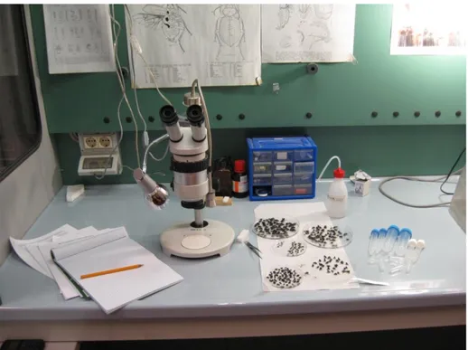
Larvae were preserved in 90% ethanol post boiling, thus ensuring the necessary procedure for the removal of bacteria and proper maintenance of the samples. In each sampling date, for all the area, the adults were sorted by families and stored in 70% ethanol. Taxa were counted and finally Calliphoridae species were identified using international identification
keys123,124,125,126 (figure 21).
Figure 21. Sorting and identification of entomological material in each sampling date (from Greco S.). 123 Rognes, 1980 124 Wallmann, 2001 125 Whitworth, 2006 126 Marshall et al, 2011
49 Data concerning environmental temperatures and rainfall for the sampling date were found on the official website of ARPACAL.
Data analysis
Statistical analysis for sampling data of necrophagous species, on their phenology and abundance comparison in the three area, were performed with IBM SPSS Statistic v. 20 and Past v.2.17 softwares.
Pearson’s Chi-square and Kruskall-Wallis tests were calculated in order to evaluate the statistically significance of the species abundance.
Correspondence Analysis (CA) were performed to evaluate the species
distribution in the areas, comparing also the seasonal abundances of the species.
Moreover, the Synanthropy Index (SI), proposed by Nuorteva (1963) was calculated to evaluate which species were more related to a human environment, with the formula:
When:
a = abundance (%) of species sampled in urban area b = abundance (%) of species sampled in rural area
50 c = abundance (%) of species sampled in wild area
The value of the index is between +100 (species totally synanthopic) and -100 (species totally forestry).
Finally correlations between abundance of species and environmental temperatures were made in order to evaluate the relation between these variables.
51
Chapter 3
RESULTS
E
NTOMOLOGICAL SUCCESSIONSAutumn investigation
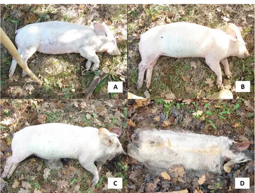
During the autumn investigation, four thanatological stages were identified: fresh, bloated, decay and dry (figure 22).
Figure 22. Stages of decomposition of the carrion in autumn. A) Fresh; B) Bloated; C) Decay; D) Dry (from Bonacci T.).
52 The entire decomposition process lasted 60 days, influenced by environmental conditions. The graph 1 shows the trend of average daily temperatures and rainfalls during the experimental period.
Graph 1. Trend of temperatures and rainfall during autumn experiment.
First colonizer occurred on the carrion were Diptera Calliphoridae, represented by Calliphora vicina Robineau-Desvoidy 1830, Calliphora
vomitoria (L.), Chrysomya albiceps (Wiedeman 1819), Lucilia caesar (L.)
and Lucilia sericata (Meigen 1826), although with different abundance. Calliphoridae colonized the carcass five minutes after the corpse placement, invading the natural orifices lying eggs (figure 23). After 21 hours from placement, other egg depositions were found in the right eye, exposed to sunlight. After 95 hours from placement, larvae in the mouth cavity started to migrate inside the body; after 170 hours other egg