UNIVERSITÀ DEGLI STUDI DI SASSARI
SCUOLA DI DOTTORATO IN
Scienze Veterinarie
Direttore: Prof. Sergio Ledda
INDIRIZZO IN: Produzione, Qualità e Sicurezza Alimentare (XXVII Ciclo) Coordinatore: prof. Enrico P. L. De Santis
Evaluation of Listeria monocytogenes contamination in sheep’s
milk cheese-making plants
Docente Guida Correlatore Dott. Carlo Spanu Dott. Vincenzo Spanu
Direttore Tesi di dottorato della Prof. Sergio Ledda Dott.ssa Michela Ibba
In order to fulfil the requirements for obtain the ―Doctor Europæus‖ mention, the thesis was reviewed by two expert pertaining to higher education institutions in European Countries other than Italy.
Reviewers:
Prof. Parthena Kotzekidou, Ph.D. Department of Food Science and Technology, Faculty of Agriculture,Aristotle University of Thessaloniki. Greece.
Prof. Paula Cristina Maia Teixeira, Ph.D. Escola Superior de Biotecnologia, Universidade Católica Portuguesa, Rua Dr António Bernardino del Almeida, Spain.
CONTENTS
ABSTRACT 7
INTRODUCTION 8
1.1Taxonomy Listeria spp 10
1.1.1 The Listeria genus 10
1.1.2 Listeria monocytogenes 11
1.2Ecology of Listeria monocytogenes 12
1.2.1 Growth limits 12
1.2.2 Biofilms and Niches 14
1.2.3 Persistent contamination 18
1.3Infection and Disease 19
1.3.1 Listeriosis 19
1.3.3 Listeriosis signs and symptoms 25
1.3.4 Infective dose 27
1.4 Listeria monocytogenes in foods 27
1.4.1 International microbiological food safety criteria for Listeria monocytogenes 27
1.4.2 Ready-to-eat foods 30
1.5 Listeria monocytogenes in the dairy sector 32
1.5.1 Milk 32
1.5.2 Dairy products 34
TABLES 43
REFERENCES 45
CHAPTER 2 67
THESIS PROJECT 67
CHAPTER 3 73
LISTERIA MONOCYTOGENES CONTAMINATION IN DAIRY PLANTS: EVALUATION OF LISTERIA MONOCYTOGENES ENVIRONMENTAL CONTAMINATION IN TWO CHEESE-MAKING PLANTS USING SHEEPS MILK 73
3.1 Abstract 74
3.2 Introduction 75
3.3 Materials and methods 76
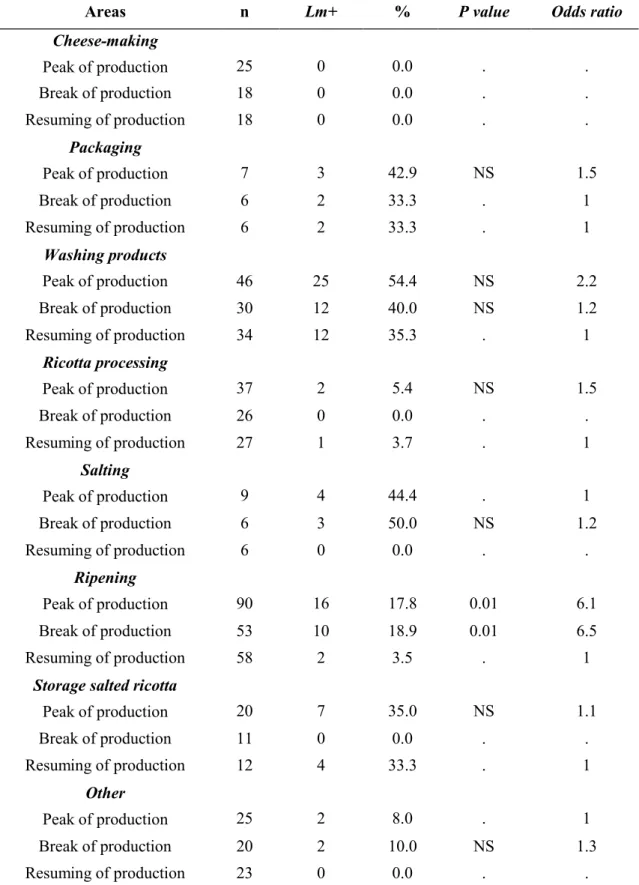
3.4 Results 77
3.5 Discussion and conclusion 79
Tables 81
REFERENCES 84
CHAPTER 4 87
OCCURRENCE AND TRACEABILITY OF LISTERIA MONOCYTOGENES STRAINS ISOLATED FROM SHEEP’S MILK CHEESE-MAKING PLANTS ENVIRONMENT. 87
4.1 Abstract 88
4.2. Introduction 89
4.3. Materials and Methods 91
4.3.1 Samples collection 91
4.3.3 Listeria monocytogenes serotyping 93
4.3.4 Listeria monocytogenes lineages 93
4.3.5 Pulsed-field gel electrophoresis 93
4.4. Results 94
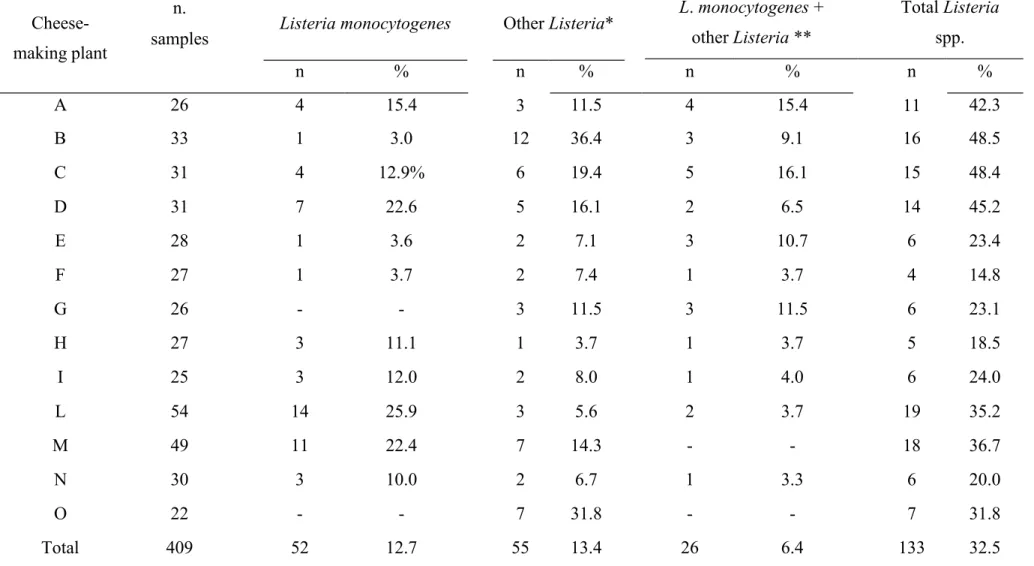
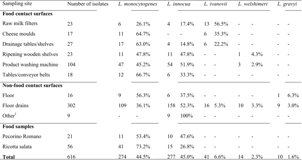
4.4.1 Prevalence of Listeria spp. and Listeria monocytogenes 94
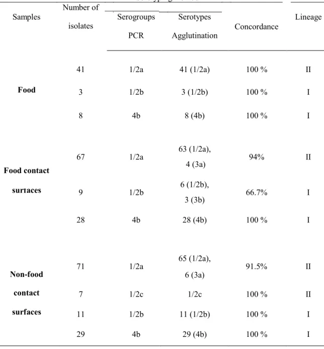
4.4.2 Listeria monocytogenes serotyping 95
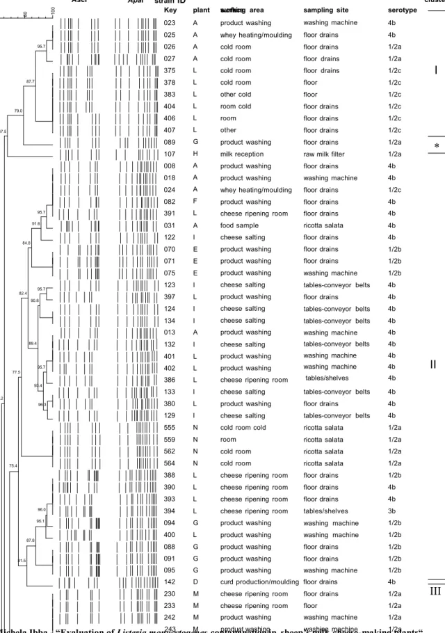
4.4.3 Strains variability and traceability 95
4.5 Discussion 96
Figures and Tables 99
REFERENCES 105
CHAPTER 5 111
GENOTYPIC CHARACTERIZATION AND VIRULENCE GENES ANALYSIS OF LISTERIA MONOCYTOGENES ISOLATED FROM SHEEP CHEESE- MAKING PLANTS 111
5.1. Introduction 112
5.2. Materials and methods 118
5.2.1 Strains selection 118
5.2.2 DNA extraction 119
5.2.3 Molecular Serogrouping 119
5.2.4 Fluorescent amplified fragment length polymorphism (fAFLP) 120
5.2.5 Next Generation Sequencing 120
5.2.6 Virulence factors analysis 121
5.3.1 Molecular Serogrouping 123
5.3.2 fAFLP 123
5.3.3 Virulence genes analysis 124
Tables 128
REFERENCES 133
CHAPTER 6 139
Abstract
The general aim of the thesis was to evaluate the pattern Listeria monocytogenes contamination in sheep’s milk industrial cheese-making plants operating in the regional territory of Sardinian (Italy). Chapter 3 describes a longitudinal study conducted in 2 cheese-making plants over one year period. The objective of the study was to identify sources, sites of persistence and route of contamination within premises. Contamination mostly occurred in salting, product washing, packaging, ricotta salata storage and cheese ripening areas. The greater persistence of contamination over time was observed in washing, salting and cheese ripening areas. In Chapter 4 is presented a cross-sectional study on Listeria spp environmental contamination in 13 sheep cheese-making plants. The objective was to investigate the genetic diversity (population study) and the route of contamination (traceability) of L. monocytogenes strains. Strains originating from the processing environment were disseminated to other sites and food samples within and among facilities. In chapter 5 the potential pathogenicity of environmental and food isolates collected in cheese-making plants was studied by DNA sequencing. In particular the modification in nucleotide sequence of virulence factors hly, InlA and InlJ was investigated. L. monocytogenes environmental strains showed 90-100% of identity with reference sequences, suggesting their potential pathogenicity.
CHAPTER 1
Introduction
1.1 Taxonomy Listeria spp
1.1.1 The Listeria genus
The genus Listeria was first placed in the Corynebacteriaceae family, but today, thanks to the sequencing of ribosomal RNA (rRNA) it has been positioned between the
Bacillus and the Lactobacillus/Streptococcus groups, within the Clostridium- Lactobacillus-Bacillus branch (Jay et al., 2009).
The members of the genus are Gram-positive rods, anaerobic facultative, not sporulated and not encapsulated bacteria. They are catalase positive, oxidase negative and beta-hemolytic. Listeria is motile if cultured between 20°C and 25°C due to the presence of peritrichous flagella. The Listeria species grow at temperature ranging between 0-45 °C (Halter et al., 2013). Although it is not able to grow below -1.5 °C,
Listeria can survive at lower temperature. Optimal range for growth is between 30-
37°C, while temperature >50°C is lethal (Rocourt and Buchrieser, 2007).
The genus Listeria comprises ten species: Listeria monocytogenes, L. innocua,
L. welshimeri, L. seeligeri, L. ivanovii and L. murray (subsp. grayi e subsp. murray)
(Rocourt, J. and Buchrieser, C., 2007), species which have long been known, and four new species that have been reported in the last years, L. rocourtiae (Leclercq et al., 2010), L. marthii, (Graves et al., 2010), L. fleischmannii (Bertsch et al., 2012) and L.
weihenstephanensis (Halter et. al, 2013). Of these species only L. innocua and Listeria monocytogenes are considered to be pathogenic. L. innocua is generally associated with
cerebral infection in ewe and in cattle (Rocha et al., 2013), even if a case of fatal bacteraemia in an old man caused by this specie was reported in 2003 (Perrin et al., 2003). One case of human infection due to L. seeligeri and two cases due to L. ivanovii
(Rocourt et al., 1987; Cummins et al., 1994; Lessing et al., 1994) have been reported in literature.
Listeria spp are widely distributed in natural environment (Sauders and
Wiedmann, 2007) and can be recovered from different sources: soil, plants, animal feed, faces of animal ad humans, water surface, effluents, food processing environment, processed food (Gravani, 1999; McLauchlin et al., 2004).
1.1.2 Listeria monocytogenes
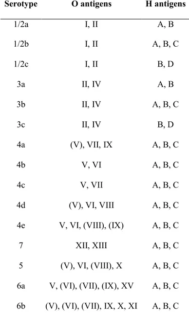
According to the serotyping method developed by Seeliger and Höhne (Seeliger and Jones, 1986), based on the reaction of somatic (O) and flagellar (H) antigens with a series of antisera, L. monocytogenes can be differentiates into 13 serotypes: 1/2a, 1/2b, 1/2c, 3a, 3b,3c, 4a, 4ab, 4b, 4c, 4d, 4e and 7 (table n.1.1). Generally only serovars 1/2a, 1/2b and 4b are involved in human cases of listeriosis (Wiedmann et al., 1997). The serotype 4b strains are generally linked with epidemic outbreaks while 1/2a and 1/2b are commonly associated to sporadic cases (McLauchlin, 1997).
Listeria monocytogenes consist of 4 evolutionary lineages: I, II, III, IV (Orsi et. al., 2011). Strains can be ascribed to the lineages through different types of genotyping
such as Pulsed-field gel electrophoresis (PFGE), rybotyping and multilocus sequence typing (MLST) Fugett et al., 2006; Nadon et al., 2001; Wiedmann et al., 1997), and phenotypic approach, such as API Listeria profiles (; however to assign more accurately isolates to lineages it is advisable the use of DNA-sequencing method (Orsi et. al, 2011). The majority of the strains isolated from human disease are included in the lineage I (serotypes 1/2b and 4b) and II (serotype 1/2a). Human listeriosis outbreaks are generally associated with serotypes included in the lineage I while sporadic clinical cases seem to be more related with strains belonging to lineage II. Also strains that are
habitually isolated from food and environment are included in the lineage II. Strains belonging to lineage III and IV are rare and frequently isolated from animal sources (Orsi et. al, 2011).
1.2 Ecology of Listeria monocytogenes
1.2.1 Growth limits
Listeria monocytogenes is a ubiquitous pathogen widespread in the environment
(Ragon et al., 2008) that can be isolated from different sources: silage, vegetation, soil, sewage, stream water, mud, slaughter-house waste, milk of normal and mastitic cows, and faeces of animals and healthy humans (Farber and Losos, 1988; Farber and Peterkin, 1991).
The ecological characteristics of L. monocytogenes allow survival and growth under extreme environmental conditions that are generally hostile and lethal to other foodborne pathogens (Zarei et al., 2012; Sleator et al., 2003).
The optimum temperature for the growth of Listeria monocytogenes range between 30°C and 37°C, but in presence of nutrients it can growth at temperature between 0°C and 45°C (Farber and Losos, 1988). The ability to grow at low temperature allows the multiplication of the microorganism in foods kept refrigerated at 0-4°C (Chan and Wiedmann, 2009; Sergelidis and Abrahim, 2009). Furthermore, even if it cannot multiply at temperature below -1.5°C, the microorganism is able to survive at freezing temperature. Heat treatments applied in food processing using temperature over 50°C, such as high-temperature short-time (HTST) pasteurization, are effective in eliminating the presence of L.monocytogenes (Cava-Roda et al., 2012). However, in literature are reported cases of listeriosis outbreaks linked with milk correctly
pasteurized (Fleming et al., 1985) and evidence of heat resistance to some extent of L.
monocytogenes has been described (Sergelidis and Abrahim, 2009; Lin and Chou, 2004;
Lou and Yousef, 1997). Several studies have demonstrated that the microorganism increases its resistance to heat treatments if previously exposed to temperatures above its maximum for growth (Hassani et al., 2007; Lin and Chou, 2004; Pagán et al., 1997).
Listeria monocytogenes can grow at values of pH ranging between 4.0 and 9.5
(Liu et. al, 2005). It seems to be moderately acid-tolerant (Lado and Yousef, 2007); this condition allows the resistance to the acid pH of the gastric content, permitting the oral transmission of L. monocytogenes (Wiedmann et al., 1998). The resistance to low pH value decreases with the increase of the temperature (Lado and Yousef, 2007).
It was demonstrated that L. monocytogenes can survive for long time at water
activity (aW) values ranging from 0.790 to 0.860 (Johnson et al. 1988), and it is able to
grow at aw value of 0.900 (Lado and Yousef, 2007). However, the optimal growth is at
aW ≥ 0.970 (Ryser and Marth, 1999).
With regard to tolerance to NaCl it is widely demonstrated that L.
monocytogenes can multiply at NaCl concentration up to 10 % and can survive at values
of 20% (Sutherland et al., 2003; Seeliger, 1987; Mc.Loure, 1989). In a study of Liu et
al. (2005) was observed a higher tolerance to NaCl; L. monocytogenes strains were
submitted to incubation for 20h in a solution saturated at 40% and they were still able to grow. This tolerance seems to be related to the ability of the organism to adapt to osmotic stress accumulating intracellular solutes such as: glutamate, glutamine, aspartate, alanine and proline (Liu et al., 2005; Patchett et al., 1992).
Listeria monocytogenes is a facultative anaerobe; even if it is predominantly
aerobic, it is also able to growth under microaerobic and aerobic conditions (Lungu et
1.2.2 Biofilms and Niches
Biofilm is a complex organization of microorganisms attached to surfaces and protected by a matrix of their own synthesis that can be found in several natural and artificial environments (Sutherland, 2001). This matrix, called ―extracellular polymeric substance‖ (EPS), consist of a mix of polysaccharides, nucleic acid and proteins (Sauer
et al., 2007); EPS allows the adhesion of microorganisms and creates a protective
structure around them. Throughout biofilms are present water channels by which nutrients, metabolites and waste products are interchanging (Sauer et al. 2007; Sutherland, 2001). Biofilms can harbour one or more species of bacteria (Shi and Zhu, 2009).
The formation of biofilm is a complex process that is conditioned by several factors, such as properties of adhesion surfaces (texture or roughness, hydrophobicity), cells surface (extracellular appendages and polymeric substance), environmental conditions (pH, temperature, nutrient components) and bacterial genetic regulation (Simões et al., 2010; Shi and Zhu, 2009; Donlan, 2002). According to Vlková et al. (2009) the formation process can be dived in three major steps: adherence of free planktonic microbial cells; colonization of the preconditioned surfaces; release of microbial cells from the biofilm structures or from the surface (figure 1.1).
Microorganisms growing in biofilms are physiologically and phenotypically different from planktonic cells of the same organism, being cells in biofilm more resistant to adverse environmental conditions (Sauer et al. 2007). In fact, biofilm provides protection from different factors such as nutrient deprivation, acidity, oxygen radicals, heat, drying, salinity, disinfectants and antimicrobial agents (Silva Meira et al., 2011; Jefferson, 2004; Møretrø and Langsrud; 2004; Spoering and Lewis, 2001;
Mittelman, 1998) and encourage the colonization of niches (Jefferson, 2004). The increased resistance of bacterial community as consequence of microorganism interaction has been demonstrated (Morton and Gaylarde, 2001). This phenomenon is called ―quorum sensing‖: biofilm population modulates its density and its gene expression accordingly; also the production of EPS seems to be related to the ―quorum
sensing‖ (Morton and Gaylarde, 2001).
Figure n.1.1. Stages of biofilm development process: (1) reversible attachment, (2) irreversible
attachment, (3) maturation-1, (4) maturation-2, and (5) dispersion (Sauer et al., 2007).
The presence of microorganism in biofilms formed on food-contact surface represents one of the main sources of contamination in food processing plants and the major cause of transmission of food-borne diseases (Bonsaglia et al., 2014; Shi and
The ability of L. monocytogenes to form biofilms has already been established (Mariani et al., 2011; Chae et al., 2006; Beresford et al., 2001).
Listeria monocytogenes is able to adhere on most of the surface present in the
food processing environment (Rieue et al., 2008): stainless steels, rubber and polymers (Beresford et al., 2001),Teflon®, nylon, and polyester floor sealant (Blackman and Frank, 1996), wood (Mariani et al., 2011) and glass (Chae et al., 2006). The areas most frequently associated with Listeria biofilms are wet surfaces such as floor, floor drains, conveyer belts (Wong, 1998), storage tanks, trucks (Shi and Zhu, 2009) and parts that are difficult to clean such as joints, crevices and gaskets (Wong, 1998).
Several authors demonstrated that the presence or the absence of Listeria in the biofilm is conditioned by the resident microbial flora (Tompkin, 2002); for example it was demonstrated that the presence of strains of the genus of Pseudomonas (Sasahara and Zottola, 1993) and Flavobacterium strains (Bremer et al., 2001) can increases the attachment of surfaces by L. monocytogenes, while Mariani et al. (2011) demonstrated that the presence in wooden shelves of an active resident microbial biofilm formed by
Lactococcus lactis and hetero-fermentative lactobacilli have an anti-Listeria action.
Also Zhao et al. (2006) and Loessner et al. (2002) showed how competitive bacteria (Lactococcus lactis and Enterococcus durans or Lactobacillus plantarum) could considerably decrease the contamination of Listeria spp. in the environment.
The adhesion of L. monocytogenes on the surface is also correlated with the serotype of the strains (Folsom et al. 2006). Listeria monocytogenes serotypes 1/2a and 1/2c strains belonging to the Lineage II, are generally considered more efficient in biofilm formation as compared to strains of serotypes 4b and 1/2b, belonging to the
Lineage I (Nilsson et al., 2011; Harvey et al., 2007; Borucki et al., 2003). However,
2002). Some studies highlight inter-strains variation which is not correlated with lineage or serotype but rather related to intrinsic properties of the single strain. Differences in adherence to surface were detected between persistent and non-persistent strains: persistent isolates from food processing environments exhibited enhanced ability to adhere and to form biofilm (Folsom et al., 2006; Lundén et al., 2000; Norwood and Gilmour, 1999).
Biofilm formation permits the survival of L. monocytogenes under various chemical and physical stresses (Harvey et al., 2007; Chae et al., 2006) and improves the opportunity of persistence in the environment (Di Bonaventura et al., 2008; Chae et al., 2006).
The ability of L. monocytogenes to attach to different materials has led some authors to consider the microorganism as able to populate and colonize environmental niches (Beresford, 2001). Niches are sites in the working areas difficult to clean and to disinfect with normal procedures (Carpentier and Chief, 2011; Tompkin, 2002), generally characterized by humidity, brines and low temperatures. These conditions are ideal for the growth of the microorganism. Niches, or harbourage sites, serve as reservoir sites from which Listeria can be transmitted from the environment to the finished products (Tompkin, 2002). Examples of harbourage sites are: floor drains, equipment sites difficult to access and to clean such as conveyors, cracks, junctions between equipment components, forklifts, equipment cleaning tools, on-off valves and switches for equipment (Tompkin, 2002). Within niches L. monocytogenes can survive and growth for months or even for several years (Unnerstad et al., 1996).
1.2.3 Persistent contamination
The presence of L. monocytogenes in foods is not due to the survival of the microorganism to listericidal treatments but instead to a recontamination of the products along the post-processing equipment and environment (Senczek et al., 2000). It is well known that Listeria is widely distributed in food premises and that can be isolated from the majority of food processing environments (Kovacevic et al., 2011). Even if hygienic conditions in industrial food production are developing and improving (e.g. high-risk design, cleaning and disinfection systems, employee food hygiene training) outbreaks of listeriosis due to contaminated foods are still an important issue at international level for consumers health (Harvey et al., 2006; Holah et al., 2004). One of the main reasons that make Listeria hard to eradicate is its ability to persist in the food processing environment, allowing the pathogen to survive within the production areas over time (Holah et al., 2004; Tompkin, 2002). The evidence of L. monocytogenes persistence has been widely reported and authors demonstrated that the microorganism can resist for periods of time that raging from few months up to several years in different kind of food premises: cheese, raw and smoked fish, mussels, pate, fresh, cooked and fermented meats, pesto sauce etc. (Unnerstad et al., 1996; Holah et al., 2004; Tomking, 2002; Senczek et al., 2000; Keto-Timonen et al., 2007). L. monocytogenes strains are considered to be persistent when isolated several times in the same processing plants in several sampling visit (Carpentier and Cerf, 2011). There is discordance between authors in the definition of number of visit, isolation and period that are necessary to indicate a strain as persistent. In their review Carpentier and Cerf (2011), according with data showed by Ragimbeau (2002), defined persistence a strain ―isolated on at least three sampling dates in a one-year period‖. However Pan et al. (2006) pointed that in some circumstances the isolation from the environment of the same strains over time
can be caused by a continuous introduction of the contamination in the plants from outsides sources. It is not fully clear the mechanisms that permit a strain to became persistent; properties that influence the possibility of L. monocytogenes to persist are: the ability to form biofilm, to establish into niches, to grow at low temperatures and to resist to sanitizers (Pan et al., 2006). It is important to underline that the relationship between the presence of persistent strains in food processing plants and listeriosis outbreaks has already been documented (McLauchlin et al., 2004; Tompkin, 2002).
1.3 Infection and Disease
1.3.1 Listeriosis
Listeria monocytogenes is the causative agent of a foodborne illness referred to
as listeriosis. Listeriosis is one of the most important foodborne disease (Schneider et
al., 2009) and the Centers for Disease Control and Prevention (CDC) has stated that it is
the 3rd leading cause of death from food poisoning.
It is generally considered a sporadic disease (Allerberger and Wagner, 2010; Swaminathan and Gerner-Smidt, 2007). However some authors agree that in some countries (especially in Asia) its incidence might be underestimated. This may be due to the fact that the cases of listeriosis are underreported because of lacks in the surveillance system, for a deficiency of the mandatory notification system and for the absence of diagnostic test capable to diagnose the illness (Barbuddhe et al., 2004; Siegman-Igra et
al., 2000).
Listeriosis has an incidence that can vary between 0.1 and 11.3 cases for 1,000,000 populations per year (WHO/FAO 2004; EFSA, 2014; Mead et al., 1999). It is responsible of an average case-fatality rate of 20-30% (Farber and Peterkin 1991; Mead
et al., 1999; WHO/FAO 2004) and shows the highest hospitalisation rates (91%)
compared to other food-borne pathogens (EFSA, 2014; CDC, 2013; Jemmi and Stephane, 2006; WHO, 2004). From 1996 to 2001 in USA was reported a decrease of Listeriosis cases by 24%, after which a relatively stable incidence rate was observed (CDC, 2013). A total of 1,642 cases of listeriosis have been reported from 26 European Union Member States in 2012 , showing an increased rate (10,5%) compare to 2011 (EFSA, 2013). Several studies and reports indicate that this increase is due mainly to a rise of cases in adults aged ≥65, regardless other predisposing conditions (EFSA 2014; CDC, 2013; Goulet, 2008).
Listeriosis is universally recognized as a foodborne disease (Rocourt and Buchrieser, 2007). However cases of human listeriosis occurred in farmers and veterinarians (cutaneous Listeriosis) due to direct transmission from animals to humans have been reported (Bortolussi and Mailman, 2010; Posfay-Barbe and Wald, 2009); vertical transmission from mother to foetus (Allerberger and Wagner, 2010) and also nosocomial cases were reported (Graham et al., 2002; Simmons et al., 1986).The first documented case of nosocomial outbreak was a cross-infection occurred in a neonatal unit due to contact with contaminated mineral oil used to bath infants (Schuchat, 1991).
The importance of the disease is also related to the population at risk; in fact listeriosis affects primary the elderly, neonates, pregnant women and people with pre- existing medical conditions such as immunocompromised (i.e. individual infected with the human immunodeficiency virus, HIV) or those receiving immunosuppressive treatments (e.g. cancer, transplantation) or affected by other underlying diseases (CDC; Doganay, 2003). Other predisposing conditions for listeriosis are represented by diabetes, alcoholism, liver and kidney diseases, drug addiction (Bortolussi and Mailman, 2010; Schlech, 2000). Listeriosis can also occur in healthy patients (30% of
adults and 54% of children and young adults) not showing such risk factors (Doganay, 2003). In 2006 Kalyani et al. presented a case of meningitis caused by Listeria
monocytogenes in a 17-years-old immunocompetent patient. In pregnant women the
possibility of contracting the disease after consumption of food is 12 times higher as compared to the general population (Hof, 2003). Generally the mother present mild flu- like illness (Swaminathan and Gerner-Smidt, 2007) but the bacteria can induce a placentitis and infects the foetus because its immune system is still not sufficiently developed (Hof, 2003). The foetus can contract the infection in utero for the passage of
Listeria through the placental barrier or, alternatively, the mother can contaminate the
respiratory tract of the baby during the passage through an infected birth canal (Allerberger and Wagner, 2010; Schlech, 2000). The results of the infection can be foetal distress, miscarriage, death or premature birth of a severely ill infant and in the case of contamination during the childbirth, infants can present meningitis up to 2 or 3 weeks after exposure (Schlech, 2000). The CDC (2013) reported that in the USA one- third of reported human listeriosis cases occur during pregnancy, while McLauchlin et
al. (2004) indicated that pregnancy and neonatal Listeria disease account for 10–20% of
UK cases. It estimated that 22% of pregnancy-related cases of listeriosis lead to the death of the baby (NHS, 2013).
1.3.2 Pathogenesis and virulence factors
Listeria monocytogenes is a facultative intracellular pathogen and its virulence
depends on the ability to adhere and enter into host cells. The processes of infection can be divided in few crucial steps: a) entry of the bacterium into the host; b) adhesion and invasion of host cells; c) lysis of the phagosomal vacuole; d) multiplication in the
cytosol; e) direct cell-to-cell spread using actin-based motility (Jemmi and Stephane, 2006;Vazquez-Boland et al, 2001).
Human mainly contract the infection through the ingestion of contaminated food, therefore the entry of L. monocytogenes in the host is generally represented by the gastrointestinal tract (Vazquez-Boland et al., 2001). In the stomach the bacteria is subjected to the action of proteolytic enzymes, hydrochloric acid and bile salts (Liu et
al, 2007). Due to the action of stress- response genes that are under the control of the
sigma factor, sigma B (σB) (Abram et al., 2008), L. monocytogenes is able to survive. As
consequence of the acid environment of the stomach, the concentration of the ingested microorganisms can be partially reduced (McLauchlin et al, 2004; Vazquez-Boland et
al., 2001). Several authors reported that treatments with antiacids which neutralize the
stomach acidity increased the susceptibility to the infection (Cobb et al., 1996; Schuchat
et al, 1992).
After passing through the stomach L. monocytogenes invades the small intestine (Vazquez-Boland et al., 2001). Once in the intestine, the entry into intestine epithelial cells is mediated by the interaction of L. monocytogenes surface proteins with host cells receptors. The surface proteins are named internalines and are encoded by different internaline genes (inl), (Bortolussi and Mailman, 2010; Jemmi and Stephane, 2006). Internalines are important virulent factors taking part in the invasion and colonization of intestinal epithelium; L. monocytogenes have 25 genes encoding for putative surface- associated internalines (Bierne et al., 2007). InlAB, InlC, InlD, InlE, InlF, InlG, InlH and InlJ are the more important virulence factors (Vazquez-Boland et al., 2001). InlA and InlB are the best characterised and the ones that show the main role with internalization in the intestinal fase (Bierne et al., 2007; Liu et al, 2007). The InlA and InlB (encoded by the inlAB operon) interact respectively with the E-cadherin receptors
and with Met receptors present on human epithelial cells. The interaction internaline/receptor determines an invading strategy typically referred as ―zipper mechanism’’ (Disson and Lecuit, 2013; Vazquez-Boland et al., 2001). Another important internalin is the InlC that is one of the most important targets of the humoral immune response against L.monocytogenes in humans (Bierne et al., 2007). The full function of the InlC is not clear but it seems to play a key role when the microorganism is already inside the host cell and during the spread from one cell to another. The InlJ instead has an important role in crossing the intestinal barrier and is involved in the successive stages of the infection (Sabet et al., 2005). The InlF increases the host cell binding and entry in cells such as fibroblasts and hepatocytes (Kirchner and Higgins, 2008).
Once inside the host cell the microorganism is entrapped into a vacuole (or phagosomes), which Listeria is able to escape from. Then it can replicate in the cytosol of the host cell (Disson and Lecuit, 2013; Schnupf and Portnoy, 2007; Vazquez-Boland
et al., 2001). Disruption of phagosomes is mediated by the secretion of pore-forming
proteins listeriolysin O (LLO, a haemolysin encoded by the gene hlyA), and two phospholipase C (PlcA and PlcB, encoded respectively by the gene plcA and the gene
plcB). These proteins are essential for the survival and the intracellular growth of Listeria (Jemmi and Stephane, 2006; Hof, 2003). Once in the cytosol Listeria induces
the production of actin and the formation of protrusion in the apical part of the host cells; this extrusion penetrates the neighbouring cells allowing the passage of the bacteria from infection to uninfected neighbour cells (Cossart and Toledo-Arana, 2008; Hof, 2003; Vazquez-Boland et al., 2001). The direct cell-to-cell spreading allows the dissemination of avoiding the host cells defences (Disson and Lecuit, 2013; Cossart and Toledo-Arana, 2008; Portnoy et al., 2002). The genes encoding for virulence factors of
L. monocytogenes are regulated by a transcriptional regulatory factor, the positive
regulatory factor A (PrfA), and they are physically located in a chromosomal island (Vazquez-Boland et al., 2001) called Virulence Gene Clusters (pVGC) or Listeria pathogenicity island 1 (LIP 1). Strains that present deletion or absence of the PrfA are considered avirulent (Posfay-Barbe and Wald, 2009).
Listeria monocytogenes, after the invasion of the enteric cells, reaches
mesenteric lymph nodes, the spleen, and the liver through the lymphatic system and the bloodstream (Vazquez-Boland et al, 2001). At this time in the liver polymorphonuclear neutrophils are recruited to eliminate the pathogens and the infected cells, causing micro abscess formation (Vazquez-Boland et al, 2001); after some days neutrophils are replaced by monocytes together with lymphocytes to form granulomas. Since most of the microorganisms are internalized into the hepatocytes, in the attempt to eliminate the infection, these cells undergo autophagy to destroy internalised L. monocytogenes (Sleator et al., 2009). Generally in the majority of the cases, such as in healthy individuals, the infection is resolved in the initial phase of invasion (McLauchlin, 2004) with the presence of only gastrointestinal symptoms (Schneider, 2009). If the infection is not properly counteracted at liver and spleen level, as in the case of immunocompromised individuals, the microorganism can cause a bacteraemia leading to the invasion of other organs. The target organs are the central nervous system (CNS) and the placenta (Lecuit, 2007; McLauchlin, 2004; Vazquez-Boland et al., 2001).
Listeria monocytogenes is able to enter and proliferate not only in enterocytes and
hepatocytes, but also in the phagocytes cells, i.e. polymorphonuclear, granulocytes and macrophages (Orndorff, 2006); with this mechanism Listeria is able to avoid antibodies, complement, or neutrophils and to be disseminated throughout the body with the
1.3.3 Listeriosis signs and symptoms
There are two forms of listeriosis: a non-invasive gastrointestinal listeriosis and an invasive listeriosis. The gastrointestinal form is more frequent in immunocompetent individuals and generally causes a self-limiting febrile gastroenteritis (Allerberger and Wagner, 2010; Vazquez-Boland et al., 2001) with nausea, vomiting and diarrhoea being the more common symptoms, occurring about 24-48 h after the ingestion of the contaminated food (Doganay, 2003). Manifestation of the invasive listeriosis, usually associated with the population at risk, can include septicaemia or meningitis and encephalitis in immunocompromised and old individuals, abortion in pregnant women and generalized infections in infants (Allerberger and Wagner, 2009; Barbuddhe et al., 2004). The population at risk of Listeria infection presents the symptoms after 3-70 days from the exposure (Lecuit, 2007). Meningoencephalitis symptoms are fever, intense headache, nausea, vomiting and signs of meningeal irritation such as nuchal rigidity, movement disorders such as tremor, ataxia and seizures disorders (Allerberger and Wagner, 2009; Doganay, 2003). Among bacterial meningitis Listeria account for 11% of all cases and has the highest mortality rate, close to 22% (Lecuit, 2007).
Infection in pregnant women occurs mainly in the third trimester of pregnancy when the immune defences are low, with the mother presenting non-specific symptoms (Allerberger and Wagner, 2009), very similar to a flu: fever (often above 39°C), headache, myalgia, arthralgia and malaise (DiMaio, 2000); in some cases the infection can be asymptomatic (Doganay, 2003). Infection with L. monocytogenes during pregnancy can result in miscarriage or stillbirth (40-50%), preterm birth (less than 35 weeks in approximately 70% of cases) and neonatal infection (Golos et al., 2013; Bortolussi and Mailman, 2010). The placenta presents chorioamnionitis, villitis,
vasculitis, fibrinoid necrosis, thrombosis and placental abscesses (Golos et al., 2013; Lecuit, 2007). In the foetus L. monocytogenes infection (or granulomatosis infantiseptica) microabscesses and granulomas are detected in various organs such as liver, spleen, brain and skin (Hof, 2003; Lecuit, 2007).
In newborns two clinical forms are distinguished: an early-onset form that occurs within the first week of life, and a late-onset form that can appear from several days to weeks after birth (Delgado, 2008). The early-onset form infants present mainly sepsis-like manifestations but can also show respiratory distress, pneumonia and, more rarely, meningitis or myocarditis, while meningitis is more common in the late-onset forms (Posfay-Barbe and Wald, 2009).
Cases of local infection are reported especially in farmers and veterinary after contact with infected animals: papules and pustules appear on the arms and the hands with associated general symptoms such as fever, myalgia and headache (Regan et al., 2005; Posfay-Barbe and Wald, 2009). Generally the symptoms are successfully resolved, but sometimes the infection can generalize and led to invasive listeriosis (McLauchlin and Low, 1994).
Other clinical manifestations of listeriosis have been reported in literature: aortic aneurysms, prosthetic joints and brain abscesses (Cone et al., 2008), endocarditis and myocarditis (Lecuit, 2007), conjunctivitis, lymphadenitis, cholecystitis, peritonitis, pleuropulmonary infection, joint infection, osteomyelitis, arteritis, necrotizing fasciitis and endophthalmitis (Allerberger and Wagner, 2009).
The incubation period is variable: for serious forms of listeriosis ranges from a few days up to three weeks, while for gastrointestinal forms in healthy people ranges
1.3.4 Infective dose
The dose necessary to cause the invasive listeriosis depends on a number of factors, including the virulence of the microorganism, the number of cells ingested, the host susceptibility and its immune status, and the characteristics of the food matrix (FDA, 2003).
The minimum dose required to cause clinical infection in humans has not been exactly determined. The long incubation period that occurs in listeriosis cases rarely allows the testing of the food responsible for the infection (CFSPH, 2005). However, several studies indicate that it is necessary a high dose to cause infection: microbiological test in foods implicated in epidemic and sporadic cases detected levels of L. monocytogenes between 102 to 106 cfu/g of ingested product (Posfay-Barbe and Wald, 2009; Jemmi and Stephane, 2006; Vazquez-Boland et al, 2001).
Foods in which the levels do not exceed 100 cfu/g seems to have no effect on healthy people, but in people with associated risk factors a dose of 1000 cells can be enough to cause illness (CFSPH, 2005; FDA, 2003).
1.4 Listeria monocytogenes in foods
1.4.1 International microbiological food safety criteria for Listeria monocytogenes In the European Union the microbiological food safety criteria concerning L.
monocytogenes are provide by Regulation (EC) No 2073/2005 of 15 November 2005 on
microbiological criteria for foodstuffs. The microbiological criteria define the acceptability of foodstuff products placed on the market; if the criteria are not met,
According to Article 4 of Regulation (EC) No 852/2004, food business operators are responsible for the compliance with microbiological criteria. Food safety criteria for
L. monocytogenes are indicated in the Annex I of the Regulation where are also
indicated the food category, sampling plan, microbiological limits, analytical methods and stage where the microbiological food safety criteria is to be applied.
Ready-to-eat (RTE) foods are the food category that should be tested for L.
monocytogenes as indicated in the annex. Among RTE foods the Regulation distinguish
in three categories:
- RTE foods intended for infants and ready-to-eat foods for special medical purposes:
the criteria is applied for products placed on the market during their shelf-life and contemplates the absence in 25 g (n = 10, c = 0; where n = number of units comprising the sample; c = number of sample units giving values between m and M). The reference analysis method indicated by the Regulation is the EN / ISO 11290-1.
- RTE foods able to support the growth of L. monocytogenes, other than those
intended for infants and for special medical purposes: for products placed on the market during their shelf-life is contemplated a limit of 100 ufc/g (n = 5, c = 0), if the manufacturer is able to demonstrate that the product will not exceed the limit of 100 cfu/g throughout the shelf-life. The operator may fix intermediate limits during the process that should be low enough to guarantee that the limit of 100 cfu/g is not exceeded at the end of the shelf-life. The reference analysis method indicated by the
Before products have left the immediate control of the food business operator when no evidence can be provided to demonstrate that the product will not exceed the limit of 100 cfu/g throughout the shelf-life, the absence criteria in 25 g (n = 5, c = 0) is applied. The reference analysis method indicated by the Regulation is the EN / ISO 11290-1.
- Ready-to-eat foods unable to support the growth of L. monocytogenes, other than
those intended for infants and for special medical purposes: foods in this category
include products with pH ≤ 4.4 or aW ≤ 0.92, or pH ≤ 5.0 and aW ≤ 0.94 and those
with a shelf-life of less than 5 days. If they are placed on the market during their shelf-life is set a limit of 100 ufc/g (n = 5, c = 0). For these products, placed on the market during their shelf-life is expected a limit of 100 cfu/g (n = 5, c = 0). The reference method of analysis of is the EN / ISO 11290-2.
In the United States the responsibility for food regulation are shared by the Food and Drug Administration (FDA) and United States Department of Agriculture (USDA). Food Drug Administration (FDA) applies a "zero-tolerance" policy for L.
monocytogenes in RTE foods. Since 1995 with a sentence of United States District
court decision, United States vs. Union Cheese Co., the detection of any Listeria
monocytogenes in either of two 25g samples of a food renders the food adulterated
(Federal Food, Drug, and Cosmetic Act. 21 U.S.C. 342). Similar policy is applied by the Food Safety and Inspection Service (FSIS) branch of the USDA responsible for controls on meat and poultry products and certain egg products (Federal Meat Inspection Act
In Canada the Listeria policy for RTE foods is based on the application of Good Manufacturing Practices (GMP) and on the HACCP principle. The control of the risk for Listeria contamination was developed using a health risk assessment approach, taking into account the combination of several factors such as inspection, environmental sampling and finished products testing. The Canada’s authority classified RTE foods in two categories based on the health risk (Canadian Food Inspection Service, 2011):
- Category 1: RTE products in which the growth of L. monocytogenes can occur. These should receive the highest priority for industry control, as well as regulatory inspection and compliance activities. The presence of L. monocytogenes in these Category 1 ready- to-eat foods will likely trigger a Health Risk 1 concern. Limit expected: absence in 125 g on 5 sample units of 25 g each;
- Category 2: include two subgroups: 2a) RTE products in which limited growth of L.
monocytogenes to levels not greater than 100 CFU/g can occur before the end of the
stated shelf-life; and 2b) RTE food in which the growth of L. monocytogenes cannot occur throughout the expected shelf life. These products should receive a lower priority with regards to industry control, as well as regulatory inspection and compliance action. Limit expected: 100 cfu/g enumeration in 50 g on 5 sample units of 10 g each.
1.4.2 Ready-to-eat foods
The Scientific Committee on Veterinary Measures relating to Public Health (SCVPH) in 1999 published data that supported the idea that L. monocytogenes can contaminate almost all food categories. Listeriosis infection are associated with meat
products (Selby et al., 2006), dairy products (Leite et al., 2005), fish products (Jallewar
et al., 2007) and vegetables (Crepet et al., 2007).
In the last years many categories of food have been linked with listeriosis; however the majority of the cases were due to the consumption of RTE (Garrido et al., 2010; Uyttendaele et al., 2009). RTE foods are defined by the EC Regulation No. 2073/2005 as ―products that are intended by the producer for direct consumption by humans without the need for thermal treatment such as cooking that will eliminate or reduce to acceptable level micro-organisms of concern‖. Some examples of RTE foods include: soft cheeses such as ricotta, brie, feta, blue-veined (e.g. Gorgonzola) and Mexican-style soft cheeses such as queso fresco; hot dogs, luncheon meats, cold cuts; sandwiches; pâtés/meat spreads; refrigerated smoked seafood products such as smoked salmon; deli-type salads such as coleslaw, macaroni, tuna; pre-packed raw vegetables and mixed raw vegetable salad and pre-cut fresh fruits. The application of heat treatment during food processing can reduce the risk of L. monocytogenes contamination in the finished product. However, the re-contamination of RTE cooked can occur during post-processing steps (Zhu et al., 2005). After recontamination, in the presence of favourable conditions, Listeria can multiply in the product to level representing a potential risk to human health (EFSA, 2013).
A survey on the prevalence of L. monocytogenes in RTE at the end of their shelf-life from retail markets, conducted by the European Food Safety Authority (EFSA) during 2010-2011 in 26 EU states members, showed a prevalence of contamination of 10.3 % in fishery products, of 2.07% in meat products and of 0.47% in cheese samples. The rate of samples exceeding the 100 cfu/g food safety limit imposed by the European Commission was 1.7 %, 0.43 % and 0.06 % for fishery products, for cooked meat and soft and semi-soft cheeses samples, respectively. Although the rate of
samples exceeding 100 cfu/g was very low, they still represent a serious concern for public health due to the large distribution of such products in the market.
The Food Drug Administration (FDA, 2003) reported that in the United State 90% of the listeriosis cases were linked to meat and dairy RTE products. Gombas et al. (2003) in a survey conducted in retails markets in the USA found prevalence of L.
monocytogenes ranging from 0.17 to 4.7% in eight RTE food categories and levels of
contamination from <0.3 MPN (most probable number) per g to 1.5 x 10⁵ cfu/g.
1.5 Listeria monocytogenes in the dairy sector
Milk and dairy products are largely consumed in Europe (about 132 kg per person annually) and are consumed by all categories of people (Lunden et al., 2004), including high-risk groups (European Commission, 2000).
In Europe dairy products have been implicated in approximately half of all cases of outbreaks and of large part of the sporadic cases of listeriosis (Lunden et al., 2004). Most of the cases are linked to the consumption of raw milk and products made with unpasterizated milk. Cases of listeriosis have also been reported after the ingestion of dairy products obtained from pasteurised milk as consequence of post process contamination (Cumming et al., 2008; Fleming et al., 1985).
1.5.1 Milk
The shedding of L. monocytogenes with milk of infected dairy ruminants is considered a rare finding. The sanitary state of the animals (encephalitis, abortions, mastitis) usually makes them unsuitable for milking. Level of the microorganism up to 104 cfu/ml in milk of infected animals have been reported; in some circumstances the
microorganism can be eliminated with milk of healthy animals for several months representing a great concern for public health (Ryser, 2011). The pasteurization applied for fluid milk, which has become a routine practice in USA and in Europe since the 1950s ensure the total destruction of L. monocytogenes. In the last 10 years an increase in the trade and consumption of raw (unpasteurized) milk was registered (Van Kessel et
al., 2004). The prevalence of L. monocytogenes contamination in raw milk is generally
low and can vary from 0% to 6.5% (D'Amico and Donnelly, 2010; Vilar et al., 2007; Van Kessel et al., 2004; Meyer-broseta et al., 2003). Some authors (Hassan et al., 2000) in a survey conducted in New York showed higher prevalence of contamination (12.7%). Contamination levels are generally below 0.1cfu/ml (Meyer-Broseta et al., 2003).
Two studies conducted on L. monocytogenes prevalence in sheep’s milk showed a prevalence of contamination between 3.6 % (Rahim et al., 2014) and 10-12.7% (Al- Tahiri et al., 2008). MacDonald and Sutherland (1993) demonstrated that in whole
sheep milk inoculated at concentration of 106 cfu/ml Listeria spp. can survive to heath
treatment at 65°C for 15 min. Finding that suggest a protective effect of sheep’s milk fat which was not reported in cow’s and goat’s milk. However Listeria is effectively killed by other treatments, e.g. high temperature short time pasteurization (72 °C for 15 s). Contamination of raw milk with L. monocytogenes is rarely due to mastitis or to faecal contamination, but it is generally caused by environmental contaminants such as feeds, e.g. spoiled silage, (Vazquez-Boland et al, 2001) and drinking water provided to the animals where the bacterium can proliferate (Venegas et al., 2004).
Because of the ubiquitous nature of L. monocytogenes may be found in natural and urban environments; however it seems to be considerably more frequently isolated
Listeria monocytogenes strains isolated from raw milk belong to serotypes 1/2a,
1/2b, 4b, 3b and 4c (Van Kessel et al., 2004).
1.5.2 Dairy products
Listeria monocytogenes in dairy products rarely presents level greater than 100
cfu/g (EFSA, 2010), however they have been linked with several cases of listerioris outbreaks reported worldwide (Almeida et al., 2013), accounting for 35% of the all
Listeria monocytogenes outbreaks (FDA, 2003). For this reason cheeses are included in
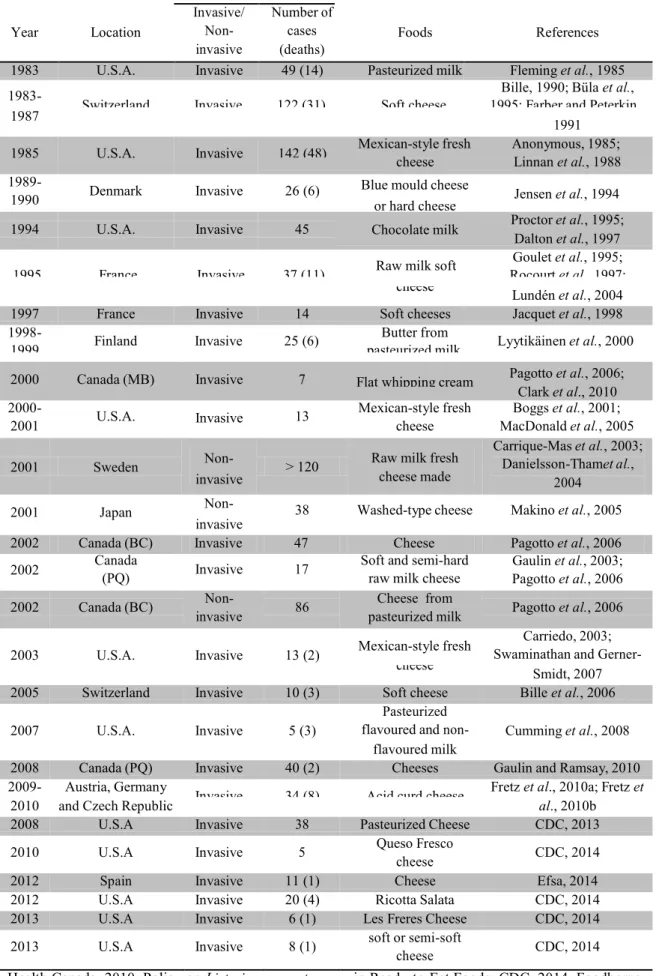
the ―FDA Top Ten‖ riskiest foods report (CSPI, 2009). In table n. 1.2 are reported the outbreaks liked with L. monocytogenes in dairy products from 1983 to date (Health Canada, 2011; Marler Clark, 2014). It is included a recent outbreak occurred in USA caused by Ricotta salata cheese produced by a Sardinian dairy company.
The risk of L. monocytogenes contamination could involve both raw-milk cheeses and those made with pasteurised and micro filtered milk because post-process contamination is equally possible in these products (Rudolf and Scherer, 2001). Some authors reported that L. monocytogenes is most often detected in cheeses made from raw milk (Loncarevic et al., 1995) while others observed an higher prevalence in cheeses made from pasteurized milk (Rudolf and Scherer, 2001), data confirmed by the EFSA report on L.monocytogenes contamination in cheeses (EFSA, 2013).
Soft-cheeses are frequently associated with listeriosis outbreaks; due to their characteristics of high water activity (> 0.920) and pH they are an excellent substrate for
Listeria growth (Rudolf and Scherer, 2001). In particular mould-ripened (e.g. Stilton,
Gorgonzola, Roquefort) and blue-veined (e.g. Brie and Camembert cheeses) are at risk because of the rise of pH during ripening. The pH of these cheeses arrive close the neutrality during ripening, enhancing the possibilities of survival and growth of L.
monocytogenes (Carminati et al., 2004). Conversely fermented lactic cheeses seems to
be less favourable for the microorganism since during the production the combination of competition with the starter culture, low water activity, and a low pH prevent to a certain level the survival and the growth of L. monocytogenes (Morgan et al., 2001). The prevalence of L. monocytogenes reported for cheeses made from cows, sheep and goat milk is very similar comparing whole wheels or cheese cut into smaller wedges (Rudolf and Scherer, 2001; Loncarevic et al., 1995). Data on the prevalence of L.
monocytogenes contamination in cheeses reported in bibliography differs considerably
depending on the cheese type. Rudolf and Scherer (2001) in a survey conducted on European red-smear cheeses and Loncaveric et al. (1995) in a survey conducted on various types of cheeses have reported a prevalence of contamination of 6.4% and 6%, respectively. In traditional soft cheeses made from raw ewe’s milk in Portugal, Pintado
et al. (2005) showed a prevalence of L. monocytogenes of 29% while Silva et al. (2003)
demonstrated an occurrence of 26.7% of Minas-type cheeses. In blue-veined cheese rinds Bernini et al. (2013) revealed a prevalence of 55% with an increase in presence and levels associated with a longer ripening time.
1.5.3 Dairy processing environments
Outbreak investigations provide evidence indicating that L. monocytogenes contamination of pre-packaged industrial food products is not a result of the survival of the organism to processing operation, instead is due to recontamination originating from the processing environment (Widemann, 2003). In dairy product processing plants the use of heat treatments (pasteurization, thermization) on raw milk guarantee an
The environmental contamination of the product is supported by the fact that even if the raw milk is found to be positive for the presence of L. monocytogenes the curd samples are usually negative (Cagri-Mehmetoglu et al., 2011) and by the fact that the strains recovered in raw milk are not usually recovered in the environment or in the products (Pak et al., 2002).
Listeria monocytogenes is widespread in several kind of environment, including
natural and urban environment. The microorganism can be introduced into a food processing environment in a number of different ways and in any point of the processing chain (Ryser, 1999). The main sources of contamination are: raw materials, equipment and people. Listeria can be introduced in the food processing environment through the raw materials and subsequently contaminate foods and equipment if appropriate control measures are not in place (Almeida et al., 2013). Employees or other people visiting food facilities can introduce Listeria into the processing environment via shoes, clothing and personal items. Authors showed levels of L. monocytogenes contamination of about 7% in the hands of food production workers (Kells and Gilmour, 2004). Lomonaco et al. (2009) detected L. monocytogenes contamination in the toilet and in the changing rooms, supporting the idea that the employers play an important role in the Listeria dissemination in the environment. Interestingly, the dairy processing plants that are associated with other processing plants (e.g. shared personnel, exchange of products) show a higher prevalence of positive samples respect to independent facilities (Fox et al., 2011; Pak et al., 2002).
Listeria monocytogenes presents characteristics that allow the survival in food
processing plants: ability to grow at low temperature, adaptability to stress conditions (e.g. acidity, alkalinity and high salt concentration) and competition with other environmental microorganisms (Lou and Yousef, 1999). The dairy processing plants
environment is characterised by conditions which are particularly favourable to L.
monocytogenes growth, e.g. refrigeration temperatures, moisture and humidity and
presence of nutrition for the microorganism (Tompkin, 2002; Unnerstad et al., 1996). Longitudinal studies demonstrated that Listeria contamination in food processing plants is related both to transient and persistent strains (Wiedmann, 2003). The persistence of L. monocytogenes it is of particular concern in the food industry (Kells and Gilmour, 2004). Various studies indicated that just a limited number of strains with similar genetic profile are found within the same food processing plant for several months or years (Keto-Timonen et al., 2007; Møretrø and Langsrud, 2004). Persistent strains in the environment can act as reservoir, providing a continual source for products contamination (Kells and Gilmour, 2004); this is demonstrated by the fact that persistent strains generally are the main cause for finished products contamination (Wiedmann, 2003). Niches that can harbour L. monocytogenes are sites generally hard to reach and clean (e.g. hollows part, cracks in flooring, worn gaskets) with the routine cleaning and sanitizing procedures.
Once L. monocytogenes become established in a niche the routine sanitizing operations are ineffective in eliminating the microorganism from the environment (Tomking, 1999), even though routinely cleaning and disinfection are implemented (Carpentier and Cerf, 2011); for this reason it is important to control the predisposing condition that allowed the creation of niches or that facilitate the diffusion of Listeria, as moisture, presence of nutrients, areas not accessible to cleaning (Kornacki, 2006). Fox et al. (2011) in a study on the prevalence of L. monocytogenes cheese making plants found that in 5 out 16 of the facilities the microorganism was not detected, even if the cleaning and sanitation procedures were the same applied by premises where
Studies on the prevalence of Listeria in cow’s and sheep’s milk cheese making plants showed a prevalence of contaminated sites ranging from 20% to 90% (Pritchard
et al., 1995; Pilo et al., 2008; Parisi et al., 2010; Ibba et al., 2013; Spanu et al., 2015)
and a period of persistence up to 7 years (Unnerstad et al., 1996). Several areas can harbour the microorganism, but the sites with the higher risk of contamination are those where the unpackaged food is exposed to direct food contact (e.g. conveyor belts, equipment and utensils), indirect contact (e.g. ceiling) or non-food contact surfaces (e.g. floors, drains, walls) sites between the lethality treatment and packaging areas (Tompkin, 1999). The sites more exposed to L. monocytogenes contamination are: floors, drains, walls, trolleys (Tompkin, 2002). According to several studies the site with the higher prevalence of contamination is the floor drain (Tompkin, 2002; Pilo et
al., 2008; Parisi et al., 2012; Cagri-Mehmetoglu et al., 2011). For this reason they can
be used as indicator site for the presence of L. monocytogenes, in food processing plant. Floor drains in fact represent an important source of dissemination of the contamination to other sites in the processing environment. For instance, during cleaning procedures (use of pressurized water) the microorganism could be spread on the equipment present in adjacent areas (Kells and Gilmour, 2004; Almeida et al, 2013). Another site that is frequently contaminated in cheese making plants and that can serve as an important source of contamination is the equipment used for cheese brushing and washing (Jaquet
et al., 1996; Almeida et al, 2013). The presence of Listeria monocytogenes in sites so
close to the end of the production line gives further support to the post-processing contamination theory (Pritchard et al., 1995).
The use of molecular typing methods, such as pulsed-field gel electrophoresis (PFGE) and fluorescent amplied fragment length polymorfism (fAFLP), is important to identify the source of contamination, the niches of persistence and to trace
contamination patterns within premises (Parisi et al., 2012). Knowledge on the pathways of contamination is an essential part in tracking action for controlling L.
monocytogenes environmental contamination (Jadhav et al., 2012). Well-designed
control and sanitation procedures, as sampling and cleaning programs, are important to decrease the incidence of L. monocytogenes and to avoid the diffusion of the contamination in the environment and in the products (Ryser et al., 2011). While the harbourage sites are often recognised in the floor, to avoid cross contamination of the adjacent equipment, authors suggest that floor should be cleaned and disinfected before proceeding with the other sites (Carpentier and Cerf, 2011).
It is important to underline that processing plants in which the L. monocytogenes contamination is ascertained, also for long period, not always are implicated in listeriosis cases. The probability that cases of listeriosis occurs seems to be related to food contamination with more virulent strains (Tompkin, 2001).
The ubiquitous nature allows constant reintroduction of L. monocytogenes in the plant environment. Despite it is not realistic to expect that food premises can be continuously maintained free of Listeria contamination (Swaminathan and Gerner- Smidt, 2007), it is possible to control and prevent the food contamination avoiding the establishment of the microorganism in the environments and prevent the diffusion of the pathogen to other areas and sites. Tompkin (2002) suggests a L. monocytogenes control program for food processing environments consisting in six strategies: 1) prevent the formation of niches, 2) implement sampling program to assess the operation of the control program, 3) rapid and effective response when the results of the sampling are positive, 4) use of follow-up sampling to check the effective detection and elimination of the source of contamination, 5) short- term assessment to early detection of problems
and trends (last 4-8 sampling), 6) long-term assessment to evaluate improvement in the processing plants.
Sampling programs are the key to provide a continuous evaluation of the environmental contamination and for its control. Food business operator should establish their own L. monocytogenes monitoring program according with the plant, the products made, the processes which take place in and previous experience (Tompkin, 2001). Sampling operation should be performed during the processing operation, at least 2 hours after the beginning of the production, or at the end of the production before the cleaning procedure (Carpentier and Barre, 2012). In this manner the possibility to detect
Listeria is increased. In fact, during the production activities, movements and vibrations
of the tools and the equipment cause the detachment of Listeria cells present in biofilms and niches; in this way the microorganism is more accessible to sampling and it is possible to detect the presence of niches and biofilm (Tompkin, 2004). Another reason of the sampling during the processing phases is that after the cleaning procedure is possible to collect cells that are injured; they are still alive but non-culturable and then it is possible to have false negative sample (Carpentier and Barre, 2012). However, pre- operational samplings should be conducted since they are useful to verify the effective action of the cleaning and sanitation programs (Kornacki, 2012).
Understanding where L. monocytogenes and its niches are settled help to apply appropriate interventions aimed to prevent and to eliminate Listeria contamination. L.
monocytogenes is the only species of the genus Listeria that has been involved in food-
borne outbreaks, however the presence of any Listeria species in food premises is considered as a useful indicator of a decline of process hygiene conditions during food production (McLauchlin, 1997). Since all Listeria spp share the same ecological niches, the recovery of any Listeria species, also in absence of L. monocytogenes, is indicative
of the presence of favourable conditions that increase the risk of contamination with L.
monocytogenes (Tompkin, 1999; Fox et al., 2011). When environmental monitoring is
conducted, it is strategic to find any type of Listeria as these act as markers for the likelihood of the presence of Listeria monocytogenes and allow taking preventive action for its establishment in the environment (Lakićević et al., 2010).
The site of sampling has to be chosen according to historical data collected by the premises; it should include points that have been found to be good indicators for
Listeria contamination and those that are known to harbour most frequently the
microorganism (e.g. floor drains). The selection of areas and sites to be sampled should be risk based and should identify potential growth niches and points where there is a high potential for transfer Listeria contamination from one area to another (Carpentier and Barre, 2012; Kornachi, 2012). It is useful to use the zoning concept in order to track environmental contamination. Zones are defined based on the probability of product contamination if the microorganism is present in the zone. Based on the risk it is possible to divide the sampling area into 4 zones (Kornacki, 2012; FDA, 2014). The zone one includes direct and indirect product contact areas; these are areas of high risks in which are present favourable conditions for the growth of Listeria in surface close to the production line. Surfaces that can be include in the zone 1 are conveyors, sliders, utensils, racks, work table, valves, pipes that transports food, packaging material, hands, gloves. Contamination of zone 1 areas means that some product contamination is likely to have occurred. The zone two includes areas adjacent to Zone 1 where the product is processed and handled (equipment guard and framework). Zone 3 includes other surfaces within the production line, as floors, drains and walls that if contaminated with the pathogen, could lead to contamination of zone 1 and 2 via actions of employers or movement of equipment. Zone 4 includes remote areas not involved in processing as the
warehouse, locker room and break rooms etc. Most of the environmental samples collected during the sampling should be taken from zone 1-2, and to a lesser degree zone 3. Very few, if any, environmental samples should be taken from zone 4.
The number of sampling points and the sampling may be decided according to the data collected over time by the single company. The number is related to the complexity of the plant, of the production process and the food produced. Subsequently to repeat negative results the frequency of sampling for the site and area can be decreased (Tompkin, 1999).
A good sampling program should allow the assessment of Listeria contamination sites and to implement control strategies aimed to prevent potential contamination of the product before it becomes a problem for the human health