Critical Reviews in Oncology/Hematology 84 (2012) 109–121
Review
Comparative analysis of multiparametric magnetic resonance and PET-CT
in the management of local recurrence after radical prostatectomy for
prostate cancer
Andrea Alfarone
a, Valeria Panebianco
b, Orazio Schillaci
c, Stefano Salciccia
a,
Susanna Cattarino
a, Gianna Mariotti
a, Alessandro Gentilucci
a, Magnus Von Heland
a,
Roberto Passariello
b, Vincenzo Gentile
a, Alessandro Sciarra
a,∗aDepartment Urology, University sapienza, Rome, Italy bDepartment Radiology, University sapienza, Rome, Italy
cDepartment of Biopathology and Diagnostic Imaging, University of Rome “Tor Vergata” and IRCCS Neuromed, pozzilli, Italy Accepted 25 January 2012
Contents
1. Introduction: the clinical point . . . 110
2. Evidence acquisition . . . 110
3. Imaging techniques: MRI and PET-CT . . . 111
3.1. PET-CT for the diagnosis of local recurrence . . . 111
3.2. Multiparametric MRI for the diagnosis of local recurrence . . . 111
4. Studies in the literature . . . 112
4.1. PET-CT . . . 112
4.2. Critical analysis and level of evidence for PET-CT . . . 116
4.3. Multiparametric MRI . . . 116
4.4. Critical analysis and level of evidence for multiparametric MRI . . . 118
5. Conclusions . . . 119 Conflict of interest . . . 119 Funding . . . 119 Reviewers . . . 119 References . . . 120 Biography . . . 121 Abstract
Purpose: Our aim was to assess whether multiparametric magnetic resonance and PET-CT can have a role in detecting local recurrence in
patients with biochemical recurrence after radical prostatectomy.
Methods: We reviewed the recent international literature by carrying out a PUBMED search.
Results: We critically reviewed 11 recent original studies about the use of PET-CT and 5 recent studies about the use of multiparametric
magnetic resonance. PET-CT has not shown significant results in terms of detection rate for local recurrence in patients with low level of PSA. Multiparametric magnetic resonance showed encouraging results to detect local recurrence in patients with low PSA and with small diameter lesions.
∗Corresponding author at: Prostate Cancer Unit, Department Urology, Policlinico Umberto I, University sapienza, Viale Policlinico 155, 00161 Rome, Italy.
E-mail addresses:sciarrajr@hotmail.com,sciarra.md@libero.it(A. Sciarra). 1040-8428/$ – see front matter © 2012 Elsevier Ireland Ltd. All rights reserved. doi:10.1016/j.critrevonc.2012.01.006
110 A. Alfarone et al. / Critical Reviews in Oncology/Hematology 84 (2012) 109–121
Conclusions: Currently, most important urological societies do not consider multiparametric magnetic resonance and PET-CT in the follow-up
of patients with suspected local recurrence after radical prostatectomy. We can assert that multiparametric magnetic resonance seems to have excellent results in detecting local recurrence in patients submitted to radical prostatectomy and PSA < 1.5 ng/ml.
© 2012 Elsevier Ireland Ltd. All rights reserved.
Keywords: Prostate neoplasm; Local recurrence; Radical prostatectomy; Imaging
1. Introduction: the clinical point
A persistently elevated PSA level after radical prostatec-tomy (RP) means that PSA producing tissue remained and this is generally thought to be residual cancer due to either micro-metastases that were not detected or are undetectable, or to residual disease in the pelvis possibly due to positive surgical margins[1–4]. Tumour recurrence is common after primary therapy, occurring in 20–50% after RP during a 10 years follow-up[3,4], manifested by a rising PSA level, often without clinical or radiological evidence of disease. More-over 16–35% of patients will receive second line treatments within 5 years from the initial therapy[5]. Freedland et al.[6] showed that biochemical recurrence precedes clinical recur-rence of a median of 5 years and that time to biochemical recurrence represents a predictive value for cancer specific survival.
Treatment failure after RP is defined as a rising PSA level, in particular two consecutive values of PSA > 0.2 ng/ml appear to represent biochemical recurrent cancer [7–10]. Once PSA relapse has been diagnosed, it is of major impor-tance to determine whether the recurrence has developed at local or at distant sites. For this reason the urologist must use some parameters that can help to differentiate from local to distant relapse. One parameter that is used is the time to PSA increase after surgery: if the PSA level develop within the first 2 years following surgery is more likely to be associated with distant recurrence[11]. PSA doubling time (DT) is a second used parameter: if PSA DT is <4 months might be associated with distant relapse, whereas a median PSA DT >12 months predicts local failure.
Other informations can be obtained from the pathologi-cal examination after RP. The TNM staging system of the International Union Against Cancer (UICC) recommends to report not only the location but also the extension of extrapro-static invasion because extension is related to the risk of recurrence[12]. For what concern the surgical margin status, even if there is insufficient evidence to prove a relationship between the extension of positive surgical margins and the risk of recurrence[12,13], surgical margin status is consid-ered an independent risk factor for biochemical recurrence, particularly local recurrence.
The way of treatment for PSA recurrence after RP remains controversial and different therapeutic options are available: in the absence of metastatic disease a rising PSA level is interpreted as locally persistent or recurrent disease and salvage radiotherapy could theoretically control the local
disease. However if metastatic disease is found, radiotherapy to the prostate bed would be unnecessary, with higher risk of morbidity for the patient. With the development of new radiotherapy modalities, nowadays there is the possibility to escalate the dose of radiation in areas of known disease recur-rence, so that an accurate identification of local recurrence with imaging might improve results of tumour eradication [14,15].
For all these reasons there is a strong need for imag-ing techniques able to recognize small recurrent lesions and to describe the nature of these (inflammation or scar tis-sue, healthy prostate tissue or residue of neoplastic tissue). These techniques should be able to detect residual disease when the PSA is very low (<1 ng/ml), so to early perform the right therapeutic plane. Nowadays several imaging tech-niques are available but they all have limitations and often fail to produce reliable results. Scattoni et al.[16] demon-strated that Trans-rectal Ultrasound (TRUS) guided biopsies to detect local recurrence after RP have a limited sensitivity of 25–54%, particularly when the PSA is <1.0 ng/ml. Kane et al.[17]in a retrospective analysis concluded that patients with biochemical recurrence after RP have a low probability of a positive bone scan (9.4%) or a positive CT scan (14.0%) within 3 years of biochemical recurrence. Most patients with a positive bone scan have a high PSA level and a high PSA velocity.
In the last years new technological acquisitions have allowed the development of imaging techniques able to detect changing of cellular metabolism and angiogenesis, from healthy to cancer tissue. Multiparametric magnetic resonance imaging (MRI) and positron emission tomography/computed tomography (PET-CT), more than other imaging procedures, have been proposed as useful tools in the diagnosis of mor-phological and functional changes. Both methods have had promising results in the diagnosis of PC, in particular of local recurrence of PC after primary therapy.
The aim of our review is to critically analyze and com-pare the current evidence of these two different techniques, multiparametric MRI and PET-CT, for the diagnosis of local recurrence after RP.
2. Evidence acquisition
We searched in the Medline and Cochrane Library databases (primary fields: prostate neoplasm and local recurrence after RP and MRI or PET-CT) without lan-guage restriction from the literature of the last 10 years.
A. Alfarone et al. / Critical Reviews in Oncology/Hematology 84 (2012) 109–121 111 Table 1
Level of evidence according to European Association of Urology Guidelines. Level Type of evidence
1a Evidence obtained from meta-analysis of randomized trials 1b Evidence obtained from at least one randomized trial 2a Evidence obtained from one well-designed controlled study
without randomization
2b Evidence obtained from at least one other type of well-designed quasi experimental study
3 Evidence obtained from well-designed non-experimental studies, such as comparative studies, correlation studies and case reports
4 Evidence obtained from expert committee reports or opinions or clinical experience of respected authorities
Original articles and review articles, were included and clin-ically reviewed. Additional references were identified from reference lists of these articles. We have not included edi-torials, abstracts and reports from meetings. We included in the present review only articles specifically and primarily focused on the role of MRI or PET-CT in the detection of local recurrence after RP. For all studies we critically evalu-ated the level of evidence according to European Association of Urology (EAU) guidelines[18](Table 1). In our research we have found 15 original studies on the use of PET-CT and 5 studies on the use of MRI, primarily focused on the detec-tion of recurrence after primary therapy, in particular RP. In order to obtain a better comparison between the two different imaging procedures we particularly included in the analysis studies who clearly reported data on local recurrence detec-tion rate, PSA level, number of patients submitted to RP and validation criteria for the imaging findings confirmation.
3. Imaging techniques: MRI and PET-CT
In recent years, new cellular biology acquisitions permit to better understand different tumour metabolism. Imaging technological improvements give the possibility to perform imaging procedures that combine anatomic, biologic and functional information. Both MRI and PET-CT nowadays are able to detect differences of cellular metabolites; the MRI with spectroscopy shows the relative concentrations of metabolites in the prostate as choline, citrate and creatine. PET-CT is able to visualize cellular metabolism using differ-ent radiotracers: Fluorodeoxyglucose (FDG), 11C metionine, 11C acetate, 11C and 18F choline. The large amount of lit-erature shows that choline is not only the most analysed metabolite, but probably the most useful for the identifica-tion of prostate tumour cells[19–21]. The aim of metabolic imaging should be to have an optimal spatial resolution so to detect very small lesions like local recurrences after RP, particularly in patients with biochemical failure and very low levels of PSA.
3.1. PET-CT for the diagnosis of local recurrence
The technique positron emission tomography (PET) is a molecular imaging that is able to create images from physi-ological and metabolic processes. PET use positron emitters to create quantitative tomographic images. PET images are volumetric set of data that can be displayed as tomographic images in the transaxial, coronal or sagittal planes. At the same time PET has a limitation: the lack of an anatomical reference frame. For this reason the combination of PET with the computerized tomography (CT) offers optimal fusion of images with an excellent morphological imaging with anatomical resolution.
Malignant tumours are usually characterized by enhanced glucose use. Increased fluorodeoxyglucose (FDG) use is cor-related with increased cellular proliferation in tumours and this can be imaged by PET and 18F-FDG. However FDG in PC is a challenge, because of urinary excretion and low uptake. For this reason new PET tracers have been investi-gated in recent years. One of these new tracers is the choline, a substrate for the synthesis of phosphatidylcholine which is the major phospholipid in the cell membrane[19]. Choline has been the most used tracer to study PC with PET-CT[22]. A target of the PET-CT procedure is the possibility to show with good sensitivity the staging of the disease in the whole body in one step.
3.2. Multiparametric MRI for the diagnosis of local recurrence
In the last 20 years several progresses have been done in the use of MRI. Endorectal coil MRI is able to pro-duce a morphological imaging of the prostate, particularly T2 weighted (T2w) MRI [23]. Other recent complemen-tary techniques improve the staging and the detection of PC. Dynamic contrast-enhanced MRI (DCE-MRI) is a technique based on the assessment of neoangiogenesis, therefore it can detect those tumours in which an angiogenic pathway has been turned on[24]. T1 weighted (T1w) sequences are used during the passage of a contrast agent within the prostatic tissue. The peri-anastomotic fibrosis appears hypointense on T2 weighted images, with absent enhancement on DCE-MRI. After DCE-MRI all benign nodules show signal enhancement of less than 50% in the early phase, whereas all recurrences showed fast signal enhancement in the early phase followed by a plateau or washout. Recurrences appear as masses with intermediate signal intensity on T2 weighted images, enhanc-ing after intravenous injection of contrast medium.
Diffusion weighted magnetic resonance imaging (DW-MRI) yields qualitative and quantitative information about tissue cellularity and cell membrane integrity and proton diffusion properties within water are used to obtain image contrast. Intraductal and extracellular water molecules move freely. In PC extracellular space is decreased, therefore the water molecule movement is restricted and the so-called apparent diffusion coefficient values are low. DW-MRI can
112 A. Alfarone et al. / Critical Reviews in Oncology/Hematology 84 (2012) 109–121 be produced without the administration of exogenous
con-trast medium and can be considered the most practical and simple to use[25].
Magnetic resonance spectroscopic imaging (MRSI) is a three dimensional data set of the prostate, with volume ele-ments (voxels) ranging from 0.24 cm to 0.34 cm [26]This technique produces the relative concentration of metabolites within voxels, such as citrate, choline and creatine. Recent studies demonstrate that in PC citrate levels are reduced, creatine and particularly choline are elevated. The ratio of choline plus creatine to citrate can distinguish from PC tis-sue to healthy tistis-sue[27]. On the basis of the literature each voxel can be categorized as follows: fibrotic or scar tissue when the ratio is <0.2, residual healthy prostatic gland tissue when ratio is >0.2 and <0.5, probably recurrent PC when ratio is >0.5 and <1; definitively recurrent PC tissue when ratio is >1[28]. MRSI technique is more complex when compared with DWI or DCE-MRI and it also requires longer acquisition times.
Multiparametric MRI has the advantage to have a very good spatial resolution so to localise and characterise PC, to detect very small lesions and to better differentiate from healthy to neoplastic areas. It is a complex technique and needs experienced and trained radiologists, in particular if we consider the MRSI. Multiparametric MRI does not have at now a large availability in hospitals, it is a procedure that requires time and it does not have good detection rates for nodal and distant metastasis.
4. Studies in the literature
4.1. PET-CT
Of all studies reviewed on the use of PET-CT, we par-ticularly describe 11 articles that appeared to be the most extensive in terms of data on local recurrence following RP. A single-centre retrospective study from Heinisch et al. [29]suggested that using 18F-choline PET-CT is possible to have positive findings in patients with biochemical progression after RP, also with PSA level <5 ng/ml. In this study the results of PET-CT were mostly compared with other imaging modalities (CT and/or MRI), and only in few cases local tumour progression was histologically confirmed at biopsy, as well as the course of disease was clinically defined. They analysed 31 patients after RP and they found that 8/17 patients (47%) with biochemical recurrence and with PSA <5 ng/ml, had positive results at 18F-choline PET-CT. In their analysis they found malignancy in 7/8 men positive at PET-CT exam and with a PSA <5 ng/ml, confirmed by either correlating imaging methods (CT and/or MR), biopsy/histology, or the course of the disease. Median PSA in 18F-choline PET-CT positive patients was 6.1 ng/ml (mean PSA 17.1 ng/ml) and median PSA in 18F-choline PET-CT negative patients was 2.3 ng/ml (mean PSA 3.4 ng/ml). In this study authors did not report other
data about the detection rate of PET-CT particularly focused on prostate bed recurrences. At the same time they did not reported data on the maximal diameter of recurrence lesions. Statistical analysis in terms of sensitivity, specificity or accuracy is not provided (level of evidence 3; seeTable 2).
In a single-centre retrospective study, Rinnab et al.[30] analysed 50 consecutive patients with biochemical recur-rence after primary therapy for PC (RP in 40 patients), using 11C choline PET-CT. The results were correlated with a histopathology examination. Mean PSA serum lev-els were 3.62 ng/ml (range 0.5–13.1 ng/ml) in patients with a positive PET-CT and 0.90 ng/ml (range 0.41–1.4 ng/ml) in patients with negative PET-CT. Of 13 patients with a PSA <1.5 ng/ml, 7 had a positive PET-CT and in 9/13 his-tology was positive for PC recurrence. In 17 patients with PSA > 1.5 < 2.5 ng/ml, all of them were positive at PET-TC and 13/17 had a positive histology. In 11 patients with PSA > 2.5 < 5.0 ng/ml, all had positive PET-CT and only 1/11 had negative histology. The last group was composed by 9 patients with PSA > 5.0 ng/ml: also in this group all patients had positive PET-CT and only 1/9 had a negative histology. The overall sensitivity of PET-CT for detecting recurrence was 95% and PPV was 86%; the overall speci-ficity was 40%, NPV was 67% and the accuracy was 84%. The authors also considered the sensitivity and specificity of PET-CT only in patients with PSA < 2.5 ng/ml, reporting 91% and 50%, respectively. Unfortunately authors did not determined the diameter of lesions (level of evidence 2b; see Table 2).
Vees et al. [31] in a multicentre retrospective study, evaluated 20 patients divided in two different groups with biochemical recurrence or suspected residual tumour after RP. In the first group they used 18F choline PET-CT to detect local recurrence whereas in the second group they used 11C acetate PET-CT. Two patients were submitted to both PET-CT exams; therefore the study analysed 22 PET-CT exams. Only patients with a PSA level < 1.0 ng/ml (range 0.11–0.73 ng/ml) were included. At PET-CT they found abnormal local tracer uptake in 5/10 and 6/10 patients, respectively in the two groups. Authors did not give more information regarding the maximal diameter of prostate bed lesions. As control they used endorectal coil MRI that was positive in 15/18 cases (2 patients did not perform MRI scan). All patients received salvage radiotherapy after PET-CT and a decrease by half in the PSA level after salvage radiotherapy was considered the endpoint of the response, confirming the presence of local disease. This endpoint was achieved in 8/10 patients in the first group and in 7/10 cases in the second group. They reported a sensitivity of 60% and 66% for 18F choline PET-CT and 11C acetate PET-CT, respectively. Authors did not estimate specificity because there were too few patients with stable or increasing PSA after salvage radiotherapy. They summarized that PET-CT cannot be recommended as standard diagnostic tool for early relapse or suspicion of min-imal persistent disease after surgery (level of evidence 3, see Table 2).
A. Alfar one et al. / Critical Re vie ws in Oncolo gy/Hematolo gy 84 (2012) 109–121 113 Table 2
Characteristics and level of evidence of the different reviewed studies on multiparametric MRI and PET-CT for the diagnosis of local recurrence after RP.
Authors Imaging used Study Number cases Mean PSA Mean lesion size Validation used Sensibility, specificity,
PPV, NPV, accuracy Level of evidence Heinisch[29] 18 F choline Pet-CT Retrospective 31 17.1 ng/ml in positive pts− 3.4 ng/ml in negative pts
Not available CT-MRI-histology, course of disease
Not available 3
Rinnab[30] Retrospective 40 3.62 ng/ml in positive
pts− 0.9 ng/ml in negative patients
Not available Hystology Sensitivity 95%,
Specificity 40%, PPV 86%, NPV 67%, Accuracy 84% 2b Vees[31] 18F choline Pet-CT+ 11C choline Pet-CT
Retrospective 20 <1 ng/ml Not available MRI, clinical validation
with PSA following radiotherapy Sensitivity of 18F choline 60% Sensitivity of 11C choline 66% 3 Reske[32] 11C choline Pet-CT Retrospective 36 + 13 control group 2 ng/ml 1.7 cm Hystology Sensitivity 73%, Specificity 88%, PPV 92%, NPV 61%, Accuracy 78% 2b Rinnab[33] 11C choline Pet-CT
Retrospective 41 2.8 ng/ml Not available Hystology Sensitivity 93%,
Specificity 36%, PPV 80%, NPV 67%
2b
Castellucci[34] 11C choline Pet-CT
Retrospective 190 4.2 ng/ml Not available Hystology Sensitivity 73%
Specificity 69%
2b Giovacchini[35] 11C choline
Pet-CT
Retrospective 170 3.24 ng/ml Not available Not available Sensitivity 87%,
Specificity 89%, PPV 87%, NPV 89%, Accuracy 88% 2b Giovacchini[36] 11C choline Pet-CT
Retrospective 358 3.77 ng/ml Not available Hystology or clinical
validation with PSA following radiotherapy Sensitivity 85%, Specificity 93%, PPV 91%, NPV 87%, Accuracy 89% 2b Giovacchini[37] 11C choline Pet-CT
Retrospective 109 1.31 ng/ml 8.7 mm for nodal
lesions, not available for prostatectomy bed recurrences
Hystology Not available 2b
Castellucci[38] 11C choline Pet-CT
Retrospective 102 <1.5 Not available Hystology Sensitivity 53%
Specificity 100%
2a
Sella[40] Endorectal MRI Retrospective 48 2.18 ng/ml 1.4 cm Hystology or clinical
validation with PSA following radiotherapy or follow up with MRI
Sensitivity 95% Specificity 100%
114 A. Alfar one et al. / Critical Re vie ws in Oncolo gy/Hematolo gy 84 (2012) 109–121 Table 2 (Continued)
Authors Imaging used Study Number cases Mean PSA Mean lesion size Validation used Sensibility, specificity,
PPV, NPV, accuracy
Level of evidence
Casciani[41] MRI with DCE Retrospective 46 1.9 ng/ml 1.5 cm Hystology or clinical
validation with PSA following radiotherapy Sensitivity 88%, Specificity 100%, PPV 100%, NPV 88%, Accuracy 94% 2b
Sciarra[42] MRSI + DCEMRI Prospectic 77 (10 pts as
control) 1.26 ng/ml in group A 0.8 ng/ml in group B 13.3 mm in group A 6 mm in group B Hystology (group A) or clinical validation with PSA following radiotherapy (Group B) In Group A Sensitivity 87%, Specificity 94%, PPV 96%, NPV 79% 2a In Group B Sensitivity 86%, Specificity 100%, PPV 100%, NPV 75%
Cirillo[43] CE-MRI Retrospective 72 1.23 ng/ml 1.7 cm Hystology, choline
PET-CT, follow up with PSA Sensitivity 84.1%, Specificity 89.3%, PPV 92.5%, NPV 78.1%, Accuracy 86.1% 2a
Panebianco[44] MRSI + DCEMRI and 18F choline Pet-CT Prospectic 84 1.1 ng/ml in group A 1.9 ng/ml in group B 6 mm in group A 13.3 mm in group B Hystology (group B) or clinical validation with PSA following radiotherapy (Group A)
In Group A with MRI Sensitivity 92%, Specificity 75%, PPV 96%, NPV 60%, Accuracy 89%
2a
In Group A with PET-CT Sensitivity 62%, Specificity 50%, PPV 88%, NPV 18%, Accuracy 60% In Group B with MRI Sensitivity 94%, Specificity 100%, PPV 100%, NPV 57%, Accuracy 94% In Group B with PET-CT Sensitivity 92%, Specificity 37%, PPV 98%, NPV 43%, Accuracy 91%
A. Alfarone et al. / Critical Reviews in Oncology/Hematology 84 (2012) 109–121 115 Reske et al.[32]in a single-centre retrospective study,
ana-lysed 49 patients into two groups. The group A included 36 patients with biochemical recurrence after RP and the group B (control group) 13 patients submitted to RP without bio-chemical relapse. All patients enrolled in this study had a 11C choline PET-CT examination. In group A mean PSA levels at the day of the exam was 2.0 ng/ml (range 0.3–12.1 ng/ml). A TRUS-guided biopsy of the prostate bed showed local recur-rence PC at histology in 33 cases and only scar tissue in 3 cases. 11C choline PET-CT was positive in 23/33 (70%) patients with histological confirmation of local recurrence disease and in one of the 3 patients without histological con-firmation of PC. The median maximal diameter of PET-CT lesions was 1.7 cm (range 0.9–3.7 cm). In the control group only 1/13 patient was positive at PET-CT exam. They con-cluded that 11C choline PET-CT had a sensitivity of 73%, specificity 88%, Positive Predictive Value (PPV) 92%, Neg-ative Predictive Value (NPV) 61% and an accuracy of 78% (level of evidence 2b, seeTable 2).
In a recent single-centre retrospective study, Rinnab et al. [33]enrolled 41 patients with biochemical failure following RP (9/41 patients received an adjuvant treatment with radio-therapy). All patients were submitted to 11C choline PET-CT and then, in cases with suspected local recurrences, a TRUS-guided biopsy was performed. Mean PSA was 2.8 ng/ml (range 0.41–11.6 ng/ml) for all the population, 3.1 ng/ml in patients with positive findings and 0.86 ng/ml in patients with negative findings. Authors divided the population on the basis of PSA levels so to analyse PET-CT detection rate in each group. Overall the sensitivity of PET-CT was 93%, specificity 36%, PPV 80%, NPV 67%. In the group of cases (12 patients) with a PSA < 1.5 ng/ml sensitivity was 67%, specificity 67%, PPV 67% and NPV 67%. In the group of cases (28 patients) with a PSA < 2.5 ng/ml sensitivity was 89%, specificity 72%, PPV 40% and NPV 67%. Authors did not reported specific data only for local recurrence and did not report diameters of local recurrence lesions (level of evidence 2b, seeTable 2).
In single-centre retrospective study, Castellucci et al. [34] enrolled 190 patients previously treated with RP and with biochemical recurrence. All cases were ana-lysed with 11C choline PET-CT and the mean PSA serum level was 4.2 ng/ml (range 0.2–25.4 ng/ml). Patients were divided into 4 different groups on the basis of the PSA level: PSA < 1.0 ng/ml (51 patients), PSA > 1.0 < 2.0 ng/ml (39 patients), PSA > 2.0 < 5.0 ng/ml (51 patients) and PSA > 5.0 ng/ml (49 patients). PET-CT was positive in 74/190 patients (38.9%), mean PSA value was 2.4 ng/ml in PET-TC negative and 6.7 ng/ml in PET-CT posi-tive patients. The percentage of posiposi-tive 11C choline PET-CT scans was 19% in the group of patients with PSA < 1.0 ng/ml, 25% in patients with PSA >1.0 < 2.0 ng/ml, 41% in patients with PSA >2.0 < 5.0 ng/ml and 67% in patients with PSA > 5.0 ng/ml. The ROC analysis demon-strated an area under the curve (AUC) of 0.76, a sensitivity of 73% and specificity of 69% for the prediction of PET-CT in terms of recurrence positive results. The analysis also
demonstrated an optimal cut-off value for PSA to indicate a PET-CT of 2.43 ng/ml. Authors found that there were no false positive results at PET-CT. Unfortunately, of the 74 PET-CT positive patients, only 5 had a local recurrence disease val-idated at histology after a TRUS guided biopsy. Moreover, they did not reported any data about the dimension of the local recurrence lesion (level of evidence 2b, seeTable 2).
In the first of three studies (retrospective single-centre), Giovacchini et al.[35]analysed 170 patients with biochem-ical failure after RP. All patients were submitted to 11C choline PET-CT, mean PSA serum level at the time of the exam was 3.24 ng/ml (range 0.23–48.6 ng/ml) and mean PSA-DT was 9.37 months. In this study 44% of patients (75/170) had a positive PET-CT and these cases had signif-icantly higher PSA levels and shorter PSA-DT. Only local recurrences at PET-CT were found in 24 patients, whereas other 12 patients had local recurrence with concomitant nodes or distant metastases. Including all patients, PET-CT showed a sensitivity of 87%, specificity of 89%, PPV of 87%, NPV of 89% and accuracy of 88%. They found 3 false positive and 7 false negative regarding the analysis of the prosta-tectomy bed. In this study PET-CT positive finding were confirmed using: histological analysis of lymph node spec-imen, vescicourethral anastomosis biopsy, progression on PET-CT follow-up studies associated with increased PSA level, confirmation with conventional imaging, disappear-ance or sizable reduction of choline uptake after local or systemic treatment, PSA decrease greater than 50% after selective irradiation of the unique site of pathological choline uptake. Authors did not explain how they specifically vali-dated local recurrences and also they did not reported the diameters of recurrence lesions. In this study authors also proposed a nomogram to predict the probability of positive finding at 11C choline PET-CT (level of evidence 2b, see Table 2).
In a second single-centre retrospective study, Giovac-chini et al. [36] enrolled 358 patients with biochemical recurrence after RP. 11C choline PET-CT was performed in all patients, results were validate by histological analysis, clinical follow-up was reported in patients that under-went salvage radiotherapy. The mean PSA value was 3.77 ng/ml (range 0.23–45.2 ng/ml) and 141 patients had a PSA level < 1.0 ng/ml. The 11C choline PET-CT resulted positive in 161/358 patients (45%) and in 55 patients at prostatectomy bed site. Of these 55 patients, only 32 patients had local recurrence alone, whereas the others had local recur-rence associated with nodes or distant metastases. Analysing PET-CT, authors reported an overall sensitivity of 85%, speci-ficity 93%, PPV 91%, NPV 87% and accuracy 89%. They found statistically significant correlation between PSA lev-els and PET-CT results. In particular 19% of patients with PSA < 1.0 ng/ml had a positive PET-CT, 46% of patients with PSA levels between 1.0 and 3.0 ng/ml had positive results at PET-CT and 82% of patients with PSA > 3.0 ng/ml had positive PET-CT results. Patients with positive PET-CT had significantly higher levels of PSA than patients with negative
116 A. Alfarone et al. / Critical Reviews in Oncology/Hematology 84 (2012) 109–121 PET-CT. The best cut-off value to distinguish between
pos-itive and negative PET-CT exams was a PSA of 1.4 ng/ml. For this PSA cut-off sensitivity was 73% and specificity 72%. The detection rate of positive PET-CT was 24% with a PSA level < 1.4 ng/ml and 68% with a PSA level >1.4 ng/ml. Authors specified that with a PSA < 1.0 ng/ml and with a PSA between 3 and 5 ng/ml the proportions of patients with patho-logical 11C choline uptake at prostatectomy bed level were 5% and 12%, respectively (level of evidence 2b, seeTable 2). In the third single centre retrospective study Giovacchini et al.[37]from a database of 2124 patients, retrospectively analysed 109 patients with biochemical recurrence, previous RP and pelvic lymph node dissection, no androgen adju-vant therapy, no positive nodes at surgery and no evidence of metastatic disease at conventional imaging. All patients were submitted to 11C choline PET-CT, with a mean PSA before imaging of 1.31 ng/ml (range 0.22–16.76 ng/ml). They reported positive findings at 11C choline PET-CT in 12/109 patients, as local recurrence in 4 patients and as pelvic nodal metastases in 8 cases. Authors determined the max-imal diameter for nodal lesions (mean 8.7 mm with a range of 5.4–9.7 mm), but not for prostatectomy bed lesions. Vesi-courethral anastomosis biopsy was done in 13 patients: 3/13 had focal choline uptake in the prostatectomy bed and histol-ogy was positive for PC in all 3 patients, 10/13 patients had negative PET-CT results but in 2 of these histology resulted positive for PC.
PSA and PSA velocity were significantly higher in patients with positive PET-CT findings and PSA-DT was significantly shorter in patients with positive PET-CT findings. Overall PET-CT was positive respectively in 5%, 15% and 28% of patients with PSA between 0.2 and 1.0 ng/ml, between 1.0 and 2.0 ng/ml and >2.0 ng/ml. Authors summarised that the resolution of 11C choline PET-CT, which is about 6 mm, limits the ability to detect small lymph node metastasis and small local recurrences. This contributes to the low positive detection rate of 11C choline PET-CT and accounts for the 2 patients with false negative findings in the prostatectomy bed compared to biopsy findings (level of evidence 2b,Table 2). In a recent single-centre retrospective study, Castellucci et al. [38]analysed 102 patients with biochemical relapse after RP submitted to 11C choline PET-CT examination (16 patients were under antiandrogen therapy). They included all patients with a PSA serum level at the time of PET-TC < 1.5 ng/ml (0.2–1.5 ng/ml). Choline PET-CT showed positive findings in 28% of patients (29/102 cases) and local recurrence disease was described in 7 of the 29 positive patients. All suspected local recurrences at PET-CT have been confirmed at TRUS-guided biopsy: 6 patients that were con-sidered negative for local recurrence at PET-CT were found histologically positive for PC recurrence. Regarding local recurrence the sensitivity of 11C choline PET-CT was 53.8% and specificity was 100% (no false positive were recorded). Authors did not reported data on the dimension of the local recurrence lesions. In patients with positive PET-CT the mean PSA-DT was 4.34 months whereas in patients with negative
PET-CT it was 13.3 months. The ROC analysis demonstrated that an optimal cut-off for PSA-DT in prediction of PET-CT results is 7.25 months. Using this value, PSA-DT specificity was 74%, sensitivity 93%, PPV 60% and NPV 96% (level of evidence 2b, seeTable 2).
4.2. Critical analysis and level of evidence for PET-CT All these studies were designed in a retrospective way and most of them were single-centre. The number of patients enrolled ranges from 20 to 358 and often the sample size is limited. Another important issue is the value of PSA at PET-CT examination. In the different studies mean PSA level ranges from 0.33 ng/ml and 5.9 ng/ml and in most of the stud-ies it is high (>3 ng/ml). Moreover, in the studstud-ies where the mean PSA level is low (<1.5 ng/ml) the detection rate of PET-CT for local recurrence is definitely poor, probably because of PET spatial resolution (5–6 mm) which does not allow the detection of small lesions. In a review article Picchio et al.[39]affirm that the routine use of choline PET-CT scan-ning for localisation of the site of PC recurrence cannot be recommended for PSA values < 1 ng/ml.
A relevant limit of all these studies is also the lack of information on local recurrence dimensions.
In our analysis PET-CT predictive values for PC recur-rence or progression after RP show a sensitivity between 60% and 95%, specificity between 36% and 93%, PPV between 60% and92%, NPV between 61% and 96% and accuracy between 78% and 89%. Unfortunately, from these results is difficult to isolate values referred only to local recurrence detection. Differences in predictive values among the differ-ent studies may be related to the differdiffer-ent validation criteria used (histologic confirmation of PET-CT findings is rarely available), but also to different PSA values at the imaging examination.
In summary all these studies do not obtain high levels of evidence because of the small sample sizes, the retrospective analysis, the lack of standardised validation criteria and the differences in clinical and pathological samples.
At the same time it is relevant that different studies demon-strate a significant relationship between PET-CT detection rate of recurrence and PSA levels or PSA kinetics (PSA-DT and PSA velocity) at the imaging examination. As proposed by Giovacchini et al.[35]nomograms including the use of PSA and its kinetic can help to better recognize patients can-didates to PET-CT scan. Finally we suggest that, according to the results of the reviewed studies, evidences are very low to consider PET-CT as a standard diagnostic tool for early relapse, local recurrence or suspicion of subclinical mini-mally persistent disease after surgery.
4.3. Multiparametric MRI
Of all studies reviewed on the use of multiparametric MRI, we particularly describe 5 articles that appeared to be the most extensive in terms of data on local recurrence following RP.
A. Alfarone et al. / Critical Reviews in Oncology/Hematology 84 (2012) 109–121 117 All the articles analysed only the aspect of local recurrence
after RP.
In a single-centre retrospective study Sella et al.[40] ana-lysed 48 patients with biochemical progression after RP. All patients were submitted to endorectal coil MRI to investi-gate the presence of a local recurrence of PC. Seven patients showed a negative MRI imaging whereas 41 patients resulted positive for local recurrence at MRI. In 39/41 cases the MRI demonstrated positive imaging in the post-prostatectomy fossa. Of these, 15 patients were submitted to a biopsy that confirmed PC recurrence, 17 patients had a significant reduc-tion of PSA after radiareduc-tion salvage therapy and 7 patients had a follow-up with MRI that revealed an increase in the size of local recurrence. In the 2/41 patients with a negative MRI, the images demonstrated post-surgical fibrosis. These data resulted in a sensitivity of 95% and a specificity of 100% for detection of local relapse at MRI imaging. Authors described also the exact location of the local recurrence: 12 lesions were peri-anastomotic, 17 retrovescical, 9 at seminal vescicle level and 4 lesions were in the anterior and lateral surgical margins. All local recurrences seen on MRI images were isointense to muscle in T1W images and slightly hyperintense to muscle in T2W images. The mean size of lesions (max-imum diameter) was 1.4 cm (range 0.8–4.5) and the mean PSA of patients before MRI imaging was 2.18 ng/ml (range 0–10 ng/ml). It is important to underline that 25 patients (64%) had a PSA serum level < 1.5 ng/ml (level of evidence 2b, seeTable 2).
Casciani et al.[41]in a single centre retrospective study described the use of MRI with DCE in 46 patients previ-ously submitted to RP and with biochemical recurrence. All patients were submitted to both endorectal MRI and DCE-MRI. This study particularly analysed the difference between a simple MRI scan and a DCE-MRI. Of all patients, 25 resulted positive for a local recurrence as established using a TRUS guided biopsy in 22 cases and on the basis of a reduc-tion in PSA following salvage radiotherapy in 3 patients. In the 25 positive patients the endorectal MRI showed negative results in 3 patients, 11 patients had dubious findings and 11 patients had positive findings. In the 21 patients without local recurrence, the endorectal MRI showed positive results in 2 patients, dubious results in 8 and negative findings in 11 patients. Therefore MRI correctly classified 23 of 46 patients with an accuracy of 48%; 21 patients without local recurrence and 12 with local recurrence were correctly classified with a sensitivity of 48% and a specificity of 52% (PPV = 54% and NPV = 46%). The DCE-MRI identified as negative all the 21 patients without local recurrence and identified as posi-tive 22 of 25 patients with local recurrence; the accuracy was 94%, sensitivity was 88% and specificity was 100%, PPV was 100% and NPV was 88%. The mean diameter of the lesions was 1.5 cm (range 0.4–4.0 cm) and the mean PSA level was 1.9 ng/ml (range 0.1–6.0 ng/ml). Authors summarised that DCE-MRI improved diagnostic accuracy and is able to bet-ter define nodules that appear markedly hyperintense at T2W MRI as residual prostatic healthy tissue or fibrotic residual of
seminal vesicle or peri-anastomotic scars (level of evidence 2b, seeTable 2).
In a prospectic single-centre study, Sciarra et al. [42] analysed the accuracy of MRSI and DCE-MRI in the def-inition of local recurrence after RP in 77 patients divided in three groups. Group A enclosed 47 patients in which, for the higher level of PSA and probable bigger size of local recurrence lesions, a TRUS-guided biopsy was performed to validate MRI results. Group B enclosed 20 patients in which, for the lower level of PSA and probable smaller size of local recurrence, a reduction in PSA (>50%) after sal-vage radiotherapy was used to validate MRI results. Group C enclosed 10 patients used as control group. In Group A, at MRSI, a local recurrence was correctly observed in 26 of 47 patients, 5/47 were false negative, 14/47 were true nega-tive and 2/47 were false posinega-tive. Sensitivity, specificity, PPV, NPV of MRSI resulted 84%, 88%, 93%, 74%, respectively. In the same group, the analysis with DCE-MRI revealed 22/47 true positive, 9/47 false negative, 15/47 true negative, 1/47 false positive cases. Sensitivity, specificity, PPV, NPV of DCE-MRI were 71%, 94%, 96%, 63% respectively. The combination of MRSI and DCE-MRI produced 27/47 true positive, 4/47 false negative, 15/47 true negative, 1/47false positive with a sensitivity of 87%, specificity of 94%, PPV of 96% and NPV of 79%. In this group the mean PSA value was 1.26 ng/ml (range 0.9–1.9 ng/ml) and the maximal transverse dimension of a lesion representing local recurrence averaged 13.3 mm.
In group B the MRSI analysis showed 10/20 true positive, 4/20 false negative, 5/20 true negative and 1/20 false positive with a sensitivity, specificity, PPV, NPV of 71%, 83%, 91%, 56%, respectively. The DCE-MRI analysis showed 11/20 true positive, 3/20 false negative, 6/20 true negative and 0 false positive with a sensitivity, specificity, PPV, NPV of 79%, 100%, 100%, 67%, respectively. The combined MRSI and DCE-MRI analysis showed 12/20 true positive, 2/20 false negative, 6/20 true negative and 0 false positive with a sen-sitivity, specificity, PPV, NPV of 86%, 100%, 100%, 75%, respectively. The mean PSA level at the time of MRI was 0.8 ng/ml (range 0.4–1.4 ng/ml) and the mean maximal trans-verse diameter of suspected local recurrence was 6 mm. In the control group any patients resulted positive in all the MRI dif-ferent studies. All these findings suggest that the combined use of MRSI and DCE-MRI can have a higher diagnostic accuracy, in particular in patients with low PSA serum level and with small volume local disease (level of evidence 2b, seeTable 2).
Cirillo et al. [43] in a retrospective single-centre study, enrolled 72 patients with a mean PSA serum level of 1.23 ng/ml (range 0.2–8.8 ng/ml) after RP. All patients were submitted to MRI scan with and without contrast enhance-ment (CE). To validate MRI data they used different clinical parameters: TRUS-guided biopsy, decrease of PSA level following radiotherapy, positive findings at choline PET-TC, PSA stability during active surveillance. They found 44 patients positive for PC local recurrence (mean PSA
118 A. Alfarone et al. / Critical Reviews in Oncology/Hematology 84 (2012) 109–121
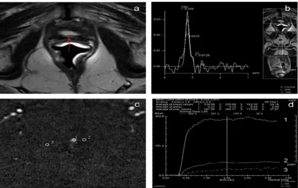
Fig. 1. Case with biochemical progression after radical prostatectomy. PSA level: 0.8 ng/ml. (a) T2-weighted MR images show 5 mm intermediate-signal intensity nodule (arrow) in a perianastomotic location. (b) MR-Spectroscopy analysis with reference images on the nodule. (c) Dynamic contrast-enhanced (DCE) MRI demonstrates a hypervascular lesion (arrow) and malignant patterns. (d) DCE-MRI intensity/time curve of the same area shows in (c) a hypervascular lesion.
1.51 ng/ml) and 28 negative patients (mean PSA 0.77 ng/ml). Using MRI imaging without CE, sensitivity, specificity, PPV, NPV and accuracy for evidencing a local recurrence were 61.4%, 82.1%, 84.4%, 57.5% and 69.4%, respectively. Authors showed that the diameter of the local recurrences detected varied from 0.8 to 4.2 cm (average 1.9 cm). Using MRI imaging with CE, sensitivity was 84.1%, specificity was 89.3%, PPV 92.5%, NPV 78.1% and accuracy was 86.1%. At CE-MRI the diameter of the sites of local recurrence detected varied from 0.8 to 3.5 cm (average 1.7). This comparison showed a statistically significant lower diagnostic accuracy of unenhanced MRI in comparison with CE-MRI, a statisti-cally significant lower sensitivity but no significant specificity differences. Authors moreover asserted that CE-MRI is able to better differentiate between tumour relapse and postopera-tive scar tissue of fibrosis. They also underlined that 75.6% of patients with positive findings had a PSA < 1.5 ng/ml (level of evidence 2b, seeTable 2).
Panebianco et al.[44]reported the first single-centre com-parative study of MRI and PET-CT in the early detection of local recurrence after RP. They enrolled 84 patients with biochemical recurrence after RP using both 18F choline PET-CT and MRI (MRSI and DCE-MRI) with a 3 Tesla (T) magnet. On the basis of the used validation criteria, patients were divided in two groups. Group A included 28 patients with >50% PSA reduction following radiation therapy used as validation criteria. This group showed an average size of lesions of 6 mm (range 5–7.2 mm) and a mean PSA of 1.1 ng/ml (range 0.8–1.4 ng/ml). Group B included 56 patients with a lesion average size of 13.3 mm (range
7.6–19.4 mm), mean PSA 1.9 ng/ml (range 1.3–2.5 ng/ml) and TRUS-guided biopsy used as validation criteria. In Group A results in identifying a local recurrence demonstrated for multiparametric MRI a sensitivity of 92%, specificity of 75%, PPV of 96%, NPV 60% and accuracy of 89% and for PET-CT a sensitivity of 62%, specificity of 50%, PPV of 88%, NPV 18% and accuracy of 60%. In Group B results in identifying a local recurrence showed for multiparametric MRI a sensitiv-ity of 94%, specificsensitiv-ity of 100%, PPV of 100%, NPV 57% and accuracy of 94% and for PET-CT a sensitivity of 92%, speci-ficity of 37%, PPV of 98%, NPV 43% and accuracy of 91%. PET-CT revealed distant metastases in 25 patients of Group B and in 5 patients of Group A. Authors compared also the MRI results of this study obtained with a 3 T MRI with those of a previous study using a 2 T MRI (18226441). The use of a 3 T magnet improved results of MRI in terms of sensitivity and specificity in patients of Group B and of specificity in patients of Group A. They concluded that MRSI-DCE-MRI combined techniques is a valid tool to detect local recurrence following RP and it is more accurate than PET-CT in the iden-tification of smaller lesions in patients with low biochemical failure (PSA < 2 ng/ml) (level of evidence 2b, seeTable 2).
4.4. Critical analysis and level of evidence for multiparametric MRI
Most of the studies using multiparametric MRI were designed in a retrospective way and they were single-centre. The number of patients enrolled in these studies ranges from 46 to 84 and therefore sample size is always limited. Mean
A. Alfarone et al. / Critical Reviews in Oncology/Hematology 84 (2012) 109–121 119
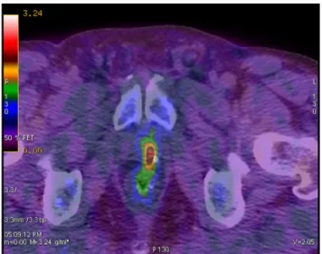
Fig. 2. F 18 choline fused PET-CT axial views show pathological uptake at the left level of the prostate bed in a case submitted to radical prostatectomy with biochemical progression (PSA = 1.5 ng/ml).
PSA level ranges from 0.8 ng/ml and 2.18 ng/ml; therefore in the MRI studies PSA levels at inclusion are definitely lower if compared with those in PET-CT studies. This is a fun-damental aspect related to the need of an early diagnosis of low dimension local recurrences. MRI studies show values of sensitivity between 84% and 95%, specificity between 75% and 100%, PPV between 92% and 100%, NPV between 57% and 88% and accuracy between 86% and 94%. The mean diameter of local recurrence lesions is always described in these studies and ranges from 6 mm and 15 mm.
In summary, we found a limited number of studies on the use of MRI for the detection of local recurrence following RP. The level of evidence is not very high because of the small sample sizes, the retrospective analysis and the lack of standardised validation criteria.
Overall MRI seems to have a high detection rate for local recurrence also in patients with low PSA levels and with small volume recurrent diseases, in particular using the combined modality of MRSI and DCE-MRI. The 3 T magnet, even if it has been used only in the recent study of Panebianco et al. [44], could offer promising better results associated to his higher spatial resolution. According to the reviewed studies we suggest that multiparametric MRI can be a valid diagnostic tool for the early biochemical failure or suspi-cion of subclinical minimally persistent disease following RP, able to detect local recurrence in patients with low PSA lev-els (<1.5 ng/ml). However, larger prospective studies should better confirm these evidences.
5. Conclusions
The problem of local recurrence after radical prostatec-tomy is topical because of its high frequency. We know the relevance to have a very rapid diagnosis in terms of PSA and also a sure localization of the recurrence. All the most
important guidelines of urological and oncological societies do not have significant recommendations regarding the use of specific imaging techniques for the early diagnosis of local recurrence after RP. The NCCN guidelines[45]provide the use of different imaging techniques as PET, MRI, CT and AUA guidelines[46]do not have specific recommendations for the use of imaging in patients with biochemical relapse and possibility to have local recurrence after RP. EAU guide-lines [47] indicate PET-CT and MRI as possible tools in patients with PSA recurrence between 1.0 and 2.0 ng/ml, but they are not considered as routine procedures.
Published data indicate an emerging role for multipara-metric MRI and for PET-CT in different aspects of PC management. Even if few studies are available, they sug-gest for MRI a significant detection rate for local recurrence also with very low PSA serum levels, in particular with the combined use of MRSI and DCE-MRI. The 3T modality can again improve the spatial resolution and scanning conditions even at very low PSA values, so to detect very small tumours (Fig. 1: a case).
PET-CT scanning, in particular with the use of choline, can provide a systemic localisation of the site of PC recurrence in a single step (Fig. 2: a case). On the basis of published data this procedure cannot be recommended for routine use to specifi-cally detect local recurrence, particularly in patients with low level of PSA (<1.5 ng/ml). At the same time we can assert that PET-CT could be indicated when PSA-DT is <3/6 months and PSA velocity is very high; in these situations patients have a high risk of nodal micrometastases and PET-CT has higher detection rate if compared with multiparametric MRI[48].
Only one study with a direct comparison between mul-tiparametric MRI and PET-CT in the same population has been produced (44). Moreover the level of evidence provides from all the studies in the literature is not very high. For these reasons more prospective comparative studies and larger mul-ticentre trials are needed to confirm promising results of these two techniques and to establish guidelines for the imaging in patients with biochemical failure after RP.
Conflict of interest
None of the authors has any financial or personal relation-ships with organizations that could inappropriately influence the work.
Funding
None of the authors has a funding source for the work.
Reviewers
Cosimo De Nunzio, MD PhD, University, Department of Urology, Ospedale Sant’Andrea, Rome, Italy.
120 A. Alfarone et al. / Critical Reviews in Oncology/Hematology 84 (2012) 109–121 Theo M. de Reijke, MD PhD FEBU, Urologist,
Aca-demic Medical Center, Urology, Meibergdreef 9, NL-1105 AZ Amsterdam, Netherlands.
Jean-Pierre Papazyan, MD FMH, Clinique de Genolier, Nuclear Medicine, 1 route du Muids, CH-1272 Genolier, Switzerland.
References
[1] Bill-Axelson A, Holmberg L, Filén F, et al. Scandinavian Prostate Can-cer Group Study Number 4. Radical prostatectomy versus watchful waiting in localized prostate cancer: the Scandinavian prostate can-cer group-4 randomized trial. J Natl Cancan-cer Inst 2008;100(August (16)):1144–54 [Epub 2008 August 11].
[2] Bianco Jr FJ, Scardino PT, Eastham JA. Radical prostatectomy: long-term cancer control and recovery of sexual and urinary function (“trifecta”). Urology 2005;66(November (5 Suppl.)):83–94. [3] Freedland SJ, Presti Jr JC, Amling CL, et al. SEARCH Database Study
Group. Time trends in biochemical recurrence after radical prosta-tectomy: results of the SEARCH database. Urology 2003;61(April (4)):736–41.
[4] Han M, Partin AW, Zahurak M, Piantadosi S, Epstein JI, Walsh PC. Biochemical (prostate specific antigen) recurrence probability follow-ing radical prostatectomy for clinically localized prostate cancer. J Urol 2003;169(February (2)):517–23.
[5] Grossfeld GD, Stier DM, Flanders SC, et al. Use of second treatment following definitive local therapy for prostate cancer: data from the caPSURE database. J Urol 1998;160(October (4)):1398–404. [6] Freedland SJ, Humphreys EB, Mangold LA, Eisenberger M, Partin AW.
Time to prostate specific antigen recurrence after radical prostatectomy and risk of prostate cancer specific mortality. J Urol 2006;176(October (4 Pt 1)):1404–8.
[7] Pound CR, Partin AW, Eisenberger MA, Chan DW, Pearson JD, Walsh PC. Natural history of progression after PSA elevation following radical prostatectomy. JAMA 1999;281(May (17)):1591–7.
[8] Moul JW. Prostate specific antigen only progression of prostate cancer. J Urol 2000;163(June (6)):1632–42.
[9] Amling CL, Bergstralh EJ, Blute ML, Slezak JM, Zincke H. Defining prostate specific antigen progression after radical prostatectomy: what is the most appropriate cut point? J Urol 2001;165(April (4)):1146–51. [10] Stephenson AJ, Kattan MW, Eastham JA, et al. Defining biochemical recurrence of prostate cancer after radical prostatectomy: a proposal for a standardized definition. J Clin Oncol 2006;24(August (24)):3973–8. [11] Roach 3rd M, Hanks G, Thames Jr H, et al. Defining biochemical failure following radiotherapy with or without hormonal therapy in men with clinically localized prostate cancer: recommendations of the RTOG-ASTRO Phoenix Consensus Conference. Int J Radiat Oncol Biol Phys 2006;65(July (4)):965–74.
[12] Marks RA, Koch MO, Lopez-Beltran A, Montironi R, Juliar BE, Cheng L. The relationship between the extent of surgical margin positivity and prostate specific antigen recurrence in radical prostatectomy specimens. Hum Pathol 2007;38(August (8)):1207–11 [Epub 2007 May 8]. [13] Stephenson AJ, Scardino PT, Kattan MW, et al. Predicting the outcome
of salvage radiation therapy for recurrent prostate cancer after radical prostatectomy. J Clin Oncol 2007;25(May (15)):2035–41.
[14] Boccon-Gibod L, Djavan WB, Hammerer P, et al. Management of prostate-specific antigen relapse in prostate cancer: a European Con-sensus. Int J Clin Pract 2004;58(April (4)):382–90.
[15] King CR, Spiotto MT. Improved outcomes with higher doses for sal-vage radiotherapy after prostatectomy. Int J Radiat Oncol Biol Phys 2008;71(May (1)):23–7 [Epub 2008 Jan 22].
[16] Scattoni V, Roscigno M, Raber M, et al. Multiple vesico-urethral biop-sies following radical prostatectomy: the predictive roles of TRUS,
DRE, PSA and the pathological stage. Eur Urol 2003;44(October (4)):407–14.
[17] Kane CJ, Amling CL, Johnstone PA, Pak N, et al. Limited value of bone scintigraphy and computed tomography in assessing biochemical fail-ure after radical prostatectomy. Urology 2003;61(March (3)):607–11. [18] Parsons KF, Irani J, Fall M, et al. European Association of urology
guidelines; 2011.
[19] Zeisel SH. Dietary choline: biochemistry, physiology, and pharmacol-ogy. Annu Rev Nutr 1981;1:95–121.
[20] Ackerstaff E, Pflug BR, Nelson JB, Bhujwalla ZM. Detection of increased choline compounds with proton nuclear magnetic resonance spectroscopy subsequent to malignant transformation of human pro-static epithelial cells. Cancer Res 2001;61(May (9)):3599–603. [21] Sutinen E, Nurmi M, Roivainen A, et al. Kinetics of [(11)C]choline
uptake in prostate cancer: a PET study. Eur J Nucl Med Mol Imaging 2004;31(March (3)):317–24 [Epub 2003 Nov 20].
[22] Souvatzoglou M, Weirich G, Schwarzenboeck S, et al. The sensitivity of [11C]Choline PET/CT to localize prostate cancer depends on the tumor configuration. Clin Cancer Res 2011;17(June (11)):3751–9 [Epub 2011 April 14].
[23] McNeal JE. The zonal anatomy of the prostate. Prostate 1981;2(1):35–49.
[24] Knopp MV, Giesel FL, Marcos H, von Tengg-Kobligk H, Choyke P. Dynamic contrast-enhanced magnetic resonance imaging in oncology. Top Magn Reson Imaging 2001;12(August (4)):301–8.
[25] Somford DM, Fütterer JJ, Hambrock T, Barentsz JO. Diffusion and perfusion MR imaging of the prostate. Magn Reson Imaging Clin N Am 2008;16(November (4)):685–95, ix.
[26] Seitz M, Shukla-Dave A, Bjartell A, et al. Functional magnetic reso-nance imaging in prostate cancer. Eur Urol 2009;55(April (4)):801–14 [Epub 2009 Jan 21].
[27] Fuchsjäger M, Akin O, Shukla-Dave A, Pucar D, Hricak H. The role of MRI and MRSI in diagnosis, treatment selection, and post-treatment follow-up for prostate cancer. Clin Adv Hematol Oncol 2009;7(March (3)):193–202.
[28] Kirkham AP, Emberton M, Allen C. How good is MRI at detecting and characterising cancer within the prostate? Eur Urol 2006;50(December (6)):1163–74, discussion 1175 [Epub 2006 Jun 30].
[29] Heinisch M, Dirisamer A, Loidl W, et al. Positron emission tomog-raphy/computed tomography with F-18-fluorocholine for restaging of prostate cancer patients: meaningful at PSA < 5 ng/ml? Mol Imaging Biol 2006;8(January–February (1)):43–8.
[30] Rinnab L, Mottaghy FM, Blumstein NM, et al. Evaluation of [11C]-choline positron-emission/computed tomography in patients with increasing prostate-specific antigen levels after primary treatment for prostate cancer. BJU Int 2007;100(October (4)):786–93.
[31] Vees H, Buchegger F, Albrecht S, et al. 18F-choline and/or 11C-acetate positron emission tomography: detection of residual or progressive sub-clinical disease at very low prostate-specific antigen values (<1 ng/mL) after radical prostatectomy. BJU Int 2007;99(June (6)):1415–20 [Epub 2007 Apr 8].
[32] Reske SN, Blumstein NM, Glatting G. [11C]choline PET/CT imaging in occult local relapse of prostate cancer after radical prostatectomy. Eur J Nucl Med Mol Imaging 2008;35(January (1)):9–17 [Epub 2007 Sep 9].
[33] Rinnab L, Simon J, Hautmann RE, et al. [(11)C]choline PET/CT in prostate cancer patients with biochemical recurrence after radical prostatectomy. World J Urol 2009;27(October (5)):619–25 [Epub 2009 Feb 21].
[34] Castellucci P, Fuccio C, Nanni C, et al. Influence of trigger PSA and PSA kinetics on 11C-Choline PET/CT detection rate in patients with biochemical relapse after radical prostatectomy. J Nucl Med 2009;50(September (9)):1394–400 [Epub 2009 Aug 18].
[35] Giovacchini G, Picchio M, Scattoni V, et al. PSA doubling time for pre-diction of [(11)C]choline PET/CT findings in prostate cancer patients with biochemical failure after radical prostatectomy. Eur J Nucl Med Mol Imaging 2010;37(June (6)):1106–16 [Epub 2010 Mar 20].
A. Alfarone et al. / Critical Reviews in Oncology/Hematology 84 (2012) 109–121 121 [36] Giovacchini G, Picchio M, Briganti A, et al. [11C]choline positron
emission tomography/computerized tomography to restage prostate cancer cases with biochemical failure after radical prostatectomy and no disease evidence on conventional imaging. J Urol 2010;184(September (3)):938–43.
[37] Giovacchini G, Picchio M, Coradeschi E, et al. Predictive factors of [(11)C]choline PET/CT in patients with biochemical failure after rad-ical prostatectomy. Eur J Nucl Med Mol Imaging 2010;37(February (2)):301–9 [Epub 2009 Sep 15].
[38] Castellucci P, Fuccio C, Rubello D, et al. Is there a role for11C-choline PET/CT in the early detection of metastatic disease in surgically treated prostate cancer patients with a mild PSA increase < 1.5 ng/ml? Eur J Nucl Med Mol Imaging 2011;38(January (1)):55–63 [Epub 2010 Sep 17].
[39] Picchio M, Briganti A, Fanti S, et al. The role of choline positron emis-sion tomography/computed tomography in the management of patients with prostate-specific antigen progression after radical treatment of prostate cancer. Eur Urol 2011;59(January (1)):51–60 [Epub 2010 Sep 15].
[40] Sella T, Schwartz LH, Swindle PW, et al. Suspected local recurrence after radical prostatectomy: endorectal coil MR imaging. Radiology 2004;231(May (2)):379–85 [Epub 2004 Apr 2].
[41] Casciani E, Polettini E, Carmenini E, et al. Endorectal and dynamic contrast-enhanced MRI for detection of local recurrence after radical prostatectomy. AJR Am J Roentgenol 2008;190(May (5)):1187–92. [42] Sciarra A, Panebianco V, Salciccia S, et al. Role of dynamic
contrast-enhanced magnetic resonance (MR) imaging and proton MR spectroscopic imaging in the detection of local recurrence after rad-ical prostatectomy for prostate cancer. Eur Urol 2008;54(September (3)):589–600 [Epub 2007 Dec 31].
[43] Cirillo S, Petracchini M, Scotti L, et al. Endorectal magnetic resonance imaging at 1.5 Tesla to assess local recurrence following radical prosta-tectomy using T2-weighted and contrast-enhanced imaging. Eur Radiol 2009;19(March (3)):761–9 [Epub 2008 Sep 30].
[44] Panebianco V, Sciarra A, Lisi D, et al. Prostate cancer: 1HMRS-DCEMR at 3T versus [(18)F]choline PET/CT in the detection of local prostate cancer recurrence in men with biochemical progression after radical retropubic prostatectomy (RRP). Eur J Radiol 2011;(February) [Epub ahead of print].
[45] NCCN guidelines Version 4. 2011 Panel Members Prostate Cancer. [46] American Urological Association. Prostate cancer clinical guideline
update panel members and consultants; 2007.
[47] Heidenreich A, Bolla M, Jomiau S, et al. European Association of Urology guidelines on prostate cancer; 2011.
[48] Scattoni V, Picchio M, Suardi N, et al. Detection of lymph-node metastases with integrated [11C]choline PET/CT in patients with PSA failure after radical retropubic prostatectomy: results confirmed by open pelvic-retroperitoneal lymphadenectomy. Eur Urol 2007;52(August (2)):423–9 [Epub 2007 Mar 20].
Biography
Alessandro Sciarra, medical doctor, received his title of
specialist in Urology in the School of Urology, University Sapienza of Rome, Italy. Now he is working in the Depart-ment of Urology “U. Bracci” of the University Sapienza of Rome and is responsible for a Prostate Unit there. He is currently a Professor in Medicine either for the School of Medicine or for the Schools of Urology and Nephrology at the University Sapienza of Rome. He has been member of the scientific committee of the Italian Society of Urology (SIU); referee for the European Association of Urology (EAU) and the European Urology Journal. His scientific activity has been published in the form of 109 contributions to various indexed international journals, most of them as the first author, and has edited three books. He has been an investigator or primary investigator in different international trials, in particular in urological oncology and benign prostatic hypertrophy, and he is a manuscript reviewer for a number of international journals. Current areas of research interest include urological oncology, mainly prostate cancer and kidney cancer, either as basic research or clinical studies.