RESEARCH ARTICLE
An analysis of the prevalence of peripheral
giant cell granuloma and pyogenic granuloma
in relation to a dental implant
Nieves Román‑Quesada
1, Beatriz González‑Navarro
2,3, Keila Izquierdo‑Gómez
2,3, Enric Jané‑Salas
2,3,
Antonio Marí‑Roig
3,4, Albert Estrugo‑Devesa
2,3*and José López‑López
2,3,5*Abstract
Background: The aim of the present investigation was to evaluate the literature recurrence of peripheral giant cell granuloma and pyogenic granuloma associated with dental implants. It’s important to know the characteristics pre‑ sent in these lesions and possible effects on the prognosis of dental implants.
Methods: An electronic search without time restrictions was done in the databases: PubMed/Medline. With the keywords "Granuloma" OR "Granuloma, Giant Cell" OR "peripheral giant cell" OR "Granuloma, Pyogenic” AND "Dental implants" OR "Oral implants”.
Results: After applying the inclusion and exclusion criteria, a total of 20 articles were included, which reported 32 lesions (10 pyogenic granulomas, 21 peripheral giant cell granulomas and one peripheral giant cell granuloma com‑ bined with peripheral ossifying fibroma, all associated with implants). According to our review, these lesions are more frequent in males and in the posterior region of the mandible. Both excision and curettage of the lesion, compared to only excision, presented similar recurrences (40%). Explantation of the implant was performed in 41% of cases without additional recurrences. The results are not statistically significant when comparing one lesion to the other in terms of explantation (p = 0.97), recurrence (p = 0.57) or bone loss (p = 0.67).
Conclusions: The main therapeutic approach is tissue excision. The lesions show a high recurrence rate (34.4%), which often requires explantation of the associated implant. This recurrence rate is not affected by curettage after excision.
Keywords: Dental implant, Oral implant, Pyogenic granuloma, Peripheral giant cell granuloma, Reactive oral lesions
© The Author(s) 2021. Open Access This article is licensed under a Creative Commons Attribution 4.0 International License, which permits use, sharing, adaptation, distribution and reproduction in any medium or format, as long as you give appropriate credit to the original author(s) and the source, provide a link to the Creative Commons licence, and indicate if changes were made. The images or other third party material in this article are included in the article’s Creative Commons licence, unless indicated otherwise in a credit line to the material. If material is not included in the article’s Creative Commons licence and your intended use is not permitted by statutory regulation or exceeds the permitted use, you will need to obtain permission directly from the copyright holder. To view a copy of this licence, visit http:// creat iveco mmons. org/ licen ses/ by/4. 0/. The Creative Commons Public Domain Dedication waiver (http:// creat iveco mmons. org/ publi cdoma in/ zero/1. 0/) applies to the data made available in this article, unless otherwise stated in a credit line to the data.
Background
The replacement of missing teeth with dental implants has a high success rate, but it is still a technique which involves risks and requires good evaluation and planning to minimize failures [1, 2]. However, the increasing num-ber of dental implants used leads the dentist to encounter
biological and technical complications [3, 4]. The most prominent biological complications are peri-implant mucositis and peri-implantitis [3, 5, 6]. In a 2017 system-atic review, Lee et al. [7] demonstrated that the rates of mucositis and peri-implantitis were 29.48% and 9.25%, respectively.
The appearance of so-called peri-implant reactive lesions is another complication that must be taken into consideration. Despite its lower incidence, its presence could imply the need for explantation [2, 3, 8]. Reactive lesions are characterized by excessive proliferation of
Open Access
*Correspondence: [email protected]; [email protected]
2 Department of Odontoestomatology, Faculty of Medicine and Health
Sciences (Dentistry), Bellvitge Campus, University of Barcelona, Barcelona, Spain
connective tissue in response to chronic irritation [9]. Among these lesions, those most frequently observed in the oral cavity are pyogenic granuloma (PG), traumatic fibroma, fibroepithelial hyperplasia, peripheral ossifying fibroma and peripheral giant cell granuloma (periheral giant cell lesion, -PGCG-) [4, 9]. PG and PGCG are the reactive lesions most frequently associated with teeth and implants [2, 9]. Some factors such as chronic inflamma-tion, the accumulation of foreign bodies or corrosion of the implant surface could cause a chronic irritative pro-cess and act as contributing factors not only for mucositis and peri-implantitis but also for PG and PGCG [3, 4, 10].
Several factors have been studied that could interfere with osseointegration and therefore the survival of the placed implant. Factors such as smoking, diabetes and periodontal disease have been studied [9, 11]. However, with regard to reactive lesions, such as PG and PGCG, the prevention technique is the maintenance of healthy peri-implant tissue [9] (Fig. 1).
PG can occur in response to irritants, trauma, hormo-nal changes or certain medications. Although classically called PG, the most correct name would be capillary lobular hemangioma, since the lesion is not strictly a granuloma or an infection [9, 10]. Clinically, oral PG is characterized as an exophytic mass with a smooth or lobulated appearance that can be sessile or peduncu-lated. The epithelium is frequently ulcerated and varies in color from pink to red and/or purple, depending on the evolution time and the vascularization of the lesion. The lesion tends to grow rapidly in a short period of time, with a tendency to bleed spontaneously or after a minor trauma [10, 12]. Its incidence varies between 3.81 to 7% of the histopathological results of biopsies performed in the oral cavity [3, 12]. According to the literature, it is more frequent in young women (3:2), more common
in the maxilla than in the mandible, and in the anterior areas when associated with teeth [9]. Histologically, PG is composed of proliferating blood vessels and granulation tissue, often organized in lobular aggregates, hence the name capillary lobular hemangioma [3]. It is character-ized by prominent capillary growth in hyperplastic gran-ulation tissue, suggesting high angiogenic activity. Blood vessels often show a grouped or separated pattern by less vascular fibrotic septa, leading some authors to consider PG as a polypoid form of capillary hemangioma [12].
PGCG is believed that its pathogenesis includes an excessive activation of osteoclasts, which is associated with a proliferation of macrophages and can cause sig-nificant bone resorption [9]. Clinically, PGCG may pre-sent as a firm or soft, sessile or pedunculated nodule, color ranging from bluish to purple, with a frequently ulcerated surface, confined to the alveolar and/or gingival mucosa. Swelling is the most frequent sign and its clinical course is usually asymptomatic [4, 13]. It tends to show a progressive increase in size and can cause pressure on the adjacent teeth, which could lead to malocclusion or interference with mastication. Erosion of the underlying bone or periodontium can also occur and in edentulous areas, it can often cause a radiolucent cup-shaped image in intraoral radiography [4]. PGCG does not have an age predilection. It seems to have a greater predilection in women and tends to appear more frequently in the molar area [4, 9, 11]. It consists of a non-encapsulated lobulated tumor of proliferating vascular connective tissue with a large number of osteoclast-type multinucleated giant cells. It is common to find signs of bleeding and hemosi-derin deposits inside the lesion. Fibroblastic proliferation and/or significant formation of osteoid material and even bone is common. Blood biochemistry shows no altera-tions [3, 13]; however, if alteration of phosphorus-cal-cium metabolism appears, hyperparathyroidism should be suspected and a brown parathyroid tumor should be ruled out [14–16].
The treatment of both PG and PGCG includes com-plete surgical excision until bone level and comcom-plete curettage, as well as removal of the causative agent if identified [17]. Since it is rarely reported it’s important to know the characteristics present in these lesions and pos-sible effects on the prognosis of dental implants in order to take the appropriate course of action in the clinical practice.
The aim of the present study was to search the associa-tion of the available data published in the literature on PG and PGCG associated with dental implants, analyzing their recurrences, associated etiological factors and dif-ferent treatment options. For this we asked ourselves the following PICO question: What is the prevalence, recur-rence, etiological factors and treatment of PG and PGCG Fig. 1 Pyogenic granuloma associated with a dental implant
around dental implants? P: Dental implants; I: develop-ment of a reactive lesion; C: PG vs. PGCG, type of treat-ment carried out (excision vs. excision and curettage); O: Prevalence of PG and PGCG around dental implants, recurrence of the lesions and bone loss around dental implants associated with the lesions.
Methods
Search strategy
The present study followed the Preferred Reporting Items for Systematic Reviews and Meta-Analysis (PRISMA) guidelines [18]. An electronic search was conducted in February 2020, with no time restrictions in the database: PubMed/Medline. The following terms were used for the search strategies: ["Granuloma" OR "peripheral giant cell granuloma" OR "Granuloma, Giant Cell" OR "peripheral giant cell" OR "Granuloma, Pyogenic" OR "peripheral giant cell reparative granuloma” OR "peripheral giant cell epulis”] AND ["Dental implants" OR "Oral implants”].
Article selection and inclusion criteria
The inclusion criteria were publications on cases of PG and PGCG associated with dental implants, with suf-ficient clinical, radiological and histological information to confirm the diagnosis (at least 8 out of the 10 out-comes of the table should be included); case series and single case studies. Articles that were written in English or Spanish. Articles with other lesions or syndromic con-ditions with similar characteristics, review, book chapter and letter to the editors were excluded.
Data extraction and analysis
For each of the included studies, the following data were extracted (whenever available): age, sex, location of the lesion, implants involved, presence of bone loss, time between implant placement and the appearance of the lesion, treatment performed, recurrence, implant failure and possible associated pathologies observed. Regard-ing bone loss, we made a subjective assessment; classi-fying severe as exposure of more than 3 implant threads or bone loss of ≥ 4 mm, moderate to less than 3 exposed threads (between 3–4 mm) and mild as a loss of less than 3 mm of bone based on radiographs and intraoral photo-graphs or on the authors’ criteria.
All cases are initially selected and analysed by NRQ & BGN, discrepancies are consulted with EJS, KIG & AMR and in case of doubt they are finally validated by AED and JLL.
All statistical analyses were conducted using Statistical Package for the Social Sciences (SPSS 12.0, Chicago, IL). Mean values of age and time of diagnosis of the lesions were estimated. Fisher’s exact test was used to analyze
recurrence of lesions and bone loss. All reported values (P values) were compared to a significance level of 5%.
Results
141 articles were obtained with the search strategy used. After excluding duplicate articles, 91 articles were selected and reading the title and abstract, 32 were left. Out of 32 articles, 4 were excluded because they were not written in English or Spanish, 6 due to lack of data and 2 because data was related to teeth instead of implants. Finally, 20 articles that met the inclusion and exclusion criteria previously specified were selected (Fig. 2).
The 20 papers analyzed [4, 9, 11, 17, 19–34] reported 30 patients with 32 lesions around 51 implants involved, and 2 patients reported two lesions. Histologically, 10 lesions were diagnosed as PG [9, 22, 24–26, 28, 30, 31], 21 as PGCG [4, 9, 11, 17, 19–21, 23, 29, 32–34] and one combined PGCG with an ossifying peripheral fibroma [24]. The epidemiological and clinical characteristics are summarized in Table 1.
Lesions were more prevalent (in this review) in men than in women (20:12), more in the mandible than in the maxilla, with a mean age of 51 years (range between 21 and 75 years). Information on the time between implant placement surgery and diagnosis of the lesion was avail-able in 27 lesions with a mean of 3.5 years (range between 1 month and 12 years) [9, 11, 17, 19–32].
Regarding the first therapeutic approach, excision of the lesion was performed in all cases. In 5/32 cases [11, 20, 23, 27, 32], only the excision of the lesion was per-formed, presenting two cases of recurrence [20, 23]. In 19 out of 32 cases [9, 11, 17, 19, 23–25, 29, 31, 33] the treat-ment was accompanied by curettage of the underlying bone, with recurrences in 8 out of 19 cases [11, 17, 23, 31] and 6 of these 8 cases finally required explantation [11, 17, 23, 31] due to this recurrence and bone loss. One case [28] was treated using laser technique without recurrence and another [30] with electrocautery, which did recur. Finally, in the remaining 6 cases [4, 21, 22, 26], excision of the lesion was performed in association with the explan-tation, without later recurrence. Based on these data, we can say that of the 13 cases in which explantation was not performed, the rate of recurrence of the lesions surgically excised with curettage was not significant compared to only the removal of the lesion, with a p = 0.97. Due to the lack of data, we do not know if the lesions that were only eliminated were the smallest. According to these data, clinically, we would choose to excise the lesion without curettage, avoiding further bone loss and damaging the implant surface. However it should be taken into account that the patients sample who do not undergo excision plus curettage is significantly smaller than those who undergo curettage.
A total of 11 out of 32 (34.4%) lesions [11, 17, 20, 23, 30, 31] with recurrences were found, of which 4 [11, 23, 31] reported more than 3 recurrences, requiring an explanta-tion to solve the issue. Of these 11 lesions, 9 were diag-nosed as PGCG [11, 17, 20, 23] and 2 as PG [30, 31]. In this review, recurrence rate is not statistically significant for one lesion or another, with a p = 0.57.
In 75% of cases [4, 9, 11, 17, 19, 21–24, 28–31, 33, 34] bone loss was observed (Table 2). 10 cases (41.6%) reported severe [4, 11, 17, 21, 22, 29, 31], 9 cases (37.5%) reported moderate [9, 11, 19, 28, 30, 31, 33] and 5 cases (20.8%) reported mild bone loss [23, 24, 34]. Of the 24 cases with bone loss, implants involved were explanted in 45.8% [4, 11, 17, 21–23, 31]; of which 81.8% had severe
bone loss [4, 11, 17, 21, 22, 29, 31] and 81.8% were PGCG [4, 11, 17, 21, 23] (Table 2). 8 cases (out of 32) did not pre-sent bone loss, comprising of 5 PGCG compared to 3 PG, and there was no statistical significance with a p = 0.65. If we correlate total severe bone loss [10, of which 8 are PGCG and 2 PG] with moderate or mild bone loss [14 of which 9 are PGCG and 5 PG], we also do not obtain sig-nificant results, with a p = 0.4.
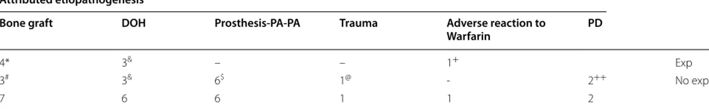
The attributed etiopathogenesis is varied (Table 3). In 71.8% (23 of 32) of the cases, observations that inform about a possible etiology were made. 26% (6 of 23) had poor oral hygiene, and 50% (3 of 6) required explantation. After the correction of these prostheses, there were no recurrences, and therefore explantation of the implants Fig. 2 PRISMA flowchart
Table
1
Summar
y of the most impor
tant charac
ter
istics of PGC
G and PG associat
ed with dental implants
Ref er enc es NP Sex Ag e Type of lesion Lo c A ppear anc e Time No . of I mp Bone L oss 1º T rea tmen t 2º T rea tmen t Recurr enc e/ follo w up Obser va tion H irshber g et al . [23] 3 ♂ 31 PGC G M d post months (*) 1 M ild Ex cision + cur ettage Ex cision with Ar g laser 1º. 1 2º. No NR ♂ 69 PGC G Mx ant 14 months 1 M ild Cur ettage Ex
cision with laser
+ exp 1º. 1 2º. No M ild congestiv e car diac insuffi ‑ cienc y ♂ 44 PGC G M d post 6 years 1 M ild Ex cision + cur ettage Ex cision + cur et ‑ tage + exp 1º. 3 2º. No NR Cloutier et al . [21] 1 ♂ 21 PGC G M d post 6 years 2 Se ver e ( + in 4.6) Ex cision + exp (4.6) + cur ettage – 1º ‑No NR Her nandez et al . [11] 3 ♀ 45 PGC G M d post 3 years 2 Se ve re Ex cision + cur ettage Ex ci ‑ sion + exp + cur et ‑ tage 1º. 5 2º. No NR ♀ 36 PGC G Mx post 2 years 1 Se ve re Ex cision + cur et ‑ tage + CLX Ex ci ‑ sion + exp + cur et ‑ tage 1º. 4 2º. No Calculus r emains ♀ 62 PGC G M d post 1 month 1 M oderat e Ex
cision of the lesion
– 1º. No A dvanced chr onic per iodontitis Olmedo et al . [24] 2 ♀ 75 PG M d post 2 months 1 No Ex cision + cur ettage – 1º. No NR ♀ 64 PGC G Mx ant 12 y ears 3 M ild Ex cision + cur ettage – 1º. No Br ok en scr ew in implant 21 M etallic par ticles D ojcino vic et al . [25] 1 ♂ 32 PG Mx post 6 months 1 No Ex cision + cur ettage – 1º. No Sinus lif t + aut olo ‑ gous iliac cr est graf t Peñar rocha ‑Diago et al . [19] 1 ♀ 54 PGC G M d post 3 years 2 M oderat e Pr osthesis remo val + ex ci ‑ sion + cur et ‑ tage + implant o‑ plast y – 1º. No Post ex trac tion implants Pr osthesis r et ention zone Et öz et al . [26] 1 ♀ 55 PG M d post 1 month 1 No Ex ci ‑ sion + exp + cur et ‑ tage – 1º. No Xenog raf t ® + PRF membrane Kang et al . [22] 1 ♂ 68 PG + CH Mx post 5 years 1 Se ve re Ex cision + exp – 1º. No 4 years af ter implant placement: infar ct + war far in
Table 1 ( continued) Ref er enc es NP Sex Ag e Type of lesion Lo c A ppear anc e Time No . of I mp Bone L oss 1º T rea tmen t 2º T rea tmen t Recurr enc e/ follo w up Obser va tion Jané ‑Salas et al . [9] 2 ♂ 52 PG Mx post 3 years 2 M oderat e Ex cision + cur et ‑ tage + CLX – 1º. No Chr onic per iodontal disease + plaque deposition M d post 3 years 1 M oderat e Ex cision + cur et ‑ tage + CLX – 1º. No Chr onic per iodontal disease + plaque deposition ♂ 64 PGC G M d post 8 years 1 M oderat e Ex cision + cur et ‑ tage + CLX – 1º. No Per iodontal disease O gbur ek e et al . [27] 1 ♂ 44 PGC G + POF M d post 3 months 2 No Ex cision – 1º. No NR Ka ya et al . [28] 1 ♂ 34 PG Mx ant 7 years 2 M oderat e Per iodontal tr eat ‑ ment + ex cision with laser – 1º. No A ut ogenous graf t + alv eolar ost eogenic dis ‑ trac tion Br own et al . [20] 1 ♂ 46 PGC G M d post 1 year 1 No Ex cision Ex cision + cur ettage 1º. 1 2º. No NR Pacifici et al . [29] 1 ♂ 60 PGC G Mx ant 5 years 1 Se ve re Ex cision + cur ettage – 1º. No D eficient oral hyg iene Truschnegg et al . [30] 1 ♂ 67 PG M d ant 3
months post loading
2 M oderat e (in 34) Elec tr ocaut er ization Ex
cision with laser
+ cur et ‑ tage + abutment change 1º. 2 2º. No D eficient oral hyg iene . Abut ‑
ment in poor condition
G efr er er et al . [31] 1 ♀ 55 PG M d post 9 w eeks 2 Se ve re Ex cision + cur ettage Exp I mplant 1º. 4 2º. No A ut ologous iliac cr est g raf t M d post 1 year 2 M oderat e Ex cision + cur ettage Exp I mplant 1º. 2 2º. No A ut ologous iliac cr est g raf t G alindo ‑M or eno et al . [32] 1 ♂ 74 PGC G Mx ant 6 years 1 No Bar r emo val + ex ci ‑ sion + CLX – 1º. No Per iodontitis , ex ‑ smok er (14 y ears) Bar r et ention z one Scarano et al . [4] 3 ♂ 26 PGC G M d ant – 1 Se ve re Ex ci ‑ sion + exp + cur et ‑ tage – 1º. No NR ♀ 52 PGC G Mx post – 1 Se ve re Ex ci ‑ sion + exp + cur et ‑ tage – 1º. No NR ♂ 31 PGC G M d ant – 6 Se ve re Ex ci ‑ sion + exp + cur et ‑ tage – 1º. No D eficient oral hyg iene
was not required. In 30% (7 of 23) of the cases, implant placement was performed with bone grafting, 57.1% (4 of 7) required explantation and in 2 of them (with asso-ciated PG) it is specified that iliac crest grafts were used [31], of the other 2, one presented PG and the other PGCG. Although in all 7 cases, there could be other associated factors, the authors preferred to attribute the occurrence to the graft. Finally, in 2 of the 23 cases the etiology was supposed to be periodontal disease [9, 11], both being PGCG and without explantation. One of the cases included in which the prosthesis was the cause of the lesion, was not included here because the author stated that the most relevant cause was the design of the prosthesis [17].
Discussion
PG and PGCG are inflammatory reactive lesions gener-ally found around teeth, root stumps and dental implants. The main feature helping to differentiate PGCG from PG is the abundant presence of multinucleated giant cells [35]. Although PG is considered more frequent than PGCG as a lesion, in this review PGCG is more frequently reported than PG in association with dental implants, data also supported the study by Brown et al. [20]. Based on this review, implant-associated PG and PGCG are rare peri-implant soft tissue complications, initially reported by Hirshberg et al. [23] and as men-tioned by Peñarrocha-Diago et al. [19], due to few docu-mented cases, the etiology and incidence of these lesions are not well established.
With respect to location, both in our review and in the cases reported in the literature, PG and PGCG associ-ated with implants have a predilection for the poste-rior regions of the jaws (20:12), mainly in the mandible (19:13) [9, 19]. This can be explained by the fact that the implants placed in the posterior regions hinder proper oral hygiene, which seems to be a factor that favors the appearance of these lesions [2, 9]. Although Hernández et al. [11] suggest that the occlusal forces experienced in
Table 1 ( continued) Ref er enc es NP Sex Ag e Type of lesion Lo c A ppear anc e Time No . of I mp Bone L oss 1º T rea tmen t 2º T rea tmen t Recurr enc e/ follo w up Obser va tion Bidra et al . [17] 3 ♂ 49 PGC G Mx ant 10 y ears 1 No Ex cision + cur ettage – 1º. No Bone g raf t ♀ 64 PGC G Mx ant 10 y ears 1 Se ve re Ex cision + cur ettage Exp + cur ettage 1º. 1 2º. No Bone g raf t + defi ‑ cient oral h yg iene ♂ 65 PGC G M d ant 2 w eeks 4 No Ex cision + cur ettage Ex cision + cur et ‑ tage + fluo ‑ cinolone + ne w pr osthesis 1º. 1 2º. No Post ‑ex trac tion and immediat e loading . P rosthesis with r et entiv e zone Baesso et al . [33] 1 ♂ 53 PGC G M d post – 1 M oderat e Ex cision + cur ettage – 1º. No Pr
osthesis with retentiv
e z one M or dini [34] 1 ♀ 39 PGC G Mx ant 7 months 1 M ild Ex cision + cur ettage – 1º. No Bone Alo ‑ Graf t + Trauma NP : number of pa tien ts , L oc: loca tion, No . of I mp: number of implan ts in volv ed , PGC G: per ipher al g ian t c ell g ranuloma, NR: No r ef er enc e; M d: mandible , post: post er ior , PG: p yogenic g ranuloma, A rg: ar gon, Mx: maxilla, an t: an ter ior , e xp: e xplan ta tion, CLX: chlor he xidine , PG: p yogenic g ranuloma, PRF : pla telet ‑r ich fibr in, CH: capillar y hemang ioma, POF : per ipher al ossifying fibr oma, Ex o: e xtr ac tion, (*): D
oes not specify
Table 2 Distribution of cases with bone loss
There is no statistical significance
(*) 8 are PGCG and 1 is PG; (#) It is a PG; (&) It is a PGCG; Exp: Explantation; NO Exp: No explantation; ($) 5 are PGCG and 3 are PG
Bone loss
Yes No
Severe Moderate Mild
11 9* 1# 1& Exp
21 1 8 4 8$ No exp
the posterior area are also a risk factor for the develop-ment of reactive lesions around the implants. Or simply the fact that more implants are placed in these locations, as different authors report while analyzing the literature [2, 9, 31].
The literature refers that when these lesions (PG and PGCG) affect the peridental areas, they are more fre-quent in women, although some studies have shown an equal prevalence for both sexes [2, 9]. This is in contrast with our findings that suggest that when these lesions are associated with implants, they are more frequent in men (20:12). According to Woelber et al. [36], this finding could be due to the fact that men have a higher plaque index than women, probably due to a lower level of hygiene, however, this is an inconsistent conclusion.
Dental implants are most commonly used in elderly patients; therefore, reactive lesions are expected to appear in older patients compared to those seen around natural teeth [2, 20]. In this review, it was observed that the age range was from 21 to 75 years, with an aver-age aver-age of 51 years, similar to the studies by Atarbashi-Moghadam et al. [2] with a mean age of 51.28 ± 14.48 and Brown et al. [20] with an average age of 50.9 years.
A possible pathogenesis of PG and PGCG associated with dental implants is a chronic local irritation that acts on the gingival tissue. In this sense, the local irritating factors involved may include accumulation of plaque or calculus or the presence of foreign bodies, such as pos-sible traces of dental cement or metal particles [2, 4, 19]. Burbano et al. [37] reported the presence of five types of dental cement particles in biopsies of peri-implanti-tis using scanning electron microscopy. Furthermore, Olmedo et al. [24] reported that the presence of metal particles in the tissues plays a role in the corrosion cess of the metallic component of the implant. They pro-posed that macrophages capture these metal particles, stimulating the release of cytokines, which play a role in bone resorption by activating osteoclasts and suppress-ing osteoblast function, thereby reducsuppress-ing bone formation and promoting osteolysis. This deposit can occur during
implant placement or due to the abutment connection [10, 20, 38]. Rodrigues et al. [39] supported this in a study that reported a PG and a PGCG associated with dental implants, and confirmed the presence of particles, pos-sibly of titanium. However, the implant technique is com-mon and there are few reference cases of PG and PGCG. This could possibly be influenced by the fact that some professionals surgically remove the peri-implant tis-sues and do not send them for a histopathological study [13], or simply do not publish the cases. An interesting study, without conclusive results, found some differ-ences in microvascular density, proliferative activity, and CD68 and Bcl-2 expression, between conventional and implant-associated granulomatous lesions [40]. In this study, histological and immunohistochemical aspects of peri-implant granulomatous lesions were analyzed and 13 new cases were presented, 2 in men and 11 in women. Although this series is not included in this review due to the lack of individualized clinical data and not meeting our inclusion criteria, it is worth noting that the average age in this case series was 57.5 years, with a higher preva-lence in women [2 of 13, 85%] and the posterior part of the mandible being the most affected area (10 of 13).
Peri-implantitis and marginal bone loss have been sug-gested to expose the unpolished portion of the implant neck, which can cause chronic irritation to the adjacent mucosa and lead to the development of reactive lesions [2, 13, 20]. Data from the articles selected in this review suggest that peri-implant bone loss is more commonly associated with PGCG than PG (17:7). Several authors [11, 21, 41, 42] support this hypothesis. In addition, Hir-shberg et al. [23] in their review of 25 peri-implant biopsy samples indicate that the PGCC around dental implants can causes loss of crestal bone that can eventually lead to implant failure. Surgical excision down to the bone level and aggressive curettage can cause additional bone loss around the implant [17, 19]. Furthermore, Hernández et al. [11] have also speculated whether bone loss is the cause or consequence of the granuloma and believe that the higher prevalence of these lesions in the posterior
Table 3 Results of attributed etiopathogenesis
DOR: Deficient Oral Hygiene; Prosthesis‑PA‑PA: Prosthesis Poorly adapted with plaque accumulation; PD: Periodontal Disease; Exp: Explantation; No Exp: No explantation
(*) 3 are PG and 1 is PGCG; (#) 2 are PG; (&) 2 are PG and 1 PGCG; ($) 5 are PGCG; (@) It is a PGCG; (+) It is a PG; (++) They are PGCG
Attributed etiopathogenesis
Bone graft DOH Prosthesis-PA-PA Trauma Adverse reaction to
Warfarin PD
4* 3& – – 1+ Exp
3# 3& 6$ 1@ ‑ 2++ No exp
regions may be due to the greater bone loss that occurs due to the greater implant overload caused by the occlusal forces. Data from the selected articles suggest that peri-implant bone loss is more commonly associ-ated with PGCG; in the present study, 10 of the 24 lesions with the presence of bone loss were severe and 8 of these lesions were PGCG [9].
Bischof et al. [42] described a PGCG in a 56-year-old woman with three posterior mandibular implants. It seems that the angulation of the implants was inap-propriate, which led to the healing abutments of two of them being poorly adjusted and juxtaposed. In addition, the patient reported the difficulty in cleaning the mouth, increasing the level of plaque in the area. This corrobo-rates the study by Özden et al. [41], who described a case of PGCG associated with a dental implant that occurred due to a poorly fitted prosthesis, which led to the accu-mulation of dental plaque and irritation of the gums. Regarding poorly designed prostheses, Bischof et al. [42], Özden et al. [41], and Peñarrocha-Diago et al. [19] replaced the prostheses or temporarily removed it, which allowed better plaque control in these cases and the absence of recurrences. In our review, authors such as Bidra et al., Peñarrocha-Diago et al. and Baesso et al. [17, 19, 33] included removal of the prosthesis and replace-ment with a healing abutreplace-ment, thus performing careful curettage and removal of the irritant factor; subsequently making a new prosthesis. We did not find justification in the literature to relate the appearance of the lesion to Warfarin medication, as suggested by Hirshberg et al. [23].
Günhan et al. [43] suggested that the appearance of these lesions could be due to the influence of sex hor-mones, since multinucleated giant cells are a target for estrogens. Immunohistochemical investigation of estrogen receptors in PGCGs turned out positive. How-ever, it can be suggested that in the context of implant-associated PGCG, the hormonal influence would be less important, since most of the patients are postmenopausal and, therefore, serum estrogen levels are low [20]. Recur-rences of PG and PGCG have been described, especially associated with teeth due to a hormonal imbalance dur-ing pregnancy, especially in cases of PG [9, 33]. Scarano et al. [35] article had as hypothesis that the multinucle-ated cells present in central giant cell granuloma of the jawbones may simply represent a reactive component of the lesion and is a response to stimuli from the stroma. These cells have been shown to present a positivity to estrogen and to show a phenotype different from that of other giant cells found in sites of chronic inflammation and may be true osteoclasts.
The length of time between implant placement and ini-tial presentation of PGCG and PG in our review ranged
from 1 month to 12 years. Although there is controversy, since tooth extraction has been described as an etio-logical factor for the development of PGCG, as demon-strated by Hirshberg et al. [23], who reported that the PGCG developed after tooth extraction in 8–11% of the cases examined, appearing up to one year after the proce-dure. Scarano et al. [35] also indicate that the occurrence of OGCG may be related to teeth extraction carried out before the implant insertion, Given this, surgical removal must be done otherwise the affected implant will fail. Therefore, it would be interesting to investigate whether the cases shown in Table 1 underwent a dental extraction before implant placement and, if so, the time between extraction and implant placement. In this sense, only Bidra et al. [17] and Peñarrocha-Diago et al. [19] speci-fied that the implants were placed post-extraction [20].
The recurrence rate of PGCG associated with teeth is varied, but it has been shown to range from 15 to 20% in most studies compared to PG that ranges from 0 to 16% [2]. In our review, the recurrence rate for PGCG and PG associated with implants was 34.4%. Therefore, it appears that the recurrence rate of these implant-associated lesions is higher than those unrelated to them [9, 19]. The reason for this difference in recurrence rates is unclear, but it could be due to difficulty faced in the removal of peri-implant tissues. However, curettage after excision does not seem to affect the recurrence rate, since the results are not significant [2, 13].
Several treatments for implant-associated PG and PGCG have been described, with conservative surgical extirpation with total removal of the lesion base and bone curettage being the treatment of choice [2, 9, 20]. Any irritating factors that may be causing or promoting the appearance of PGCG and PG must also be eliminated. Hernández et al. [11] also recommend polishing the implant surface [19, 20]. Photodynamic therapy (PDT) has been used as a complementary treatment for peri-implant diseases. Its main objective is controlling disease progression through decontamination of infected sur-faces. PDT is a simple and non-invasive technique that has proven to have antibacterial effects. Although fur-ther studies are needed to prove its efficiency in cases of PGCG around dental implants. [33]
Due to the aggressive nature of the lesion and the high recurrence rate, implants can fail unless the lesion is detected early and proper surgical removal is performed along with close follow-up. [23] The recommendation to remove an involved implant should be based on the clini-cal judgment of the practitioner balanced with the risk of recurrence of the lesion due to potential inadequate removal if access is jeopardized by the continued physical presence of a dental implant. [21]
The use of the Er-YAG, carbon dioxide or diode laser have been suggested as other treatment modalities and may offer advantages compared to conventional surgical techniques, especially by reducing the risk of postopera-tive bleeding, pain, and edema and also by eliminating the need to suture at the end of the procedure [2, 9].
In the study of Kaya et al. [28], to avoid a peri-implant reactive lesion, they decontaminated the exposed implant surface using the Er:YAG laser. There was no scarring or recurrence at the 2-year follow-up. Hence, an Er:YAG laser appears to be a good therapeutic option for intraoral pyogenic granulomas.
As limitations we have found that the works are clinical cases and the longest series of cases found is 3 cases. In many cases the incidence and etiology are not well estab-lished. Even in some reports the diagnosis is only clinical, which may represent an added bias. In some cases not all the data needed to perform the analysis is specified.
Conclusions
Implant-associated granulomas are more frequent in men, the mandible, and the most commonly diagnosed and referenced lesion is the PGCG.
The main focus of treatment is excision without tissue curettage with subsequent histopathological study. The lesions show a high recurrence rate that often require explantation, partly due to large bone loss or multiple recurrences. This recurrence rate is not affected by curet-tage after excision.
Histopathological diagnosis is important, since if the result is a PGCG, the dentist will know that there is a higher risk of bone loss and a higher recurrence rate. Oral hygiene instructions and care by professionals in avoiding leaving "foreign" materials during treatment or rehabilitation should be considered in this entity. These conclusions should be interpreted with caution due to the low number of published cases.
Abbreviations
PG: Pyogenic granuloma; PGCG : Periapheral giant cell granuloma; PICO: Population, or Problem, Intervention or Exposure, Comparison (if appropriate), Outcome you would like to measure or archieve.
Acknowledgements
We thank the entire Oral Medicine group of the Faculty of Medicine and Health Sciences [Dentistry] for their support and in a special way to Mayra Schemel for correcting the grammatical style.
Authors’ contributions
NRQ and JLL conceived the idea. NRQ, BGN and KIN screened the articles. Allauthors (NRQ, BGN, KIG, EJS, AMR, AED & JLL) worked together to review thearticles until agreement was reached. All authors then created the table of papers. NRQ, BGN and KIG led the writing. EJS, AMR and AED checked and revised the text. NRQ and JLL led the final editing stages of the manuscript. All authors read and approved the final manuscript.
Funding
This study has not had additional funding. It is financed with own funds of the research group.
Availability of data and materials
All data generated or analyzed during this study are included in this published article.
Declarations
Ethics approval and consent to participate
Not applicable.
Consent for publication
Not applicable.
Competing interests
The authors declare that they have no competing interests.
Author details
1 Faculty of Medicine and Health Sciences (Dentistry), University of Barcelona,
Barcelona, Spain. 2 Department of Odontoestomatology, Faculty of Medicine
and Health Sciences (Dentistry), Bellvitge Campus, University of Barcelona, Barcelona, Spain. 3 Oral Health and Masticatory System Group, Institut
D’Investigació Biomédica de Bellvitge (IDIBELL, Bellvitge Institute of Biomedi‑ cal Research), L’Hospitalet de Llobregrat, Barcelona, Spain. 4 Department
of Maxillofacial Surgery, Bellvitge University Hospital, L’Hospitalet de Llobre‑ grat, Barcelona, Spain. 5 Odontology Hospital University of Barcelona (HOUB),
Barcelona, Spain.
Received: 8 August 2020 Accepted: 14 April 2021
References
1. Han HJ, Kim S, Han DH. Multifactorial evaluation of implant fail‑ ure: a 19‑year retrospective study. Int J Oral Maxillofac Implants. 2014;29:303–10.
2. Atarbashi‑Moghadam F, Atarbashi‑Moghadam S, Namdari M, Shahrabi‑ Farahani S. Reactive oral lesions associated with dental implants. A systematic review. J Investig Clin Dent. 2018;9:e12342.
3. Halperin‑Sternfeld M, Sabo E, Akrish S. The pathogenesis of implant‑ related reactive lesions: a clinical, histologic and polarized light micros‑ copy study. J Periodontol. 2016;87:502–10.
4. Scarano A, Lorusso C, Mortellaro C, Limongelli L, Tempesta A, Favia G. Peripheral giant cell granuloma associated with dental implants. J Craniofac Surg. 2018;29:e196–9.
5. Froum SJ. The team approach to managing dental implant complica‑ tions: the periodontist’s point of view. Compend Contin Educ Dent. 2013;34:4–9.
6. Zitzmann UN, Berglundh T. Definition and prevalence of peri‑implant diseases. J Clin Periodontol. 2018;35:286–91.
7. Lee CT, Huang YW, Zhu L, Weltman R. Prevalences of peri‑implantitis and peri‑implant mucositis: systematic review and meta‑analysis. J Dent. 2017;62:1–12.
8. Derks J, Tomasi C. Peri‑implant health and disease. A systematic review of current epidemiology. J Clin Periodontol. 2015;42:S158–71. 9. Jané‑Salas E, Albuquerque R, Font‑Muñoz A, González‑Navarro B,
EstrugoDevesa A, López‑López J. Pyogenic granuloma/peripheral giant cell granuloma associated with implants. Int J Dent. 2015;2015:839032. 10. Al‑Shamiri HM, Alaizari NA, Al‑Maweri SA, Tarakji B. Development of
pyogenic granuloma and hemangioma after placement of dental implants: a review of literature. J Int Soc Prev Community Dent. 2015;5:77–80.
11. Hernandez G, Lopez‑Pintor RM, Torres J, de Vicente JC. Clinical out‑ comes of peri‑implant peripheral giant cell granuloma: a report of three cases. J Periodontol. 2009;80:1184–91.
12. Anitua E, Pinas L. Pyogenic granuloma in relation to dental implants: clinical and histopathological findings. J Clin Exp Dent. 2015;7:e447–50.
•fast, convenient online submission
•
thorough peer review by experienced researchers in your field
• rapid publication on acceptance
• support for research data, including large and complex data types
•
gold Open Access which fosters wider collaboration and increased citations maximum visibility for your research: over 100M website views per year
•
At BMC, research is always in progress. Learn more biomedcentral.com/submissions Ready to submit your research
Ready to submit your research ? Choose BMC and benefit from: ? Choose BMC and benefit from: 13. Chrcanovic BR, Gomes CC, Gomez RS. Peripheral giant cell granuloma
associated with dental implants: a systematic review. J Stomatol Oral Maxillofac Surg. 2019;120:456–61.
14. Smith BR, Fowler CB, Svane TJ. Primary hyperparathyroidism present‑ ing as a “peripheral” giant cell granuloma. J Oral Maxillofac Surg. 1998;46:65–9.
15. Alarcón‑Arratia A, Muñoz‑Repetto M, Schnettler K, Ulloa‑Marin C. Peripheral giant cell granuloma and hyperparathyroidism. Case report. Int J Odontostomat. 2019;13:266–70.
16. Wilson JJ, Schwartz HC, Tehrany GM. Brown tumor of the posterior maxilla as initial manifestation of primary hyperparathyroidism: case report. J Oral Maxillofac Surg. 2013;71:886–90.
17. Bidra AS, Persenaire MJ, Natarajan E. Management of peripheral giant cell granuloma around complete‑arch fixed implant‑supported pros‑ thesis: a case series. J Prosthet Dent. 2019;122:181–8.
18. Moher D, Liberati A, Tetzlaff J, Altman DG, PRISMA Group. Preferred reporting items for systematic reviews and meta‑analyses: the PRISMA statement. PLoS Med. 2009;6:e1000097.
19. Peñarrocha‑Diago MA, Cervera‑Ballester J, Maestre‑Ferrín L, Peñar‑ rocha‑Oltra D. Peripheral Giant cell granuloma associated with dental implants: clinical case and literature review. J Oral Implantol. 2012;38:527–32.
20. Brown AL, Camargo de Moraes P, Sperandio M, Borges Soares A, Araújo VC, Passador‑Santos F. Peripheral giant cell granuloma associated with a dental implant: a case report and review of the literature. Case Rep Dent. 2015;2015:697673.
21. Cloutier M, Charles M, Carmichael RP, Sándor GK. An analysis of periph‑ eral giant cell granuloma associated with dental implant treatment. Oral Surg Oral Med Oral Pathol Oral Radiol Endod. 2007;103:618–22. 22. Kang YH, Byun JH, Choi MJ, Lee JS, Jang JH, Kim YI, Park BW. Co‑devel‑
opment of pyogenic granuloma and capillary hemangioma on the alveolar ridge associated with a dental implant: a case report. J Med Case Rep. 2014;8:192.
23. Hirshberg A, Kozlovsky A, Schwartz‑Arad D, Mardinger O, Kaplan I. Peripheral giant cell granuloma associated with dental implants. J Peri‑ odontol. 2003;74:1381–4.
24. Olmedo DG, Paparella ML, Brandizzi D, Cabrini RL. Reactive lesions of peri‑implant mucosa associated with titanium dental implants: a report of 2 cases. Int J Oral Maxillofac Surg. 2010;39:503–7. 25. Dojcinovic I, Richter M, Lombardi T. Occurrence of a pyogenic
granuloma in relation to a dental implant. J Oral Maxillofac Surg. 2010;68:1874–6.
26. Etöz OA, Soylu E, Kiliç K, Günhan Ö, Akcay H, Alkan A. A reactive lesion (pyogenic granuloma) associated with dental implant: a case report. J Oral Implantol. 2013;39:733–6.
27. Ogbureke EI, Vigneswaran N, Seals M, Frey G, Johnson CD, Ogbureke KU. A peripheral giant cell granuloma with extensive osseous meta‑ plasia or a hybrid peripheral giant cell granuloma‑peripheral ossifying fibroma: a case report. J Med Case Rep. 2015;9:14.
28. Kaya A, Ugurlu F, Basel B, Sener CB. Oral pyogenic granuloma associ‑ ated with a dental implant treated with an Er:YAG laser: a case report. J Oral Implantol. 2015;41:720–3.
29. Pacifici A, Carbone D, Marini R, Sfasciotti GL, Pacifici L. Clinical management of a peri‑implant giant cell granuloma. Case Rep Dent. 2015;2015:976756.
30. Truschnegg A, Acham S, Kqiku L, Beham A, Jakse N. CO2 laser exci‑
sion of a pyogenic granuloma associated with dental implants: a case report and review of the literature. Photomed Laser Surg. 2016;34:425–31.
31. Gefrerer L, Popowski W, Perek JN, Wojtowicz A. Recurrent pyogenic granuloma around dental implants: a rare case report. Int J Periodon‑ tics Restorative Dent. 2016;36:573–81.
32. Galindo‑Moreno P, Hernández‑Cortés P, Ríos R, Sánchez‑Fernández E, Cámara M, O’Valle F. Immunophenotype of dental implant‑associated peripheral giant cell reparative granuloma in a representative case report. J Oral Implantol. 2016;42:55–60.
33. Baesso RCP, de Lima Jacy Monteiro Barki MC, de Souza Azevedo R, da Costa Fontes KBF, Pereira DL, Tucci R, Pires FR, Picciani BLS. Peripheral giant cell granuloma associated with a dental implant. BMC Oral Health. 2019;19:283.
34. Mordini L. Esthetic management of peripheral giant cell granuloma affecting a dental implant in the esthetic zone. Clin Adv Periodontics. 2019;9:77–82.
35. Scarano A, Iezzi G, Artese E, Cimorelli E, Piattelli A. Peripheral giant cell granuloma associated with a dental implant. A case report. Miner Stomatol. 2008;57:529–34.
36. Woelber JP, Bienas H, Fabry G, Silbernagel W, Giesler M, Tennert C, Stampf S, Ratka‑Krüger P, Hellwig E. Oral hygiene‑related self‑efficacy as a predictor of oral hygiene behaviour: a prospective cohort study. J Clin Periodontol. 2015;42:142–9.
37. Burbano M, Wilson TG Jr, Valderrama P, Blansett J, Wadhwani CP, Choud‑ hary PK, Rodriguez LC, Rodrigues DC. Characterization of cement particles found in peri‑implantitis‑affected human biopsy specimens. Int J Oral Maxillofac Implants. 2015;30:1168–73.
38. Olmedo D, Fernández MM, Guglielmotti MB, Cabrini RL. Macrophages related to dental implant failure. Implant Dent. 2003;12:75–80. 39. Rodrigues DC, Valderrama P, Wilson TG, Palmer K, Thomas A, Sridhar S,
Adapalli A, Burbano M, Wadhwani C. Titanium corrosion mechanisms in the oral environment: a retrieval study. Materials. 2013;6:5258–74. 40. Morais TM, Soares CD, Aguirre Urizar JM, Alberdi‑Navarro J, Almeida
OP, Pires FR. Peri‑implant peripheral giant cell lesions: report of 13 new cases and comparative histological and immunohistochemical analysis with peripheral and central giant cell lesions. Med Oral Patol Oral Cir Bucal. 2019;24:e739–74.
41. Özden FO, Ozden B, Kurt M, Gündüz K, Günhan O. Peripheral giant cell granuloma associated with dental implants: a rare case report. Int J Oral Maxillofac Implants. 2009;24:1153–6.
42. Bischof M, Nedir R, Lombardi T. Peripheral giant cell granuloma associated with a dental implant. Int J Oral Maxillofac Implants. 2004;19:295–9.
43. Günhan M, Günhan O, Celasun B, Mutlu M, Bostanci H. Estrogen and progesterone receptors in the peripheral giant cell granulomas of the oral cavity. J Oral Sci. 1998;40:57–60.
Publisher’s Note
Springer Nature remains neutral with regard to jurisdictional claims in pub‑ lished maps and institutional affiliations.