Dottorato di ricerca in Oncologia e Patologia Sperimentale Progetto Nr.2 - Patologia Sperimentale
CICLO XXIII MED/05
Tesi di Dottorato
RELEVANCE OF CELL CYCLE REGULATORS
ON CHEMOTHERAPY RESPONSE
IN BREAST CANCER
Presentata da: Dott.ssa Elisa Brighenti
Coordinatore Dottorato:
Relatore:
Chiar.mo Prof. Chiar.mo Prof.
Sandro Grilli Massimo Derenzini
TABLE OF CONTENTS
LIST OF ABBREVIATIONS
...IINDEX OF FIGURES
...VINDEX OF TABLES
...VIIINTRODUCTION
1. BREAST CANCER
1.1 FEATURES………...……….…...…1
1.2 EPIDEMIOLOGY AND RISK FACTORS………...2
1.3 DISEASE ONSET AND PROGRESSION……….12
1.4 CLASSIFICATION AND CLINICAL PATHOLOGY……….…..15
1.5 TYPES AND SUBTYPES……...……….22
1.5.1 TRIPLE-NEGATIVE BREAST CANCER………....26
1.6 THERAPY...……....………….…………...27
2. CHEMOTERAPY
2.1 FEATURES ...……….332.2 CHEMOTHERAPEUTIC DRUGS AND MECHANISM OF ACTION……35
3.
CELL CYCLE
3.1 CELL CYCLE AND CANCER………403.2 THE p53, pRb AND p16INK4a PATHWAYS IN CANCER………..48
EXPERIMENTAL DESIGN
4. AIMS OF THE THESIS
………...585. MATERIALS AND METHODS
5.1. Patients………...……….60
5.2. Adjuvant treatments………61
5.3. Immunohistochemical assessment…………...……….………..61
5.4. Cell lines and growth conditions...63
5.5. Production of HCT-116-derived cells with stably inactivated p53...64
5.6. Drugs and cell treatment protocols...64
5.7. Genes silencing by RNAi transfection...65
5.8. RNA extraction, cDNA synthesis and real-time RT–PCR analysi...66
5.9. Proteins extraction and Western blot analysis...67
5.10. Immunocytochemical analysis...68
5.11. Evaluation of cell population growth...69
5.12. Evaluation of cell death rate...70
5.13. Cell cycle progression analysis by dual-parameter flow cytometry...71
5.14. Effect of drug treatment on p53 activation and DNA double-strand breaks accumulation...72
5.15. Statistical analysis...72
5.16. RNAi sequences...73
6. RESULTS
6.1. The p53-mediated sensitivity of cancer cells to chemotherapeutic agents is conditioned by status of the pRb protein 6.1.1. Assessment of pRb and p53 status...74
6.1.2. Relationship between p53 and pRb in tumor prognosis...77
6.1.3. Evaluation of p53-mediate chemosensitivity and pRb pathway
status in cancer cells...80
6.2. Loss of pRb protein makes human breast cancer cells more sensitive to antimetabolites exposure
6.2.1. Immunohistochemical definition of pRb status and determination of its prognostic value in a large series of primary breast
cancer patients...90
6.2.2. Prognostic value of pRb expression and phosphorylation...91
6.2.3. The absence of pRb expression is the only predictive factor of good clinical outcome in patients treated with adjuvant
chemotherapy...92
6.2.4. 5-FU and MTX treatment hindered cell population growth of
RB1- silenced MCF-7 and HCT-116 cells...95
6.2.5. 5-FU and MTX treatment caused a cell cycle arrest in control but not in RB1-silenced cells...98
6.2.6. The p53/p21 pathway was normally activated in RB1-silenced
cells treated with 5-FU and MTX...100
6.2.7. RB1-silenced cells accumulated DNA double-strand breaks...101
6.3. High prevalence of retinoblastoma protein loss in triple-negative breast
cancers and its association with a good prognosis in patients treated with adjuvant chemotherapy
6.3.1. Valuation of pRb status and its association of the clinical outcome of chemotherapy-treated patients with triple-negative tumors...103
6.3.2. pRb status and the clinical outcome of triple negative tumors treated with chemotherapy...106
6.3.3. Relevance of pRb status on sensitivity to doxorubicin in
MDA-MB-231 triple-negative derived cells...108
7. DISCUSSION
...111 7.1. In breast cancer with a normally function of pRb pathway, the p53 status was the only independent factor capable to predicting the patient clinical outcome after adjuvant chemotherapy treatment...112
7.2. The absence but not inactivation of pRb predicted the clinical outcome of patients treated with 5-FU and MTX adjuvant therapy...113
7.3. Lack of pRb expression was the only independent factor predicting a good clinical outcome in patients treated with adjuvant
chemotherapy...114
7.4. The greater sensitivity of pRb deficient cells to 5-FU plus MTX exposure was due to the absence of a DNA damage checkpoint and DNA
repair mechanisms...116
7.5. High prevalence of retinoblastoma protein loss in triple-negative breast cancers was responsible for a good prognosis in patients treated
with adjuvant chemotherapy...118
8. NOTES
...123I
LIST OF ABBREVIATIONS
5-FU: 5-Fluorouracile
ADH: Atypical Ductal Hyperplasia
ATCC: American Type Culture Collection ATM: Ataxia Telangiectasia Mutated BC: Breast Cancer
BRCA1: Breast Cancer 1 BRCA2: Breast Cancer 2 BrdUrd: Bromodeoxyuridine BSA: Bovine Serum Albumin CDK: Cyclin-Dependent Kinase
CDKI: Cyclin-Dependent Kinases Inhibitor cDNA: copy of DNA
CMF: Cyclophosphamide plus Methotrexate plus 5-Fluorouracil CSPG2: Chondroitin Sulfate Proteoglycan 2
DAB: Diaminobenzidine
DAPI: 4',6-Diamidino-2-phenylindole dihydrochloride DCIS: Ductal Carcinoma In Situ
DEPC: Diethylpyrocarbonate treated water DFS: Disease-Free Survival
DHFR: Dihydrofolate Reductase
DMEM: Dulbecco‟s Modified Eagle‟s Medium DNA: DesossiriboNucleic Acid
II
EDTA: EthyleneDiamine Tetra-acetic Acid EGF: Epidermal Growth Factor
EGFR: Epidermal Growth Factor Receptor ER: Estrogen Receptor
ErbB-2: Human Epidermal growth factor Receptor 2 ERD: Estrogen Receptor Downregulators
FBS: Fetal Bovine Serum
FITC: Fluorescein IsoThioCyanate HCT-116: Colon cancer cell line
HELU: Hyperplastic Enlarged Lobular Units HepG2: Human liver carcinoma cell line
HER2/neu: Human Epidermal Growth Factor Receptor 2 IBC: Invasive Breast Cancer
IL-2: Interleukin-2
LCIS: Lobular Carcinoma In Situ LI: Labeling Index
MCF-7: estrogen receptor positive breast cancer cell line
MDA-MB-231: estrogen receptor negative breast cancer cell line MDM2: Murine Double Minute 2
MI: Mitotic Index
mAbs: Monoclonal Antibodies mRNA: messenger RNA MTX: Methotrexate
III
PBS: Phosphate Buffered Saline
PDGF: Platelet-Derived Growth Factor PR: Progesterone Receptor
pRb: Retinoblastoma protein
PTEN: Phosphatase and Tensin Homolog
RB1: Retinoblastoma gene
RNA: ribonucleic acid RNAi: RNA interference
RPMI: Roswell Park Memorial Institute RT: Room Temperature
RT-PCR: Reverse Transcriptase-Polymerase Chain Reaction SD: Standard Deviation
SDS-PAGE: Sodium Dodecyl Sulfate PolyAcrilammide Gel Electrophoresis SERM: Selective Estrogen Receptors Modulators
TDLU: Terminal Duct Lobular Unit TGF-β: Transforming Growth Factor-β TLI: Thymidine Labeling Index
TNBC: Triple-Negative Breast Cancer TNM: Tumor Nodes Metastasis
TP53: p53 gene
VEGF: Vascular Endothelial Growth Factor WHO: World Health Organization
WT: Wild Type
IV
µl: microlitre µm: micrometer µM: micromolar
V
INDEX OF FIGURES
FIGURE 1………...……….……… 1 FIGURE 2………...……….………….3 FIGURE 3………...……….………….4 FIGURE 4………...………….……...13 FIGURE 5……….…….….23 FIGURE 6………....……...40 FIGURE 7………...….……...42 FIGURE 8………....……...44 FIGURE 9……….……...…...48 FIGURE 10………..……….………...49 FIGURE 11……….……….………...…50 FIGURE 12……….……...….53 FIGURE 13……….……...…….54 FIGURE 14………...……..57 FIGURE 15……….…………....75 FIGURE 16……….……...…….75 FIGURE 17……….……...…….78 FIGURE 18……….……….…………...78 FIGURE 19………...79 FIGURE 20……….…………82 FIGURE 21……….…………83 FIGURE 22………..……….…………..84 FIGURE 23……….……….………...85VI FIGURE 24………...86 FIGURE 25……….…………87 FIGURE 26……….…………88 FIGURE 27………..……….…………..89 FIGURE 28……….……….………...95 FIGURE 29……….…………96 FIGURE 30………..……….…………..98 FIGURE 31……….……….………...99 FIGURE 32……….………..101 FIGURE 33………..……….…………102 FIGURE 34……….……….……….105 FIGURE 35...107 FIGURE 36...109 FIGURE 37...110 FIGURE 38...122 FIGURE 39...122
VII
INDEX OF TABLES
TABLE 1……….……….…..……….17 TABLE 2……….……….…….……..44 TABLE 3……….……….…….….….76 TABLE 4……….……….…….……..77 TABLE 5……….……….…………...80 TABLE 6……….……….…………...91 TABLE 7……….……….…………...92 TABLE 8……….……….…………...93 TABLE 9……….……….…………...94 TABLE 10……….……….………...103 TABLE 11……….……….………...104 TABLE 12……….……….………...105 TABLE 13……….……….………...106 TABLE 14……….……….………...1081
INTRODUCTION
1. BREAST CANCER
1.1 . FEATURES
Breast cancer (BC) is the most frequent carcinoma in females and the second most
common cause of cancer-related mortality in women, after lung cancer. According to
the American Cancer Society, it is expected that the 3 most commonly diagnosed
types of cancer among women in 2010 will be cancers of the breast, lung and
bronchus, and colorectum, accounting for 52% of estimated cancer cases in women.
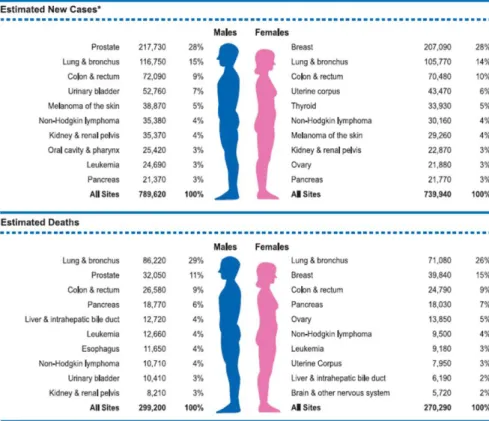
Breast cancer alone is expected to account for 28% (207,090) of all new cancer cases
among women, more than 1 in 4 women (Figure 1) (Jemal et al., 2010).
Figure 1: Ten Leading Cancer Types for the Estimated New Cancer Cases and Deaths by Sex, 2010. *Excludes basal and squamous cell skin cancers and in situ carcinoma except urinary bladder. Estimates are rounded to the nearest 10.
2
The decrease in breast cancer incidence, and in particular mortality, has been
attributed to the combination of early detection with screening programmes, breast
cancer prevention interventions, a decrease in the use of post-menopausal
hormone-replacement therapy and the advent of more efficacious adjuvant systemic therapy
(Jemal et al., 2007). Continued advances in our understanding of the molecular
biology of breast cancer progression have aided in the discovery of novel
pathway-specific targeted therapeutics, and the emergence of such effective therapeutics is currently driving the „patient-tailored‟ treatment planning. Knowledge gained from studying the molecular pathology of human breast cancer progression, integration
and implementation of this knowledge in the clinical setting, promises to further
reduce breast cancer morbidity and mortality.
1.2. EPIDEMIOLOGY AND RISK FACTORS
The cause of breast cancer is still relatively unknown, although researchers have
accumulated a considerable amount of information on the factors, which may
increase one's risk of developing the disease. Today, the disease, like all other forms
of cancer, is considered to be the end result of many factors, both environmental and
hereditary. These factors include gender, age, family history of the disease especially
if there are first degree relatives affected, age at menarche and at menopause, number
of full term pregnancies, the use of both oral contraceptives and hormone therapies
and mutation in specific genes. Also the industrialization accompanied by
3
1.2.1. Gender
Breast cancer is predominantly a disease that occurs in women even if, in rare
circumstances, it can develop in men. In fact, approximately one out of every 150
breast cancer cases occurs in male. It seems likely that estrogens have some role in
the development of breast cancer; in fact the difference in incidence may be because
estradiol is able to exert a direct biological effect on breast cells in females, whereas
in males testosterone needs to be converted to estradiol before exerting any biologic
effect (Endogenous Hormones and Breast Cancer Collaborative Group, 2002).
1.2.2. Age
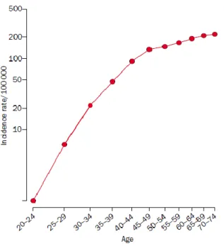
The incidence of breast cancer, in the reproductive years, increases rapidly with age
then increases at a slower rate after about the age of 50, which is average age at
menopause (Figure 2).
Figure 2. Age-incidence curve of breast cancer; log-log plot (from data for England and Wales 1983–87).
4
Younger women are not generally considered to be at risk for breast cancer: only 7%
of all breast cancer cases occur in women under 40 years old, even if these women
tend to have more aggressive breast cancers than older women, which may explain
why often survival rates are lower among younger women. The incidence rates
increased up to 10-fold by the age of 40 (Hulka and Moorman, 2001).
1.2.3. Effects of migration and geographical factors
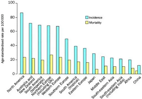
Among populations around the world the incidence and the mortality of breast cancer
vary greatly, also five-fold (Figure 3). In most of more developed countries the rates
are high while in less developed countries and in Japan they are low, probably
because of differences in reproductive factors. Among the migrants, the rates of
those who migrate from countries with low incidence to countries with high
incidence take on the higher rates of the new host country (Buell, 1973).
Figure 3. Worldwide variation in breast cancer rates (data from International Agency for Research on Cancer 1990).
5
1.2.4. Reproductive factors
Menarche and the menstrual cycle
At menarche a woman's body undergoes changes in order to accommodate the
monthly cycling of sex steroid hormones and to prepare the body for childbearing.
The age at menarche is inversely related to the risk for development of breast cancer
(women who begin menstruating before age 13 years, have a two-fold increased risk
of cancer). Some researchers have suggested that certain characteristics of the
menstrual cycle, such as the time it takes for regular menstrual cycles, the length of
menstrual cycles and the age at which these cycles begin, may increase the likelihood
of developing breast cancer (Butler et al., 2000): for example, a short menstrual cycle
of less than 28 days confers a greater risk of breast cancer than longer cycles of 28
days (Whelan et al., 1994). This is because women who have short menstrual cycles,
would have more cycles throughout a year, and have more time spent in the luteal
phase of the menstrual cycle and therefore an increase in time spent on cell
proliferation. Moreover, if fertilization does not occur, there could also be effects on
apoptosis that would occur more frequently determining a major cancer risk.
Pregnancy, breastfeeding and abort
Pregnancy and related factors, such as the age at first full term pregnancy, the
number of full term births, interruptions in pregnancy (such as abortions) and
breastfeeding have opposite influences on the risk of developing breast cancer.
Childbearing seems to have a dual effect on risk of breast cancer: it is increased in
the period immediately after a birth, but this excess risk gradually diminishes and, in
the longer term, the effect of a birth is to protect against the disease (Beral and
6
women who have had at least one full-term pregnancy have, on average, around a
25% reduction in breast-cancer risk. (Layde et al., 1989). The age at first full term
pregnancy is related to breast cancer risk. The reason is that the pregnancy induces
changes in the hormonal profile and these changes could result in alterations in the
tissues that are under hormonal control. This renders the breast tissue less susceptible
to carcinogenic stimuli and thus protects from cancer induction (Lambe et al., 1994).
Furthermore, the protection rises with increasing of full-pregnancies number (Layde
et al., 1989).
About the effect of breastfeeding, recent studies in less developed countries, in which
the total duration of breastfeeding can be much longer, have reported substantial
protective effects (women who had breastfed for a total of 25 months had a 33%
lower risk of breast cancer than those who had never breastfed) (Layde et al., 1898).
Regarding the incomplete pregnancies, arising from spontaneous or induced
abortions, the risk of breast cancer may be increased because the birth does not go to
term, and would no longer have a protective effect. During pregnancy there is the
interplay between prolactin, estrogen and progesterone which all act to promote
breast growth and differentiation. If the pregnancy is interrupted, the growth and
differentiation would also be incomplete and the undifferentiated structures of breast
would render the breast susceptible to carcinogenesis (Russo and Russo, 1980).
Menopause
In the breast of postmenopausal women the cellular proliferation tends to be less than
that of premenopausal women and this reduction of proliferation rate may be due to
the decline of plasma estrogen concentrations during the menstrual cycle. The age at
7
menopause at a late age have a higher risk of breast cancer than those who cease
menstruating earlier (Collaborative Group on Hormonal Factors in Breast Cancer,
1997).
A combination of early age at menarche and a late age at menopause would therefore
prolong the time of the menstrual cycling of sex hormones, and thus would
substantially increase a woman's risk of breast cancer development (Rosner et al.,
1994).
1.2.5. Hormone therapies
Hormone therapies are used throughout a woman's reproductive life and decline of
reproductive years, to combat a variety of ailments. They include oral contraceptives
and hormones for menopausal women.
Oral contraceptives
The use of combined oral contraceptives increases the risk of breast cancer of around
25%, and the risk falls after cessation of use (10 or more years after use stops, no
significant increase in risk is evident); risk does not vary significantly with duration
of use, with the effect of combined oral contraceptives or with the type of estrogen or
progestagen used. Women with several years of oral contraceptive use before age 25
and/or before the first full-term pregnancy, women who use oral contraceptives at
age 45 or older, women with early menarche and women with a family history of
breast cancer have an increased risk of breast cancer (Vessey et al., 1989).
Hormonal therapy for the menopause
Hormone replacement therapies (HRTs) are routinely prescribed for menopausal
8
associated with postmenopausal osteoporosis. Their use determines a higher risk of
breast cancer than that of women who have never used these therapies and this risk
increases with increasing duration of HRT use (Magnusson et al., 1999).
1.2.6. Breast tissue composition
Breast density reflects variations in breast tissue composition and can be strongly
associated with breast cancer risk. Breast density is assessed by mammography and
expressed as the percentage of the breast that is occupied by radiologically dense
tissue. Researchers found that a major extension of mammographic density percent
was associated with an increased risk of breast cancer (McCormack and dos Santos
Silva, 2006). For many women, breast density will change with age or be related to
factors such as relative body mass index, age at first childbirth, postmenopausal
hormone replacement use and/or genetic make-up.
1.2.7. Alcohol and smoking
Observational studies have repeatedly shown that alcohol consumption is associated
with only a moderate increase in the risk of breast cancer, although it depends on the
amount and on the type of alcohol taken (Rohan and Bain, 1987). It has been
suggested that alcohol may induce changes in the liver, which in turn may affect
estrogen metabolism or may affect the level of steroid binding globulins, or for the
increased secretion of pituitary stimulated hormones, such as prolactin and thyroid
stimulating hormone, which would increase mitotic activity in target tissues, and
hence lead to an increased susceptibility to malignancy. Another hypothesis is that
9
increased estrogen levels in premenopausal and postmenopausal women (Ginsburg et
al., 1959).
Carcinogens found in tobacco smoke pass through the alveolar membrane and into
the blood stream, by means of which they may be transported to the breast via
plasma lipoproteins. Due to the fact that they are lipophilic, tobacco-related
carcinogens can be stored in breast adipose tissue and then metabolized and activated
by human mammary epithelial cells (MacNicoll et al., 1980). As is well known,
tobacco smoke contains potential human breast carcinogens (including PAHs,
aromatic amines, and N-nitrosamines); in fact an higher prevalence of
smoking-specific DNA adducts and p53 gene mutations were found in the breast tissue of
smokers compared with that in nonsmokers, supporting the biological plausibility of
a positive association between cigarette smoking and breast cancer risk, depending
by dose and duration (Palmer and Rosenberg, 1993).
1.2.8. Diet
Foods may have several effects on the breast cancer risk. It has been demonstrated
that aliments rich in omega-3 fatty acids, such as fish, suppress mammary tumour
growth by blocking the tumour promoting properties of carcinogens or by inhibiting
prostaglandin synthesis. Conversely, foods rich in omega-6 fatty acids, such as oil,
are thought to stimulate mammary tumour growth. Both saturated and unsaturated
fats are thought to act during the promotional stages of carcinogenesis and this
promotion is largely dependent on the amounts and sources of fat in the diet.
A link between red meat consumption and risk for breast cancer have been reported
10
fibre, vegetables and breast cancer risk have been reported in several case-control
studies because they are important sources of antioxidants, which may help protect
against the tissue damage linked to increased cancer risk (Fund WCRL, 1997).
Antioxidants include vitamin C, vitamin E, and Vitamin A such as carotenoids.
Regarding to caffeine, in a prospective studies, it has not been seen correlation
between caffeine intake and breast cancer risk (Vatten et al., 1990).
1.2.9. Height, weight and exercise
Adult height shows a positive association with breast cancer risk. Average height is
substantially greater in populations with high rates of breast cancer than in
populations with low rates. Within populations, a 10 cm greater height is typically
associated with an increase in risk of about 10%. (Hunter and Willett, 1993).
Probably because height is positively correlated with energy during growth and with
early menarche, and it might be a marker for the number of susceptible breast cells.
In postmenopausal women, obesity increases the risk of breast cancer; risk is about
50% higher in obese women (body-mass index >30 kg/m2) than in lean women (body
mass index 20 kg/m2) and this association is not observed in premenopausal women
(Hunter and Willett, 1993). Several studies have reported that moderate physical
activity is associated with a lower risk of breast cancer. The size of the effect of high
physical activity has varied widely between studies, but a typical result is a reduction
in risk of around 30% in association with a few hours per week of vigorous activity
11
1.2.10. Family history and genetic factors
Environmental and lifestyle factors rather than inherited genetic factors account for
most cases of breast cancer, even if most women with the disease do not have a
family history of it, and most women with affected relatives never develop breast
cancer.
Family history
The evidence for genetic predisposition to breast cancer derives originally from
observations of cancer clustering in families and cancer risk increasing in individuals
with some genetically determined syndromes.
Most studies on familial risk of breast cancer have found about two-fold relative
risks for first-degree relatives (mothers, sisters, daughters) of affected patients
(Pharoah et al., 1997). About 13% of all patients have a first-degree relative with
breast cancer. A significant increased in breast cancer risk has been observed even in
second (grandmothers, aunts, grand-daughters) to fifth degree (Amundadottir et al.,
2004).
High-risk mutations
About 5-10% of all breast cancers are caused by germ-line mutations in well-identified breast cancer susceptibility genes (inherited from one‟s mother or father). So far at least five germ line mutations that predispose to breast cancer have been
identified. These include mutations in the genes BRCA1, BRCA2, TP53, PTEN, and
ATM. Mutations in BRCA1 and BRCA2 can cause high risks of breast cancer because
they are tumor suppressor genes and their inactivation causes genetic defects and
genetic instability. Germ line mutations in TP53 predispose to the Li-Fraumeni
early-12
onset breast cancer) and those in PTEN are responsible for Cowden disease (of which
breast cancer is a major feature). High-risk alleles probably account for most of the
families with four or more breast cancer cases, for around 20–25% of the familial
breast cancer risk overall, and for around 5% of all breast cancers (Easton, 1999).
The ATM (ataxia telangiectasia mutate) gene control cell cycle and mutations of this
gene are closely linked to a childhood disorder of the nervous system called Ataxia
Telangiectasia and to breast cancer susceptibility.
1.3. DISEASE ONSET AND PROGRESSION
Breast cancer is a group of related conditions, characterized by differing microscopic
appearance and biologic behavior, in which the cells of the breast escape the normal
replication, growing and dividing rapidly and uncontrollably (Coe and Steadman,
1995). It is believed that this capacity of evade from the replication cycle involves
the accumulation of mutations, usually in genes that regulate cell division and the
accurate replication of DNA (Davis and Bradlow, 1995). Also hormones and other
substances located in close proximity of the cell can stimulate abnormal cell
multiplication. There are many models of human breast cancer evolution.
Cytogenetic and molecular genetics analysis have revealed that the development of a
primary breast carcinoma derives from a multistep process involving initiating or
promoting factors characterized by the accumulation of various genetic alterations
which may invoke a transformation of normal cells into malignant cells (Beckmann
et al., 1997)
One of the most well-established models, published by Wellings and Jensen over 30
13
normal terminal duct lobular unit (TDLU), the basic histopathologic and
physiologic unit of breast, and there is an apparently continuous but non-obligatory
progression from TDLUs to cancers through a series of increasingly abnormal stages
over long periods of time also decades in most cases (Figure 4) (Wellings and
Jensen, 1973).
Figure 4. Revised Wellings and Jensen model of human breast cancer evolution. The original Wellings and Jensen model proposed an apparently continuous but non-obligatory linear progression from normal TDLU to IBC through a series of increasingly abnormal stages over long periods of time.
The key stages in this progression, in today‟s terminology, are called: ◊ hyperplastic enlarged lobular units (HELU);
◊ atypical ductal hyperplasia (ADH);
◊ ductal carcinoma in situ (DCIS) or lobular carcinoma in situ (LCIS) so called when the tumor remain confined within the basement membrane of the duct or lobule
14
If the breast cancer remains within the basement membrane and does not invade
surrounding tissue or metastasize to distant organs it is said to be in situ
(non-invasive)
◊ invasive breast cancer (IBC) when the tumor increases in size and the invade (or infiltrate) the normal adjacent tissue (Allred et al., 2004). When the cancer cells
break away from the site of origin and penetrate the basement membrane of the
epithelium, they enter the bloodstream or lymphatics located in connective tissue and
may metastasize to distant organs and form secondary tumors. The major route of
metastases via the lymphatic system is through the axillary nodes. Hence, the tumor
extends into the central lymphatic terminus and the cancer cells enter into the venous
stream. These cells can then be carried through the heart to lungs. Tumor fragments
that may break loose from pulmonary vein are then carried off, back to heart and
enter the bloodstream. Organs with a rich blood supply, such as the liver, spleen,
adrenals and bone, are the targets for blood-bone metastases (Lu and Kang, 2007).
.
Several characteristics distinguish the breast cancer types. The transition from TDLU
to HELU is characterized by increased growth due to epithelial hyperplasia.
Alterations of cell adhesion and polarity distinguish ADH from HELU as the
hyperplastic epithelium begins to pile up and distends acini. DCIS is characterized by
further expansion of tumor volume, intraductal spread into other areas of the breast,
and, most importantly, the appearance of increased histologic and biological
diversity compared with earlier precursors. Invasion into surrounding stroma defines
the transition of DCIS to IBC. Evidences support that most high-grade DCIS
15
random accumulation of genetic defects, which are propagated to IBC in a manner
that is largely independent of progression to invasion. Since the DCIS are the
precursor of nearly all ductal IBCs (which account for 85-90% of all IBCs), then
ADH is probably also a risk factor for the development of DCIS independent of its
histologic and biological characteristics (Allred et al., 2008).
Since the major breast cancers evolve from precursors, identifying of biological
alterations associated with early precursors, before the cancer development, may
reveal strategies for the prevention of the majority of cancers or treated them early.
1.4. CLASSIFICATION AND CLINICAL PATHOLOGY
When cancer is present, a number of tests are performed to assess the behavior of the
cancer, and to determine the most effective treatments.
Prognosis is defined according to several parameters: tumor size and grade, the
presence/ absence of estrogen (ER) and/or progesterone (PR) receptors, HER2/neu
(HER2, c-erbB2) protein, lymph node metastases and vascular or perineural tumor
invasion. Other parameters, such as the proliferating index, the presence of p53,
BRCA1 and 2 or EGFR alterations, may also be useful for prognostic evaluation or
as predicting therapeutic response.
The TNM Classification of Malignant Tumors (TNM) is a cancer staging system for
all solid tumors that describes the extent of cancer in a patient‟s body. It was devised
by Pierre Denoix between 1943 and 1952 using the size and extension of the primary
tumor, its lymphatic involvement, and the presence of metastases to classify the
16
● T (range from 1 to 4) describes the size of the primary tumour and whether it has invaded nearby tissue:
o T1: No evidence of primary tumour
o T2: Tumor 2 cm or less
o T3: Tumour more than 5cm
o T4: Tumour of any size with extension to adjacent tissue
● N (range from 0 to 3) describes regional lymph nodes that are involved and the
degree of spread:
o N0: tumor cells absent from regional lymph nodes
o N1: regional lymph node metastasis present; (at some sites: tumor
spread to closest or small number of regional lymph nodes)
o N2: tumor spread to an extent between N1 and N3 (N2 is not used at
all sites)
o N3: tumor spread to more distant or numerous regional lymph nodes
(N3 is not used at all sites);
● M (0-1) represents the presence of metastasis (spread of cancer from one body
part to another).
o M0: no distant metastasis
o M1: metastasis to distant organs (beyond regional lymph nodes)
1.4.1. Tumour Stage
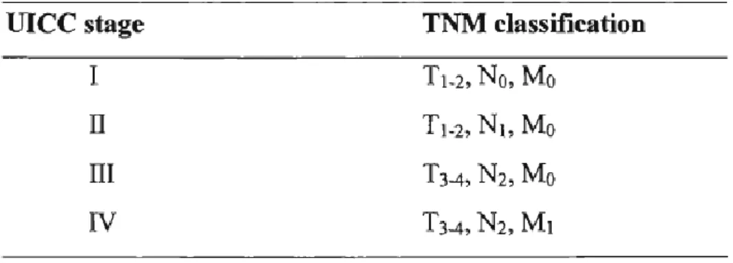
Once a TNM classification is available for a tumour, the tumour is then classified
17
Table 1. The correlation of the tumour, nodes, metastases (TNM) system and the Unio Internationale Contra Cancrum (UICC) system of classification for tumours.
Survival from breast cancer is largely dependent on the stage at presentation, and the
prescription of appropriate treatment is based on stage.
1.4.2. Tumour Grade
On microscopic examination, a tumour can be graded according to the degree of
differentiation of the tumour from adjacent "normal" cells. The most common
grading system used by pathologists is the Scarff, Bloom, and Richardson (SBR)
classification and is usually used as a preference to tumour staging.
Tumour Grade Definition:
◊ Tumor grade 1: tumor well-differentiated ◊ Tumor grade 2: tumor moderately-differentiated ◊ Tumor grade 3: tumor poorly-differentiated
Grade 1 tumors are small, round, have regular nuclei and very few mitoses.
18
Survival studies show that grade 1 tumors have a good prognosis, and thus a good
response to treatment, whilst grade 3 tumors would have a poor prognosis and the
response to treatment would be less successful (Elledge and McGufre, 1993).
1.4.3. Tumour size
The size of the primary tumour and the involvement of axillary nodes (which,
combined, constitute the stage of the disease) in cancer development, are the most
important indicators of prognosis. A good prognosis is associated with a small
tumour (less than 1cm in diameter); whilst a poor prognosis accompanies a large
tumour (a diameter greater than 5cm) (Stockdale, 19889. Results from the SEER
program (Surveillance, Epidemiology and End Results program of the National
Cancer Institute) suggest that if tumors are less than 1cm in diameter and have not
progressed from the initial site of development, then there is a relatively high chance
of survival, after 5 years, from the time of primary diagnosis, in comparison, tumors
of greater than 5 cm in diameter, have an 82% chance of survival after 5 years from
the initial time of diagnosis (Carter. Et al., 1989).
1.4.4. Estrogen and progesterone receptors
Hormone receptor assays are considered to be essential tools for the assessment,
prognosis and treatment of breast cancer. Approximately 50 to 85% of breast cancers
cells contain receptors that specifically bind estrogen and progesterone.
Estrogen receptors (ER) and progesterone receptors (PR) are present in higher
concentrations in breast cancer tissue than in "normal" breast tissue, and are thus
19
are estrogen receptor-positive (ER-positive, or ER+). About 65% of ER-positive
breast cancers are also progesterone receptor-positive (PR-positive, or PR+). Cells
that have receptors for one of these hormones, or both of them, are considered
hormone receptor-positive. Patients with breast cancers that are shown to be ER
positive, respond favorably to hormone treatments such as tamoxifen, in
approximately 60-65% of cases. On the contrary, patients with negative ER assays
have a less than 10% response rate to hormone therapy (Stockdale, 1988). Therefore
a high concentration of these receptors is highly predictive of the response hormonal
therapy.
1.4.5. Proliferation index
The proliferation index is a measure of the number of cells in a tumor that are
dividing, and thus proliferating. Cell proliferation can reasonably be supposed to be
related to tumor aggressiveness. Proliferative activity can be determined using
various methods based on different rationales:
Ki-67 protein is an indicator strictly associated with cell proliferation. During
interphase, the Ki-67 antigen can be detected only within the nucleus of cells, while
in mitosis the majority of the protein is relocated to the surface of chromosomes. The
Ki-67 protein is present during all active phases of cell cycle (G1, S, G2 and M) but
it is absent from resting cells (G0). Ki-67 is an excellent indicator to determine the
fraction of development given population of cells. The fraction of Ki-67 positive
tumor cells (Ki-67 labeling index) is often correlated with the clinical course of
20
The mitotic index (MI) is the fraction of cells in mitosis at any given time. It
consists in counting the number of mitotic figure on a constant sample of cells (1000
or 10000) per mm2 of epithelium. Mitotic activity is currently used mainly as part of
the tumor grading system, for women with infiltrating breast carcinoma. Several
studies have indicated that mitotic activity is an important imprint of tumor evolution
as it exerts a determining influence on long-term clinical outcome, regardless of type
of treatment, but also they suggested that mitotic activity does not provide predictive
information on response to systemic therapy (Medri et al, 2003).
The thymidine labeling index (TLI) is a method, which involves the incubation of
fresh tissue with tritium-labeled thymidine, provides an estimate of the fraction of
tumor cells that are in the S (DNA synthesis) phase of the cell cycle. Because DNA
synthesis is an integral part of each cell division cycle, TLI gives an indication of the
amount of proliferation taking place in a tumor and it is a strong independent
predictor of survival and relapse-free survival.
Both Ki-67 and TLI are high in cancers with high nuclear and histologic grade and
are higher in cancers from premenopausal women than in those from postmenopausal
women (Gentili et al., 1981; McGurrin et al, 1987); tumors with high TLI or Ki-67
are frequently estrogen receptor negative (Gerdes et al, 1987).
1.4.6. HER2-neu
HER2/neu (Human Epidermal growth factor Receptor 2, also known as ErbB-2) is a
member of the ErbB protein family, more commonly known as the epidermal growth
factor receptor family, and it is encoded by the ERBB2 gene. It is a cell membrane
21
transduction pathway leading to cell growth. In breast cancer approximately 30%
have an amplification of HER2/neu gene or overexpression of its protein product,
giving higher aggressiveness, increased disease recurrence and worse prognosis of
breast cancer patients.
1.4.7. p53
p53 is a tumor suppressor protein that regulates the cell cycle and plays a role in
genetic stability and inhibition of angiogenesis; it exerts its anti-cancer role through
several mechanisms (activates DNA repair proteins, induces growth arrest and
initiates apoptosis). More than 50% of human tumors contain mutations or deletions
of the TP53 gene. While the prognostic and predictive value of p53 is still matter of
debate, there is an increased interest for p53-based therapies.
1.4.8. BRCA1 and BRCA2
BRCA1 and BRCA2 are two tumor suppressor genes with several functions such as
repair DNA double-strand breaks, protein ubiquitylation and cell cycle checkpoint
control. Germ line mutations of these two genes confer strong lifetime risks of breast
cancer and the risks are influenced by the position of mutation within the gene
sequence (Easton, 1997). Researchers have identified hundreds of mutations in the
BRCA1 and BRCA2 genes, many of which are associated with an increased risk of
cancer. Women with a family history of breast cancer are screened for mutations in
22
1.4.9. EGFR
The Epidermal Growth Factor Receptor (EGFR) is a cell-surface receptor for
members of epidermal growth factor family (EGF-family) of extracellular protein
ligands. The binding by ligands activates EGFR dimerization and stimulates intrinsic
intracellular protein-tyrosine kinase activity. The downstream signaling proteins
initiate several signal transduction cascades, principally MAPK, Akt and JNK
pathways leading to DNA synthesis and cell proliferation.
The expression of EGFR in models of breast cancer is associated with increased
proliferation and resistance to apoptosis and with poorer prognosis. Mutations that
lead to EGFR overexpression or over-activity have been associated with breast
cancer: it is overespressed in 35-60% of breast cancers.
1.5. TYPES AND SUBTYPES
The normal female adult breast consists of a mixture of epithelial and stromal
elements. The epithelial elements of the breast contain a series of branching ducts,
which extends from the nipple, and terminates into the functional units of the breast,
the lobules (DiSaia, 1993). Each breast is composed of 15-20 lobules, containing a
cluster of alveoli, which are responsible for the secretion of milk during lactation.
The stroma contains variable amounts of interspersed adipose tissue and fibrous
connective tissue, which constitutes most of the breast volume in a non-lactational
state (Carola et al., 1992; DiSaia, 1993).
The two most common types of breast cancer are named after the parts of the breast
23
Figure 5. Anatomy of breast
● Ductal Carcinoma in situ (DCIS): it is the most common type of non invasive
breast cancer, in fact between 85% and 90% of all breast cancers are ductal. It starts
inside the milk ducts, beneath the nipple and areola and it is well contained, hasn‟t
spread beyond the milk duct into any normal surrounding breast tissue, and it can be
very successfully treated. The DCIS cancers have a higher risk for recurrence (most
recurrences happen within the 5 to 10 years after initial diagnosis and the chances of
a recurrence are under 30%) and for developing a new breast cancer.
● Lobular Carcinoma: about 8% of breast cancers are lobular. LCIS begins in the
lobes, or glands which produce milk in the breast and the cancer is limited within the
lobe and has not spread to surrounding tissues. Despite the fact that its name includes
the term “carcinoma,” LCIS is not a true breast cancer. Rather, LCIS is an indication
that a person is at higher-than-average risk for getting breast cancer at some point in
the future. LCIS is usually diagnosed often between the ages of 40 and 50.
These two cancer types are usually removed during a lumpectomy if the tumor
24
cancer has broken into nearby breast tissue (invasive cancer) then a mastectomy may
be needed, and also chemotherapy.
Second most common is a group of breast cancers that invade nearby tissue:
● Invasive (Infiltrating) Breast Cancer has the potential to spread out of the
original tumor site and to invade other parts of your breast, the lymph nodes and
other areas of the body. There are several types and subtypes of invasive breast
cancer such as invasive ductal carcinoma and invasive lobular carcinoma. The
treatments fall into two broad categories: local (surgery and radiation) or systemic
(chemotherapy, hormonal and target therapy).
Other breast cancer types are:
● Inflammatory Breast Cancer: is the least common (1-5% of all breast cancer),
but most aggressive of breast cancers, taking the form of sheets or nests, instead of
lumps. It can start in the soft tissues of the breast, just under the skin, or it can appear
in the skin. Unlike ductal and lobular cancers, it is treated first with chemotherapy
and then with surgery. When caught early, inflammatory breast cancer can be a
manageable disease, and survival rates are increasing.
● Paget's disease of the nipple/areola is a rare form of breast cancer, often looks
like a skin rash, or rough. The itching and scabs are signs that cancer may be under
the surface of the skin, and is breaking through. The cancer usually affects the ducts
of the nipple first (small milk-carrying tubes), then spreads to the nipple surface and
the areola. The disease usually develops after age 50 and is usually treated with a
mastectomy, because the cancer has by then invaded the nipple, areola, and the milk
25
● Rare types of breast cancer include:
- Medullary breast cancer (5%)
- Mucinous (mucoid or colloid) breast cancer (2%) - Tubular breast cancer (1%)
- Adenoid cystic carcinoma of the breast (1%)
- Metaplastic breast cancer (is a mixture of two cell types; 1%)
Human breast cancer is a heterogeneous disease, encompassing a number of distinct
biological entities that are associated with specific morphological and
immunohistochemical features and clinical behavior and, therefore, no golden
standard therapy exists suitable for all tumors of the mammary gland (Lacroix et al.,
2004). For many decades, breast carcinomas were only classified according to
histological type, grade, and expression of hormone receptors as described above.
However, this classification proved to be limiting for it was unable to define
subgroups sharing similar prognostic and therapeutic aspects. A more recent
approach to classify breast cancer subgroups is gene expression profiling, based on
cDNA microarrays (Care et al., 2006; Sorlie et al., 2001), which suggests the
presence of multiple molecular subtypes of breast cancer. Based on transcriptomic similarity, breast carcinomas can be distinguished into five “intrinsic” main distinct subtypes:
- Luminal A (ER positive, and/or PR positive, HER2 negative)
- Luminal B (ER positive and/or PR positive, HER2 positive)
- Triple negative (or also basal like) (ER negative, PR negative, HER2 negative)
- HER2 positive (ER negative, PR negative, HER2 positive)
26
Known as the „intrinsic subtypes of breast cancer‟, these groups of tumors have revealed critical differences in incidence (Millikan et al., 2008), survival (Cheang et
al., 2009; Hu et al., 2006), and response to treatment (Prat et al., 2010; Nielsen et al.,
2010). For example, luminal tumors have been associated with the most favorable
prognoses, while HER2-overespressing and triple-negative have been associated with
the worst prognoses.
1.5.1 TRIPLE-NEGATIVE BREAST CANCER
Triple-negative breast cancers (TNBC) account for 10–17% of all breast carcinomas
(Reis-Filho and Tutt, 2008) are reported to be more commonly seen in younger
women, often in pre-menopausal women (<50 years), of African-American and
Hispanic ethnicity (Morris et al., 2007), with BRCA1 mutations (Dent et al., 2007),
an increased body weight (Trivers et al., 2009). It have been characterized by several
aggressive clinicopathologic features including higher mean tumor size, higher
histologic grade tumors, elevated mitotic count, ductal or mixed histology, and, in
some cases, a higher rate of node positivity (Dent et al., 2007; Irvin and Carey,
2008). TNBC have a worse prognosis than the other breast cancer subtypes, high
recurrence, occurring within three years of diagnosis and mortality rates are
increased for five years after diagnosis, and development of recurrence and distant
metastasis with a specific metastatic pattern (meninges, brain, liver and lung) (Rakha
et al., 2007). Due to the absence of hormone receptors and HER2 expression, these
tumors cannot take advantage from the endocrine therapy or trastuzumab treatment,
chemotherapy remaining the only potential adjuvant therapeutic approach. As far as
27
objective response to neoadjuvant chemotherapy than other tumor types (Reis-Filho
and Tutt, 2008), thus suggesting that biological features present more frequently in
this group are responsible for the increased sensitivity to chemotherapy. In general,
adjuvant therapeutic options for TNBC can be divided into two groups: cytotoxic
agents (as anthracycline agents or platinum-containing agent) and targeted therapies
(as PARP1 and EGFR or VEGF inhibitors). Although triple-negative cancers are
report to have excellent response rates to neoadjuvant chemotherapy (Rouzier et al.,
2005), survival of patients with such tumors is still poor and their management may
therefore require a more aggressive alternative intervention and it remains an urgent
need to understand the molecular and biological features of these tumors in order to
develop novel therapeutic strategies to improve their clinical outcome.
1.6. THERAPY
The mainstay of breast cancer is surgery when the tumor is localized, followed by
chemotherapy, radiotherapy and hormonal therapy for ER positive tumor, depending
on clinical criteria. Treatments are given with increasing aggressiveness according to
the prognosis and risk of recurrence.
1.6.1 Surgery
Surgery is usually the first line of attack against breast cancer. Some of the lymph
nodes under the arm are usually taken out and looked at under a microscope to see if
they contain cancer cells. Several types of surgery exist to remove breast cancer.
Breast-conserving surgery, an operation to remove only the cancer but not the breast
28
Lumpectomy: Surgery to remove a tumour (lump) and a small amount of normal
tissue around it.
Partial mastectomy: Surgery to remove the part of the breast that has cancer and
some normal tissue around it.
Other types of surgery include the following:
Total mastectomy: Surgery to remove the whole breast that has cancer. Some of
the lymph nodes under the arm may be removed for biopsy.
Modified radical mastectomy: Surgery to remove the whole breast that has
cancer, many of the lymph nodes under the arm, the lining over the chest
muscles, and sometimes, part of the chest wall muscles.
Radical mastectomy: Surgery to remove the breast that has cancer, chest wall
muscles under the breast, and all of the lymph nodes under the arm
Radiation therapy
Radiation therapy is a cancer treatment that uses high-energy x-rays or other types of
radiation (gamma rays). This radiation is very effective in killing cancer cells that
may remain after surgery or recur where the tumor was removed.
There are two types of radiation therapy. External radiation therapy uses a machine
outside the body to send radiation toward the cancer. Internal radiation therapy (or
brachytherapy) uses a radioactive substance sealed in needles, seeds, wires, or
catheters that are placed directly into or near the cancer. The way the radiation
therapy is given depends on the type and stage of the cancer being treated. Although
radiation therapy can reduce the chance of breast cancer recurrence, it is much less
29
United States' National Cancer Institute, none of them found a survival benefit for
radiation therapy (Porter et al., 1993).
Chemotherapy
Chemotherapy is a cancer treatment that uses drugs to stop the growth of cancer
cells. The mechanism of action of chemotherapy is to destroy fast growing or fast
replicating cancer cells either by causing DNA damage upon replication or other
mechanisms; these drugs also damage fast-growing normal cells where they cause
serious side effects. Chemotherapy is used to treat: early-stage invasive breast cancer
to get rid of any cancer cells that may be left behind after surgery and to reduce the
risk of the cancer coming back; advanced-stage breast cancer to destroy or damage
the cancer cells as much as possible. In some cases, chemotherapy is given before
surgery to shrink the cancer.
When chemotherapy is taken by mouth or injected into a vein or muscle, the drugs
enter the bloodstream and can reach cancer cells throughout the body (systemic
chemotherapy). When chemotherapy is placed directly into the cerebrospinal fluid,
an organ, or a body cavity such as the abdomen, the drugs mainly affect cancer cells
in those areas (regional chemotherapy). The way the chemotherapy is given depends
on the type and stage of the cancer being treated. Some protocols call for a cycle of
treatment every three weeks; others may be more frequent.
It predominately is used for stage 2-4 disease, but may also be used to treat types of
early-stage breast cancer. Many different types of chemotherapy drugs are used to
30
One of the most common treatments is cyclophosphamide plus doxorubicin
(Adriamycin), known as AC. Sometimes a taxane drug, such as docetaxel, is added,
and the regime is then known as CAT; taxane attacks the microtubules in cancer
cells. Another common treatment, which produces equivalent results, is
cyclophosphamide, methotrexate, and fluorouracil, known as CMF.
Hormone therapy
Hormones are substances produced by glands in the body and circulated in the
bloodstream. Some hormones can cause certain cancers to grow. Hormonal therapy
medicines treat hormone-receptor-positive breast cancers in two ways: by lowering
the amount of the hormone estrogen in the body or by blocking the action of estrogen
on breast cancer cells, stopping their growth.
If tests show that the cancer cells have places where hormones can attach (receptors),
drugs, surgery, or radiation therapy are used to reduce the production of hormones or
block them from working. The hormone estrogen, which makes some breast cancers
grow, is made mainly by the ovaries. Treatment to stop the ovaries from making
estrogen is called ovarian ablation.
Hormone therapy with tamoxifen is often given to patients with early stages of breast
cancer and those with metastatic breast cancer. Hormone therapy with tamoxifen or
estrogens can act on cells all over the body and may increase the chance of
developing endometrial cancer. Hormone therapy with an aromatase inhibitor is
given to some postmenopausal women who have hormone-dependent breast cancer.
Hormone-dependent breast cancer needs the hormone estrogen to grow. Aromatase
31
turning androgen into estrogen. For the treatment of early stage breast cancer, certain
aromatase inhibitors may be used as adjuvant therapy instead of tamoxifen.
Targeted therapy
Targeted therapy is a type of treatment that uses drugs or other substances to identify
and attack specific cancer cells without harming normal cells. Monoclonal antibodies
and tyrosine kinase inhibitors are two types of targeted therapies used in the
treatment of breast cancer.
Monoclonal antibody therapy is a cancer treatment that uses antibodies made in the
laboratory, from a single type of immune system cell. These antibodies can identify
substances on cancer cells or normal substances that may help cancer cells grow. The
antibodies attach to the substances and kill the cancer cells, block their growth, or
keep them from spreading. Monoclonal antibodies are given by infusion. They may
be used alone or to carry drugs, toxins, or radioactive material directly to cancer cells
ant they may be used in combination with chemotherapy as adjuvant therapy.
Trastuzumab (Herceptin) is a monoclonal antibody that blocks the effects of the
growth factor protein HER2, which sends growth signals to breast cancer cells.
About one-fourth of patients with breast cancer have tumors that may be treated with
trastuzumab combined with chemotherapy.
Another important monoclonal antibody used for the antiangiogenic therapy is
Bevacizumab that blocks the VEGF receptor protein, which is involved in forming
tumor blood vessels.
Tyrosine kinase inhibitors are targeted therapy drugs that block signals needed for
32
other anticancer drugs as adjuvant therapy. Lapatinib is a tyrosine kinase inhibitor
that blocks the effects of the HER2 protein and other proteins inside tumor cells. It
may be used to treat patients with HER2-positive breast cancer that has progressed
following treatment with trastuzumab.
PARP inhibitors are a type of targeted therapy that block DNA repair and may cause
cancer cells to die. PARP inhibitor therapy is being studied for the treatment of
triple-negative breast cancer.
Stage 1 cancers (and DCIS) have an excellent prognosis and are generally treated
with lumpectomy and sometimes radiation. HER2+ cancers should be treated with
the trastuzumab (Herceptin) regime (Gonzalez-Angulo et al., 2009) chemotherapy is
uncommon for other types of stage 1 cancers. Stage 2 and 3 cancers with a
progressively poorer prognosis and greater risk of recurrence are generally treated
with surgery (lumpectomy or mastectomy with or without lymph node removal),
chemotherapy (plus trastuzumab for HER2+ cancers) and sometimes radiation
(particularly following large cancers, multiple positive nodes or lumpectomy). Stage
4, metastatic cancer, (i.e. spread to distant sites) has poor prognosis and is managed
by various combination of all treatments from surgery, radiation, chemotherapy and
33
2. CHEMOTHERAPY
2.1. FEATURES
Chemotherapy for the treatment of cancer was introduced into the clinic more than
fifty years ago. Chemotherapy refers to antineoplastic drugs or chemical used to treat
cancer. Chemotherapeutic drugs acts by killing cells that divide rapidly, one of the
main properties of most cancer cells. Since malignant cells divide without control or
order, these drugs effectively target cancerous growths. Ideally, chemotherapeutic
drugs should specifically target only neoplastic cells and should decrease tumor
burden by inducing cyto-endotoxic and/or cytostatic effects with minimal “collateral damage” to normal cells. Indeed, chemotherapy inadvertently also harms healthy cells that divide rapidly under normal circumstances: cells in the bone marrow,
digestive tract and hair follicles; this results in the most common side effects of
chemotherapy: myelosuppression (decreased production of blood cells, hence also
immunosuppression), mucositis (inflammation of the lining of the digestive tract),
and alopecia (hair loss).
There are various types of cancer those need different type of drugs that kill cancer
cell in different ways at various phases in the cell cycle. Depending on the type, size,
and location of the cancer, as well as your overall health, there are different strategies
in the administration of chemotherapeutic drugs:
● Neoadjuvant Chemotherapy: refers to the administration of therapeutic agents
prior to the main treatment, that usually it is the surgery. The aim is to reduce the size
or extent of the cancer before employing radical treatment intervention, thus making
procedures easier and more likely to be successful, and reducing the consequences of
34
● Adjuvant chemotherapy: refers to additional treatment, usually given after
primary therapy (surgery or radiotherapy) where all detectable disease has been
removed, but where there remains a statistical risk of relapse due to occult disease.
This treatment strategy permit to kill any remaining cancer cells in the body.
● Palliative chemotherapy: is given to patients who develop metastatic disease
(cancer that spreads throughout the body) which are generally not curable. New
advances in drug therapies, however, can help shrink tumors, prolong survival, and
improve quality of life. Palliative treatments are also used to help relieve cancer-related symptoms, improving the patient‟s quality of life.
First line chemotherapy is treatment with chemotherapeutic drugs that has, through
research studies and clinical trials, been determined to have the best probability of
treating a given cancer. This may also be called “standard therapy”.
Second line chemotherapy: is chemotherapy that is given if a disease has not
responded or reoccurred after first line chemotherapy. In some cases, this may also
be referred to as “salvage therapy”.
Multiple chemotherapeutic agents may be used in combination to treat patients with
breast cancer. Determining the appropriate regimen to use depends on many factors;
such as, the character of the tumor, lymph node status, and the age and health of the
patient. In general, chemotherapy has increasing side effects as the patient's age
35
2.2. CHEMOTHERAPEUTIC DRUGS ANS MECHANISM OF
ACTION
Currently there are many drugs, about a hundred, which can be used in cancer
treatment. The majority of chemotherapeutic drugs can be divided into:
Alkylating agents: are drugs that act directly on DNA, causing cross-linking of
DNA strands, abnormal base pairing, or DNA strand breaks, thus preventing the cell
from dividing. Alkylating agents are generally considered to be cell cycle phase
non-specific, meaning that the kill the cell in various and multiple phases of the cell
cycle. Although alkylating agents may be used for most types of cancer, they are
generally of greatest value in treating slow-growing cancers. Examples of these
drugs are:
- classical alkylating agents, that are drugs with true alkyl groups, which including
three subgroups: nitrogen mustards such as cyclophosphamide and melphalan,
nitrosoureas such as carmustine, and alkyl sulfonates such as busulfan;
- alkylating-like agents that are platinum-based drugs, don‟t have an alkyl group but
nevertheless damage DNA (Cruet-Hennequart et al., 2008) and including cisplatin,
oxaliplatin and carboplatin.
Antimetabolites: are chemical that interfere with the formation or use of a normal
cellular metabolites, interfering with DNA or RNA production and therefore cell
division and the tumor growth. Antimetabolites are cell cycle specific, in fact they
are most effective during S-phase of cell division because they primarily act upon
36
antimetabolites masquerade as a purine or a pyrimidine chemicals which become the
building blocks of DNA and they prevent these substance becoming incorporated in
to DNA during S phase stopping normal development and division. The toxicities
associated with these drugs are seen in cells that are growing and dividing quickly.
Examples of antimetabolites include:
- purine antagonists (act by mimicking the structure of metabolic purines) such as 6-
mercaptopurine;
- pyrimidine antagonists (act by mimicking the pyrimidine structures) such as 5-
fluorouracil, Gemcitabine and Cytarabine;
- folate antagonists (impair the acid folic function) such as Methotrexate.
Methotrexate is one of the most commonly used chemotherapy agents and works on
the S-phase of the cell cycle. It is an analogous of folic acid and acts by inhibiting
dihydrofolate reductase (DHFR) and, therefore, the metabolism of folic acid required
for DNA synthesis and also for RNA and proteins.
5-Fluorouracil (or 5-FU) is a pyrimidine analogous which works through non
competitive inhibition of thymidylate synthase, blocking the synthesis of the
thymidine required for DNA replication, inducing cell cycle arrest.
Anti-tumor antibiotics: have several mechanisms of action to block cell growth, by
interfering with DNA and RNA synthesis, and they work in all phase of the cell
cycle. Example of anti-tumor antibiotics including:
- anthracyclines (act by inhibiting DNA and RNA synthesis by intercalating between
base pairs of DNA/RNA strand preventing the replication of rapidly-growing