UNIVERSITÀ DEGLI STUDI DI ROMA
"TOR VERGATA"
FACOLTA' DI MEDICINA E CHIRURGIA
DOTTORATO DI RICERCA IN MEDICINA PRENATALE
CICLO DEL CORSO DI DOTTORATO
XIXTitolo della tesi
PRODUZIONE SCIENTIFICA DURANTE IL DOTTORATO DI RICERCA IN MEDICINA PRENATALE
Dottorando
GIOVANNI LARCIPRETE
Docente Guida/Tutor: Prof. DOMENICO ARDUINI
Coordinatore: Prof. DOMENICO ARDUINI
INTRODUZIONE
In questo elaborato finale per il Dottorato di Ricerca in Medicina Prenatale ho voluto raccogliere la produzione scientifica che ho portato a termine durante l’intera durata del Dottorato stesso.
Presento qui di seguito tutti i lavori editati a mezzo stampa e prodotti sotto l’egida del Dottorato in oggetto, omettendo gli atti congressuali (ove si eccettuino quelli prodotti su riviste indicizzate).
La produzione scientifica è relativa ai due campi di ricerca perseguiti nel corso degli ultimi 3 anni:
1. Ruolo della plicometria ultrasonografica fetale nello sviluppo e nel miglioramento degli algoritmi di stima del peso fetale. Cambiamenti dei compartimenti corporei fetali in condizioni patologiche.
2. Epidemiologia delle trombofilie in una popolazione ostetrica e correlazione con gli
esiti avversi della gravidanza.
Le pubblicazioni inerenti al primo settore sono 4: la prima, in ordine di tempo, definisce sistematicamente la metodica della psicometria ecografia fetale e presenta le tavole di riferimento biometriche dei parametri plicometrici in gravidanza normale ed in gravidanza affetta da diabete gestazionale. Il lavoro in oggetto è stato pubblicato da Ultrasound Obst/Gyn (UOG) nel 2003. La seconda pubblicazione è del 2005, è stata editata sempre da UOG e mostra le differenze, in termini di tessuto sottocutaneo fetale, tra feti sani e feti con ritardo di crescita sin dall’inizio del terzo trimestre. La terza e la quarta pubblicazione in ambito di plicometria ultrasonografica fetale sono state prodotte su Journal of Obstetrics and Gynaecology Research (Elsevier) entrambe nel 2007, e trattano dell’introduzione dei parametrici plicometrici fetali negli algoritmi di stima del peso fetale. Inoltre in questi due ultimi lavori vengono prodotti due nuovi algoritmi per la stima del peso fetale: una formula matematica ed un modello tabulare a lettura visiva, per la predizione 3
del peso fetale a termine di gravidanza. Uno dei due lavori è in corso di stampa e viene qui presentato come manoscritto con lettera di accettazione da parte degli editori.
Le pubblicazioni inerenti al secondo ambito di ricerca da me seguito in corso di Dottorato riguardano gli studi di associazione tra condizioni di trombofilia ereditaria ed esiti avversi della gravidanza. La prima pubblicazione è del 2007 ed è stata editata su Journal of Obst/Gyn Research e mostra i risultati di uno studio condotto su due gruppi di pazienti gravide, con o senza patologie della gravidanza. In questo set di pazienti si andava a controllare l’associazione tra difetti trombofilici singoli ed esiti avversi. Anche il secondo lavoro è dello stesso tenore, ed è stato pubblicato si International Journal of Biomedical Sciences nel 2007. Anche in quest’ultima fatica si sono studiati i difetti trombofilici singoli, ma con approccio statistico differente.
Due lavori sono tuttora in mano ai referee: il primo rappresenta un ampio studio di associazione tra patologie della gravidanza su base microangiopatica e difetti trombofilici multipli (Am J Obst/Gyn, inviato, non ancora accettato) ed il secondo riguarda l’incidenza di difetti trombofilici in pazienti gravide con danno renale accertato (Acta scand, inviato). Di questi due ultimi lavori non mostro il manoscritto originale, in mancanza delle bozze di stampa.
Sempre in ambito di produzione letteraria, riporto anche alcuni case report pubblicati nello stesso periodo del Dottorato di Ricerca.
Al di la delle mere note bibliografiche, questi tre case report rappresentano un interessante modo di approcciare alla letteratura scientifica. Tutti e tre questi lavori nascono dalla curiosità scaturita dall’osservazione di casi clinici gestiti personalmente nel corso della abituale e routinaria attività clinica assistenziale. La stessa attività di pratica clinica esce dall’ambito routinario se da essa si traggono spunti di studio e di approfondimento e se si desidera portare alla conoscenza degli altri operatori nello stesso settore casi che altrimenti rimarrebbero chiusi nell’oblio di cartelle cliniche accatastate.
E’ recente la pubblicazione (2007) sul Journal of Ultrasound in Meidicne di un raro caso di gemelli torcosternopaghi a tipo Giano bifronte. Nella tesi riporto il lavoro con il ricco corredo iconografico ultrasonografico e post-natale.
E’ poi presente un case report che tratta lo spinoso argomento della responsabilità professionale in ambito ostetrico e l’istituto della procedura riconvenzionale ed un lettera all’editore con note di tecnica di cervicometria ecografia. Entrambi questi ultimi lavori appartengono alla rivista ufficiale della Società Italiana di Ginecologia ed Ostetricia.
Sono persuaso che niente meglio di un lavoro pubblicato possa esprimere i risultati di una osservazione clinica o di un set di pazienti paragonabili per caratteristiche epidemiologiche.
Per questo motivo ho deciso di compilare la mia tesi riportando i lavori da me pubblicati durante il Dottorato in Medicina Prenatale. In realtà dietro ognuno di questi articoli c’è un lavoro intenso di risposta ai referee e di rielaborazione del manoscritto originario, anche più e più volte. Questa è senz’altro la parte più stimolante e più interessante di ogni pubblicazione e spesso comporta la preparazione di epistolari lunghi ed articolati che prolungano anche di molti mesi (o anni) l’uscita a stampa del proprio lavoro. Il più delle volte non si tratta di sterili discussioni dottrinali, ma di un costruttivo lavoro di revisione volto alla presentazione di un manoscritto più appetibile per la rivista ed i lettori.
Giovanni Larciprete 5
INDICE DELLE PUBBLICAZIONI RIPORTATE NELLA TESI DI DOTTORATO
1.
G. LARCIPRETE, H. VALENSISE, B. VASAPOLLO, G. P. NOVELLI, E. PARRETTI, F. ALTOMARE, G. DI PIERRO, S. MENGHINI, G. BARBATI, G. MELLO and D. ARDUINI. Fetal subcutaneous tissue thickness (SCTT) in healthy and gestational diabetic pregnancies. Ultrasound Obstet Gynecol 2003; 22: 591-597 (Wiley and Sons, The Atrium, Southern Gate, Chichester, West Sussex, UK). Pagina 82.
G. Larciprete, H. Valensise, G. Di Pierro et al. Intrauterine growth restriction and fetal body composition. Ultrasound Obstetrics and Gynaecology 2005; 26:258-262. Pagina 153.
Larciprete G, Valensise H, Barbati G, Di Pierro G, Jarvis S, Deaibess T, Gioia S, Giacomello F, Cirese E, Arduini D. Ultrasound-determined fetal subcutaneous tissue thickness for a birthweight prediction model. J Obstet Gynaecol Res. 2007 Oct;33(5):635-640. Pagina 204.
Larciprete et al. COULD THE BIRTHWEIGHT PREDICTION MODELS BE IMPROVED BY ADDING FETAL SUBCUTANEOUS TISSUES? JOG Res, in press 2007. Pagina 265.
Larciprete G, Gioia S, Angelucci PA, Brosio F, Barbati G, Angelucci GP, Frigo MG, Baiocco F, Romanini ME, Arduini D, Cirese E. Single inherited thrombophilias and adverse pregnancy outcomes. J Obstet Gynaecol Res. 2007 Aug;33(4):423-30. Pagina 51pregnancy. Case report and review of the literature. Med Sci Monit 2003 May;9(5):CS29-33 (Albertson, NY, USA). Pagina 66
8.
Larciprete G, Romanini ME, Arduini D, Cirese E. Umbilical cord segmental hemorrage and fetal Di stress. International Journal of Biomedical science 2006, Vol 2 (2) : 184-186. Pagina 719.
Larciprete G, Arduini D, Cirese E. Choroid Plate Cyst: not a rare finding. Ultrasound Obstet Gynecol. 2005 (Jul); 26 (1): 99-100. Pagina 7410.
Larciprete Giovanni, Muscatello A, Agostino R, Cirese E, Arduini D. Symmetric Cephalthoracopagus “Janiceps”Twins: Sonographic Features. J Ultrasound Med 2007; 26:1635–1637. Pagina 7611.
Larciprete G, Bartuli F, Cirese E. Il caso della costola rotta che rotta non è. La spensierata accusa di malpractice e la domanda riconvenzionale. It. J. Gynaecol. Obstet 2006, 18 (4): 119. Pagina 7912.
Pitfalls in Transabdominal sonography of the pelvis. Larciprete G, Stroppolo A, Arduini D, Cirese E. Italian Journal of Gynaecology and Obstetrics 2004, 16 (3/4):122-124. Pagina 8213.
Larciprete G, Valli E, Romanini ME, Gioia S, Exacoustos C, Arduini D, Cirese E. Laparoscopy in women with previous abdominal open surgery. Safety of the ultrasound preoperative evauation of the subumbilical field. Ultrasound Obstet Gynecol. 2007 Oct;30(4):493. Pagina 85COULD THE BIRTHWEIGHT PREDICTION MODELS BE IMPROVED BY ADDING FETAL SUBCUTANEOUS TISSUES?
REF-UOG-2006-0044. R1 Giovanni Larciprete° +, MD, PhD Student
Giuseppe Di Pierro°, MD Barbara Vasapollo°, MD Giulia Barbati+, PhD Student
Therese Deaibess°, MD
Sheba Jarvis*, MBBS, BSc (Hons) Herbert Valensise°, MD
Domenico Arduini°, MD
+A.Fa.R. Associazione Fatebenefratelli per la Ricerca. Department of Obstetrics and
Gynaecology. Ospedale Fatebenefratelli Isola Tiberina, Rome, Italy. °Department of Perinatal Medicine, Tor Vergata University, Rome, Italy.
*Guys, Kings and St Thomas’ School of Medicine, Kings College London, United Kingdom
Author responsible for correspondence and reprint request:
Dr. Giovanni Larciprete, E-Mail:[email protected]
Department of Obstetrics and Gynecology.
Fatebenefratelli Hospital, Isola Tiberina 39, 00186, Rome, Italy.
COULD THE BIRTHWEIGHT PREDICTION MODELS BE IMPROVED BY ADDING FETAL SUBCUTANEOUS TISSUES?
ABSTRACT
Objectives. The aims of the study were: 1) to compare the accuracy of standard ultrasonic algorithms in the estimation of fetal weight; 2) to test two new algorithms in order to improve the global performance of birthweight prediction by adding in the fetal subcutaneous tissue thickness.
Methods. We enrolled 398 patients who were between 34-42 weeks’ gestation. Routine ultrasonographic biometric parameters as well as subcutaneous tissue thickness ultrasound parameters were measured. Correlation matrices between US parameters, in order to evaluate the degree of multicollinearity between these parameters were computed prior to develop a stepwise multiple regression birthweight (BW) predictive model.
Results. Contributions of single ultrasound measurements in predicting BW were examined, by fitting Log-transformed BW versus single US measurement; we found that the mid-thigh tissue area was able to significantly improve the performance of BW prediction when added to the other standard US measurements. We derived two new algorithms which appeared to be better at predicting BW. Furthermore there was a lower minimum absolute estimation error noted when compared to other reported formulas. Conclusions. Our algorithms showed the usefulness to add the mid-thigh tissue area evaluation in BW prediction with respect to the other reported algorithms based on routine ultrasound biometric parameters.
COULD THE BIRTHWEIGHT PREDICTION MODELS BE IMPROVED BY ADDING FETAL SUBCUTANEOUS TISSUES?
INTRODUCTION
Fetal weight is commonly evaluated through the use of both ultrasound derived estimated fetal anthropometric parameters and population-based growth charts. Among the individual fetal measurements, it is the abdominal circumference, which is the most sensitive indicator in the detection of fetal over-growth (1). Estimated fetal weight (EFW) is commonly used as an index of fetal growth and is generally computed through a combination of parameters, which also include abdominal circumference. However, it is important to note that both EFW and abdominal circumference may show a wide margin of error between 10% - 25% which may impact significantly on clinical practice (2-4).
This margin of error may be attributable to factors such as technical errors which may occur whilst carrying out the procedure. Other contributing factors may include the assumption that the body composition of the fetus remains the same throughout the gestational period and that the composition remains the same in some fetal pathologies which in actual fact may alter the normal muscle to fat ratios (5, 6).
Since fat content correlates directly with energy stores, the fat mass and lean body mass are often used in the nutritional assessment of an individual. Fat constitutes 12-14% of birth weight and has been shown to account for variations noted in neonatal weight (7). Consequently, ultrasound-generated estimates of fetal fat may be useful in the evaluation of fetal growth abnormalities. Several authors have used ultrasonography as a means in assessing anthropometric measurements of fetal body composition (5,6). Bernstein et al (6) compared fat and lean body mass measurements in healthy fetuses throughout gestation and showed significant correlations between birth weight and neonatal estimates of lean and fat mass. Recently, two works from the same research group (7, 8) stated the importance of assessing the fetal growth not on the basis of conventional ultrasound parameters, but rather using the novel concepts of individualized growth assessment (IGA) linked with the fetal mid-limbs soft tissues evaluation (fractional
measurements) (10). The author stated that the accuracy of EFW is compromised by large intra- and inter-observer variability. He recommended averaging of multiple measurements, improvements in image quality, uniform calibration of equipment, careful refinement of measurement methods, to improve the accuracy of fetal weight estimation (10).
Many new algorithms have begun to utilize soft tissue parameters in order to improve the birthweight prediction process.
Recently a clear method to ultrasonographically detect fetal soft tissue parameters was shown and reference values for fetal subcutaneous tissue thickness (SCTT) in several fetal compartments have been reported in a large 300-subject longitudinal ultrasound study (11). The cited work had other two interesting issues: reference ranges for fetal soft tissues were performed also for gestational diabetic pregnant women and the variability (intra- and inter-observer coefficient of variation) was reported for repeated measurements (11).
The authors concluded their paper speculating that “the incorporation of SCTT measurements into existing
formulae involving long bone (humerus and femur) and head and abdominal circumference measurements could reduce the amplitude of the birthweight estimation errors” (11).
On this basis, the aims of our present study were to compare the accuracy of standard ultrasonic algorithms in the estimation of fetal weight. Additionally, we wished to test two new algorithms (derived from a multiple linear regression), in the estimation of fetal weight, with the aim of improving the global performance of the process of ‘birthweight prediction’ through the addition of soft tissue parameters.
METHODS Patient selection
This was a cross-sectional study in which we enrolled 398 patients who had been admitted for delivery (either via spontaneous delivery or cesarean section) at any gestational age. The study was approved by the Institutional Review Boards at Tor Vergata University of Rome and was conducted under written informed consent.
The patients were admitted via the department of Obstetrics and Gynecology at Fatebenefratelli Hospital Isola Tiberina (Rome, Italy) throughout the period of January-December 2003. The inclusion criteria were: 1) singleton pregnancy, 2) confirmed gestational age, and 3) the absence of fetal anomalies. Women with twin pregnancy and type 1 diabetes were excluded from the study.
Ultrasound scans were performed at the admission, taking both the routine conventional and the SCTT parameters in the same session, by the same operator.
Two operators (G.L. and G.D.P.) performed the US measurements, both for conventional or SCTT parameters.
Gestational age was calculated from the first day of the last menstrual period and confirmed by either a first- or second-trimester ultrasound scan. When the ultrasound determined gestational age differed from that calculated from the last menstrual period by >7 days in the first trimester, or by >10 days in the second trimester, the ultrasound-determined gestational age was used.
Ultrasonography
A conventional ultrasound scan was carried out on the patients using the unmodified Teknos Esaote Ultrasound Machine (ESAOTE S.p.a. Headquarters, Genova, Italy) with a 3.5 MHz probe. The routine ultrasound biometric parameters collected included head circumference, abdominal circumference, femur
calculated by subtracting the central lean tissues i.e. muscle and bone from the total cross-sectional area of the limb.
The abdominal tissue thickness (ATT, millimeters, mm) was determined by measuring the thickness of the anterior abdominal subcutaneous tissue on the same axial image from which the abdominal circumference is obtained (Figure 1), as previously reported (14). Subscapular tissue thickness (SSTT, mm) was also evaluated. The intra- and inter-observer reproducibility was similar to that described previously (11) for each SCTT parameter.
Statistical analysis
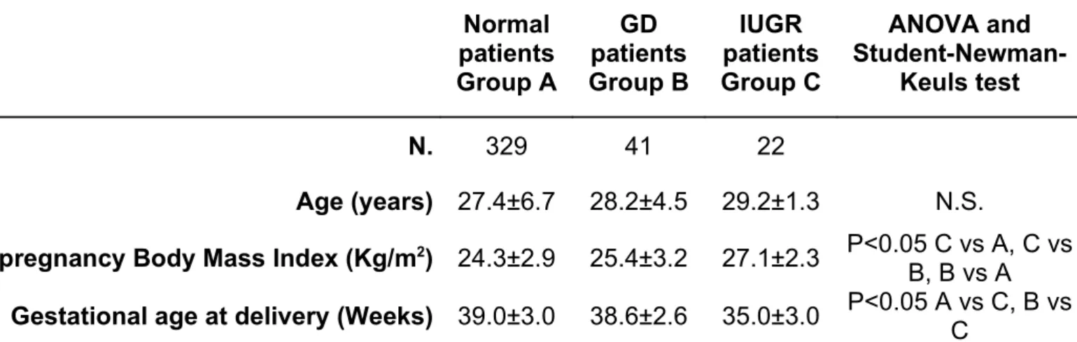
The study sample was divided into 4 main groups: Group A: who had a normal pregnancy, Group B: those with gestational diabetes, Group C: those mothers with an intrauterine growth restricted (IUGR) fetus and a small group of women with type 1 diabetes mellitus that were excluded from the analysis.
In order to test for demographic and gestational differences between the groups, we compared their characteristics in terms of age, pre-pregnancy body mass index and gestational age at delivery using the ANOVA model. Subsequently, we considered the body weight (BW) distribution across group (a dependent variable in the final regression model), and we tested for differences in BW between Groups A, B and C, through the use of both the ANOVA model and also through the post-hoc comparison between pairs of diagnostic groups.
The gaussian shape of the US measurements distribution has been evaluated by means of Kolmogorov-Smirnov normality test (all p values>0.05). A MANOVA model (Multivariate Analysis of Variance), was used to test if the US measurements, used as dependent variables in the model, were significantly different across groups, the three-levels between-subjects factor. To investigate for the presence of multicollinearity, i.e. a significant level of pairwise correlation between the US variables (a critical aspect to evaluate prior to build the final multiple regression model to estimate birthweight), Pearson correlation matrices between US measurements were computed for the entire study sample.
In order to reliably compare performances of BW prediction equations in two independent samples, we splitted our database of 392 patients into two samples, S1 and S2, using a computer generated pseudo-random selection. Measurements from 25% of patients (98 patients) formed sample group-S2, and the remaining 75% of patients formed S1 (294 patients). We used S1 to derive “our” equations.
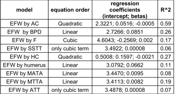
To examine contributions of single ultrasound measurements in predicting BW, equations were derived by fitting log-transformed birthweight versus individual US derived measurements in S1 (Table 1). Log-transformation of the birthweight has been used after evaluation of the Box-Cox linearity plots, in fact when
performing the linear fit of BW against single US measurements the appropriate transformation to both improve the fit and minimizing the error sum of squares was the logarithmic one, a finding also consistently with the literature on this topic.
To produce the BW predictive multiple linear regression model, a 'stepwise' approach has been used in order to select the best group of 'explicative' variables (the independent variables in the regression model, where the dependent one is the variable to be explained, i.e. the BW). This procedure takes into account the existing interactions between explicative variables.
In stepwise regression, the computer program finds the explicative variable with the highest correlation with the dependent variable; it then tries each of the remaining explicative variables in a multiple linear regression until it finds the two variables with the highest R square; then, it tries all of them until it finds the combination of three with the highest R square, and so on. The cut-off used is that a variable enter the model only if a significant F change (p<0.05) is obtained. As a consequence, if two variables are highly correlated, only one will enter in the model with a stepwise approach.
To have a benchmark, among the huge number of published birthweight prediction equations, we selected six formulas widely used in the current literature on this topic (9, 15) (table 2). Two of the selected algorithms were from Hsieh (16), two from Hadlock (17) and two from Warsof (18). A statistical comparison of errors distributions has been obtained by means of a repeated measures ANOVA model, both in S1 and S2, where in the within-subjects contrasts section the first six equations error distributions has been tested against the proposed ones (19). A repeated-measures model is one in which multiple measurements on the same subject comprise the replicate data. In the present application, we aimed to test the null hypothesis of no difference between the equation error distributions. Since there were not independent samples of women but instead each subject has been tested with the different equations, and there is consequential relationship among the data in each row, being errors of the BW estimation equations on the same subject, the within-subjects contrasts have to be used. It has to be noted that, in the case of absolute estimation errors, to evaluate the within-subjects contrasts , we used the Greenhouse-Geisser correction on the resulting p-values that do not require compound symmetry. Moreover, to further check the results obtained, we applied to these data a
RESULTS
Ultrasound scan was performed at 34-42 weeks’ gestation and the mean interval between ultrasonographic assessment and delivery was 3 days (ranging from 6 days to 1 day before delivery.
From our sample, 329 women (82.7%) had a normal pregnancy (group A), 41 women (10.3%) were diagnosed with gestational diabetes (group B) and 22 women (5.5%) had an ultrasound derived diagnosis of intrauterine growth restriction (IUGR) (Group C) (18). Diagnosis of IUGR was based on a fetal abdominal circumference <5th percentile for gestational age by local reference values, an estimated fetal
weight <10th percentile for gestational age and an umbilical artery pulsatility index of more than 2 SDs above the gestational mean compared to local reference values (20). Six women with type 1 diabetes mellitus were excluded from our study group and so a total of 392 were studied. Gestational diabetes was diagnosed using the criteria outlined by the National Diabetes Data Group (21,22).
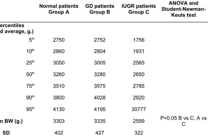
Birthweights of greater than 4000 g were considered as macrosomic (23) and 23 women from group A (7%) and 4 women from group B (9.7%) had macrosomic fetuses. The characteristics of the study groups are summarized in table 3. The percentiles of the birthweight distribution and descriptive statistics for birthweight across the three diagnostic groups are reported in table 4.
The birthweight (BW) distributions for the normal and gestational diabetic (GD) groups were similar until the 75th percentile, however at higher birthweight values a divergence was observed. Group C showed
consistently lower BW values with respect to the other groups. BW appeared to be significantly affected by diagnosis, (ANOVA F[2,389]=32.37, p<0.0001); in particular, from the post-hoc and contrasts results, birthweight resulted significantly affected by diagnosis of intrauterine growth restriction (IUGR) (group C versus group A, and group C versus group B, p<0.0001). The difference between Group A and B were not statistically significant.
All US measurements were normally distributed (Kolmogorov-Smirnov normality test, p values>0.05), therefore parametric techniques has been used in the successive statistical analysis. In the MANOVA model the US measurements, considered as dependent variables, were significantly different across groups (results shown in table 5). Only the sub-scapular tissue thickness (SSTT) and the mid-arm tissue area (MATA) were not significantly different across diagnostic groups. Using the control group (group A) as the reference category, we can see that the group C (IUGR) is significantly lower for in all the US parameters measured excluding the SSTT, MATA and the abdominal tissue thickness (ATT). Comparisons between Group A and B were not significant for all US parameters except for the abdominal tissue thickness.
Correlations between ultrasound measurements
In order to assess the multicollinearity level between explicative variables, bivariate correlations between ultrasound measurements were computed (data not shown). To summarize results obtained, significant correlation (Pearson correlation coefficient p value<0.05) were found between: biparietal diameter (BPD) with abdominal circumference (AC), femur with head circumference (HC); moreover, AC correlated with HC and mid-thigh tissue area (MTTA). HC was associated with humerus; sub-scapular tissue thickness (SSTT) with mid-arm tissue area (MATA), mid-thigh tissue area (MTTA) and abdominal tissue thickness (ATT); finally, MTTA correlated with ATT.
Given this complex situation of pairwise associations, the stepwise approach to select the most representative subset of US variables, maximally uncorrelated, was used in the multiple regression model to predict BW, as explained in the above method section.
Comparison of benchmark formulas for predicting BW
Simple linear regression models, varying from linear to cubic interpolations, selected on the basis of the best fitting of the log BW data in terms of R square, produced a clear indication that the mid-thigh tissue area (MTTA) could significantly improve performance in BW prediction.
This finding was confirmed using the method of stepwise model construction, that selected as the best subset of explicative variables the product of AC and BPD plus the MTTA measurement.
Taking into account the significant differences in BW distribution across the three groups considered, we decided to include the diagnostic group information in a separate birthweight prediction model. Therefore, two separate formulae were derived: Larciprete (a), considering only the stepwise selected ultrasound measurements, and Larciprete (b), with the stepwise selected ultrasound measurements plus the diagnostic group. These are as follows:
• Larciprete (a): Log EFW=3.030+0.001448 x (AC x BPD)+0.002099 x MTTA
• Larciprete (b): Log EFW=3.008+0.00138 x (AC x BPD)+0.002140 x MTTA + 0.02123 x DIAG where DIAG was coded as: 1=IUGR; 2=Normal; 3=GD.
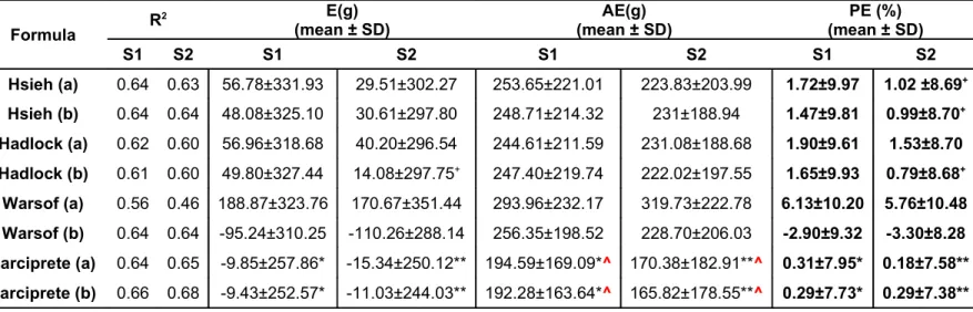
These two equations showed a fitting performance (R2) comparable to the best R2 obtained with the
benchmark equations, and the minimum absolute estimation errors with respect to the other formulas reported in table 2 (Table 6).
When the benchmark equations error distributions has been tested against the two proposed ones all contrast p values were<0.05 in sample S1 indicating a significant mean lower error for our algorithms for the three types of error distributions reported in table 6 (E, AE and PE); having obtained the same results with the Friedman's test for the absolute errors distributions, for sake of synthesis in table 6 the repeated-measures ANOVA Greenhouse-Geisser corrected p-values have been reported in the AE columns.
Some of the contrasts in sample S2, marked with a cross in table 6, indicated no significant difference, i.e. a similar error distribution to the benchmark equations in those cases.
CONCLUSIONS
The routine use of ultrasonographic measured parameters has been demonstrated to be of clinical benefit in the assessment of gestational age. Ultrasound derived parameters are usually resistant to external influence such as environmental factors, which account in part for their accuracy. However, it is also this same characteristic which reduces their suitability in the identification of fetal growth abnormalities. From the parameters discussed it is the measurement of the abdominal circumference which has been shown to be most sensitive in the detection of growth abnormalities (1, 2, 6).
Over the last 30 years, ultrasonographic fetal biometry is often assumed to be more accurate than clinical methods in the estimation of fetal weight. This is largely due to the presumption that ultrasonographic measurements of multiple linear and planar dimensions of the fetus provide sufficient parametric information to create an accurate algorithmic reconstruction of the three-dimensional fetus with varying tissue density. Thus correspondingly, a large number of studies have attempted to create ‘best-fit’ fetal biometric algorithms which can predict birth weight on the basis of obstetrical ultrasonographic measurements. Numerous studies have recently challenged the accuracy of these ultrasonographic birth weight estimations and have concluded that ultrasonography may be no more accurate in the prediction of birth weight than clinical palpation or even maternal self-estimations of fetal weight (24, 25, 26). Furthermore some studies suggest that quantitative assessment of maternal characteristics may be as accurate as obstetric ultrasonography in birthweight prediction (27, 28). Therefore, to date the most accurate method for the prediction of term birth weight has yet to be elucidated.
A study by Nahum et al (9) assessed twenty commonly used fetal US biometric algorithms in the accuracy in the prediction of term fetal weight (9). The equations were based on various combinations of the fetal measurements including the AC, BPD, and femur length (FL). The accuracy of US derived fetal weight predictions were quantified in each case through calculation of its correlation with actual birthweight, the mean absolute error, the mean absolute percentage error, and the percentage of birthweights which predicted to within ± 10% to 15% of the actual birthweight. Comparison among the equations that used the fetal AC and BPD indicated that algorithms of Hadlock et al (17), Warsof et al (18), Jordaan et al (29) and
of birthweight of a normal well-dated singleton fetus, other measurements other than AC may be superfluous. However, factors such as suboptimal fetal position, oligohydamnios, anterior placentation, and maternal obesity may confound and reduce the accuracy of ultrasonic birth-weight estimates.
It is important to remember that despite the wide degree of error that is associated with ultrasonic estimates of term fetal weight, both the technology dependent and labour-intensive nature of ultrasonic estimates may foster a false sense of reassurance among obstetric practitioners as to the projected weight of an individual fetus. This may sometimes result in the potential overuse or lack of obstetric interventions concerning both the timing and mode of delivery of a fetus, which may be potentially detrimental.
Recently, Deter (2004) stated the importance of assessing the fetal growth not on the basis of single anatomical variables, such as birthweight or abdominal circumference, but rather using the novel concepts of individualized growth assessment (IGA) and the Prenatal Growth Profiles, in which growth is assessed whereby each fetus serves as its own control (31). This is a more precise approach, since fetal growth potential is in part linked to demographic and age-specific variables (31). The ‘individualized’ evaluation of fetal growth has been derived from the concept that fetal growth is a more complex process that can be adversely affected in various ways, in different individuals (31). Therefore, the IGA (Individualized Growth Assessment), provides a comprehensive and integrated evaluation of fetal growth, correcting for differences in age and growth potential which are two primary confounding variables of growth assessment.
Deter shows that third trimester growth trajectories for a specific parameter are predicted from sonographic data obtained during the second trimester of pregnancy (31).
This new method takes into consideration the concept that soft tissues undergo early changes in abnormal growth conditions such as IUGR or macrosomia (31).
Lee et al (32) introduced the fractional thigh volume as a new soft tissue parameter for fetal growth evaluation, defining its relationship to menstrual age and developing individualized growth standards, thus applying the soft tissues to the IGA model. Moreover, they added the fractional arm volume, a soft tissue parameter, to their research (7).
These concepts highlight the importance of the evaluation of fetal subcutaneous tissue thickness (SCTT) in the assessment of fetal growth. Bernstein and Catalano used the ultrasound approach to measure subcutaneous fetal fat in the extremities and noted variations in SCTT comparable to that noted in skin fold thickness measurements in neonates (6, 33). Additionally, they report that fetal fat and lean body mass have unique growth profiles and an accelerated rate of growth is noted in late gestation. Therefore the measurement of fetal fat may well provide a more sensitive and specific means of identifying abnormal fetal growth when compared with index values of lean body mass (6).
Our previous study has provided us with the gestational reference ranges for fetal soft tissues in both normal and gestational diabetic mothers (11). We propose that adding the SCTT parameters to the
conventional ultrasound algorithms may help with the individualization approach in birthweight prediction, following the “Deter paradigm”. From our work, we describe higher birthweights in fetuses from normal and diabetic women when compared to women with growth restricted fetuses. The similarities between the normal and diabetic mothers may be attributed to the dietary regimen undertaken by the diabetic patients. From the conventional US parameters, we have seen no real differences between normal and diabetic patients except for the abdominal skin fold which was greater in fetuses from diabetic mothers. In terms of the growth restricted fetuses, both conventional and SCTT ultrasound parameters were lower with the exception of MATA and HC which correlate with findings from our previous work (11).
Generally, an absence of differences among groups for most SCTT parameters was seen.
Using the proposed algorithms we have found lower mean errors, mean absolute errors and percentage errors when compared to those algorithms which are currently used (Hsieh, Hadlock and Warsof) (16, 17, 18). Nonetheless, the quite low fitting performance for the relationship between individual SCTT parameters and birth weight has to be cited. Small differences were noted in systematic and random estimation errors when the results using weight estimation functions with a SCTT parameter are compared to the results using weight estimation functions without such parameters. Our algorithms showed the usefulness to add the mid-thigh tissue area evaluation in birthweight prediction with respect to the other reported algorithms.
But our findings need to be further clarified since we used functions from the literature in a new sample, without determining sample-specific coefficients, and this behaviour frequently gives poorer results than were obtained in the original studies.
REFERENCES
1. Rosati P, Exacoustos C, Caruso A, Mancuso S. Ultrasound diagnosis of fetal macrosomia. Ultrasound Obstet Gynecol 1992;2:23-90.
2. Chang TC, Robson SC, Boys RJ, Spencer JAD. Prediction of the small for gestational age infant: which ultrasonic measurement is best? Obstet Gynecol 1992;80:1030-8.
3. Skovron ML, Berkowitz GS, Lapinski RH, Kim JM, Chitkara U. Evaluation of early third-trimester ultrasound screening for intrauterine growth retardation. J Ultrasound Med 1991;10:153-9
4. Manning FA. Intrauterine growth retardation. In: Manning FA, ed. Fetal medicine: Principle and practice. Norwalk, CT: Appleton & Lange, 1995;30:93.
5. Galan HL, Rigano S, Radaelli T, Cetin I, Bozzo M, Chyu J, Hobbins JC, Ferrazzi E. Reduction of subcutaneous mass, but not lean mass, in normal fetuses in Denver, Colorado. Am J Obstet Gynecol 2001;185:839-44.
6. Bernstein IM, Goran MI, Amini SB, Catalano PM. Differential growth of fetal tissues during the second half of pregnancy. Am J Obstet Gynecol 1997;176:28-32.
7. Lee W, Deter RL, McNie B, Gonçalves LF, Espinoza J, Chaiworapongsa T, Balasubramaniam M, Romero R. The Fetal Arm. Individualized Growth Assessment in Normal Pregnancies. J Ultrasound Med 2005; 24:817-828.
8. Deter RL. Individualized growth assessment: evaluation of growth using each fetus as its own control. Semin Perinatol. 2004 Feb;28(1):23-32.
9. Nahum GG, Stanislaw H. "Ultrasonographic prediction of term birth weight: how accurate is it?", Am J Obstet Gynecol 2003; 188, 2, 566:574.
10. Dudley NJ. A systematic review of the ultrasound estimation of fetal weight. Ultrasound Obstet Gynecol 2005; 25: 80–89.
11. Larciprete G, Valensise H, Vasapollo B, Novelli GP, Parretti E, Altomare F, Di Pierro G, Menghini S, Barbati G, Mello G, Arduini D. Fetal subcutaneous tissue thickness (SCTT) in healthy and gestational diabetic pregnancies. Ultrasound Obstet Gynecol Dec 2003;22(6):591-7.
12. Bernstein IM, Catalano PM. Ultrasonographic estimation of fetal body composition for children of diabetic mothers. Invest Radiol 1991;26:722-6.
13. Winn HN, Holcomb WL. Fetal non-muscular soft tissue: a prenatal assessment. J Ultrasound Med 1993;4:197-9.
14. Gardeil F, Greene R, Stuart B, Turner MJ. Subcutaneous fat in the fetal abdomen as a predictor of growth restriction. Obstet Gynecol 1999;94:209-12.
15. Liang RI, Chang FM, Yao BL, Chang CH, Yu CH, Ko HC. Predicting birth weight by fetal upper-arm volume with use of three-dimensional ultrasonography. Am J Obstet Gynecol 1997; 177(3):632-8.
16. Hsieh FJ, Chang FM, Huang HC, Lu CC, Ko TM, Chen HY. "Computer-assisted analysis for prediction of fetal weight by ultrasound: comparison of biparietal diameter, abdominal circumference and femur length" J Formosan Med Assoc 1987; 86: 957-64.
17.Hadlock FP, Harrist RB, Shearman RS, Deter RL, Park SK. "Estimation of fetal weight with the use of head, body and femur measurements: a prospective study". Am. J Obstet Gynecol 1985; 151: 333-7. 18.Warsof SL, Gohan P, Berkowitz RL, Hobbins JC. "The estimation of fetal weight by computer-assisted
analysis." Am J Obstet Gynecol 1977; 128: 881-92.
19.Zar JH, "Biostatistical Analysis", International Fourth Edition, 1999, Prentice Hall.
20. Vasapollo B, Valensise H, Novelli GP, Larciprete G, Di Pierro G, Altomare F, Casalino B, Galante A, Arduini D. Abnormal maternal cardiac function and morphology in pregnancies complicated by intrauterine fetal growth restriction. Ultrasound Obstet Gynecol 2002;20: 452–457.
21. The Expert Committee on the Diagnosis and Classification of Diabetes Mellitus. Report of the Expert Committee on the Diagnosis and Classification of Diabetes Mellitus. Diabetes Care. 2001 Jan; 24(1): S5-S20.
22. National Diabetes Data Group. Classification and diagnosis of diabetes mellitus and other categories of glucose intolerance. Diabetes 1979;28:1039-57.
23. Chauhan SP, West DJ, Scardo JA, Boyd JM, Joiner J, Hendrix NW. Antepartum Detection of Macrosomic Fetus: clinical versus sonographic, including soft-tissue measurements. Obstetrics & Gynecology 2000; 5:1, 639:642.
24.Chauhan SP, Cowan BD, Magann E, Bradford T, Roberts WE, Morrison JC. Intrapartum detection of a macrosomic fetus: clinical versus 8 sonographic models. Aust N Z J Obstet Gynaecol 1995;35:266-70. 25.Hendrix NW, Grady CS, Chauhan SP. Clinical vs sonographic estimate of birth weight in term
parturients: a randomized clinical trial. J Reprod Med 2000;45:317-22.
26.Sherman DJ, Arieli S, Tovbin J, Siegel G, Caspi E, Bukovsky I. A comparison of clinical and ultrasonic estimation of fetal weight. Obstet Gynecol 1998;91:212-7.
30.Shepard MJ, Richards VA, Berkowitz RL, Warsof SL, Hobbins JC. An evaluation of two equations for predicting fetal weight by ultrasound. Am J Obstet Gynecol 1982;142:47-54.
31.Vintzileos AM, Campbell WA, Rodis JF, Bors-Koefoed R, Nochimson DJ. Fetal weight estimation formulas with head, abdominal, femur, and thigh circumference measurements. Am J Obstet Gynecol 1987;157:410-4.
32. Lee W, Deter RL, McNie B, Goncalves LF, Espinoza J, Chaiworapngsa T, Romero R. Individualized growth assessment of fetal soft tissue using fractional thigh volume. Ultrasound Obstet Gynecol 2004; 24: 766–774.
33. McGowan A, Jordan M, MacGregor J. Skinfold thickness in neonates. Biol Neonate 1975;25:66-84.
Figure 1. The a and b pictures represent the ultrasound scan showing the axial view of the extremities (MATA=mid-arm tissue area and MTTA=mid-thigh tissue area, respectively). The c picture represents the way to evaluate the abdominal tissue thickness (ATT) and the
d picture shows the subscapular tissue thickness (SSTT) measurement.
Table 1. Single-measurement regression models derived from S1. model equation order coefficientsregression
(intercept; betas)
R^2 EFW by AC Quadratic 2.3221; 0.0516; -0.0005 0.59
EFW by BPD Linear 2.7266; 0.0851 0.26
EFW by F Cubic 4.6043; -0.2569; 0.002 0.17
EFW by SSTT only cubic term 3.4922; 0.00008 0.06 EFW by HC Quadratic 0.5008; 0.1597; -0.0021 0.27
EFW by humerus Linear 3.0792; 0.0662 0.11
EFW by MATA Linear 3.4470; 0.0095 0.08
EFW by MTTA Linear 3.4113; 0.0082 0.19
EFW by ATT only cubic term 3.4878; 0.00008 0.07
MATA=mid-arm tissue area. MTTA=mid-thigh tissue area. SSTT=subscapular tissue thickness. ATT=abdominal tissue thickness. EFW=estimated fetal weight. AC=abdominal circumference. BPD=biparietal diameter. F=femur length. HC=head circumference.
Table 2. Birthweight prediction formulas commonly used in literature
Hsieh (a) 15 Log10 EFW= 5.6541 x 0.001 x AC x BPD-1.5515 x 0.0001 x (AC2) x BPD
+1.9782 x 0.00001 x (AC3) + 5.2594 x 0.01 x BPD + 2.13153
Hsieh (b) 15 Log10 EFW= 9.4962 x 0.001 x AC x BPD – 0.1432 x F – 7.6742 X 0.0001 x AC x
(BPD2) + 1.7450 x 0.001 x (BPD2) x F + 2.7193
Hadlock (a) 16 Log
10 EFW=1.335-0.0034 x AC x F + 0.0316 x BPD + 0.0457 x AC+0.1623 x F
Hadlock (b) 16 Log
10 EFW=1.326-0.00326 x AC x F + 0.0107 x HC + 0.0438 x AC + 0.158 x F
Warsof (a) 17 Log
10 EFW=-1.8367+0.092 x AC-0.000019 x (AC3)
Warsof (b) 17 Log
10 EFW= -1.599 + 0.144 x BPD + 0.032 x AC-0.000111 x ((BPD2) x AC)
EFW=estimated fetal weight. AC=abdominal circumference. BPD=biparietal diameter. F=femur length. HC=head circumference. The Warsof algorithms express the BW in kilograms.
Table 3. Characteristics of the studied populations Normal patients Group A GD patients Group B IUGR patients Group C ANOVA and Student-Newman-Keuls test N. 329 41 22 Age (years) 27.4±6.7 28.2±4.5 29.2±1.3 N.S.
Pre-pregnancy Body Mass Index (Kg/m2) 24.3±2.9 25.4±3.2 27.1±2.3 P<0.05 C vs A, C vs
B, B vs A Gestational age at delivery (Weeks) 39.0±3.0 38.6±2.6 35.0±3.0 P<0.05 A vs C, B vs C GD: gestational diabetes; IUGR: intrauterine growth restriction; N.S.: not significant.
Table 4. Percentiles of birth-weight distribution and descriptive statistics across the three diagnostic groups Normal patients Group A GD patients Group B IUGR patients Group C ANOVA and Student-Newman-Keuls test BW percentiles (weighted average, g.) 5th 2750 2752 1756 10th 2860 2804 1931 25th 3050 3005 2565 50th 3260 3280 2650 75th 3510 3575 2785 90th 3800 4028 2920 95th 4130 4195 30777 Mean BW (g.) 3303 3335 2599 P<0.05 B vs C, A vs C SD 402 427 322
GD: gestational diabetes; IUGR: intrauterine growth restriction; N.S.: not significant; BW: birth-weight; SD: standard deviation.
Table 5. Descriptive statistics for the ultrasound measurements within the three study groups (mean ± SD) Normal patients Group A n. 329 GD patients Group B n. 41 IUGR patients Group C n. 22
MANOVA Contrasts Results P value Group B vs Group A Group C vs Group A Conventional US Parameters BPD (cm) 9.25 ± 0.36 9.11 ± 0.27 8.88 ± 0.33 0.187 0.000* AC (cm) 34.16 ± 2.25 34.50 ± 2.58 30.78 ± .1.99 0.395 0.000* Femur (cm) 7.23 ± 0.37 7.09 ± 0.26 6.91 ± 0.27 0.157 0.000* HC (cm) 33.78 ± 1.44 33.47 ± 1.33 32.03 ± 1.54 0.522 0.000* Humerus (cm) 6.51 ± 0.30 6.47 ± 0.33 6.33 ± 0.36 0.381 0.009* SCTT parameters SSTT (mm) 5.69 ± 1.47 6.34 ± 1.44 5.51 ± 1.60 0.307 0.574 MATA (cm2) 6.65 ± 1.74 6.92 ± 1.50 6.38 ± 1.90 0.264 0.513 MTTA (cm2) 12.00 ± 3.07 13.30 ± 3.47 10.19 ± 2.94 0.321 0.009* ATT(mm) 6.18 ± 1.39 6.78 ± 1.27 5.74 ± 1.33 0.029* 0.151
SCTT=subcutaneous tissue thickness. MATA=mid-arm tissue area. MTTA=mid-thigh tissue area. SSTT=subscapular tissue thickness. ATT=abdominal tissue thickness. MANOVA= multivariate analysis of variance. *p<0.05
Table 6. Comparison of benchmark formulas, our formulas in estimating fetal weight in S1 and S2.
Formula R
2 E(g)
(mean ± SD) (mean ± SD)AE(g) (mean ± SD)PE (%)
S1 S2 S1 S2 S1 S2 S1 S2 Hsieh (a) 0.64 0.63 56.78±331.93 29.51±302.27 253.65±221.01 223.83±203.99 1.72±9.97 1.02 ±8.69+ Hsieh (b) 0.64 0.64 48.08±325.10 30.61±297.80 248.71±214.32 231±188.94 1.47±9.81 0.99±8.70+ Hadlock (a) 0.62 0.60 56.96±318.68 40.20±296.54 244.61±211.59 231.08±188.68 1.90±9.61 1.53±8.70 Hadlock (b) 0.61 0.60 49.80±327.44 14.08±297.75+ 247.40±219.74 222.02±197.55 1.65±9.93 0.79±8.68+ Warsof (a) 0.56 0.46 188.87±323.76 170.67±351.44 293.96±232.17 319.73±222.78 6.13±10.20 5.76±10.48 Warsof (b) 0.64 0.64 -95.24±310.25 -110.26±288.14 256.35±198.52 228.70±206.03 -2.90±9.32 -3.30±8.28 Larciprete (a) 0.64 0.65 -9.85±257.86* -15.34±250.12** 194.59±169.09*^ 170.38±182.91**^ 0.31±7.95* 0.18±7.58** Larciprete (b) 0.66 0.68 -9.43±252.57* -11.03±244.03** 192.28±163.64*^ 165.82±178.55**^ 0.29±7.73* 0.29±7.38**
EFW=estimated fetal weight; ABW=actual birth weight; E (mean error): EFW-ABW; AE (mean absolute error): |EFW-ABW|; PE (percentage error): (EFW-ABW)x100/ABW. *;** p<0.05 within-subject contrasts with respect to benchmark equations; +within-subject contrasts p value>0.05 w.r.t. our two proposed
equations. ^Greenhouse-Geisser corrected p values.
Da: [email protected] per conto di [email protected] Inviato: mercoledì 10 ottobre 2007 12.35
Oggetto: Request for copy right form JOGR-2006-0399 10-Oct-2007
JOGR-2006-0399.R2 - COULD THE BIRTHWEIGHT PREDICTION MODELS BE IMPROVED BY ADDING FETAL SUBCUTANEOUS TISSUES?
Dear Dr. Larciprete:
Greetings from Editorial Office of The Journal of Obstetrics and Gynaecology Research. This is the reminder that we haven't received the Exclusive License Form (ELF) for above manuscript yet, which is imperative for preparation on publication.
It would be grateful if you send us ELF to below address at your earliest possible;
Editorial Office, The Journal of Obstetrics and Gynaecology Research c/o WILEY-BLACKWELL Frontier Koishikawa Bldg 4F, 1-28-1 Koishikawa, Bunkyo-ku, Tokyo, 112-0002 Japan
Tel: 81-3-3830-1264 Fax: 81-3-5689-7278
If it is difficult to send by Post mail, please send them by FAX to below number by the end of this week.
81-3-5689-7278
I really appreciate your cooperation.
Exclusive License Form can be downloaded by below address: http://www.blackwellpublishing.com/pdf/jog_elf.pdf
Best regards, Editorial Office
PRODUZIONE SCIENTIFICA DURANTE IL DOTTORATO DI RICERCA IN MEDICINA PERINATALE
ABSTRACT
Nell’elaborato finale del Dottorato di Ricerca in Medicina Prenatale ho seguito principalmente due rami di ricerca clinica:
1- La ricerca sull’utilizzo e lo sviluppo della plicometria ultrasonogrfica fetale
2- Lo studio delle associazioni tra mutazioni (o condizioni) trombofiliche e l’insorgere di patologie della gravidanza.
Nella prima applicazione ho studiato alcuni dei nuovi parametri plicometrici fetali proposti in letteratura negli ultimi anni. Ho implementato la tecnica di misura, contribuendo alla sua standardizzazione. In seguito ho prodotto tavole biometriche di riferimento nelle successive epoche di gravidanza, sia in condizioni di normalità, sia in pazienti con diabete gestazionale. Ho condotto uno studio sui cambiamenti dei compartimenti corporei fetali in caso di ritardo di crescita. Ho inoltre studiato il potenziale beneficio (in termini di accuratezza diagnostica) dell’uso dei parametri plicometrici fetali nei preesistenti modelli di stima del peso fetale. Da ultimo ho prodotto un algoritmo per la predizione del peso alla nascita, introducendovi anche uno dei parametri plicometrici da me studiati. I risultati della mia ricerca in questo campo vengono esposti in 4 articoli editati su stampa internazionale dal 2003 al 2007, proprio in corso del Dottorato di Ricerca in oggetto.
Nella seconda applicazione ho studiato l’associazione tra trombofilia ed esiti avversi della gravidanza. I due lavori presentati nel seguente documento hanno timbro epidemiologico e mostrano in comune lo stesso set di pazienti, ma differente analisi statistica. In questo attuale campo la mia ricerca sta procedendo. Due manoscritti sono in coso di valutazione e trattano dell’associazione tra trombofilia ed alterata funzione renale in gravidanza.
Sempre nella stessa tesi di Dottorato di Ricerca mi preme mostrare alcuni case report relativi ad interessanti casi clinici occorsi alla mia visione nel corso di studi in oggetto, ed
SCIENTIFIC PRODUCTION DURING THE PERINATAL MEDICINE PHELLOWSHIP ABSTRACT
The results and conclusive reports of my Phellowship in Perinatal Medicine are enclosed within the final manuscript of the following thesis.
My main research application fields were:
1- The study of fetal skinfolds by means of ultrasound evaluation. 2- The study of the impact of thrombophilias in pregnancy.
My former research field dealed with the study of the fetal subcutaneous tissues and their detection by ultrasounds. I spent part of my Phellowship analyzing whether these innovative ultrasound parameters could add more informations to the worldwide used birthweight estimating conventional algorithms. Thus, the targets of my research in this first field were: to analyze the behaviour of fetal compartments in different physiological or pathological gestational conditions; to analyze the usefulness of the fetal skinfolds in ameliorating pre-existing birthweight estimating formulae; to build-up a mathematical model to predict term neonatal weight as a result of an integration between traditional and innovative fetal US parameters.
The sum of my findings is elucidated in 4 published papers edited from year 2003 to year 2007.
The latter research field deals with the correlation between connatal thrombophilias and the occurrence of adverse pregnancy outcomes. In fact by now I can show the result of this research by presenting two works that share the same setting with a different statistical viewpoint. The research is going on and other 2 manuscripts are away to be evaluated by the referees.
Moreover, within the same document I show some clinical cases published as case reports (with or without a review), most of them dealing with umbilical cord pathologies.
Eventually I bring also some proceedings from Congresses from the International Society of Ultrasound in Obstetrics and Gynaecology, also edited by the ISUOG official magazine.
PAROLE CHIAVE
Plicometria fetale, ecografia ostetrica, peso fetale, algoritmi di crescita, tabelle di riferimento, biometria, trombofilie, ritardo di crescita, preclampisa, omocisteina, cordone ombelicale, flussimetria
KEY WORDS
Fetal skinfolds, ultrasound, fetal weight, birthweight prediction, referral charts, thrombophilias, intrauterine growth restriction, preclampsia, omocysteinemia, umbilical cord, Doppler velocimetry.