142 Acta Orthop Scand 1994; 65 (2): 142-1 46
Reversibility of the inhibitory effect of salmon
calcitonin on bone resorption in rats
Ugo E Pazzaglia’, Giovanni Zatti’, Paolo Rolla’ and Amalia Di Nucci2
Inhibition of osteoclastic bone resorption has been induced in growing rats with high doses of salmon calcitonin. This effect was evaluated by measuring the perichondrial ring height of the proximal tibia1 metaphysis. The aim was to assess whether osteo- clastic activity resumed after a period of inhibition with high doses of calcitonin. 20 male Sprague-Daw- ley rats were treated for 21 days with 100
~~~ ~
units/kg/day of salmon calcitonin subcutaneously and killed after 0-60 days, together with non-treated controls at 0 and 60 days.
Arrest of metaphyseal modeling and increased height of the perichondrial ring at the end of the period of therapy
(P
0.002 versus controls) were observed. Recovery of bone resorption was evident 20 and 40 days after withdrawal of calcitonin.~ ~~~
~ ~~ ~~~
‘Clinica Ortopedica, II Facolta’ di Medicina e Chirurgia dell’universita’ di Pavia Ospedale F. Del Ponte, 1-21 100 Varese, Italy, 21stituto di Farmacologia II, Universita’ di Pavia. Tel +39-332 282 682. Fax -332 288 956
Submitted 93-04-03. Accepted 93-09-1 0
The capacity of calcitonin to inhibit bone resorption has been demonstrated in vitro (Chambers et al. 1985, Reynolds and Dingle 1970, Stewart and Stern 1987) but not in animals with physiological doses of the hor- mone (Russel et al. 1973). High doses of salmon cal- citonin were found to influence the morphology of the growth plate and metaphysis of growing rats: increased height of the perichondrial ring, persistence of cartilage intercolumnar septa in the outer part of the growth plate and failure of metaphyseal conization were observed. These changes can be explained by the inhibitory effect of the hormone on osteoclasts and the model employed can provide quantitative assessment of bone resorption inhibition (Pazzaglia et al. 1993). From these results arises the question of the reversibil- ity of calcitonin action on osteoclasts. The aim of this study was to assess whether and at what time osteo- clnstic activity resumed after a period of inhibition with high doses of calcitonin.
Material and methods
20 male Sprague-Dawley rats (Stefan0 Morini, S. Polo tl’Enza. Reggio Emilia, Italy), average weight 150 g, were in.jected for 2 I days with 100 unitslkglday of sal- mon cnlcitonin subcutaneously (Sandoz S.p.A., Milano, Italy). At the end of this period, 4 rats were killed with an overdose of ether. Groups of 4 of the remaining rats were killed at 10, 20. 40, and 60 days.
A control group of 10 rats was injected subcutane-
ously for 21 days with the same volume of the vehicle alone: 5 animals were killed after withdrawal of vehi- cle administration (control Group I ) and 5 after 60 days (control Group 2). The mean weight gain of rats treated with calcitonin and controls was similar.
Radiographs of both tibiae were taken on Kodak mammographic film. The proximal epiphysis arid metaphysis of each bone were dissected froni soft tis- sues and fixed in neutral fornialin for histological study. Evaluation of radiographs and histological slides was made blindly and independently by IWO of the authors (AN and UEP).
After decalcification in EDTA f o r 2 weeks. the specimens were embedded in paraffin and wctioiis cut in the middle coronal plane of the tnetaphysih. Herna- toxylin-eosin stained sections were evaluated for histo- rnorphometry with a Leitz M I S low-power microscope equipped with a grid ocular. The distance between 2 intersections of the grid was adjusted with a reference caliper resting o n the coverslip; the measurement on the grid was then corrected according to this factor and expressed i n min. The height of the perichondrial ring was measured i n the middle coronal plane sections of the right and left rnetaphyses of each rat. Since the effect of arrest of rnetaphyseal modeling is present occasionally on both the medial and laternl sides o f the metaphysis, hut is ii constant finding on the inedinl side, only the height of the former was meastired. The right or left tibia of each rat was randornly tired for evaluation and compared with control Group I . Con- trol Group 2 was matched against control Group I in
CopvriRhr 0 Scandinavian University Press 1994. 1SSN 00014470. Primed irr Stwtlrri - ol1 rixlrrs rr.wriw/.
Acta Orthop Downloaded from informahealthcare.com by 93.62.227.228 on 01/25/12
Acta Orthop Scand 1994; 65 (2): 142-146 143
order to check differences of perichondrial ring height determined by the rat’s growth. In each group the left and right tibia were also compared.
Osteoclasts were counted with a Leitz Aristoplan microscope (enlargement 2 5 0 ~ ) in the same coronal slide of the proximal tibia: the area utilized for the quantitative assessment included the whole epiphysis,
growth plate and metaphysis. The lower margin of this area was a line parallel to the growth plate and drawn to a distance from the top margin of the growth plate equal to twice the height of the epiphysis. The density of osteoclasts was expressed as the ratio between the number of cells and the surface area (cells/mm2). Only multinuclear cells closely associated with both trabec- ular and cortical endosteal surfaces were identified as osteoclasts.
The selection of groups for statistical evaluation was the same as for the perichondrial ring height
assessment. The Student’s t-test was used.
Figure 1. The proximal tibia in control rats.
Results
Typical changes were present in all the rats after 21
days treatment. Radiographic thickening of the margi- nal growth plate on the medial side was evident (Fig- ures 1 and 2 ) . Histologically these zones were charac-
terized by persistence of columns of hypertrophic chondrocytes (Figure 3 ) . The corresponding metaphy- sis had a cylindrical shape due to arrest of modeling (failure of conization), and the perichondrial ring was consequently higher than normal (Table 1 ). The num- ber of osteoclasts was increased, and their nuclei had a dense chromatin or were frankly pyknotic.
10 days after withdrawal of therapy, extension of growth plate cartilage into the metaphysis was no longer evident (Figure 2 ) . In the corresponding histo-
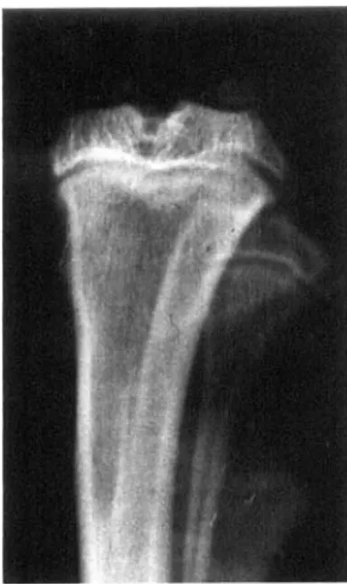
Figure 2. The proximal tibia in rats treated with salmon calcitonin 100 units/kg/day for 21 days at time 0 (left), 10 days (center) and 60 days (right) after withdrawal of therapy.
At time 0, extension of the growth plate and arrest of metaphyseal modeling on the medial side are evident
At 10 days, extension of the growth plate inside the metaphysfs is no longer seen and renewed metaphyseal modeling is present However, the medial margin is irregular and shaded
At 60 days, a denser opaque area is evident on the medial part of the meta- physis A structural alteration suggesting a giant-cell-like tumor IS present in the head of the fibula
Acta Orthop Downloaded from informahealthcare.com by 93.62.227.228 on 01/25/12
144 Acfa Orthop Scand 1994; 65 (2): 142-146
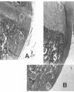
Figure 3. The medial growth piate cartilage and metaphysis in controls (A) and in rats after 21 days of treatment with salmon calcitonin (6). Arrows indicate the top and the bottom of the perichondrial ring. HE, x30.
logical slides the abnormally high columns of hyper- trophic chondrocytes had disappeared, the perichon- drial ring was still higher than in the controls, but signs of renewed metaphyseal modeling were present at the bottom end of the ring, forming a notch on its inner aspect (Figure 4). In this area, the distribution of Osteoc~asts Was Very dense: mUkinUClear Cells with a
large, finely-granular cytoplasm and nuclei with sparse chromatin were present, together with cells with pyk- notic nuclei and scanty cytoplasm (Figure 5 ) .
Figure 4. The perichondnal ring 10 days after withdrawal of calcitonin therapy is higher than in controls. However, renewed bone resorption is occurring at the bottom end of the ring (arrows). in the growth plate, the abnormally high columns of hypertrophic chondrocytes have disappeared. HE, x100.
Table 1. Mean height (mm SO) of the medial perichondrial ring and number of osteoclasts (m/mm2 SO) in rat tibias. The rats had been treated wRh salmon caicitonin 100 U/kg/day for 21 days and then killed after 0-60 days. The controls were killed after 0 or 60 days. The experimen- tal groups and the 60 days control group were compared with 0 days control group using the Student's t-test
Daysaner n Perichondrial ring P treatment Osteociasts P 0 4 3.47 0.59 0.02 10 4 3.12 0.72 e0.02 20 4 1.56 0.86 ns 40 4 1.71 0.69 ns 60 4 1.02 0.49 < 0.05 control 1 5 1.79 0.44 ns control 2 5 1.71 0.31 ns 2.33 0.35 <O.M)l 1.79 0.27 < 0.02 1.33 0.36 lls 1.21 0.36 ns 1.16 0.20 ns 1.32 0.19 ns 1.30 0.18 ns
Acta Orthop Downloaded from informahealthcare.com by 93.62.227.228 on 01/25/12
Acfa Ollhop Scand 1994; 65 (2): 142-1 46 145
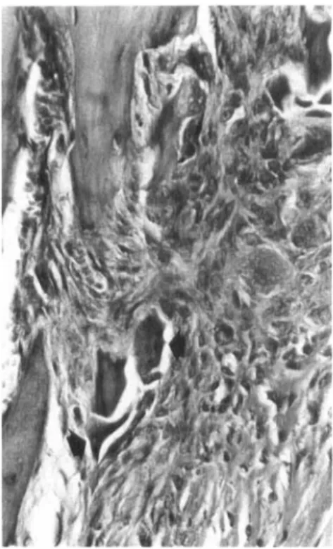
Figure 5 Detail of the area at the bonom end of perichondrial ring 10 days after withdrawal of calcitonin therapy a large number of osteoclasts with abundant, finely-gran&r cytoplasm are present, a few osteodasts with denser qtoplasm and pyknotic nuclei are still evident (arrows) HE, x400
At later intervals, the height of the perichondrial ring progressively decreased, as well as the number of osteoclasts. In the groups studied 20 and 40 days after withdrawal of therapy, no difference in perichondrial ring height was present, while i,n rats killed after 60
days, the ring was even lower than in the controls. N o differences were observed between control groups.
The mean number of osteoclasts was higher than in controls at 0 and 10 days. There were no differences from u) days onward or between the control groups. Comparisons of perichondrial ring height and mean number of osteoclasts between the right and left proxi- mal tibia i n each group and i n controls also did n o t dif- fer.
The radiographic evolution of the area of cartilage extension showed a gradual healing with time. How- ever, after 60 days, a denser, opaque area was evident o n the medial aspect of the rnetaphyses. Failure of conization was corrected from 20 days onward.
Discussion
The height of the perichondrial bone ring is a reliable indicator of activity of the resorbing cells. Since it is formed by differentiated osteoblasts at the periphery of the growth plate and is resorbed by osteoclasts at the bottom, the constant height of the ring during growth can be maintained only if apposition at the top is equal to resorption at the bottom. Therefore, an increased height of the ring nlay be due to increased apposition or reduced resorption. In another study (Pazzaglia et al. 1993), no differences in the total tibia1 length between calcitonin-treated rats and controls were observed as would be expected in case of accelerated apposition. thus confirming that resorption was reduced in our cal- citonin rats.
The observed persistence of hypertrophic chondro- cytes in the growth plate of rats treated with high doses of salmon calcitonin h.ai previously been reported (Burch and Corda 1985). but these findings have been interpreted as stimulation of chondrocytes rathei- than modeling inhibition; but again if such an interpretation is correct, an increased total length of the bone should be observed.
The arrest of modeling of the growth plate cartilage has been reported to be associated with failure of min- eralization of intercolumnar septa and primary meta- physeal trabeculae (Pazzaglia et al. 1993). The same phenomenon is present with other inhibitors of bone remodeling, I k e diphosphonates (Schenk et al. 1973) or in human pathological conditions, like rickets o r niuco1ipidoki.s 2 (Pazzaglia et al. 1989). The correla- tion betwean these two phenomena iernains obscure. and no causal relationship ha5 so far been documented. However, this association can explain the radiotrans- parent area on the medial aspect of the unmodeled metaphysis after 21 days of calcitonin administration; recovery of hone resorption activity after suspension of treatmerat is accompanied by renewed calcification, as shown by the radiographs at 10 days. Following the block of modeling, uncalcified primary metaphyseal trabeculae become denser and when recalcified they produce a more opaque area in the metaphysis. as noted at 60 days.
I n conclusion, the resorption activity of osteoclasts appears to be closely associated with the calcification process. Both must function 10 days after withdrawal of high doses of calcitonin, even i f the quantitative assessment of resorption activity (by perichondri;il ring height measurement) became significant only after 20
days.
There is also a striking resemblance of the multinu-
clear cells at the distal end of the perichondrial bone bark and the outer aspect of the rnetaphysis of rats
Acta Orthop Downloaded from informahealthcare.com by 93.62.227.228 on 01/25/12
146 Acta ofthop Scand 1994; 65 (2): 142-1 46
treated with high doses of calcitonin and those observed when diphosphonates are given (Schenk et ul. 1973). Ultrastructural study of the large polycarios present in the latter suggests that these are osteoclasts which have lost the brush border and are no longer capable o f resorbing bone (Miller et al. 1977). The conclusions drawn from the morphological changes in these cells are confirmed in the present study by the increased height of the perichondrial ring, which docu- ments the arrest of the resorbing activity. A paradox hecomes apparent where the number of osteoclasts increases, but the resorption activity is inhibited. A possible explanation rests on the fact that transient hypocalcetiiia. induced by high doses of calcitonin administration. produces secondary hyperparathyr- oidisni (Glajchen et al. 1990) with stimulation of oste- oclast formation (Baron and Vignery 198 I). However, these cells are not capable of resorbing bone because of the inhibitory effect of calcitonin itself. Other fac- tors [nay contribute to the increased number (Hedlund et al. 1983) of large polynucleated cells: their appear- ance is such that they are more easily detected in histo- logical slides or the life span of these cells is increased. Whichever hypothesis is true, these osteo-
IS are not active and no reliable assessment of bone resorption can be based on their number.
When calcitonin administration was interrupted, the recovery o f resorption activity was shown with the morphometric analysis of periochondrial ring height after 20 days and it was accompanied by a reduction in the number of osteoclasts as well as by normalization of their appearance. A possible explanation of this aspect is that a new population of young osteoclasts is recruited, while the old inactive ones progressively disappear.
The effects we have observed were obtained with very high doses of the hormone, which are never used in clinical therapy.
References
Baron R. Vignery A. Behavior of oatroclasts during rapid change i n their number induced by high doses of parathy- roid hormone or calcitonin in intact rat. Metab Bone 13s Re1 Res 198 I ; 2 : 339-46.
Burch W M. Corda G. Calcitonin stiiiiulates niaturation of mammalian growth plate cartilage. Endocrinology 19x5,
I16 ( 5 ) : 1724-X.
Chambers T J. McSheehy P M. Thonison H M. Ikller K. The effect of calcium-regulating hormones and prostsglandins on bone resorption by osteoclasts disaggregated from neonatal rabbit bones. Endocrinology I9X.5; 1 I6 ( I ):
234-9.
Glajchen N. Thomas S, Jowell P. Epstein S, Isinail I;, M l o n M. The effect of high-dose salmon calcitonin on hone m i i -
era1 metabolism in the normal rat. Calcil Tissue Int 1990. 46 ( I ): 28-32,
Hedlund T, Hulth A, Johnell 0. Early effects of parathormone and calcitonin on the number of osteoclasts and on serum calcium in rats. Acta Orthop Scand 19x3; 54 (6): 8 0 2 4 . Miller S C. Jee W S. Kinimel D €3. Woodhury 1.. Ethane-l-
hydroxy- I , I-diphosphonate (EHDP) eltects o n incorporii- tion and accumulation of osteoclast nuclei. C;ilcif Tissue Res 1977; 22 ( 3 ) : 243-52.
Pazzaglia U E. Beluffi G, Bianchi E, Castello A. Coci A. Marchi A. Study of the hone pathology i n early mucolipid- osis II (I-celldisrase). Eur J Pediatr 19x9; I4X ( 6 ) : 553-7. Pazzaglia U E, Zatti G. Di Nucci A, Coci A. Inhihitory effect
of salmon calcitonin on hone resorption: niorphological study of the tibial growth plate in rats. Calcif Tissue Int
1993; 52 (2): 125-9.
Reynolds J J , Dingle J T. A sensitive in vitro method t o r stud-
ying the induction and inhibition of hone resorption. Calcil Tissue Res 1970; 4 (4): 339-49.
Russel R G, Kislig A M, Casey P A, Fleisch H, l’hornton J . Schenk R, Williams D A. Effect of diphosponates and calcitonin in the chemistry and quantitative histology of rat hones. Calsif Tissue Res 1973; 1 I (3): 179-9s.
Schenk R. Merz W A, Muhlbauer R, Russell R G . Fleisch H. Effect of ethane-I-hydroxy-l .I-diphosphonate (EHDP) and dichloromethylene diphosphonate (CI 2 MDP) on the calcification and resorption of cartilage and hone in the tibial epiphysis and metaphysis of rats. Calcif Tissue Res 1973; I I (3): 196214.
Stewart P J, Stern P H. Vertebral bone resorption in vitro:
effects of parathyroid hormone calcitonin. I .25-dihydroxy vitamin D3, epidermal growth factor, prostaglandin E2 and estrogen. Calcif Tissue Int 1987; 40: 2 1 4 .
Acta Orthop Downloaded from informahealthcare.com by 93.62.227.228 on 01/25/12