REVIEW
Zoonotic parasites of dromedary camels:
so important, so ignored
Alireza Sazmand
1*, Anja Joachim
2and Domenico Otranto
1,3Abstract
With a global population of about 35 million in 47 countries, dromedary camels play a crucial role in the economy of many marginal, desert areas of the world where they survive under harsh conditions. Nonetheless, there is scarce knowledge regarding camelsʼ parasite fauna which can reduce their milk and meat productions. In addition, only scattered information is available about zoonotic parasites transmitted to humans via contamination (e.g. Crypto-sporidium spp., Giardia duodenalis, Balantidium coli, Blastocystis spp. and Enterocytozoon bieneusi), as foodborne infections (e.g. Toxoplasma gondii, Trichinella spp. and Linguatula serrata) or by arthropod vectors (Trypanosoma spp.). Herein, we draw attention of the scientific community and health policy-making organizations to the role camels play in the epidemiology of parasitic zoonotic diseases also in the view of an increase in their farming in desert areas worldwide.
Keywords: Camelus dromedarius, Zoonoses, One-Health
© The Author(s) 2019. This article is licensed under a Creative Commons Attribution 4.0 International License, which permits use, sharing, adaptation, distribution and reproduction in any medium or format, as long as you give appropriate credit to the original author(s) and the source, provide a link to the Creative Commons licence, and indicate if changes were made. The images or other third party material in this article are included in the article’s Creative Commons licence, unless indicated otherwise in a credit line to the material. If material is not included in the article’s Creative Commons licence and your intended use is not permitted by statutory regulation or exceeds the permitted use, you will need to obtain permission directly from the copyright holder. To view a copy of this licence, visit http://creat iveco mmons .org/licen ses/by/4.0/. The Creative Commons Public Domain Dedication waiver (http://creat iveco mmons .org/publi cdoma in/ zero/1.0/) applies to the data made available in this article, unless otherwise stated in a credit line to the data.
Background
With a worldwide population of about 35 million, cam-els are an important source of meat and milk in many regions of the world, mainly in Africa and Asia [1]. The one-humped camel, also known as dromedary (Came-lus dromedarius), account for approximately 95% of the whole population of Old World Camels and are distrib-uted in 47 countries [1], playing a crucial role in their economy. Therefore, as they are important food sources in semi-arid and arid zones the picture of dromedaries transformed from “ship of the desert” to a “food security livestock” species. The total population of the Old World Camels (OWCs) increased by about 82% from 19 million in 1997 to 35 million in 2017 [1], and the camel industry is in transition from nomadism to intensive production. Although this trend recognizes the economic value of this livestock species as a food source, it could also make camels an increasingly important source for zoonotic
disease transmission to humans, especially in resource-poor communities with improper sanitation and medi-cal access. This article reviews the current knowledge on zoonotic parasites reported from camels and gaps on the topic that should be addressed in future research.
What are camels and why they are important?
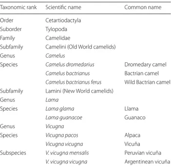
The word ‘camel’ refers to any of the members of the family Camelidae including Old World Camels (OWC) and New World Camels (syn. South American came-lids, NWC) [2]. The origin of the OWC traces back to around 40 million years ago when the first ancestors of the camelid family were found in North America before migrating via the Bering Land Bridge to the eastern hem-isphere (the “Old World”) [3] (Table 1). Compared with other animal species (e.g. dogs), the domestication of the dromedary camel took place rather late in human his-tory, approximately 3000 years ago [4]. The dromedary camel is specifically adapted to life in hot, arid areas of the world, notably the Middle East, Africa and India, with a considerable feral population in Australia [5]. Unique physiological peculiarities of dromedaries in circulatory system, respiratory system, water economy mechanism,
Open Access
*Correspondence: [email protected]
1 Department of Pathobiology, Faculty of Veterinary Science, Bu-Ali Sina University, Hamedan 6517658978, Iran
heat tolerance, etc. enable them to survive almost one week with little or no food and water [6], making them suitable also for trade and trafficking over longer dis-tances in arid areas. Indeed, they are utilized since ancient times for transportation of people, goods, war-fare and as draft animals including in agriculture and in local industry. Furthermore, they provide food (meat and dairy products) with great nutritional value, wool and leather in regions of the globe where the common rumi-nant livestock species (cattle, sheep and goat) cannot be used for this purpose. In the year 2017, camels produced 2,852,213 tons of milk and 630,210 tons of meat [1]. In this article “camel” refers only to “dromedary camel”.
Zoonoses parasites of camels
About 65% of the articles on zoonotic pathogens of cam-els published between 1970 and 2018 focused on Middle East respiratory syndrome (MERS), hydatidosis, brucel-losis and Rift Valley fever [7]. Camel echinococcosis is the most studied zoonotic parasitic infection affecting humans but Toxoplasma gondii, Cryptosporidium spp., Fasciola spp., Trichinella spp. and Linguatula serrata originating from camels are also considered as major public health risks [7]. Relatively few parasites of camels are specific for this host species [8], whereas many others that infect camels are (i) non-zoonotic but with a large host range; or (ii) of zoonotic concern. Transmission of zoonotic parasites includes different routes of infection such as faecal contamination (e.g. Cryptosporidium spp., Giardia duodenalis, Balantidium coli, Blastocystis spp.,
Enterocytozoon spp.), or consumption of raw or under-cooked infected tissues and milk (e.g. Toxoplasma gondii, Trichinella spp., Linguatula serrata).
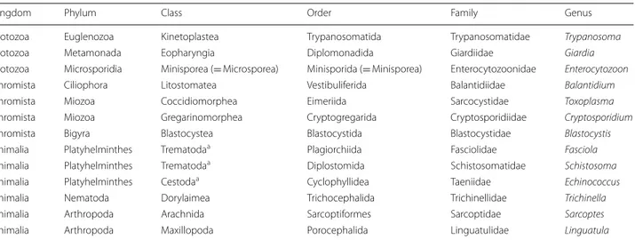
In addition, camels serve as reservoir hosts for Trypa-nosoma evansi, or may be infected by gastropod-borne trematodes (e.g. Fasciola spp., Dicrocoelium dendriti-cum and Schistosoma spp.) or metacestode larvae of zoonotic tapeworms, such as Echinococcus granulosus (s.l.). Moreover, camels are a blood source for several haematophagous ectoparasites, such as ticks and fleas, which ultimately may transmit zoonotic viral and bacte-rial pathogens (e.g. Crimean-Congo hemorrhagic fever virus, Coxiella burnetii, Anaplasma spp., Rickettsia spp., Bartonella spp. and Yersinia pestis) [9]. These parasites and infections have been detected in camels in Asia and Africa while there is not much known about the parasite fauna of camels in Australia (see section “Parasites of camels in Australia” below). The current taxonomic sta-tus of the zoonotic parasites discussed in this article is shown in Table 2.
Protozoan parasites in camel faeces
A wide range of gastrointestinal protozoan parasites develop exclusively in camels [11]. Although scientific data are available about infections of camels with several species of parasites of zoonotic importance (e.g. Crypto-sporidium spp., Giardia duodenalis, Blastocystis spp., B. coli, Enterocytozoon bieneusi) their impact on human health has not been confirmed in ad-hoc studies except only one documentation on zoonosis linked with cam-els and that is from Iran [12]. Undoubtedly, the most investigated gastrointestinal protozoan genus in cam-els is Cryptosporidium [13]. Cryptosporidiosis is one of the major zoonotic parasites associated with food-borne and water-borne outbreaks [14]. Of the 39 valid spe-cies and approximately 70 host-adapted Cryptosporid-ium genotypes (which do not yet have species names), over 20 have been identified in human patients causing asymptomatic or mild to severe gastrointestinal disease [15]. So far, C. parvum subtype IIaA17G2R1 (a common zoonotic subtype reported in humans and animals world-wide), C. parvum genetically related to the C. hominis If subtype family, C. andersoni, Cryptosporidium rat geno-type IV and a novel genogeno-type (named “camel genogeno-type”) have been confirmed in dromedary camels by PCR and sequencing [13, 16–18]. There is only one documenta-tion of zoonosis linked with camels from Iran where 24 of 100 people in long-term contact with camels were found infected with Cryptosporidium spp. [12]. Although C. parvum and C. andersoni identified in camels are potentially infectious for humans, no confirmed direct
Table 1 Taxonomic classification of camelids and other
artiodactylids [2]
Taxonomic rank Scientific name Common name Order Cetartiodactyla
Suborder Tylopoda Family Camelidae
Subfamily Camelini (Old World camelids)
Genus Camelus
Species Camelus dromedarius Dromedary camel
Camelus bactrianus Bactrian camel
Camelus bactrianus ferus Wild Bactrian camel Subfamily Lamini (New World camelids)
Genus Lama
Species Lama glama Llama
Lama guanacoe Guanaco
Genus Vicugna
Species Vicugna pacos Alpaca
Vicugna vicugna Vicuña
Subspecies V. vicugna mensalis Peruvian vicuña
association between camels and human infections have been reported, in contrast to other livestock such as cat-tle [19].
Giardia infection is extremely common in both industrialised nations and developing countries, and is responsible for about 280 million human cases of diar-rhoea every year [20]. Currently eight species with over 40 animal host species have been reported, of which only Giardia duodenalis infects humans [20]. There is only one report on microscopical diagnosis of Giardia cysts and trophozoites in dromedary camels [21] with no molecular-based study on the species and genotypes. Giardia duodenalis assemblages A and B are the pre-dominant assemblages in humans, but assemblage E is increasingly reported from human patients and assem-blages C, D and F have occasionally been identified from human patients [22]. It is predictable that camels, like other hoofed animals, are primarily infected with the zoonotic assemblage E, but the occurrence of other assemblages could clarify the zoonotic potential of camel giardiosis.
Balantidiosis caused by B. coli is a zoonotic disease and pigs, non-human primates and humans are known as pri-mary reservoirs [23]. Indeed, human populations living in close proximity to domestic pigs are naturally resistant and mostly without any clinical manifestation, though a case fatality rate of 30% has been reported in acute bal-antidiosis with intestinal perforation or fulminating haemorrhagic dysentery and shock [23]. In absence of pig raising, such as in some Middle-Eastern countries, cam-els play a major epidemiological role in the transmission of B. coli [24]. Because of the pleomorphism of balanti-dial trophozoites and the host range, taxonomy of this genus is controversial. However, as for other mammalian
hosts [25] balantidia from camels, previously named Bal-antidium cameli [26], are now referred to as B. coli, a spe-cies causing widespread infection with infection rates of up to 23% [27]. Recent studies on the genetic diversity of Balantidium spp. and Balantidium-like cyst-forming ciliates, such as Buxtonella, suggest that genetic analyses are needed to explain the real spectrum of intestinal cili-ates as the cysts are morphologically indistinguishable. Buxtonella sulcata, another ciliate with a worldwide dis-tribution, is mainly found in the cecum of cattle but also of camels [28]. Finding of Buxtonella-like ciliates in pri-mates opened the hypothesis that Buxtonella may also be a pathogen in humans [29], and the possible transmission from camels to humans should be further investigated.
The genus Blastocystis comprises at least 17 differ-ent ribosomal lineages or subtypes (ST1-ST17), which are arguably separate species [30]. These parasites are estimated to colonize between 1 and 2 billion people globally, with prevalence rates ranging from 5–15% to 50–100% in developed and developing countries, respec-tively [30, 31]. Humans become infected with ST1 to ST9; however, over 90% of reports are associated with ST1– ST4 [31]. Infections of camels with Blastocystis spp. have been reported from Australia [32], Libya [33] and Egypt [34] where ST1, ST3, ST5, ST10, ST14, ST15 or a mixture of them were identified. Interestingly, camel is the host infected with the widest range of STs out of 53 species examined [33]. Three subtypes found in camels (i.e. ST1, ST3 and ST5) can infect humans, suggesting their poten-tial role in transmitting zoonotic subtypes.
Microsporidia are diverse emerging opportunistic pathogens with 200 genera and 1500 species, 17 of which infect humans [35]. Of these, Enterocytozoon bieneusi, a ubiquitous protozoan that infects the gastrointestinal
Table 2 Taxonomic status of major zoonotic parasites of camels discussed in this article as classified by Ruggiero et al. [10]
a Neoophora sensu Ruggiero et al. [10]
Kingdom Phylum Class Order Family Genus
Protozoa Euglenozoa Kinetoplastea Trypanosomatida Trypanosomatidae Trypanosoma
Protozoa Metamonada Eopharyngia Diplomonadida Giardiidae Giardia
Protozoa Microsporidia Minisporea (= Microsporea) Minisporida (= Minisporea) Enterocytozoonidae Enterocytozoon
Chromista Ciliophora Litostomatea Vestibuliferida Balantidiidae Balantidium
Chromista Miozoa Coccidiomorphea Eimeriida Sarcocystidae Toxoplasma
Chromista Miozoa Gregarinomorphea Cryptogregarida Cryptosporidiidae Cryptosporidium
Chromista Bigyra Blastocystea Blastocystida Blastocystidae Blastocystis
Animalia Platyhelminthes Trematodaa Plagiorchiida Fasciolidae Fasciola Animalia Platyhelminthes Trematodaa Diplostomida Schistosomatidae Schistosoma Animalia Platyhelminthes Cestodaa Cyclophyllidea Taeniidae Echinococcus Animalia Nematoda Dorylaimea Trichocephalida Trichinellidae Trichinella
Animalia Arthropoda Arachnida Sarcoptiformes Sarcoptidae Sarcoptes
tract of a large number of mammals, is frequently rec-ognized in humans [36]. Currently 474 distinctive E. bieneusi genotypes from 11 groups have been differenti-ated out of which Group 1 members are mainly identi-fied in humans whereas others have been suspected [35]. In the only study on dromedaries, Group 6 genotype “Macaque1” and a novel genotype named “Camel-2” (related to members of the group 8 E. bieneusi genotypes “Macaque1”, “KB5” and “Horse2”) were identified [16]. The zoonotic potential of E. bieneusi genotypes from camels, their frequency and distribution still need to be investigated.
Toxoplasma gondii in camels: what do we know?
Due to its exceptionally wide range of warm- and cold-blooded hosts, T. gondii is one of the most successful zoonotic parasites on earth [37]. Indeed, approximately 30% of the world’s human population are infected with this cosmopolitan food- and water-borne parasite [38]. In the USA alone toxoplasmosis accounts for 32,700 dis-ability-adjusted life years (DALYs) annually, being also responsible for 8% of food-borne-illnesses hospitaliza-tions with 86,700 confirmed patients and 330 deaths [39, 40]. Like other livestock, camels acquire T. gondii infections through ingestion of sporulated oocysts shed by cats or wild felids in the environment [41]. Antibod-ies against T. gondii in sera of dromedarAntibod-ies from dif-ferent countries have been determined using various techniques, reporting seroprevalences as high as 67% [42,
43]. It has been estimated that 36% of camels in Africa have anti-T. gondii antibodies [44]. Moreover, T. gondii DNA has been detected in the blood of Iranian drom-edaries [45]. Clinical and congenital toxoplasmosis, however, are limited to a few reports and probably under-estimated in camels [46–49]. Toxoplasma gondii cysts have been isolated from camel meat [43] but predilection sites of Toxoplasma cysts have not been comprehensively investigated in this host species. The rooted habits of nomadic populations of some African and Asian com-munities of raw camel liver consumption [43, 50] sug-gest that this could represent a risk factor for infection of humans, as T. gondii is frequently isolated from the livers of domestic ruminants and horses [51]. In addition, con-sumption of camel milk is becoming increasingly popular in recent years because it is richer in vitamin C and iron than cow’s milk, with important therapeutic effects for the treatment of type 1 diabetes and reduction of aller-gies in children [52]. The implication of unpasteurized camel milk as a source of human toxoplasmosis [53] sug-gests that consuming raw milk or dairy products without pasteurization or heat treatment (e.g. Shubat) could be a risk for human health. Little is known about the genetic characteristics of T. gondii genotypes infecting camels.
Some surveys showed the occurrence of all three con-ventionally defined clonal lineages (Types I, II and III) in camel meat and milk [18, 54, 55]. All of these types have also been isolated from human patients [56]. Since the conventional nomenclature of Toxoplasma isolates does not sufficiently delineate the plethora of existing geno-types [57], multilocus PCR-RFLP genotyping should be applied to improve current understanding of the trans-mission dynamics of infected camels to people consum-ing their meat and dairy products.
Trypanosoma evansi in camels
Camels are affected by several Trypanosoma species [58]. While T. evansi, the etiologic agent of “Surra” is the more prevalent trypanosome species of camels [59], T. brucei, T. congolense and T. vivax are found at low infection rates [60, 61]. Due to a partial loss of T. evansi mitochondrial DNA, which occurred during its segregation from T. bru-cei [62], this species can be mechanically transmitted by virtually all biting flies, so its geographical distribution is potentially unlimited. Trypanosoma evansi affects a wide range of domestic and wild mammals in Africa, Asia and South America [63], and recent outbreaks of infection amongst dromedary populations on the Canary Islands, in mainland Spain and France demonstrated the poten-tial of the parasite to spread rapidly even in non-endemic areas [64]. In dromedaries, the infection may cause sig-nificant morbidity and great impairment of productiv-ity and mortalproductiv-ity [65]. It is assumed that the spread of T. evansi among camels with the consequence of fatal anae-mia weakened the Arab-African Muslim forces in their prolonged battle against Christendom, as they relied heavily on camels and equids for transport and economy [66]. Trypanosoma evansi has its highest prevalence in camels compared to other animal hosts such as buffaloes, cattle, dogs, equids and small ruminants [63], but in con-trast to other livestock species, in camels the economic burden of this infection has not been evaluated [67]. Human cases of T. evansi infection have been reported from India, Sri Lanka, Egypt and Thailand [68–70]. For a decade it was hypothesized that human susceptibility to T. evansi could be linked to insufficient or missing lev-els of human trypanocide apolipoprotein L1 (APOL1), a trypanocidal component of normal human serum [71]. However, a recent report of infection in a patient with no previous immunological risk, 2 wild-type APOL1 alleles and a normal serum APOL1 concentration suggested that T. evansi is a true zoonosis with a risk of infection for the general population [70].
Traditionally tabanids, muscids and hippoboscids are considered to be mechanical vectors of T. evansi, and several species of the genera Ancala, Atylotus, Chrys-ops, Haematobia, Haematopota, Hippobosca, Pangonia,
Philoliche, Stomoxys and Tabanus have been collected directly from camels or their surroundings [9, 72–74]. Stomoxys calcitrans, Stomoxys niger, Tabanus taeniola, Tabanus par and Tabanus subangustus collected on cat-tle have been identified to be infected with T. evansi [75]. However, there is no molecular confirmation on the role of definite fly species as vectors of T. evansi in camels.
Hydatidosis in camels
Cystic echinococcosis (CE) is a major zoonotic infection with worldwide distribution caused by the larvae of the tapeworm Echinococcus granulosus (s.l.). CE causes con-siderable medical costs and economic losses in endemic areas. Camels are intermediate hosts for several zoonotic Echinococcus species, being important in their epidemi-ology [76]. Cysts are commonly found in the lungs and, to a lesser extent, the liver of camels, resulting in carcass condemnation and, subsequently, great economic losses. In Iran where CE is endemic, the annual monetary bur-den of CE has been estimated at 232.3 million USD, out of which the loss due to condemnation of infected camel livers amounts to approximately 600,000 USD [77]. Infec-tion of camels with cysts of E. granulosus (s.s.), E. ortleppi and E. canadensis (formerly G1, G2, G3, G5, G6) have been reported [78–80], all of them being causative agents of human CE [80]. The prevalence of Echinococcus spe-cies infecting dromedaries differs in various studies, i.e. in studies from Iran and Ethiopia the most prevalent species isolated from camels was E. granulosus [81, 82], while in Nigeria and Oman most of the isolates were identified as E. canadensis [83, 84]. A comprehensive review of all the available information about Echinococcus species infect-ing dromedaries is needed to show which species occur more commonly in each region, continent and at global scale.
Linguatulosis in camels
Humans become infected with the cosmopolitan pen-tastomid Linguatula serrata by ingestion of eggs from the faeces of infected dogs or consumption of raw or undercooked infected viscera of intermediate ruminant hosts and camels [85]. Human nasopharyngeal infection by the so-called tongue-worm can affect the nasophar-ynx, throat, eyes, lymph nodes, nose, oral cavity, lungs and liver [85]. Reliable data on the rate and geographical range of the infection in dogs are unavailable and diagno-sis is challenging as infections in dogs are often asympto-matic [86]. However, clinical linguatulosis is increasingly reported in pet and stray dogs worldwide [87] and zoonotic cases of L. serrata infection are recorded from several countries in Asia, Europe, Africa and the Ameri-cas [85]. In camels, infection with L. serrata larvae in the liver, lungs, spleen, mesenteric and mediastinal lymph
nodes have been reported from Iran, Egypt and Sudan [79, 85]. Although consumption of improperly cooked and raw liver of infected intermediate hosts are major sources of zoonotic infection, the actual sources of infec-tion in human patients have not been documented suf-ficiently, and camels may play an important role in the epidemiology of human and canine linguatulosis.
Trichinellosis in camels
The genus Trichinella comprises nine species and three genotypes occurring in birds, reptiles and more than 150 domestic and wild mammalian species [88]. Some species within the genus cause a meat-borne zoonosis responsi-ble for 5751 cases and five deaths per year [88]. Although human risk for trichinellosis has historically been linked to Trichinella spiralis acquired from domestic pig or wild boar, meat of other omnivorous or carnivorous animals, but also from herbivorous domestic livestock and horses, have been implied in the occurrence of human trichinel-losis [89]. Camelus sp. was listed as host of T. spiralis in India in 1977 [90], and as a sequel to a severe outbreak of trichinellosis in Germany attributed to spiced and dried camel meat, illegally imported from Cairo, a few confirm-ing studies were conducted [91]. While, the camel origin of the exotic dish incriminated for that outbreak could not been confirmed, experimental infection of camels with T. spiralis from pork meat resulted in a high Trich-inella burden of the smooth and striated muscles [92]. Based on these finding and on the fact that meat from sheep, cattle and horse has been recognized as source of human trichinellosis in several outbreaks [89], the role of camels in the epidemiology of trichinellosis needs further investigation. This should be a priority, also consider-ing that eatconsider-ing raw camel meat is popular among camel nomads in some regions, and severe foodborne outbreaks of plague have occurred due to this habit [93].
Zoonotic gastropod‑borne trematodes in camels
Diseases caused by gastropod-borne helminths are esti-mated to affect more than 300 million people worldwide [94]. Camels can potentially play a role in the main-tenance and transmission of several gastropod-borne trematodes in areas where both parasites and hosts are present. Fasciolosis is a food- and water-borne disease caused by Fasciola hepatica and Fasciola gigantica liver flukes. Human fascioliasis is an important public health problem and is considered a highly neglected tropical disease with estimated 35 to 72 million people infected worldwide [95]. Infections of camels with both fluke spe-cies with prevalences of up to 15% have been recorded [79, 96]. Meanwhile, reports of human infections with D. dendriticum flukes are increasing, mainly due to the expansion of arid areas and the increase in anthelmintic
resistance [97]. Dicrocoeliosis in humans is poorly known and probably underestimated; however, infections have been reported from several countries [98] and many ani-mal species, including camels, have been demonstrated to harbor fertile adult flukes and excrete eggs with their faeces [99, 100].
Schistosomiasis is an infectious disease that affects more than 230 million people worldwide [101]. Four Schistosoma species, Schistosoma bovis, Schistosoma mattheei, Schistosoma indicum and Schistosoma turke-stanica (syn. Orientobilharzia turkestanicum, Ornitho-bilharzia turkestanicum), have been reported in camels [102–104]. Schistosoma bovis and S. mattheei have also been described in humans [105], as well as human cer-carial dermatitis caused by S. turkestanica has been reported [106].
Arthropods infesting camels
Camels can be infested by a wide range of external para-sites that irritate, injure or debilitate them [9]. Moreover, different ticks and flies are biological and mechanical vectors of several viruses, bacteria and parasites that can induce human infections [9]. Camel mange caused by Sarcoptes scabiei var. cameli is a major threat to camel health and production as it is extremely contagious [9]. It is considered second only to Surra in terms of losses in camels [107] and its transmission to humans, particularly camel attendants and riders, is well-known since ancient times [108]. Infestation of camels with ticks of the gen-era Rhipicephalus, Hyalomma, Dermacentor, Ixodes, Amblyomma, Argas, Otobius and Ornithodoros is often reported [9]. Almost all of the tick genera above encom-pass species of known or putative vectors for zoonotic pathogens [109]. For instance, viruses with zoonotic potential have been detected by molecular methods in ticks collected from camels, including Crimean-Congo hemorrhagic fever virus [110], Alkhurma hemorrhagic fever [111], Dhori virus and Sindbis virus [112], Kadam virus [113] and Toghoto virus [114]. In addition, some bacteria of public health importance such as Coxiella burnetii [115], Rickettsia spp. (R. aeschlimannii, R. afri-cae, R. sibirica mongolitimonae), Bartonella (B. bovis and B. rochalimae), Anaplasma phagocytophilum and Borre-lia burgdorferi (s.l.) [116] have been detected in the blood of camels and/or ticks parasitizing them.
As Yersinia pestis has been isolated from Xenopsylla cheopis rat fleas captured near camel corrals [50] it has been assumed that they may act as vectors for plague in camels, which in turn, can infect humans directly or carry infected fleas close to humans. Infection of cam-els with plague has been suspected for a long time [117], and the role of this animal species in outbreaks in differ-ent countries have been documdiffer-ented although infection
might show no overt symptoms [118]. Transmission of plague form camels to humans has been reported in Kazakhstan, where from 1907 to 2001, human plague was acquired from camels in 400 instances [119].
Parasites of camels in Australia
There is controversy about the estimated population of feral dromedary camels in Australia with an estimated number of between 300,000–1,200,000 camels [5, 120]. However, in-depth information about diseases of camels in Australia is scarce. In the only study available [121], cystic echinococcosis was reported with zero cases in 4915 camels examined during meat inspection in abat-toirs. According to the current knowledge, the most common parasites of camels in Australia are Sarcoptes scabiei, B. coli and camel nasal bot fly, Cephalopina tit-illator, whereas Trichuris tenuis, Camelostrongylus men-tulatus, Cooperia pectinata, Nematodirella dromedarii, Haemonchus sp., Trichostrongylus sp., Cooperia sp., Nematodirus sp., Nematodirella sp. and Eimeria cameli are less common. Cryptosporidium parvum was reported from a dromedary calf. Tapeworm infections and taeniid cysts were absent. Trypanosoma evansi was imported into Western Australia in 1907 with camels but was diag-nosed and quickly eradicated before spreading [13, 67,
122–124]. Of the parasites listed, S. scabiei, C. parvum and B. coli are zoonotic.
In recent years, export of live camels from Australia to the Middle East has increased [125]. Apart from ani-mal welfare issues, and possible intolerance of Austral-ian camels to the climate of the PersAustral-ian Gulf, the trade of animals on a global scale has implications for parasite spread. Recently it was suggested that treatment of inter-nal parasites in livestock in the country of origin may help in preventing entrance of helminths to barns, flocks and pastures in the country of destination [126]. Since not much is known about the parasite fauna of Austral-ian camels it is conceivable that certain parasite species are transferred to areas of the Middle East where they are currently absent. Conversely, the susceptibility of feral camels from Australia to the diverse parasite fauna of the Middle East upon their arrival remains to be discussed.
Conclusions
Due to their increasing importance as a livestock ani-mal in marginal, desert areas of developing countries, the role of camels in the epidemiology of zoonotic par-asitic infections needs to be further investigated, espe-cially in view of the risk factors associated with them. So far, research on parasites of camels has focused on case reports or prevalence surveys by microscopical examination of faecal samples or blood smears [21, 127,
tools and phylogenetic analyses are scarce. Considering this, it would be important to perform molecular inves-tigations on parasites of camels and of people having direct contact to them, in order to improve the current understanding of transmission dynamics in epidemio-logical studies. In addition, the role of camels as hosts for zoonotic pathogens such as Trichinella needs con-firmatory evidence, and further studies of infectivity, pathogenicity, muscle larvae distribution and antibody development are necessary to understand the role of camels in the maintenance, distribution and transmis-sion of this parasite. As a large portion of the camel population is kept in communities lacking equipment and trained personnel for carrying out parasitologi-cal examinations, there is a need for the development of rapid diagnostic tests for the detection of the most important camel parasites. Moreover, international and local organizations must work to increase the aware-ness of the zoonotic risk of camel parasites and the ways of pathogen transmission for people working in close contact with camels. Most importantly, the high risk of acquiring zoonotic infections by consumption of raw milk, meat and liver of infected camels, as well as their role in maintaining zoonotic transmission of hydatidosis must be communicated in the best possible way.
Abbreviations
OWC: old world camelids; NWC: new world camelids; MERS: Middle East res-piratory syndrome; PCR: polymerase chain reaction; DALY: disability-adjusted life year; APOL1: apolipoprotein L1; CE: cystic echinococcosis.
Acknowledgements
This article was planned under the academic agreement between the Bu-Ali Sina University Hamedan (Iran) and the University of Bari (Italy).
Authors’ contributions
AS, AJ and DO conceived the idea. AS wrote the manuscript and prepared an original draft. AJ and DO reviewed the manuscript. All authors read and approved the final manuscript.
Funding
Not applicable.
Availability of data and materials
All data generated or analyzed during this study are included in this published article.
Ethics approval and consent to participate
Not applicable.
Consent for publication
Not applicable.
Competing interests
The authors declare that they have no competing interests.
Author details
1 Department of Pathobiology, Faculty of Veterinary Science, Bu-Ali Sina University, Hamedan 6517658978, Iran. 2 Institute of Parasitology, Department of Pathobiology, University of Veterinary Medicine Vienna, Veterinaerplatz 1,
1210 Vienna, Austria. 3 Department of Veterinary Medicine, University of Bari, Str. prov. per Casamassima km 3, 70010 Valenzano, Bari, Italy.
Received: 6 September 2019 Accepted: 19 December 2019
References
1. FAOSTAT. Food and Agriculture Organization of the United Nations Statistics Division. 2019. http://www.fao.org/faost at. Accessed 27 Oct 2019.
2. Sazmand A. Molecular identification of vector-borne parasites in blood of camels (Camelus dromedarius) of Iran. Ph.D. thesis, University of Veterinary Medicine, Vienna; 2016.
3. Burger PA. The history of Old World camelids in the light of molecular genetics. Trop Anim Health Prod. 2016;48:905–13.
4. MacHugh DE, Larson G, Orlando L. Taming the past: ancient DNA and the study of animal domestication. Ann Rev Anim Biosci. 2017;5:329–51.
5. Camel-Scan: Large populations of feral camels in Australia. 2019. https ://www.feral scan.org.au/camel scan/defau lt.aspx. Accessed 27 Oct 2019. 6. Ouajd S, Kamel B. Physiological particularities of dromedary (Camelus
dromedarius) and experimental implications. Scand J Lab Anim Sci. 2009;36:19–29.
7. Zhu S, Zimmerman D, Deem SL. A review of zoonotic pathogens of dromedary camels. EcoHealth. 2019;16:356–77.
8. Schuster RK. Parasites of dromedaries and bactrian camels—a review part 1: stenoxenous parasites. J Camel Pract Res. 2018;25:1–8. 9. Wernery U, Kinne J, Schuster RK. Camelid infectious disorders. Paris:
World Organisation for Animal Health (OIE); 2014.
10. Ruggiero MA, Gordon DP, Orrell TM, Bailly N, Bourgoin T, Brusca RC, et al. Correction: a higher level classification of all living organisms. PLoS ONE. 2015;10(6):e0130114.
11. Dubey J, Schuster R. A review of coccidiosis in Old World camels. Vet Parasitol. 2018;262:75–83.
12. Sazmand A, Rasooli A, Nouri M, Hamidinejat H, Hekmatimoghaddam S. Prevalence of Cryptosporidium spp. in camels and involved people in Yazd Province, Iran. Iran J Parasitol. 2012;7:80–4.
13. Zahedi A, Lee GK, Greay TL, Walsh AL, Blignaut DJ, Ryan UM. First report of Cryptosporidium parvum in a dromedary camel calf from Western Australia. Acta Parasitol. 2018;63:422–7.
14. Ryan U, Hijjawi N, Xiao L. Foodborne cryptosporidiosis. Int J Parasitol. 2018;48:1–12.
15. Feng Y, Ryan UM, Xiao L. Genetic diversity and population structure of Cryptosporidium. Trends Parasitol. 2018;34:997–1011.
16. Baroudi D, Zhang H, Amer S, Khelef D, Roellig DM, Wang Y, et al. Divergent Cryptosporidium parvum subtype and Enterocytozoon bieneusi genotypes in dromedary camels in Algeria. Parasitol Res. 2018;117:905–10.
17. Gu Y, Wang X, Zhou C, Li P, Xu Q, Zhao C, et al. Investigation on Crypto-sporidium infections in wild animals in a zoo in Anhui Province. J Zoo Wildl Med. 2016;47:846–54.
18. El-Alfy ES, Abu-Elwafa S, Abbas I, Al-Araby M, Al-Kappany Y, Umeda K, et al. Molecular screening approach to identify protozoan and trichostrongylid parasites infecting one-humped camels (Camelus dromedarius). Acta Trop. 2019;197:105060.
19. Lal A, Dobbins T, Bagheri N, Baker MG, French NP, Hales S. Cryptosporid-iosis risk in New Zealand children under 5 years old is greatest in areas with high dairy cattle densities. EcoHealth. 2016;13:652–60.
20. Ryan U, Hijjawi N, Feng Y, Xiao L. Giardia: an under-reported foodborne parasite. Int J Parasitol. 2019;49:1–11.
21. Al-Jabr OA, Mohammed GE, Al-Hamdan BA. Giardiosis in camels (Came-lus dromedarius). Vet Rec. 2005;157:350–2.
22. Cacciò SM, Lalle M, Svärd SG. Host specificity in the Giardia duodenalis species complex. Infect Genet Evol. 2018;66:335–45.
23. Schuster FL, Ramirez-Avila L. Current world status of Balantidium coli. Clin Microbiol Rev. 2008;21:626–38.
24. Cox FEG. Human balantidiasis in Iran: are camels reservoir hosts? Trends Parasitol. 2005;21:553.
25. Nakauchi K. The prevalence of Balantidium coli infection in fifty-six mammalian species. J Vet Med Sci. 1999;61:63–5.
26. Hegner R. Specificity in the genus Balantidium based on size and shape of body and macronucleus, with descriptions of six new species. Am J Epidemiol. 1934;19:38–67.
27. Khodakaram-Tafti A, Maleki M, Oryan A. Pathological study of intestines and mesentric lymph nodes of camels (Camelus dromedarius) slaugh-tered in Iran. J Camel Pract Res. 2001;8:209–13.
28. Taylor MA, Coop RL, Wall RL. Veterinary Parasitology. 4th ed. Oxford: Wiley Blackwell; 2016.
29. Pomajbíková K, Obornik M, Horák A, Petrželková KJ, Grim JN, Levecke B, et al. Novel insights into the genetic diversity of Balantidium and Balantidium-like cyst-forming ciliates. PLoS Negl Trop Dis. 2013;7:e2140. 30. Scanlan PD, Stensvold CR. Blastocystis: getting to grips with our guileful
guest. Trends Parasitol. 2013;29:523–9.
31. Lepczyńska M, Białkowska J, Dzika E, Piskorz-Ogórek K, Korycińska J. Blastocystis: how do specific diets and human gut microbiota affect its development and pathogenicity? Eur J Clin Microbiol Infect Dis. 2017;36:1531–40.
32. Stenzel D, Cassidy M, Boreham P. Morphology of Blastocystis sp. isolated from circus animals. Int J Parasitol. 1993;23:685–7.
33. Alfellani MA, Taner-Mulla D, Jacob AS, Imeede CA, Yoshikawa H, Stensvold CR, et al. Genetic diversity of Blastocystis in livestock and zoo animals. Protist. 2013;164:497–509.
34. Mokhtar A, Youssef A. Subtype analysis of Blastocystis sppisolated from domestic mammals and poultry and its relation to transmission to their in-contact humans in Ismailia governorate, Egypt. Parasitol Unit J. 2018;11:90–8.
35. Li W, Feng Y, Santin M. Host specificity of Enterocytozoon bieneusi and public health implications. Trends Parasitol. 2019;35:436–51. 36. Matos O, Lobo ML, Xiao L. Epidemiology of Enterocytozoon bieneusi
infection in humans. J Parasitol Res. 2012;2012:981424.
37. Djurković-Djaković O, Dupouy-Camet J, Van der Giessen J, Dubey JP. Toxoplasmosis: overview from a one health perspective. Food Water-borne Parasitol. 2019;12:e00054.
38. Bahia-Oliveira L, Gomez-Marin J, Shapiro K. Toxoplasma gondii. In: Rose JB, Jiménez-Cisneros B. Global water pathogen project; 2017. http:// www.water patho genso rg/book/toxop lasma -gondi i. Accessed 27 Oct 2019.
39. Scallan E, Hoekstra R, Mahon B, Jones T, Griffin P. An assessment of the human health impact of seven leading foodborne pathogens in the United States using disability adjusted life years. Epidemiol Infect. 2015;143:2795–804.
40. Scallan E, Hoekstra RM, Angulo FJ, Tauxe RV, Widdowson MA, Roy SL, et al. Foodborne illness acquired in the United States—major patho-gens. Emerg Infect Dis. 2011;17:7–15.
41. Hamidinejat H, Ghorbanpour M, Rasooli A, Nouri M, Hekmatimo-ghaddam S, Mohammad Namavari M, et al. Occurrence of anti-Toxo-plasma gondii and Neospora caninum antibodies in camels (Camelus dromedarius) in the center of Iran. Turk J Vet Anim Sci. 2013;37:277–81. 42. Dubey JP. Toxoplasmosis of animals and humans. Boca Raton: CRC
Press; 2010.
43. Gebremedhin EZ, Yunus HA, Tesfamaryam G, Tessema TS, Dawo F, Terefe G, et al. First report of Toxoplasma gondii in camels (Camelus dromedar-ius) in Ethiopia: bioassay and seroepidemiological investigation. BMC Vet Res. 2014;10:222.
44. Tonouhewa ABN, Akpo Y, Sessou P, Adoligbe C, Yessinou E, Hounmanou YG, et al. Toxoplasma gondii infection in meat animals from Africa: systematic review and meta-analysis of sero-epidemiological studies. Vet World. 2017;10:194.
45. Khamesipour F, Doosti A, Iranpour Mobarakeh H, Komba EVG. Toxo-plasma gondii in cattle, camels and sheep in Isfahan and Chaharmahal va Bakhtiary Provinces, Iran. Jundishapur J Microbiol. 2014;7:e17460. 46. Hagemoser W, Dubey J, Thompson J. Acute toxoplasmosis in a camel. J
Am Vet Med Assoc. 1990;196:347.
47. Riley J, Garner MM, Kiupel M, Hammond EE. Disseminated toxoplasmo-sis in a captive adult dromedary camel (Camelus dromedarius). J Zoo Wildl Med. 2017;48:937–40.
48. Ishag MY, Majid A. Association of diarrhea with congenital toxoplasmo-sis in calf-camel (Camelus dromedarius). Int J Trop Med. 2008;3:10–1.
49. Ishag MY. Studies on Toxoplasma and Sarcocystis from camels (Camelus dromedaries) in the Sudan. Ph.D. thesis, University of Khartoum; 2003. 50. Saeed AAB, Al-Hamdan NA, Fontaine RE. Plague from eating raw camel
liver. Emerg Infect Dis. 2005;11:1456–7.
51. Belluco S, Mancin M, Conficoni D, Simonato G, Pietrobelli M, Ricci A. Investigating the determinants of Toxoplasma gondii preva-lence in meat: a systematic review and meta-regression. PLoS ONE. 2016;11:e0153856.
52. Boughattas S. Toxoplasma infection and milk consumption: meta-analysis of assumptions and evidences. Crit Rev Food Sci Nutr. 2017;57:2924–33.
53. Medani M, Mohamed H. Camel’s milk as a source of human toxoplas-mosis in Butana area-Sudan. Int J Infect Dis. 2016;45:471–2. 54. Elfadaly HA, Hassanan N, Shaapan RM, Hassanain MA, Barakat AM,
Abdelrahman KA. Molecular detection and genotyping of Toxoplasma gondii from Egyptian isolates. Asian J Epidemiol. 2017;10:37–44. 55. Tavakoli Kareshk A, Tavakoli Oliaee R, Mahmoudvand H, Keyhani A,
Mohammadi MA, Bamorovat M, et al. The first survey of isolation and molecular typing of Toxoplasma gondii by bioassay and PCR method in BALB/c mice in camels from eastern Iran. Iran J Parasitol. 2018;13:382–91.
56. Ajzenberg D, Yera H, Marty P, Paris L, Dalle F, Menotti J, et al. Genotype of 88 Toxoplasma gondii isolates associated with toxoplasmosis in immunocompromised patients and correlation with clinical findings. J Infect Dis. 2009;199:1155–67.
57. Shwab EK, Zhu X-Q, Majumdar D, Pena HF, Gennari SM, Dubey JP, et al. Geographical patterns of Toxoplasma gondii genetic diversity revealed by multilocus PCR-RFLP genotyping. Parasitology. 2014;141:453–61. 58. Roettcher D, Schillinger D, Zweygarth E. Trypanosomiasis in the camel
(Camelus dromedarius). Rev Sci Tech. 1987;6:463–70.
59. Desquesnes M, Holzmuller P, Lai D-H, Dargantes A, Lun Z-R, Jitta-plapong S. Trypanosoma evansi and surra: a review and perspectives on origin, history, distribution, taxonomy, morphology, hosts, and pathogenic effects. Biomed Res Int. 2013;2013:194176.
60. Dirie MF, Wallbanks K, Aden AA, Bornstein S, Ibrahim M. Camel trypano-somiasis and its vectors in Somalia. Vet Parasitol. 1989;32:285–91. 61. Birhanu H, Fikru R, Said M, Kidane W, Gebrehiwot T, Hagos A, et al.
Epi-demiology of Trypanosoma evansi and Trypanosoma vivax in domestic animals from selected districts of Tigray and Afar regions, northern Ethiopia. Parasites Vectors. 2015;8:212.
62. Lai D-H, Hashimi H, Lun Z-R, Ayala FJ, Lukes J. Adaptations of Trypano-soma brucei to gradual loss of kinetoplast DNA: TrypanoTrypano-soma equiper-dum and Trypanosoma evansi are petite mutants of T. brucei. Proc Natl Acad Sci USA. 2008;105:1999–2004.
63. Aregawi WG, Agga GE, Abdi RD, Büscher P. Systematic review and meta-analysis on the global distribution, host range, and prevalence of Trypanosoma evansi. Parasites Vectors. 2019;12:67.
64. Gutierrez C, Desquesnes M, Touratier L, Büscher P. Trypanosoma evansi: recent outbreaks in Europe. Vet Parasitol. 2010;174:26–9.
65. Sazmand A, Eigner B, Mirzaei M, Hekmatimoghaddam S, Harl J, Duscher GG, et al. Molecular identification of hemoprotozoan parasites in cam-els (Camelus dromedarius) of Iran. Iran J Parasitol. 2016;11:568–73. 66. Clarence-Smith WG. The historical spread of Trypanosoma evansi
(surra) in camels: a factor in the weakening of Islam? In: Emery E, edi-tor. Selected papers from the first international conference of camel cultures: historical traditions, present threats and future prospects. London: RN Books; 2013. p. 87–94.
67. Reid SA. Trypanosoma evansi control and containment in Australasia. Trends Parasitol. 2002;18:219–24.
68. Truc P, Büscher P, Cuny G, Gonzatti MI, Jannin J, Joshi P, et al. Atypi-cal human infections by animal trypanosomes. PLoS Negl Trop Dis. 2013;7:e2256.
69. Joshi PP, Shegokar VR, Powar RM, Herder S, Katti R, Salkar HR, et al. Human trypanosomiasis caused by Trypanosoma evansi in India: the first case report. Am J Trop Med Hyg. 2005;73:491–5.
70. Van Vinh Chau L, Buu Chau L, Desquesnes M, Herder S, Phu Huong Lan N, Campbell JI, et al. A clinical and epidemiological investigation of the first reported human infection with the zoonotic parasite Trypanosoma evansi in Southeast Asia. Clin Infect Dis. 2016;62:1002–8.
71. Vanhollebeke B, Truc P, Poelvoorde P, Pays A, Joshi PP, Katti R, et al. Human Trypanosoma evansi infection linked to a lack of Apolipoprotein L-I. N Eng J Med. 2006;355:2752–6.
72. Antoine-Moussiaux N, Desmecht D. Epidémiologie de lʼinfection par Trypanosoma evansi. Ann Méd Vét. 2008;152:191–201.
73. Luckins A. Epidemiology of surra: unanswered questions. J Protozool Res. 1998;8:106–19.
74. Oyieke F, Reid G. The mechanical transmission of Trypanosoma evansi by Haematobia minuta (Diptera: Muscidae) and Hippobosca camelina (Diptera: Hippoboscidae) from an infected camel to a mouse and the survival of trypanosomes in fly mouthparts and gut. Folia Vet. 2003;47:38–41.
75. Odeniran P, Macleod E, Ademola I, Welburn S. Molecular identification of bloodmeal sources and trypanosomes in Glossina spp., Tabanus spp. and Stomoxys spp. trapped on cattle farm settlements in southwest Nigeria. Med Vet Entomol. 2019;33:269–81.
76. Sadjjadi SM. Present situation of echinococcosis in the Middle East and Arabic North Africa. Parasitol Int. 2006;55:S197–202.
77. Fasihi Harandi M, Budke CM, Rostami S. The monetary burden of cystic echinococcosis in Iran. PLoS Negl Trop Dis. 2012;6:e1915.
78. Ebrahimpour M, Sadjjadi SM, Yousefi Darani H, Najjari M. Molecular studies on cystic echinococcosis of camel (Camelus dromedarius) and report of Echinococcus ortleppi in Iran. Iran J Parasitol. 2017;12:323–31. 79. Sazmand A, Joachim A. Parasitic diseases of camels in Iran (1931–
2017)—a literature review. Parasite. 2017;24:21.
80. Deplazes P, Rinaldi L, Rojas CA, Torgerson P, Fasihi Harandi M, Romig T, et al. Global distribution of alveolar and cystic echinococcosis. Adv Parasitol. 2017;95:315–493.
81. Khademvatan S, Majidiani H, Foroutan M, Hazrati Tappeh K, Aryamand S, Khalkhali H. Echinococcus granulosus genotypes in Iran: a systematic review. J Helminthol. 2019;93(2):131–8.
82. Tigre W, Deresa B, Haile A, Gabriël S, Victor B, Van Pelt J, et al. Molecular characterization of Echinococcus granulosus s.l. cysts from cattle, camels, goats and pigs in Ethiopia. Vet Parasitol. 2016;215:17–21.
83. Ohiolei JA, Yan H-B, Li L, Magaji AA, Luka J, Zhu G-Q, et al. Cystic echi-nococcosis in Nigeria: first insight into the genotypes of Echinococcus granulosus in animals. Parasites Vectors. 2019;12:392.
84. AlKitani FA, Baqir S, Mansoor MK, AlRiyami S, Hussain MH, Roberts D. Genetic survey of cystic echinococcosis in farm animals in Oman. Trop Anim Health Prod. 2019. https ://doi.org/10.1007/s1125 0-019-02019 -5. 85. Hajipour N, Tavassoli M. Prevalence and associated risk factors of
Linguatula serrata infection in definitive and intermediate hosts in Iran and other countries: a systematic review. Vet Parasitol Reg Stud Rep. 2019;16:100288.
86. Otranto D. Diagnostic challenges and the unwritten stories of dog and cat parasites. Vet Parasitol. 2015;212:54–61.
87. Nagamori Y, Ramachandran A, Kuzma C, Nafe L, Johnson EM. A zoonotic parasite, Linguatula serrata, infection in a dog imported from Ethiopia to the United States. Vet Parasitol Reg Stud Rep. 2019;16:100273. 88. Devleesschauwer B, Praet N, Speybroeck N, Torgerson PR, Haagsma JA,
De Smet K, et al. The low global burden of trichinellosis: evidence and implications. Int J Parasitol. 2015;45:95–9.
89. Rostami A, Gamble HR, Dupouy-Camet J, Khazan H, Bruschi F. Meat sources of infection for outbreaks of human trichinellosis. Food Micro-biol. 2017;64:65–71.
90. Chaturvedi Y, Kansal KC. Check-list of Indian nematodes (animal para-sites). Delhi: Controller of Publications; 1977.
91. Bommer W, Kaiser H, Mergerian H, Pottkämper G. Outbreak of trichinel-liasis in a youth centre in Neidersachsen by air-dried imported camel meat. In: Proceedings of the 1st world congress on food-borne infec-tions and intoxicainfec-tions. West Berlin, Germany; 1980. p. 441–4. 92. Eckhardt T, Abd-El-Rahman MS, Omar HM. Survey on the Trichinella
larvae in butcherʼs meat offered in Cairoan shops. First experimental Trichinella infection in camel. In: 9th international symposium of world association of veterinary food hygienists, Budapest, Hungary; 1985. p. 139.
93. Arbaji A, Kharabsheh S, Al-Azab S, Al-Kayed M, Amr Z, Abu Baker M, et al. A 12-case outbreak of pharyngeal plague following the consump-tion of camel meat, in north-eastern Jordan. Ann Trop Med Parasitol. 2005;99:789–93.
94. Giannelli A, Cantacessi C, Colella V, Dantas-Torres F, Otranto D. Gas-tropod-borne helminths: a look at the snail-parasite interplay. Trends Parasitol. 2016;32:255–64.
95. Sabourin E, Alda P, Vázquez A, Hurtrez-Boussès S, Vittecoq M. Impact of human activities on fasciolosis transmission. Trends Parasitol. 2018;34:891–903.
96. Banaja AA, Ghandour AM. A review of parasites of camels (Camelus dromedarius) in Saudi Arabia. J King Abdulaziz Univ. 1994;6:75–86. 97. Otranto D, Traversa D. Dicrocoeliosis of ruminants: a little known fluke
disease. Trends Parasitol. 2003;19:12–5.
98. Mas-Coma S, Bargues M. Human liver flukes: a review. Res Rev Parasitol. 1997;57:145–218.
99. Cirak V, Senlik B, Gulegen E. Gastrointestinal parasites of camels (Camelus dromedarius) from turkey and efficacy of doramectin against trichostrongyles. J Camel Pract Res. 2011;18:283–5.
100. Asadov S. Analysis of the helminth fauna of the dromedary (Camelus dromedarius L 1758) in Azerbaidzhán. Dokl Akad Nauk Azerb SSR. 1957;13:781–4.
101. Colley DG, Bustinduy AL, Secor WE, King CH. Human schistosomiasis. Lancet. 2014;383:2253–64.
102. Arfaa F, Sabaghian H, Ale-Dawood H. Studies on Ornithobilharzia turke-stanicum (Skrjabin, 1913), Price, 1929 in Iran. Ann Parasitol Hum Comp. 1965;40:45–50.
103. Graber M, Tabo R, Service J. Enquêtes sur les helminthes du droma-daire tchadien: étude des strongyloses gastro-intestinales et de lʼhaemoncose à Haemoncus longistipes. Rev Elev Med Vet Pays Trop. 1967;20:227–54.
104. Chauhan A, Srivastava C, Chauhan B. Studies on the trematode fauna of India. Part 6. Digenea: Schistosomatidae—a monographic aid to the identification of Indian schistosomes. J Zool Soc India. 1973;25:83–127. 105. Cox FE. Taxonomy and classification of human parasitic protozoa and
helminths. Manual of clinical microbiology. 11th ed. Washington: American Society of Microbiology; 2015. p. 2285–92.
106. Sahba GH, Malek EA. Dermatitis caused by cercariae of Orientobilharzia turkestanicum in the Caspian Sea area of Iran. Am J Trop Med Hyg. 1979;28:912–3.
107. Pegram R, Higgins AJ. Camel ectoparasites: a review. In: Allen W, Higgins A, Mayhew I, Snow D, Wade J, editors. Proceeding of the First Interna-tional Camel Conference. Dubai: R&W Publications (New Market); 1992. p. 69–78.
108. Tadjbakhsh H. Traditional methods used for controlling animal diseases in Iran. Rev Sci Tech. 1994;13:599–614.
109. Dantas-Torres F, Chomel BB, Otranto D. Ticks and tick-borne diseases: a one health perspective. Trends Parasitol. 2012;28:437–46.
110. Champour M, Chinikar S, Mohammadi G, Razmi G, Shah-Hosseini N, Khakifirouz S, et al. Molecular epidemiology of Crimean-Congo hemorrhagic fever virus detected from ticks of one humped camels (Camelus dromedarius) population in northeastern Iran. J Parasites Dis. 2016;40:110–5.
111. Charrel RN, Fagbo S, Moureau G, Alqahtani MH, Temmam S, De Lambal-lerie X. Alkhurma hemorrhagic fever virus in Ornithodoros savignyi ticks. Emerg Infect Dis. 2007;13:153.
112. Al-Khalifa MS, Diab FM, Khalil GM. Man-threatening viruses isolated from ticks in Saudi Arabia. Saudi Med J. 2007;28:1864–7.
113. Wood OL, Moussa MI, Hoogstraal H, Büttiker W. Kadam virus (Togaviri-dae, Flavivirus) infecting camel-parasitizing Hyalomma dromedarii ticks (Acari: Ixodidae) in Saudi Arabia. J Med Entomol. 1982;19:207–8. 114. Williams R, Hoogstraal H, Casals J, Kaiser M, Moussa M. Isolation of
Wanowrie, Thogoto, and Dhori viruses from Hyalomma ticks infesting camels in Egypt. J Med Entomol. 1973;10:143–6.
115. Loftis AD, Reeves WK, Szumlas DE, Abbassy MM, Helmy IM, Moriarity JR, et al. Rickettsial agents in Egyptian ticks collected from domestic animals. Exp Appl Acarol. 2006;40:67–81.
116. Sazmand A, Harl J, Eigner B, Hodžić A, Beck R, Hekmatimoghaddam S, et al. Vector-borne bacteria in blood of camels in Iran: new data and literature review. Comp Immunol Microbiol Infect Dis. 2019;65:48–53. 117. Fedorov V. Plague in camels and its prevention in the USSR. Bull World
Health Organ. 1960;23:275–81.
118. Stenseth NC, Atshabar BB, Begon M, Belmain SR, Bertherat E, Carniel E, et al. Plague: past, present, and future. PLoS Med. 2008;5:e3.
•fast, convenient online submission •
thorough peer review by experienced researchers in your field • rapid publication on acceptance
• support for research data, including large and complex data types •
gold Open Access which fosters wider collaboration and increased citations maximum visibility for your research: over 100M website views per year •
At BMC, research is always in progress. Learn more biomedcentral.com/submissions
Ready to submit your research? Choose BMC and benefit from:
119. Aikimbayew A, Meka-Mechenko T, Temiralieva G, Bekenov J, Sagiyev Z, Kaljan K, et al. Plague peculiarities in Kazakhstan at the present time. Przeg Epidemiol. 2003;57:593–8.
120. McGregor M, Hart Q, Bubb A, Davies R. Managing the impacts of feral camels across remote Australia: final report of the Australian Feral Camel Management Project. Ninti One Limited; 2013. https ://www.ninti one.com.au/resou rce/Manag ingIm pacts Feral Camel s_Final Repor tAFCM P.pdf. Accessed 27 Oct 2019.
121. Brown A. A review of camel diseases in central Australia. Department of Business, Industry and Resource Development; 2004. https ://dpir. nt.gov.au/__data/asset s/pdf_file/0014/23322 2/tb314 .pdf. Accessed 27 Oct 2019.
122. Beveridge I, Green P. Species of Trichuris in domestic ruminants in Australia. Aust Vet J. 1981;57:141–2.
123. Beveridge I, Barker I, Rickard M, Burton J. Experimental infection of sheep with Camelostrongylus mentulatus and associated gastritis. Aust Vet J. 1974;50:36–7.
124. Barton M. Nasal and gastro-intestinal parasites of the camel (Came-lus dromedarius) from Central Australia. T Royal Soc South Aust. 2008;132:40–2.
125. Australian Government. Camel coup for Ngaanyatjarra Camel Com-pany. https ://www.ilc.gov.au/ngaan yatja rraca mels. 2017. Accessed 27 Oct 2019.
126. Sazmand A. International movement of livestock and lack of regulation for internal parasites monitoring. Iran J Parasitol. 2019;14:682–3. 127. Tajik J, Nourollahi Fard SR, Paidar A, Anousheh S, Dehghani E.
Balantidia-sis in a dromedarian camel. Asian Pac J Trop Dis. 2013;3:409–12. 128. Sazmand A, Rasooli A, Nouri M, Hamidinejat H, Hekmatimoghaddam
S. Serobiochemical alternations in subclinically affected dromedary camels with Trypanosoma evansi in Iran. Pak Vet J. 2011;31:223–6.
Publisher’s Note
Springer Nature remains neutral with regard to jurisdictional claims in pub-lished maps and institutional affiliations.