ORIGINAL ARTICLE
The interleukin (IL)-31/IL-31R axis contributes to tumor
growth in human follicular lymphoma
E Ferretti1, C Tripodo2, G Pagnan1, C Guarnotta2, D Marimpietri1, MV Corrias1, D Ribatti3, S Zupo4, G Fraternali-Orcioni5, JL Ravetti5, V Pistoia1,6and A Corcione1,6
Interleukin (IL)-31A binds to an heterodimer composed of IL-31 receptor A (IL-31RA) and Oncostatin M Receptor (OSMR). The IL-31/ IL-31R complex is involved in the pathogenesis of various skin diseases, including cutaneous T-cell lymphoma. No information is available on the relations between the IL-31/IL-31R complex and B-cell lymphoma. Here we have addressed this issue in follicular lymphoma (FL), a prototypic germinal center(GC)-derived B-cell malignancy. IL-31 enhanced primary FL cell proliferation through IL-31R-driven signal transducer and activator of transcription factor 1/3 (STAT1/3), extracellular signal–regulated kinase 1/2 (ERK1/2) and Akt phosphorylation. In contrast, GC B cells did not signal to IL-31 in spite of IL-31R expression. GC B cells expressed predominantly the inhibitory short IL-31RA isoform, whereas FL cells expressed predominantly the long signaling isoform. Moreover, GC B cells lacked expression of other IL-31RA isoforms potentially involved in the signaling pathway. IL-31 protein expression was significantly higher in surface membrane than in cytosol of both FL and GC B cells. IL-31 was detected in plasma membrane microvesicles from both cell types but not released in soluble form in culture supernatants. IL-31 and IL-31RA expression was higher in lymph nodes from FL patients with grade IIIa compared with grade I/II, suggesting a paracrine and/or autocrine role of IL-31/IL-31RA complex in tumor progression through microvesicle shedding.
Leukemia (2015)29, 958–967; doi:10.1038/leu.2014.291
INTRODUCTION
Interleukin-31 (IL-31) is a member of the IL-6 cytokine superfamily originally identified as the product of activated T helper type 2 cells.1 Additional cell types such as monocytes, macrophages, immature and mature dendritic cells and mast cells produce IL-31 following activation.2–5 Increased IL-31 expression has been detected in inflamed tissues from patients with atopic dermatitis, bowel diseases, allergic asthma and rhinitis.6–12
The activity of IL-31 is mediated through an heterodimeric receptor composed of a gp130-like receptor chain, IL-31 receptor A (IL-31RA) and the Oncostatin M Receptor (OSMR).13–15 Expres-sion of IL-31RA mRNA is found in specific tissues, including the skin, testis, bone marrow, brain and thymus.1,13,15 Furthermore, tumor cell lines derived from osteosarcoma, glioblastoma, melanoma and myelomonocytic leukemia express IL-31RA mRNA,1,13–15 whereas OSMR is ubiquitously expressed in human tissues and organs.13,15 Engagement of the IL-31R with IL-31 results in the activation of Janus-activated kinase 1 (JAK1) and, to a minor extent, of JAK2 followed by activation of signal transducer and activator of transcription factor 1 (STAT1)/3/5, mitogen-activated protein kinase (MAPK), and phosphatidylinositol 3′-kinase (PI3K) signaling pathways.10,15–17 Co-expression of the IL-31RA and OSMR chains is essential for IL-31 signaling, because IL-31RA activates STAT phosphorylation while the activation of phosphatidylinositol 3′-kinase and mitogen-activated protein kinase pathways is mediated by OSMR.18
Different IL-31RA isoforms have been so far identified, of which a long signaling (745 aa residues) and a short non-signaling (560 aa residues) isoforms are the best characterized.13,19,20
Recent studies have linked IL-31 to human malignant lympho-mas of T-cell lineage. It has been shown that malignant T-cell populations from cutaneous T-cell lymphoma produce IL-31, whose serum levels are increased in the same patients.21,22No information is so far available on the relationship between IL-31 and B-cell lymphoma.
Here we have investigated the expression and function of the IL-31/IL-31R complex in follicular lymphoma (FL) as a prototypic model of mature B-cell malignancy.23,24
MATERIALS AND METHODS Patients and controls
Invaded lymph nodes from 25 FL patients (15 males and 10 females, age range: 46–61 years), biopsied for diagnostic purposes, were obtained from the San Martino Hospital-Istituto Scientifico Tumori Biobank (Genova, Italy). Diagnosis of FL was established according to the criteria of the Revised European-American Classification of Lymphoid Neoplasms.23–25 Histo-pathological analysis showed that 11 FL patients had grade I; 7 FL patients grade II; and 7 FL patients grade III disease. All patients were studied at diagnosis and were untreated.
Lymph node mononuclear cells (MNCs) were isolated from FL patients as reported.26 Staining for immunoglobulin (Ig) light chains showed that monoclonal B cells expressing eitherκ (10 of 25 cases) or λ (15 of 25 cases)
1
Laboratorio di Oncologia, Istituto Giannina Gaslini, Genova, Italy;2Tumor Immunology Unit, Department of Health Science, Human Pathology Section, University of Palermo,
Palermo, Italy;3
Department of Basic Medical Sciences, Neuroscience and Sensory Organs, University of Bari Medical School, Bari and National Cancer Institute "Giovanni Paolo II",
Bari, Italy;4
Laboratory of Diagnostics of Lymphoproliferative Disorders, National Cancer Research Institute, Genova, Italy and5
Unit of Pathology, IRCCS Azienda Ospedaliera Universitaria San Martino—IST—Istituto Nazionale per la Ricerca sul Cancro, Genova, Italy. Correspondence: Dr A Corcione, Laboratorio di Oncologia, Istituto Giannina Gaslini, Via Giannina Gaslini 5, Genova 16148, Italy.
E-mail: [email protected] 6
These two authors shared last authorship.
Received 31 March 2014; revised 19 August 2014; accepted 30 September 2014; accepted article preview online 6 October 2014; advance online publication, 28 October 2014
light chain represented at least 85% of CD19+cells in all samples. FL MNCs were cryopreserved in a freezing solution composed of 50% RPMI 1640 (Sigma Chemical Co., St Louis, MO, USA), 40% fetal bovine serum (Sigma) and 10% dimethyl sulfoxide (Sigma). Cells were kept in liquid nitrogen until tested.
Ten tonsil samples surgically removed for localized inflammation and three peripheral blood samples from healthy donors were obtained following informed consent. The study was approved by the Institutional Review Board of Istituto Giannina Gaslini, Genova, Italy on 27 October 2005. Four reactive lymph nodes with follicular hyperplasia were obtained from San Martino Hospital-Istituto Scientifico Tumori Biobank (Genova, Italy).
Cell isolation and cultures
After thawing, lymphoma B cells were purified by removing: (i) residual normal B cells according to the expression of the Ig light chain not expressed by the malignant clone and (ii) other contaminant cell types (that is, CD3+, CD56+ and CD14+cells), through immunomagnetic bead manipulation (Miltenyi Biotec, Bergisch Gladbach, Germany). Tonsil B cells were isolated as reported27and enriched for germinal center (GC) B cells (CD19+, CD38high, CD10+, CD44−cells) by centrifugation on a discontinuous (60–30%) Percoll (Pharmacia, Uppsala, Sweden) density gradient followed by depletion of CD39+naive and memory B cells.28,29B cells, purified as above contained consistently 497% GC B cells, with a centroblast/ centrocytes ratio of approximately 3/1, as assessed by staining for the CD77 marker, specific for centroblast. All of the steps for GC B-cell isolation were performed at 4 °C to prevent spontaneous apoptosis. GC B cells were also isolated from the lymph nodes of individuals with reactive follicular hyperplasia as reported above.28,29In some experiments, non-B cells were isolated from FL patients (n = 4) and tonsil MNCs (n = 4) by negative selection with a CD20 mAb (Beckman Coulter, Brea, CA, USA) followed by immunomagnetic bead manipulation (Miltenyi Biotec). CD4+T cells were isolated from peripheral blood MNCs using anti-CD4 microbeads (Myltenyi Biotec) following the manufacturer’s protocol and stimulated in vitro for 48 h with an anti-CD3 mAb (OKT3, coated overnight on 96-well plates).
The details for the U937, EA.hy926, DU145, T84 and K562 cell lines used in the study are reported in Supplementary Methods.
Antibodies forflow cytometry
The list of monoclonal antibodies (mAbs) used throughout the study is shown in Supplementary Methods. Cells were stained with fluorochrome-conjugated or unfluorochrome-conjugated antibodies followed by secondary reagents. Isotype and fluorochrome-matched antibodies were tested as controls. Cells were run on a Gallios Instrument (Beckman Coulter), and data were analyzed using the Kaluza software (Beckman Coulter). Data were expressed as the percentage of positive cells.
Reverse transcriptase–PCR (RT-PCR)
Total RNA was isolated using the RNeasy kit (Qiagen, Milano, Italy) according to the manufacturer’s instructions. RNA was assessed for integrity by gel electrophoresis and quantified by spectrophotometry (Nanodrop Products, Wilmington, DE, USA). One microgram of total RNA was reverse transcribed using the High Capacity cDNA Reverse Transcrip-tion kit (Life Technologies, Monza, Italy), according to the manufacturer’s instructions.
The primer sequences for human IL-31, IL-31RA, OSMR, IL-31RA short and long isoforms and GAPDH (glyceraldehyde 3-phosphate dehydrogen-ase) mRNA and the relative PCR conditions were as described.1,19,30,31 Primers’ sequences designed to identify the five different variants of IL-31RA mRNA are shown in Figure 3a (NCBI Unigene database),20together with the sequences of the primers for IL-31RA short and long isoforms. All the primers were purchased from TIB Molbiol (TIB MolBiol S.r.L., Genova, Italy). The amplified products were visualized by electrophoresis on a 2% agarose gels. mRNA expression levels of IL-31RA short and long isoforms were quantified by scanning densitometry using the VersaDoc instrument (BioRad Laboratories, Segrate, Italy). PCR reactions for each sample were performed at least twice.
Immunohistochemistry and immunofluorescence
Formalin-fixed and paraffin-embedded tissue sections from four tonsils with reactive follicular hyperplasia and four lymph nodes with FL were collected from the archives of the Human Pathology Section, Department of Health Sciences, University of Palermo, Palermo, Italy. The immuno-histochemistry techniques are detailed in Supplementary Methods. In
addition, 20 FL lymph node samples (n = 6 grade I; n = 6 grade II; n = 8 grade III) were selected for tissue microarray (TMA) preparation. TMAs were used to investigate the expression of IL-31 and IL-31RA in relation to FL grading. See Supplementary Methods for TMA construction and immunofluorescence assay.
Cell signaling
Phosphorylation was investigated byflow cytometry in FL and tonsil GC B cells. See Supplementary Methods for detailed technique.
Cell proliferation and apoptosis
Purified FL cells were cultured in RPMI 1640 medium for 72 h with or without 20, 50 and 100 ng/ml hrIL-31 in combination or not with 100 ng/ml hrCD40L (Immunotools, Friesoythe, Germany) and tested for proliferation by overnight pulse with 0.5μCi/well tritiated thymidine (3H-TdR) or for apoptosis. See Supplementary Methods for detailed technique.
Enzyme-linked immunosorbent assay (ELISA)
IL-31 production was tested by ELISA in the supernatants of FL and tonsil and reactive lymph node GC B cells. See Supplementary Methods for detailed technique.
IL-31R internalization assay
FL and tonsil GC B cells were cultured at 37 °C for 15, 30, 60 min, 3 h and 6 h with or without 100 ng/ml hrIL-31 and then was investigated for IL-31RA and OSMR internalization. See Supplementary Methods for detailed technique.
Western blotting analysis
Total cell lysates were prepared using the Cell Extraction Buffer (Life Technologies) containing a protease inhibitor cocktail (Sigma) and analyzed by western blotting as previously described.32To obtain subcellular fractions, cells were processed with the Qproteome Cell Compartment kit according to the manufacturer’s instructions (Qiagen) and resuspended with the above-mentioned lysis buffer. Additional details are provided in Supplementary Methods.
Microvesicles (MVs) purification, analysis and signaling activity on FL cells
MVs were isolated from culture supernatants of FL cells, tonsil GC B cells and the EA.hy926, Oci-Ly8 and K562 cell lines by differential centrifugation.33,34 Additional technical details are provided in Supplementary Methods. The signaling activity of MVs was subsequently analyzed. FL cell suspensions from three patients (two with grade II and one with grade IIIa disease) were stimulated for 0 and 30 min with 100 ng/ml hrIL-31 or different dilutions (1:1, 1:2, 1:5) of MVs isolated from the Oci-LY8 and EA.hy926 cell lines. The signaling activity was evaluated as STAT-1 phosphorylation in target cells.
Statistical analysis
Data are reported in box plot in terms of medians,first and third quartiles and minimun and maximum values. For some experiments, data are represented in histograms as mean ± s.d. The Mann–Whitney U test was used to compare quantitative variables between the two groups of observation. All statistical tests were two tailed, and a P valueo0.05 was considered statistically significant. Statistical analyses were performed using the Graph Pad Prism 5 software (La Jolla, CA, USA).
RESULTS
Characterization of IL-31 receptor in FL B cells
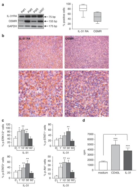
First, we investigated the expression of IL-31RA and OSMR mRNAs in malignant B cells purified from five invaded lymph nodes of FL patients. As shown in Figure 1a left panel, RT-PCR amplification products of the expected length (159 and 155 bp for IL-31RA and OSMR, respectively) were detected in FL cell suspensions, as well as in the U937 cell line tested as a positive control.13 Flow cytometric analysis showed the expression of both IL-31RA
and OSMR proteins in FL cell suspensions (median percentage of IL-31RA+cells = 82.0, range 47–99; median percentage of OMSR+ cells = 48.5, range 22–69; n = 25) (Figure 1a, right panel). A representative histogram for both IL-31RA and OSMR chains is shown in Supplementary Figure S1A. The U937 and T84 cell lines were tested as positive and negative10 controls, respectively (Supplementary Figures S1B and C).
Expression of IL-31R was then assessed by immuno-histochemistry in six lymph node samples from FL patients. In the representative experiment shown in Figure 1b, both IL-31RA and OSMR were diffusely detected in lymphomatous cells with a combined cytoplasmic and membrane staining pattern (Figure 1b, left and right panels, respectively). % positiv e cells IL-31 RA OSMR 0 20 40 60 80 100 IL-31RA Pt#1 Pt#2 Pt#3 U937 75 bp GAPDH a b c d 175 bp 155 bp OSMR 0 10 20 30 40 50 0 20 40 60 80 0 20 40 60 80 0 20 40 60 80 100 120 % p-ERK1/2 + cells % p-ST A T 1 + cells % p-ST A T 3 + cells % p-Akt + cells 0’ 1’ 10’ 30’ 60’ IL-31 0’ 1’ 10’ 30’ 60’ IL-31 0’ 1’ 10’ 30’ 60’ IL-31 0’ 1’ 10’ 30’ 60’ IL-31 cpm * **** ** * ** ** ** ** ** *** *** medium CD40L IL-31 0 1000 2000 3000 4000 5000 6000 7000 IL-31 RA OSMR
Figure 1. Expression and function of IL-31R in malignant FL cells. (a) Left panel. mRNA expression for IL-31RA, OSMR and GAPDH genes was detected by RT-PCR in FL cells isolated from patient (Pt) lymph nodes. Thefigure shows the results obtained with three FL cell suspensions out of thefive tested. The U937 cell line was tested as a positive control. Right panel. Surface expression of IL-31RA and OSMR was determined on purified FL cells by flow cytometry. Results are expressed in box plot as the median percentage of positive cells, first and third quartiles and maximum and minimum values, from 25 different FL patients. (b) Immunohistochemical analysis of IL-31RA and OSMR in lymph node tissue section from FL patients (one representative staining out of the six performed). Original magnification, × 200 (upper panels) and × 630 (lower panels). (c) Purified FL cells were incubated for 0, 1, 10, 30 and 60 min with 100 ng/ml hrIL-31 and subjected to flow cytometric analysis using antibodies to p-Erk1/Erk2, -STAT1, -STAT3 and -Akt. Results are shown as the mean percentage of positive cells± s.d. from six different experiments. p-Erk1/Erk2: *P= 0.015 at 1 min; **P = 0.002 at 10 min; **P = 0.004 at 30 min. p-STAT1: **P = 0.008 at 30 min. p-STAT3: *P = 0.015 at 1 min; **P= 0.002 at 10 min; **P = 0.004 at 30 min. p-Akt: P = 0.004 at 1 min; **P = 0.004 at 10 min; **P = 0.004 at 30 min. (d) Purified FL cells were cultured without (medium) or with 100 ng/ml hrIL-31 or 100 ng/ml hrCD40L for 72 h and tested for proliferation by3H-TdR incorporation. Results are the mean percentage of positive cells± s.d. from 17 different experiments. ***P = 0.0001 for hrCD40L and ***P = 0.0006 for hrIL-31. 960
The signaling pathway initiated by the binding of IL-31 to its receptor was then investigated in six different experiments. FL cells were incubated for different time intervals with hrIL-31 and then analyzed byflow cytometry using mAbs to phosphorylated (p) and non-p extracellular signal–regulated kinase 1 (Erk1)/Erk2, STAT1, STAT3 and Akt. As shown in Figure 1c, constitutive phosphorylation of all these molecules was detected in FL cells at time 0. Following incubation with hrIL-31, p-Erk1/Erk2, p-STAT1, p-STAT3 and p-Akt increased significantly, although with different kinetics. In particular, p-Erk1/Erk2, p-Akt and STAT-3 expression increased after 1 min, peaked between 10 and 30 min and returned to basal levels at 60 min. p-STAT1 increased significantly after 30 min of incubation and returned to baseline levels at 60 min (Figure 1c). One representative histogram for each phosphorylated molecule is shown in Supplementary Figure S2. In contrast, staining of FL cells incubated with hrIL-31 for the indicated times with mAbs to non-p Erk1/Erk2, STAT1, STAT3 and Akt did not reveal any change in the expression of these molecules compared with time 0 (not shown). These results were confirmed by western blotting analysis of primary FL cells incubated with hrIL-31. One representative experiment out of the three performed is shown in Supplementary Figure S3, where the densitometric values of the bands are shown.
Next, FL cells from 17 patients were cultured for 72 h without or with hrIL-31 or hrCD40L, which has been previously shown to induce proliferation of neoplastic B cells of GC origin.35As shown in Figure 1d, both IL-31 and CD40L significantly stimulated the proliferation of FL cells. No additive or synergistic effects on proliferation were found when FL cells were costimulated with hrCD40L and hrIL-31 (data not shown). Moreover, survival of lymphoma cells was not affected by stimulation with IL-31 in the presence or absence of CD40L (data not shown).
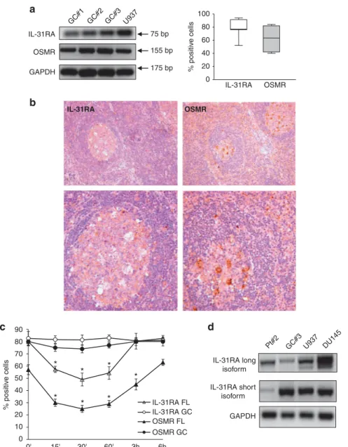
Characterization of IL-31 receptor in human GC B lymphocytes IL-31RA and OSMR expression was next investigated in human GC B cells, which represent the normal counterpart of FL cells.26 GC B cells purified from five tonsils were subjected to RT-PCR and flow cytometric analyses. IL-31RA and OSMR expression was detected at both mRNA (Figure 2a, left panel) and protein (Figure 2a, right panel) levels (median percentage of IL-31RA+ cells = 77.0, range 52–94, median percentage of OSMR+
cells = 63.5, range 40–84, n = 7) (a representative histogram for both the receptor chains is shown in Supplementary Figure S1B). In addition, both IL-31RA and OSMR were found to be expressed in tonsil tissue sections (n = 6) by scattered immunoreactive cells with lymphoid and monocytic morphology (Figure 2b, left and right panels, respectively).
The functional activity of IL-31R was next investigated by culturingfive freshly isolated GC B-cell suspensions with hrIL-31 or medium for 1–60 min. AnnexinV staining showed that sponta-neous apoptosis of GC B cells under these conditions was o5% (data not shown). Moreover, no phosphorylation of Erk1/Erk2, STAT1, STAT3 and Akt was detected in GC B cells at any time and condition tested (data not shown).
In order to gain more insight into the differences in IL-31-driven signaling detected in normal GC B cells vs FL cells, we next investigated the internalization of the IL-31/IL31R complex in both cell types. Following incubation with hrIL-31, FL cells (n = 4) showed a significant reduction in surface expression of both IL-31RA and OSMR chains at 15 min, 30 min, 60 min and 3 h, indicative of ongoing internalization of both receptor chains. In contrast, GC B cells (n = 4) did not internalize IL-31RA or OSMR at any time tested, as assessed byflow cytometric analysis, following gating on AnnexinV− cells in order to exclude apoptotic cells (Figure 2c).
Previous studies have identified two different IL-31RA isoforms: a long one of 745 aa residues and a short one of 560 aa residues. IL-31R-driven signaling appears to be regulated by the ratio between the long and the short isoforms, the latter being considered as a dominant negative one.19Thus, wefirst analyzed the mRNA expression level of the previously described long and short IL-31RA isoforms in FL and GC B cells in comparison with those expressed in U937 and DU145 cells tested as controls.19FL cells expressed predominantly the long isoform (Pt: 73.5% vs 22.5%), similarly to the DU145 control cell line (long vs short: 60% vs 40%) (Figure 2d). In contrast, GC B cells expressed predomi-nantly the short isoform (short vs long: 77.5% vs 22.5%), similarly to the U937 control cell line (short vs long: 62% vs 38%) (Figure 2d, representative samples out of the three tested are shown).
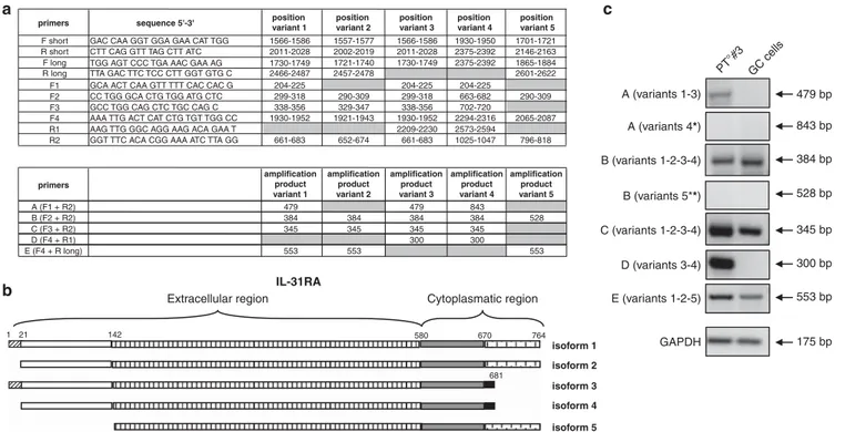
IL31R protein, however, can be translated from five different IL-31RA mRNA variants (Unigene Database from NCBI). The primers for the short isoform recognize all thefive mRNA variants, whereas the primers for the long isoform do not discriminate among mRNA variants 1, 2 and 5, as indicated in Figure 3a. Thus we decided to get further insight on the predominant mRNA variant expressed by FL and GC B cells by designing a new set of primers described in Figure 3a. As shown in Figure 3b, IL-31R mRNA variants 1 and 2 codify for a long cytoplasmic region spanning from aa 580 to aa 764. These two variants differ because variant 2 lacks aa 1–21. IL-31R mRNA variants 3, 4 and 5 are translated into short isoforms; however, the products of variants 3 and 4 lack the terminal part of the cytoplasmic region, whereas the product of variant 5 lacks the terminal part of the extracellular region (Figure 3b). Variants 3 and 4 differ because variant 4 lacks aa 1–21, but both are devoid of the three tyrosine residues necessary for STAT activation.20
When the RNA from three FL and three GC B cells were tested with the panel of primers, the results indicated that the IL-31R mRNA variants expressed by neoplastic and normal cells were different (Figure 3c, where representative samples are shown). In particular, GC B cells did not express variants 1, 3, 4 and 5, expressing variant 2. Conversely, FL cells did not express variants 4 and 5, while expressed high level of variants 3 and 1.
The experiments on the expression of long and short isoforms and of the five mRNA variants strongly suggest that FL cells express IL-31R proteins with a complete extracellular region and a complete or incomplete cytoplasmic region. Conversely, GC B cells express IL-31R protein devoid of aa 1–21 but with a complete cytoplasmic region. These quantitative and qualitative differences may explain the lack of IL-31R-mediated signaling in GC B cells. IL-31 expression in FL and GC B cells
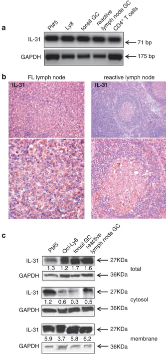
The potential involvement of autocrine or paracrine loops related to the IL-31/IL-31R axis in FL cells was next investigated. To this end, IL-31 mRNA expression wasfirst assessed in FL and GC B-cell fractions isolated from tonsil or reactive lymph nodes. As shown in Figure 4a, IL-31 mRNA was detected in FL cells, GC B cells and the Oci-Ly8 FL cell line, as well as in activated CD4+T cells tested as a positive control.36
Immunohistochemical analysis of four FL and six reactive lymph nodes showed a diffuse and intense expression of IL-31 in FL lymph nodes compared with reactive ones, in which the cytokine was detected more abundantly in the GC than in perifollicular areas (Figure 4b).
IL-31 protein secretion was next evaluated by ELISA in the supernatants from FL cells (n = 18), as well as from tonsil (n = 10) and reactive lymph node (n = 4) GC B cells that had been cultured for 24 h with hrCD40L and hrIL-4 to prevent spontaneous apoptosis.28 The EA.hy926 cell line was tested as a positive control. IL-31 was detected in the supernatant from the latter cell line but not in the other supernatants tested (data not shown).
Western blotting analysis of total cellular lysates from FL cells (n = 3), Oci-Ly8 cell line and tonsil (n = 3) and reactive (n = 3) lymph node GC B cells showed similar IL-31 protein expression levels, as assessed by densitometric analysis (Figure 4c). When cytosolic and membrane cellular fractions were separately analyzed, all samples showed significantly higher expression of membrane vs cytosolic IL-31, as indicated by densitometric analysis (Figure 4c).
The higher IL-31 expression detected by immunohistochemistry in FL vs reactive lymph nodes, together with the similar levels of cytokine detected by western blotting in purified FL and normal GC B cells, suggested that non-B cells present in the FL microenvironment contributed to IL-31 production. This latter
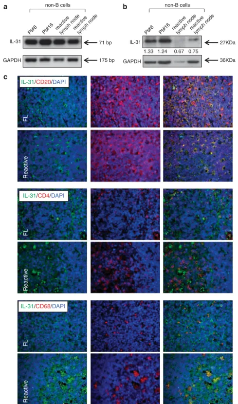
hypothesis was also supported by the finding that such micro-environment is enriched for IL-4,37which is a well-known inducer of IL-31 expression.38 To address this issue, we isolated non-B cells from FL and reactive lymph nodes by immunomagnetic bead manipulation and tested at the mRNA and protein levels by RT-PCR and western blotting, respectively. As shown in Figures 5a and b, all samples expressed IL-31 mRNA and protein; however, IL-31 protein expression was definitely higher in tumor vs reactive samples, indicating that non-B cells indeed contributed to the higher expression of the cytokine in FL lymph nodes.
Immunofluorescence experiments were next performed to elucidate what non-B cell type(s) expressed IL-31 in FL vs reactive 0 10 20 30 40 50 60 70 80 90 0' 15' 30' 60' * * * * * * * 3h 6h IL-31RA FL IL-31RA GC OSMR FL OSMR GC % positiv e cells IL-31RA OSMR 0 20 40 60 80 100 IL-31RA a b c d GC#1 GC#2 GC#3 U937 GAPDH 75 bp 175 bp 155 bp OSMR IL-31RA short isoform IL-31RA long isoform GAPDH Pt#2 GC#3 U937 DU145 % positiv e cells IL-31RA OSMR
Figure 2. Expression and function of IL-31R in human GC B lymphocytes. (a) Left panel. GC B cells were purified from tonsil CD19+B cells and subjected to RT-PCR to determine mRNA expression levels for IL-31RA, OSMR and GAPDH genes. Three different GC B-cell suspensions out of thefive tested are shown. The U937 cell line was tested as a positive control. Right panel. Surface expression of IL-31R was determined on purified GC B cells by flow cytometry. Results are expressed in box plot as the median percentage of positive cells, first and third quartiles and maximum and minimum values, from seven different GC B-cell suspensions. (b) Immunohistochemical analysis of IL-31RA and OSMR chains in tonsils tissue section (one representative staining out of the six performed). Original magnification, × 200 (upper panels) and × 630 (lower panels). (c) FL and GC B cells purified from four FL lymph nodes and four tonsils, respectively, were incubated with 100 ng/ml hrIL-31 for various time intervals (0 min, 15 min, 30 min, 1 h, 3 h, 6 h) and subsequently stained with antibodies to IL-31RA and OSMR. Results are the mean percentage of positive cells± s.d. IL-31RA: *P = 0.05 at 15, 30, 60. OSMR: *P = 0.05 at 15 min, 30 min, 60 min, 3 h. (d) Expression of both short and long isoforms of IL-31RA mRNA levels in FL and tonsil GC B cells. One representative experiment out of the three performed for each cell fraction is shown. The DU145 and U937 cell lines were tested as controls.
lymph nodes. As shown in Figure 5c, CD4+ T cells and CD68+ macrophages were found to express IL-31 in both types of lymph nodes, where CD20+B cells proved to be the major source of the cytokine with comparable expression in FL and reactive follicles. Of note, in reactive GCs, CD68+macrophages displayed IL-31 expression within tingible body vesicles, suggesting possible IL-31 uptake from GC B-cell removal, while FL-associated CD68+ macrophages dis-played a more homogeneous IL-31 expression pattern.
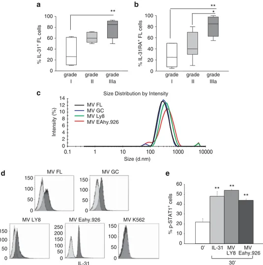
In vivo evidence for IL-31/IL-31RA involvement in FL growth IL-31-driven stimulation of FL cell proliferation pointed to a role of the IL-31/IL-31R complex in FL progression. To gain more insight into this issue, we investigated IL-31 and IL-31RA protein expression in two TMAs from FL lymph nodes with different histological grading. The overall percentage of IL-31+ cells was significantly higher in FL grade IIIa (median percentage of IL-31+ cells = 85, range 50–90, n = 8) than FL grade I (median percentage of IL-31+cells = 26, range 10–60, n = 6) but not FL grade II (median percentage of of IL-31+cells = 60, range 50–70, n = 6) (Figure 6a and Supplementary Figure S4).
A statistically significant increment of IL-31RA+cells in FL grade IIIa (median percentage of IL-31RA+cells = 85, range 55–100, n = 8) compared with FL grade II (median median percentage of IL-31RA+ cells = 40, range 10–80, n = 6) and FL grade I (median percentage of IL-31RA+ cells = 25, range 5–50, n = 6) was also detected (Figure 6b and Supplementary Figure S4).
The increased expression of IL-31 and IL31RA in grade IIIa FL lymph nodes suggested the possible involvement of this complex in tumor progression, but this hypothesis was not supported by the failure to detected soluble IL-31 in FL cell supernatants. An alternative hypothesis was that the intercellular communication in FL lymph nodes might occur through release of IL-31 containing MVs.39Therefore we isolated plasma membrane MVs from grade IIIa FL cell suspensions (n = 3) and, for comparison, GC B-cell
fractions (n = 3), the Oci-Ly8 FL cell line and the EA.hy926 cell line were tested as control. Dynamic light scattering analysis of MVs from all the above cell sources showed a bell-shaped size distribution profile, with a peak ranging from 334.5 ± 33.5 to 411.7 ± 21.6 nm and a polydispersity factor ranging from 0.155 ± 0.033 to 0.388 ± 0.062, indicative of homogeneous MV preparations (Figure 6c). AnnexinV staining confirmed the MV nature of the particles because of phosphatydylserine expression (data not shown).40
The presence of IL-31 in all MV preparations was next investigated by flow cytometry. As shown in Figure 6d, IL-31 was detected on MVs from all the samples analyzed (mean relative fluorescence intensity (MRFI) MVs FL: 3.9; MRFI MVs GC: 2.7; MRFI MVs Oci-Ly8:3.1;7. MRFI MVs EA.hy926: 7.2). These results are consistent with the hypothesis that IL-31 released by FL in MVs is involved in autocrine/paracrine loops supporting tumor growth. MVs isolated from FL patients with different grades (n = 8, 2 FL with grade I, 3 FL with grade II and 3 FL with grade III) did not differ for IL-31 content evaluated as MRFI (data not shown). In contrast, exosomes isolated from the Oci-Ly8 FL cell line and the EA.hy926 cell line, the latter tested as a control, did not contain IL-31 (data not shown).
We next investigated the signaling activity of MVs, isolated from the Oci-LY8 and Eahy.926 cell lines, on primary FL cells (n = 3). FL cells incubated for 30 min with or without the above MVs displayed a significant phosphorylation of STAT-1, indicating that MVs were biologically active (Figure 6e).
DISCUSSION
IL-31 is a recently discovered cytokine produced by different cell types, including T helper type 2 lymphocytes and mast cells, that has a role in the pathogenesis of cutaneous allergic diseases through itch stimulation6,7,41 by binding to IL-31RA+ sensory neurons.36 A (variants 1-3) B (variants 1-2-3-4) C (variants 1-2-3-4) D (variants 3-4) E (variants 1-2-5) GAPDH 479 bp PT°#3 GC cells 384 bp 345 bp 300 bp 553 bp 175 bp
Extracellular region Cytoplasmatic region
isoform 1 isoform 2 isoform 3 isoform 4 isoform 5 IL-31RA 1 21 142 580 670 764 681 A (variants 4*) B (variants 5**) 843 bp 528 bp primers amplification product variant 1 amplification product variant 2 amplification product variant 3 amplification product variant 4 amplification product variant 5 A (F1 + R2) 479 479 843 B (F2 + R2) 384 384 384 384 528 C (F3 + R2) 345 345 345 345 D (F4 + R1) 300 300 E (F4 + R long) 553 553 553 primers a c b
sequence 5'-3' positionvariant 1 positionvariant 2 positionvariant 3 positionvariant 4 positionvariant 5
F1 GCA ACT CAA GTT TTT CAC CAC G 204-225 204-225 204-225
F2 CC TGG GCA CTG TGG ATG CTC 299-318 290-309 299-318 663-682 290-309
F3 GCC TGG CAG CTC TGC CAG C 338-356 329-347 338-356 702-720
F4 AAA TTG ACT CAT CTG TGT TGG CC 1930-1952 1921-1943 1930-1952 2294-2316 2065-2087
R1 AAG TTG GGC AGG AAG ACA GAA T 2209-2230 2573-2594
R2 GGT TTC ACA CGG AAA ATC TTA GG 661-683 652-674 661-683 1025-1047 796-818
R long TTA GAC TTC TCC CTT GGT GTG C 2466-2487 2457-2478 2601-2622
R short F short F long
CTT CAG GTT TAG CTT ATC
1566-1586 GAC CAA GGT GGA GAA CAT TGG
2011-2028 1730-1749 TGG AGT CCC TGA AAC GAA AG
1557-1577 2002-2019 1721-1740 1566-1586 2011-2028 1730-1749 1930-1950 2375-2392 2375-2392 1701-1721 2146-2163 1865-1884
Figure 3. Expression of IL-31RA isoforms in FL and GC B cells. (a) Upper table: primers’ sequences for IL-31RA mRNA variants and their position. Lower table: amplification products of primers in the five different IL-31RA variants. * or ** = amplification products with different bp for isoforms 4 and 5, respectively. (b) Schematic representation of the amino-acidic sequences for IL-31RA protein isoforms. (c) Patterns of amplification products obtained with the primers for the five different IL-31RA mRNA variants in three FL and three GC B-cell samples. One representative experiments out of the three performed is shown.
Limited information is available on the expression and/or function of the IL-31/IL-31R complex in malignant diseases. Different tumor cell lines have been reported to express IL-31 mRNA.1,13–15 Furthermore, IL-31 was found to be produced by malignant T-cell populations from patients with cutaneous T-cell lymphomas,22 with increased serum levels of the cytokine correlating to itch.21 No information was so far available on the expression and the possible role of the IL-31/IL-31R complex in B-cell lymphomas.
Here we have addressed this issue by choosing FL as prototypic mature B-cell malignancy, as it represents the second most frequent B-cell lymphoma after diffuse large B-cell lymphoma.23,24In malignant B cells purified from FL patients IL-31R, composed of the IL-31RA and OSMR chains, was expressed both at mRNA and protein levels. Signal transduction studies showed that hrIL-31, upon binding to its receptor on primary FL cells, triggered phosphorylation of STAT1/3, ERK1/2 and Akt, similarly to that reported for other cell types.10,16–18,20,30 Furthermore, hrIL-31 increased significantly proliferation of cultured FL cells over background levels. In contrast, GC B cells, although expressing IL-31RA and OSMR, did not signal in response to IL-31.
The different responsiveness of FL cells and GC B cells to IL-31 prompted additional studies addressing such issue. As ligand-induced internalization of signaling receptors usually correlates with initiation of signal transduction,42 FL and GC B cells were cultured with hrIL-31 and tested at different times for the surface expression of IL-31RA and OSMR. FL cells displayed ligand-induced internalization of both IL-31RA and OSMR, whereas GC B cells did not, suggesting that such defect may be involved in lack of responsiveness of the latter cells to IL-31.
Previously it was shown that, despite co-expression of both IL-31R chains, only cell lines expressing predominantly the long isoform of IL-31RA signaled in response to IL-31 and that the short IL-31RA isoform operated as a dominant-negative receptor.19 A similar dicotomy has been reported for other receptors such as those for prolactin, growth hormone and leptin, whose short isoforms ‘silenced’ the respective long isoforms.43–45 Consistent with this background, signaling FL cells displayed prevalent expression of the long IL-31RA isoform, whereas non-signaling GC B cells expressed predominantly the short isoform.
Next, the expression of five IL-31RA mRNA variants was investigated in FL vs GC B cells. FL cells appeared to express mRNA variants 1, 2 and 3, whereas GC B cells appeared to express variant 2 only. Lack of mRNA variants translated into signaling IL-31RA isoforms might contribute to defective responsiveness of GC B cells to IL-31.
Experiments on lymph nodes from FL patients with different histological grading showed that the proportion of both IL-31+ and IL31RA+FL cells was significantly higher in grade III compared with grade I/II FL. These results indicate that the progressive disruption of lymph node architecture is accompanied by increased expression of both IL-31 and IL-31RA. Additional experiments were performed to investigate whether other cell populations of the tumor microenvironment expressed IL-31. Both CD4 T cells and CD68 macrophages were found to express the cytokine.
In spite of IL-31 mRNA expression in FL cells as well as in GC B cells, soluble IL-31 was not detected in their culture supernatants. However, western blotting analysis of lysates from FL cells and GC B cells revealed the presence of IL-31 protein, with a significantly higher membrane vs cytosolic expression.
The majority of secretory proteins are externalized through the classical pathway, whereas others lacking hydrophobic signal sequence, such as IL-31, are released through different non-classical secretory pathways,46including MV shedding. MVs are small membrane vesicles shed from normal and malignant cells, either constitutively or following stimulation, that trans-port molecules involved in cell signalling and intercellular
FL lymph node reactive lymph node
total cytosol membrane IL-31 IL-31 IL-31 GAPDH GAPDH GAPDH 27KDa
Pt#5 Oci-Ly8tonsil GCreactivelymph node GC
36KDa 27KDa 36KDa 27KDa 36KDa IL-31
Pt#5 Ly8 tonsil GCreactivelymph node GCCD4 + T cells GAPDH IL-31 1.3 1.2 1.7 1.6 1.2 0.6 0.3 0.5 5.9 3.7 5.8 6.2 IL-31 a b c 175 bp 71 bp
Figure 4. Expression of IL-31 in FL and GC B cells. (a) IL-31 and GAPDH mRNA expression were determined by RT-PCR in one FL patient (out of the three tested), Oci-Ly8 cell line, one tonsil and one reactive lymph node GC B-cell fractions (out of the three tested each) and one activated CD4+ T-cell fraction (out of the three tested), tested as a positive control. (b) Immunohistochemical analysis of IL-31 in FL (one representative staining out of the six performed) and reactive (one representative staining out of the six performed) lymph node tissue sections. Original magnification, × 200 (upper panels) and × 630 (lower panels). (c) IL-31 protein expression was tested by western blotting analysis in FL cells (lane 1), Oci-Ly8 cell line (lane 2), tonsil (lane 3) and reactive lymph node (lane 4) GC B cells. One representative experiment out of the three performed for each cell fraction is shown. Total cytosolic and membrane cell lysates were subjected to western blotting with an antibody against hIL-31. GAPDH was used as a loading control. Numbers indicated the ratio between the intensity of the specific bands and that of the housekeeping protein bands, as assessed by densitometry. Membrane vs cytosolic fractions: Po0.0001 for both FL and GC cells and Ly8 cell line; P= 0.0002 for reactive GC B cells. 964
communications.39 MVs secreted from primary tumor cells mediate interactions with the tumor microenvironment and participate in the formation of metastatic niches.39 Here we show that plasma membrane MVs from primary FL cells contained
IL-31, and FL-derived MVs activated STAT-1 phosphorylation in primary FL cells. These data support the hypothesis that cell to cell transfer of the cytokine through MVs is involved in autocrine/ paracrine loops promoting tumor growth.
IL-31/CD20/DAPI Reactiv e FL IL-31/CD68/DAPI IL-31/CD4/DAPI Reactiv e FL Reactiv e FL IL-31 a c b Pt#8 Pt#16 reactiv e lymph nodereactiv
e
lymph node Pt#8 Pt#16 reactiv e lymph nodereactiv
e lymph node GAPDH IL-31 GAPDH 27KDa 36KDa non-B cells 1.33 1.24 0.67 0.75 non-B cells 175 bp 71 bp
Figure 5. Expression of IL-31 in non-B cells from FL and reactive lymph nodes. (a) IL-31 and GAPDH mRNA expression were determined by RT-PCR in non-B cells from two FL patients (out of the four tested), two reactive lymph node (out of the four tested). (b) IL-31 protein expression was tested by western blotting analysis in non-B cells from FL (lanes 1 and 2) and reactive lymph node (lanes 3 and 4) GC B cells. Two experiments out of the four performed are shown. GAPDH was used as a loading control. Numbers indicated the ratio between the intensity of the specific bands and that of the housekeeping protein bands, as assessed by densitometry. (c) Double-marker immunofluorescence analysis of IL-31 (green) cells in combination with CD20 (red), CD4 (red) or CD68 (red) within neoplastic infiltrates of FL and reactive tissue. Original magnification, × 200. DAPI, 4,6-diamidino-2-phenylindole.
CONFLICT OF INTEREST
The authors declare no conflict of interest.
ACKNOWLEDGEMENTS
This work was supported by grants from Associazione Italiana Ricerca Cancro (A.I.R.C.), Milano, Italy to VP (No. 13003), from Cinque per mille IRPEF–Finanziamento Ricerca Sanitaria and from Ricerca Corrente Ministeriale. EF is a recipient of a Fondazione Umberto Veronesi fellowship. We thank Dr Francesco Bertoni for help in discussion. European Union Seventh Frame Programme (FP7/2007-2013) under grant agreement N° 278570 to DR.
REFERENCES
1 Dillon SR, Sprecher C, Hammond A, Bilsborough J, Rosenfeld-Franklin M, Presnell SR et al. Interleukin 31, a cytokine produced by activated T cells, induces dermatitis
in mice. Nat Immunol 2004;5: 752–760.
2 Cornelissen C, Brans R, Czaja K, Skazik C, Marquardt Y, Zwadlo-Klarwasser G et al. Ultraviolet B radiation and reactive oxygen species modulate interleukin-31
expression in T lymphocytes, monocytes and dendritic cells. Br J Dermatol 2011; 165: 966–975.
3 Niyonsaba F, Ushio H, Hara M, Yokoi H, Tominaga M, Takamori K et al. Antimicrobial peptides human beta-defensins and cathelicidin LL-37 induce the secretion of a pruritogenic cytokine IL-31 by human mast cells. J Immunol 2010; 184: 3526–3534.
4 Ishii T, Wang J, Zhang W, Mascarenhas J, Hoffman R, Dai Y et al. Pivotal role of mast cells in pruritogenesis in patients with myeloproliferative disorders. Blood
2009;113: 5942–5950.
5 Siraganian RP. Mast cell signal transduction from the high-affinity IgE receptor.
Curr Opin Immunol 2003;15: 639–646.
6 Sonkoly E, Muller A, Lauerma AI, Pivarcsi A, Soto H, Kemeny L et al. IL-31: a new
link between T cells and pruritus in atopic skin inflammation. J Allergy Clin
Immunol 2006;117: 411–417.
7 Bilsborough J, Leung DY, Maurer M, Howell M, Boguniewicz M, Yao L et al. IL-31 is associated with cutaneous lymphocyte antigen-positive skin homing T cells in
patients with atopic dermatitis. J Allergy Clin Immunol 2006;117: 418–425.
8 Raap U, Wichmann K, Bruder M, Stander S, Wedi B, Kapp A et al. Correlation of IL-31 serum levels with severity of atopic dermatitis. J Allergy Clin Immunol 2008; 122: 421–423. 0 20 40 60 80 100 a c d e b 0 20 40 60 80 100 grade I grade II grade IIIa ** * grade I grade II grade IIIa % IL-31 + FL cells % IL-31RA + FL cells ** MV Eahy.926 MV LY8 IL-31 MV GC MV FL 150 100 50 0 150 100 50 0 150 200 250 100 50 0 150 100 50 0 150 100 50 0 0 2 4 6 8 10 12 14 0.1 1 10 100 1000 10000 Intensity (%) Size (d.nm) Size Distribution by Intensity
MV EAhy.926 MV Ly8 MV GC MV FL MV K562 % p-STAT1 + cells MV LY8 MV Eahy.926 IL-31 0’ 30’ 0 10 20 30 40 50 60 ** ** **
Figure 6. (a, b) IL-31/IL-31RA expression in relation to FL grading. Expression of IL-31 (a) and IL-31RA (b) in FL lymph node samples from patients with different histological grading was investigated by immunohistochemistry in TMA. Results are shown in box plot as the median percentage of positive cells,first and third quartiles and maximum and minimum values, from 20 different FL tissue sections. **P = 0.008 for IL-31 grade I compared with grade III; **P= 0.002 for IL-31RA grade I compared with grade III; *P = 0.014 for IL-31RA grade II compared with grade III. (c, d) IL-31 expression in MVs from FL and GC B cells. (c) Dynamic light scattering analysis of MVs from FL cells (black line), GC B cells (blu line), Oci-LY8 (green line) and EA.hy926 cell lines (red line). One representative profile out of the three performed is shown. (d) Flow cytometric analysis of IL-31 content in MV preparations from FL cells, GC B cells (upper panels), LY8, EA.hy926 and K562 cell lines (lower panels). Grey histograms: IL-31 staining with a rabbit polyclonal antibody. Light grey histograms: rabbit serum control. One representative histogram for each MV preparation studied is shown. (e) Purified FL cells were incubated for 0 or 30 min with 100 ng/ml hrIL-31 or with MVs isolated from Oci-LY8 and EA.hy926 and subjected toflow cytometric analysis using an antibody to p-STAT1. Results are shown as the mean percentage of positive cells± s.d. from three different experiments. **P = 0.002 for MVs from both OCI-LY8 and EA.hy926.
9 Andoh A, Yagi Y, Shioya M, Nishida A, Tsujikawa T, Fujiyama Y. Mucosal cytokine
network in inflammatory bowel disease. World J Gastroenterol 2008; 14:
5154–5161.
10 Dambacher J, Beigel F, Seiderer J, Haller D, Goke B, Auernhammer CJ et al. Interleukin 31 mediates MAP kinase and STAT1/3 activation in intestinal epithelial cells and its expression is upregulated in inflammatory bowel disease. Gut 2007; 56: 1257–1265.
11 Lei Z, Liu G, Huang Q, Lv M, Zu R, Zhang GM et al. SCF and IL-31 rather than IL-17 and BAFF are potential indicators in patients with allergic asthma. Allergy 2008; 63: 327–332.
12 Okano M, Fujiwara T, Higaki T, Makihara S, Haruna T, Noda Y et al. Characterization of pollen antigen-induced IL-31 production by PBMCs in patients with allergic
rhinitis. J Allergy Clin Immunol 2011;127: 277–279, 279e.1–279e.11.
13 Diveu C, Lelievre E, Perret D, Lak-Hal AH, Froger J, Guillet C et al. GPL, a novel cytokine receptor related to GP130 and leukemia inhibitory factor receptor. J Biol
Chem 2003;278: 49850–49859.
14 Ghilardi N, Li J, Hongo JA, Yi S, Gurney A, de Sauvage FJ. A novel type I cytokine receptor is expressed on monocytes, signals proliferation, and activates STAT-3
and STAT-5. J Biol Chem 2002;277: 16831–16836.
15 Zhang Q, Putheti P, Zhou Q, Liu Q, Gao W. Structures and biological
functions of IL-31 and IL-31 receptors. Cytokine Growth Factor Rev 2008;19:
347–356.
16 Chattopadhyay S, Tracy E, Liang P, Robledo O, Rose-John S, Baumann H. Interleukin-31 and oncostatin-M mediate distinct signaling reactions and
response patterns in lung epithelial cells. J Biol Chem 2007;282: 3014–3026.
17 Kasraie S, Niebuhr M, Werfel T. Interleukin (IL)-31 activates signal transducer and activator of transcription (STAT)-1, STAT-5 and extracellular signal-regulated kinase 1/2 and down-regulates IL-12p40 production in activated human
macro-phages. Allergy 2013;68: 739–747.
18 Cornelissen C, Luscher-Firzlaff J, Baron JM, Luscher B. Signaling by IL-31 and
functional consequences. Eur J Cell Biol 2012;91: 552–566.
19 Diveu C, Lak-Hal AH, Froger J, Ravon E, Grimaud L, Barbier F et al. Predominant expression of the long isoform of GP130-like (GPL) receptor is required for
interleukin-31 signaling. Eur Cytokine Netw 2004;15: 291–302.
20 Horejs-Hoeck J, Schwarz H, Lamprecht S, Maier E, Hainzl S, Schmittner M et al. Dendritic cells activated by IFN-gamma/STAT1 express IL-31 receptor and release
proinflammatory mediators upon IL-31 treatment. J Immunol 2012; 188:
5319–5326.
21 Ohmatsu H, Sugaya M, Suga H, Morimura S, Miyagaki T, Kai H et al. Serum IL-31 levels are increased in patients with cutaneous T-cell lymphoma. Acta Derm
Venereol 2012;92: 282–283.
22 Singer EM, Shin DB, Nattkemper LA, Benoit BM, Klein RS, Didigu CA et al. IL-31 is produced by the malignant T-cell population in cutaneous T-Cell lymphoma and
correlates with CTCL pruritus. J Invest Dermatol 2013;133: 2783–2785.
23 Hartmann EM, Ott G, Rosenwald A. Molecular biology and genetics of lymphomas.
Hematol Oncol Clin North Am 2008;22: 807–823, vii.
24 Kurtin PJ. Indolent lymphomas of mature B lymphocytes. Hematol Oncol Clin
North Am 2009;23: 769–790.
25 A clinical evaluation of the International Lymphoma Study Group classification of
non-Hodgkin's lymphoma. The Non-Hodgkin's Lymphoma Classification Project.
Blood 1997;89: 3909–3918.
26 Corcione A, Ottonello L, Tortolina G, Facchetti P, Airoldi I, Guglielmino R et al. Stromal cell-derived factor-1 as a chemoattractant for follicular center lymphoma
B cells. J Natl Cancer Inst 2000;92: 628–635.
27 Corcione A, Ferretti E, Bertolotto M, Fais F, Raffaghello L, Gregorio A et al. CX3CR1 is expressed by human B lymphocytes and mediates [corrected] CX3CL1 driven
chemotaxis of tonsil centrocytes. PLoS One 2009;4: e8485.
28 Corcione A, Baldi L, Zupo S, Dono M, Rinaldi GB, Roncella S et al. Spontaneous production of granulocyte colony-stimulating factor in vitro by human B-lineage
lymphocytes is a distinctive marker of germinal center cells. J Immunol 1994;153:
2868–2877.
29 Dono M, Burgio VL, Tacchetti C, Favre A, Augliera A, Zupo S et al. Subepithelial B cells in the human palatine tonsil. I. Morphologic, cytochemical and phenotypic
characterization. Eur J Immunol 1996;26: 2035–2042.
30 Fossey SL, Bear MD, Kisseberth WC, Pennell M, London CA. Oncostatin M promotes STAT3 activation, VEGF production, and invasion in osteosarcoma
cell lines. BMC Cancer 2011;11: 125.
31 Di Paolo D, Ambrogio C, Pastorino F, Brignole C, Martinengo C, Carosio R et al. Selective therapeutic targeting of the anaplastic lymphoma kinase with liposomal siRNA induces apoptosis and inhibits angiogenesis in neuroblastoma. Mol Ther
2011;19: 2201–2212.
32 Pagnan G, Di Paolo D, Carosio R, Pastorino F, Marimpietri D, Brignole C et al. The combined therapeutic effects of bortezomib and fenretinide on neuroblastoma
cells involve endoplasmic reticulum stress response. Clin Cancer Res 2009;15:
1199–1209.
33 Jayachandran M, Miller VM, Heit JA, Owen WG. Methodology for isolation,
iden-tification and characterization of microvesicles in peripheral blood. J Immunol
Methods 2012;375: 207–214.
34 Marimpietri D, Petretto A, Raffaghello L, Pezzolo A, Gagliani C, Tacchetti C et al.
Proteome profiling of neuroblastoma-derived exosomes reveal the expression
of proteins potentially involved in tumor progression. PLoS One 2013; 8: e75054.
35 Johnson PW, Watt SM, Betts DR, Davies D, Jordan S, Norton AJ et al. Isolated follicular lymphoma cells are resistant to apoptosis and can be grown in vitro in
the CD40/stromal cell system. Blood 1993;82: 1848–1857.
36 Cevikbas F, Wang X, Akiyama T, Kempkes C, Savinko T, Antal A et al. A sensory neuron-expressed IL-31 receptor mediates T helper cell-dependent
itch: involvement of TRPV1 and TRPA1. J Allergy Clin Immunol 2013; 133:
448–460.
37 Calvo KR, Dabir B, Kovach A, Devor C, Bandle R, Bond A et al. IL-4 protein expression and basal activation of Erk in vivo in follicular lymphoma. Blood 2008; 112: 3818–3826.
38 Stott B, Lavender P, Lehmann S, Pennino D, Durham S, Schmidt-Weber CB. Human IL-31 is induced by IL-4 and promotes TH2-driven inflammation. J Allergy Clin
Immunol 2013;132: 446–454 e445.
39 Lee Y, El Andaloussi S, Wood MJ. Exosomes and microvesicles: extracellular vesicles for genetic information transfer and gene therapy. Hum Mol Genet 2012; 21: R125–R134.
40 Frey B, Gaipl US. The immune functions of phosphatidylserine in membranes of
dying cells and microvesicles. Semin Immunopathol 2011;33: 497–516.
41 Raap U, Weissmantel S, Gehring M, Eisenberg AM, Kapp A, Folster-Holst R. IL-31
significantly correlates with disease activity and Th2 cytokine levels in children
with atopic dermatitis. Pediatr Allergy Immunol 2012;23: 285–288.
42 Thiel S, Behrmann I, Dittrich E, Muys L, Tavernier J, Wijdenes J et al. Internalization of the interleukin 6 signal transducer gp130 does not require activation of the
Jak/STAT pathway. Biochem J 1998;330(Pt 1): 47–54.
43 Ross RJ, Esposito N, Shen XY, Von Laue S, Chew SL, Dobson PR et al. A short isoform of the human growth hormone receptor functions as a dominant negative inhibitor of the full-length receptor and generates large amounts of
binding protein. Mol Endocrinol 1997;11: 265–273.
44 Schuler LA, Lu JC, Brockman JL. Prolactin receptor heterogeneity: processing and signalling of the long and short isoforms during development. Biochem Soc Trans
2001;29: 52–56.
45 White DW, Tartaglia LA. Evidence for ligand-independent homo-oligomerization of leptin receptor (OB-R) isoforms: a proposed mechanism permitting productive long-form signaling in the presence of excess short-form expression. J Cell
Biochem 1999;73: 278–288.
46 Eder C. Mechanisms of interleukin-1beta release. Immunobiology 2009;214:
543–553.
