CHRONIC FATIGUE SYNDROME: CIRCADIAN RHYTHM AND
HYPOTHALAMIC-PITUITARY-ADRENAL (HPA) AXIS IMPAIRMENT.
D. RACCIATTI, M. T. GUAGNANOI, J. VECCHIET,
P. L. DE REMIGISl, E. PIZZIGALLO, R. DELLA VECCHIAI,
T. DI SCIASCIO, D. MERLITTP and S. SENSP
Clinic of Infectious Diseases - 'Clinic of Internal Medicine Department of Internal Medicine and Aging, Chieti University, ItalyReceived November8,2000 - Accepted January 5,2001
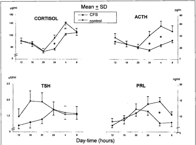
Chronic Fatigue Syndrome (CFS) is a clinical condition characterized by a persistent or relapsing debilitating fatigue at rest, lasting> 6 months, and made worse by exercise. At the present moment, there are three potential etiopathogenic factors: immunologic, viral and neuroendocrine. The purpose of our study was to evaluate possible alterations of the hypothalamic-pituitary-adrenal (HPA) axis in our CFS patients by studying the circadian rhythms of prolactin (PRL) , thyrotropic hormone (TSH), adrenocorticotropic hormone (ACTH), and cortisol (CS). A total of 36 patients were enrolled according to the Centers for Disease Control and Prevention case-definition criteria. Twenty healthy subjects were included as controls. Blood samples were taken every 4 hours during a single 24-hour period. We performed a fluorometric enzyme immunoassay with serum PRL, cortisol and TSH, and an immunoradiometric assay with plasma ACTH. The circadian rhythms ofPRL, TSH, ACTH and CS were statistically significant in both CFS and control groups. At 24:00 and 04:00 hrs the CFS patients showed lower ACTH levels than healthy subjects (p<0.001); the PRL levels were higher at 04.00 h in CFS patients than in healthy subjects.
In recent years, Chronic Fatigue Syndrome (CFS) has become a recognized clinical entity, but the etiology of CFS is still poorly understood. However, interesting hypotheses as to its pathogenesis have been advanced: viral, immunologic and neuroendocrine. The latter is due to the presence of a significant number of patients complaining of atypical depression and symptoms of hypothalamic dysfunction, such as menstrual alterations; fluid retention; changes in appetite, body weight and sleep; and excessive sweating (1). The diagnosis is confirmed by at least 4 other symptoms included in the Centers for Disease Control and Prevention (CDC) case-definition criteria and by exclusion of other well-defined diseases and psychiatric disorders (2).
Furthermore many clinical features of CFS
patients are also characteristic of glucocorticoid insufficiency. Demitrack showed an impaired activation of the hypothalamic-pituitary-adrenal (HPA) axis in CFS patients (3). Since the hypothalamus represents the center of a chronobiological clock, we decided to study the circadian rhythm of prolactin (PRL), thyrotropic (TSH), adrenocorticotropic (ACTH) and cortisol (CS) hormones, to verify possible modifications of this hypothalamic function as a further sign of HPA axis involvement in CFS patients.
MATERIALS AND METHODS
We studied 36 patients (16 males) who met the CDC criteria (2) with a mean age of 38.3± 10.9 (SD) years (table 1). The illness was precipitated by an acute
Key words: Chronic Fatigue Syndrome, circadian rhythm, hypothalamic-pituitary-adrenal axis Mailing address: Prof. Sergio Sensi
Clinica Medica - Dipartimento diMed. Interna e Scienze dell'lnvecchiamento Policlinico Colle dell'Ara - Via dei Vestini - 66100 Chieti - Italy Tel. and fax: 0871-551562
e-mail: [email protected] 11
0394-6320 (200 I)
Copyright!) hy BIOLIFE."'.5..
This publication and/orarticleis for individual use onlyand maynot be further reproduced withoutwrittenpermission fromthe copyrightholder. Unauthorized reproduction mayresultin financial andotherpenalties
12
D. RACCIATTI ET AL.virus-like infection in 55.6% of them. Controls were 20 subjects (6 males), aged34.2±2.6; they were considered healthy according to their medical history, physical examination and routine laboratory tests. None of the subjects were taking any medication. Prolactin, CS, ACTH and TSH samples were collected at 4-hour intervals over a 24 h period. PRL, CS and TSH serum concentrations were determined by fluorometric enzyme immunoassay (Baxter Diagnostic Inc.), and ACTH plasma concentrations were determined by immunoradiometric assay (Henning Berlin GmbH). All subjects were synchronized for seven days with diurnal-wake period between 07:00 hand 23:00 hand a nocturnal rest period between 23.00 hand 07.00 h. Subjects were given three similar meals each day at 08:00 h, 12:00 hand 20:00 h, with a total daily calorie intake of 1600-1800 Kcal (CHO 50%, proteins 20%, lipids 30%). All groups were evaluated at same time. The blood collections were performed before the meals at the following hours: 08:00 h, 12:00 h, 16:00 h, 20:00 h, 24:00 hand 04:00 h. The serum and plasma samples were stored at-200
C until assayed. All subjects gave their informed consent.
Statistical analysis: The data were analyzed by conventional statistical methods (mean, standard deviation, unpaired Student'st test between CFS and control groups). The raw data of each time point measurement were averaged and plotted as a function of the time, in order to draw the circadian chronograms. Furthermore, a computerized program analyzed the data by the least-square method, to find the best-fitting cosine function [y=M+A cos (rot+<1»]approximating the data collected throughout the 24-hour cycle. The Mesor (M) is the rhythm adjusted average; the Amplitude (A) represents the difference between maximum and mesor of a best fitting cosine;tis the period of oscillation; the Acrophase (<I» is the peak time of the cosine function used to approximate the rhythm, with the
Tab. I.Characteristics of study subjects (mean±SD)
CONTROLS
corresponding 95% confidence limits (95% C.L.), expressed in hour:minutes. It was thus possible to validate the 95% statistical significance of each rhythm (4).
RESULTS
The chronograms of TSH, PRL, ACTH and cortisol are shown in Fig. 1. The levels of TSH document no significant differences to any time of the survey between controls and CFS subjects. Increased levels of PRL are documented at 04:00 h of the morning (p <0.001) without any statistically significant variation in other periods of the day, among CFS patients compared with controls. The cortisol and ACTH values result decreased at each time, but are significant only at 24:00 h and at 04:00 h of the morning (p < 0.001) in the CFS subjects respect to the controls.
The circadian microscopic analysis documents significant oscillations of TSH (p<O.OOl and p<O.OOl), PRL (p<0.02 and p<O.OOl), cortisol (p<O.OOl and p<O.OOl), and ACTH (p<O.OOl and p<O.OOl), both in controls and CFS subjects, respectively (Tab. II). The mesor and the amplitude of TSH, PRL and cortisol show no significant differences between the two groups; while, among CFS patients in comparison to the controls, ACTH levels showed a weakly significant decrease of mesor (p < 0.05) and amplitude of oscillation whether expressedas absolutevalueor expressedas percentage of the mesor (31% versus 42%, respectively) (Tab. II). The subjects with CFS present respect to the controls a displacement of the acrophase for the PRL levels (01: 18 h respect to 21: 19 h), for cortisol (09:31 h respect to 05:19 h) and for ACTH (10:40 h respect to 05:44 h), respectively. The greatest displacement of acrophase is documented for TSH (02:49 h respect to 19:03 h) (table 2).
CFS Age (years)
Sex (M/F) Body weight (kg) Body Mass Index (kg/rrr') Duration of disease (years)
34.25 ± 2.67 6/20 66.10 ± 10.06 23.71 ±2.82
o
38.33 ± 10.99 16/36 69.70 ± 12.81 25.14 ± 3.88 7.36 ± 8.20 CFS: Subjects with chronicfttigue syndrome.Tab. II. Circadian rhythms by microscopic analysis (Cosinor test) of TSH, Prolactin (PRL),
Cortisol and ACTH in36 subjects with Chronic Fatigue Syndrome (CFS) and in 20 control subjects.
MESOR AMPLITUDE ACROPHASE#
h:min P<
(mean ±",SEM) (mean ±"'SEM) (95%c.r.;
TSH CONTROLS 1.69 ± 0.24 0.21 ± 0.05 19:03 (17:22 - 20.45) 0.001 (/lV/ml) CFS 1.42 ± 0.13 0.29 ± 0.06 02:49 (01:50 - 03:47) 0.001 PRL CONTROLS 9.13 ± 0.81 2.30 ± 0.72 21:19 (19:23 - 23:14) 0.02 (ng/ml) CFS 11.75 ± 0.97 3.71 ± 0.75 01: 18 (00:07 - 02:28) 0.001 CORTISOL CONTROLS 90.15± 4.96 52.27 ± 5.38 05: 19 (04:30 - 06:08) 0.001 (ug/ml) CFS 84.72 ± 4.83 53.51 ± 3.42 09:31 (08:54 - 10:07) 0.001 ACTH CONTROLS 36.22 ± 6.03 15.15 ± 2.47 05:44 (04:47 - 06:42) 0.001 (pg/ml) CFS 23.55 ± 2.50* 7.39 ± 1.60** 10:40 (08:59 - 12:21) 0.001
# Phase reference: local midnight (360"=24 h); 95% C.L.: 95% Confidence Limits (given whether the rhythm detection level was found to reject the null hypothesis of zero-amplitude at a significant level of probability: p < 0.001).
Comparison between control subjects and subjects with Chronic Fatigue Syndrome: * p < 0.05; ** p < 0.01 (unpaired Student's t test).
Fig. 1. 24-hour profile of TSH, Prolactin (PRL), Cortisol and ACTH in 36 subjects with Chronic Fatigue
Syndrome (CFS) and in 20 control subjects. (*p< 0.001).
j1g!ml Mean±SD pg/m 180 •
c---CFS]
!80 COHTISOL - - t I ACTH 140 1 ~ conro ! 60 100 >C ~~/r
40 60, . A
20 j!U/ml 2.5 12 16 20 TSH 24 12 16 20 PRL 24 4 ng/ml r20 2,0 1,5 15 10 12 16 2C 24 12 Day-time (hours) 16 20 2414
D. RACCIATTI ET AL.DISCUSSION
The results of the temporal macroscopic and the circadian microscopic analyses suggest some remarks. The macroscopic analysis of the circadian hormone levels documented a normal rhythms of TSH and PRL during the 24-hour period, while ACTH and cortisol levels were decreased during the night and the first hours of the morning among CFS patients respect to the controls. Neuroendocrine studies on CFS are few and conflicting. Demitrack documented a significant reduction in both plasma and urinary glucocorticoid levels, and these findings suggest a possible impairment of HPA axis (3,5-6). Poteliakhoff et al. observed a reduction in diurnal levels of cortisol in patients with chronic fatigue and in a group of subjects with acute fatigue in comparison to non-fatigued controls (7). Salivary and urinary levels of cortisol did not show any variation in the 4-hour interval determinations over the diurnal period (from 08.00 a.m. to 08.00 p.m.) both in patients and in controls (8). Moldofsky et al. (9) described chronobiological modifications in CFS patients: they found altered diurnal patterns in cortisol, prolactin and NK cells, with low levels of each one of these parameters. These observations were also associated with alpha EEG sleep disorder, and daytime fatigue, sleepiness and pain. These findings could support the hypothesis of circadian rhythm disturbances in CFS(l0). Ourneuroendocrine studies on CFS patients documented some interesting findings on circadian microscopic analysis: all the examined hormones preserved the circadian rhythm in CFS patients as well as in controls, but when the CFS subjects were compared with controls they showed an acrophase displacement in all endocrine rhythms studied.
Some years ago a similar behaviour was described as expression of affective disorders (11-13). Moreover, patients with unipolar depression showed a temporal advancement of PRL circadian levels (14-15).
So, these data regarding an acrophase displacement of some circadian hormonal rhythms could represent a sign of cyclicity "derailment" of HPA axis.
Furthermore, it is remarkable that the circadian mean levels of ACTH and cortisol showed a decrease in CFS subjects. We agree with the excellent review of Demitrack on neuroendocrine aspects in
CFS (16), particularly the suggestion of this author to continue neuroendocrine studies in CFS patients since until now they raise more questions than answers to CFS etiopathogenesis..
The temporal microscopic circadian analysis of four hormones seems to suggest an altered cyclicity of HPA axis, even though each hormone preserves the circadian rhythm, and these alterations are already reported in patients with unipolar depression (14-15). Besides, these observation do not exclude the potential pathogenetic role of other factors (immunologic, virologic, toxic and behavioural), where the neuropsychiatric and/or neuroendocrine aspect could simply represent an epiphenomenon or an element of comorbidity.
Finally, the weak decrease of two important hormones, such as ACTH and cortisol documented in our CFS patients, could represent a further sign and so could confirm literature data on the possible existence of an HPA axis impairment in CFS patients, of which we could point to an early involvement with our studies.
ACKNOWLEDGEMENTS
We are indebted to Mrs. Patrizia Di Renzo for assistance in the preparation of the manuscript.
REFERENCES
1) Bakheit A.M.O., P.O. Behan, W.S. Watson and J.J. Morton. 1993. Abnormal arginine-vasopressin secretion and water metabolism in patients with postviral fatigue syndrome. Acta Neurol. Scand. 87:234.
2) Fukuda K.J., S.E. Straus, I. Hickie, M.C. Sharpe, J.G. Dobbins and J.G. Komroff. 1994. The chronic fatigue syndrome: a comprehensive approach to its definition and study. International Chronic Fatigue Syndrome Study Group. Ann. Intern. Med. 121:953. 3) Demitrack M.A., J.K. Dale, S.E. Straus, L. Laue, S.J.
Listwak, M.J. Kruesi, G.P. Chrousos and P.W. Gold. 1991. Evidence for impaired activation of the hypothalamic-pituitary-adrenal axis in patients with chronic fatigue syndrome. J.Clin. Endocrinol. Metab. 73:1224.
4) Halberg F. 1969. Chronobiology. Ann. Rev. Physiol.
31:675.
5) DemitrackM.A.1994. In: Neuroendocrineaspectsofchronic fatigue syndrome: implications for diagnosis and research. Chronicfatigue syndrome.S.E.Straus,ed. New York,p. 285.
6) Demitrack M.A. 1997. Neuroendocrine correlates of chronic fatigue syndrome: a brief review. J. Psychiat. Res. 31:69.
7) Poteliakhoff A.1981. Adrenocortical activity and some clinical findings in acute and chronic fatigue.J.Psychosom. Res. 25:91.
8) Young A.H., M. Sharpe, A. Clements, B. Dowling, K.E. Hawton and P.J. Cowen. 1998. Basal activity of the hypothalamic-pituitary-adrenal axis in patients with the chronic fatigue syndrome (Neurasthenia). Biol.
Psychiatry 43:236.
9) Moldofsky H., F.A. Lue and J. Dickstein. 1999. Disordered circadian sleep-wake neuroendocrine and immune functions in chronic fatigue syndrome. InChronic Fatigue Syndrome. Advances in Epidemiologic, Clinical, and Basic Science Research. R. Patarca-Montero, ed. The Haworth Medical Press New York, p. 129. 10) Scott L.V. and T.G. Dinan. 1999. The
neuroendocrinology of chronic fatigue syndrome: focus on the hypopituitary-adrenal axis.Funct. Neurol. 14:3. 11) Wehr T.A. and F.K. Goodwin. 1983. Biological
rhythm in maniac-depressive illness. In: Circadian Rhythms in Psychiatry. T.A. Wehr, F.K. Goodwin, ed, Pacific Grove Boxwood Press, p.129.
12) Van Cauter E. and F.W. Turek. 1986. Depression: A disorder of timekeeping?Perspect. BioI. Med. 29:510. 13) Modolfsky H., S. Musisi and E.A. Phillipson. 1986.
Treatment of advance sleep phase syndrome by phase advance chronotherapy.Sleep 9:61.
14) Linkowski P., J. Mendlewicz, R. Leclercq, M. Brasseur, P. Hubain, J. Golstein, G. Cop ins chi and E. Van Cauter. 1985. The 24-hour profile of adrenocorticotropin and cortisol in major depressive illness.J.CUn. Endocrinol. Metab.61 :429.
15) LinkowskiP.,E. Van Cauter, M. L'Hermlte-Balerlaux, M. Kerkhofs, P. Hubain, M. L'Hermite and J. Mendlewicz. 1989.The 24-hourprofile of plasma prolactin in men with major endogenous depressive illness. Arch. Gen. Psychiatry 46:813.
16) Demitrack M.A. 1998. Neuroendocrine Aspects of Chronic Fatigue Syndrome: A Commentary. Am. J.