Anticipation of somatosensory and motor events increases centro-parietal
functional coupling: An EEG coherence study
Claudio Babiloni
a,b,*, Alfredo Brancucci
a,c,d, Fabrizio Vecchio
a,b, Lars Arendt-Nielsen
c,
Andrew C.N. Chen
e, Paolo M. Rossini
b,fa
Dipartimento di Fisiologia Umana e Farmacologia, Universita` ‘La Sapienza’, Rome, Italy
b
A.Fa.R., Ospedale FBF, Isola Tiberina, Rome, Italy
c
Human Brain Mapping and Cortical Imaging Laboratory/SMI, University of Aalborg, Aalborg, Denmark
dIstituto di Medicina e Scienza dello Sport, CONI Servizi, Rome, Italy
eCenter for Functional Human Brain Mapping, Capital University of Medical Sciences, Beijing, China fClinica Neurologica, Universita` ‘Campus Biomedico’, Rome, and IRCCS ’S. Giovanni di Dio-FBF, Brescia, Italy
Accepted 25 December 2005 Available online 3 March 2006
Abstract
Objective: Does functional coupling of centro-parietal EEG rhythms selectively increase during the anticipation of sensorimotor events composed by somatosensory stimulation and visuomotor task?
Methods: EEG data were recorded in (1) ‘simultaneous’ condition in which the subjects waited for somatosensory stimulation at left hand concomitant with a Go (or NoGo) visual stimulus triggering (50%) right hand movements and in (2) ‘sequential’ condition where the somatosensory stimulation was followed (C1.5 s) by a visuomotor Go/NoGo task. Centro-parietal functional coupling was modeled by spectral coherence. Spectral coherence was computed from Laplacian-transformed EEG data at delta–theta (2–7 Hz), alpha (8–14 Hz), beta 1 (15–21 Hz), beta 2 (22–33 Hz), and gamma (34–45 Hz) rhythms.
Results: Before ‘simultaneous’ sensorimotor events, centro-parietal coherence regions increased in both hemispheres and at all rhythms. In the ‘sequential’ condition, right centro-parietal coherence increased before somatosensory event (left hand), whereas left centro-parietal coherence increased before subsequent Go/NoGo event (right hand).
Conclusions: Anticipation of somatosensory and visuomotor events enhances contralateral centro-parietal coupling of slow and fast EEG rhythms.
Significance: Predictable somatosensory and visuomotor events are anticipated not only by synchronization of cortical pyramidal neurons generating EEG power in parietal and primary sensorimotor cortical areas (Babiloni C, Brancucci A, Capotosto P, Arendt-Nielsen L, Chen ACN, Rossini PM. Expectancy of pain is influenced by motor preparation: a high-resolution EEG study of cortical alpha rhythms. Behav. Neurosci. 2005a;119(2):503–511; Babiloni C, Brancucci A, Pizzella V, Romani G.L, Tecchio F, Torquati K, Zappasodi F, Arendt-Nielsen L, Chen ACN, Rossini PM. Contingent negative variation in the parasylvian cortex increases during expectancy of painful sensorimotor events: a magnetoencephalographic study. Behav. Neurosci. 2005b;119(2):491–502) but also by functional coordination of these areas.
q2006 International Federation of Clinical Neurophysiology. Published by Elsevier Ireland Ltd. All rights reserved.
Keywords: High-resolution electroencephalography (EEG); Spectral coherence; Expectancy; Motor Go/NoGo task; Sensorimotor interactions; Primary sensorimotor cortex; Posterior parietal cortex
1. Introduction
Sensorimotor events modulate the activity of centro-parietal areas not only during ongoing sensorimotor events but also during their anticipation. Recent studies have reported marked negative event-related potentials over www.elsevier.com/locate/clinph
1388-2457/$30.00 q 2006 International Federation of Clinical Neurophysiology. Published by Elsevier Ireland Ltd. All rights reserved. doi:10.1016/j.clinph.2005.12.028
* Corresponding author. Address: Dipartimento di Fisiologia Umana e Farmacologia, Sezione Di EEG Ad Alta Risoluzione, Universita` Degli Studi Di Roma ‘La Sapienza’, P. le Aldo Moro 5, 00185 Rome, Italy. Tel.: C39 06 4991 0989; fax: C39 06 4991 0917.
posterior midline and bilateral central areas during the expectancy of painful (Babiloni et al., 2004b, 2005a) or non-painful (Babiloni et al., 2005c) sensorimotor inter-actions comprising somatosensory stimulations at the left arm and motor Go/NoGo task triggering right hand movements. In the case of painful sensorimotor inter-actions, the cortical potentials increase in amplitude at secondary somatosensory cortex also (Babiloni et al., 2005b). In parallel to the event-related potentials, the anticipation of the sensorimotor interactions modulates brain electroencephalographic (EEG) rhythms. In this respect, the alpha range (about 8–14 Hz) has been particularly investigated as alpha event-related desynchro-nization/synchronization (ERD/ERS; Pfurtscheller and Lopes da Silva, 1999). It has been shown that the anticipatory alpha ERD was higher in amplitude during painful sensorimotor events as compared to the alpha ERD preceding non-painful sensorimotor events or the simple pain anticipation (Babiloni et al., 2005a). The modulation of alpha rhythms has also been implicated during the expectancy of cognitive and visuomotor events (Babiloni et al., 2004a; Gomez et al., 2004; Klimesch, 1996, 1997, 1999; Klimesch et al., 1996, 1998). It has been shown that not only alpha but also slow (about 2–7 Hz), beta (about 15–33 Hz), and gamma (about 34–45 Hz) rhythms are modulated during cortical information processing related to a vast bulk of sensorimotor transformations (Pfurtscheller and Lopes da Silva, 1999).
The mentioned EEG studies suggest that a putative role of parietal areas is to integrate exteroceptive and proprio-ceptive information within a compatible reference frame (Tomberg and Desmedt, 1999). Together with central motor systems, parietal areas would also contribute to the transformation of sensory information into operative motor commands (Fogassi and Luppino, 2005). However, these studies have just disclosed the topographical distribution of the cortical activity, without testing whether the activity of the central and parietal areas was functionally interrelated by a functional coupling of their EEG rhythms. Keeping in mind these notions, the present study tested the working hypothesis that parietal and central cortical areas specifically increase the functional coupling of their EEG rhythms during the expectancy of contralateral somatosen-sory or motor events. For this aim, two conditions were included in the experimental design. In the ‘simultaneous’ condition, subjects expected parallel somatosensory stimu-lus to left hand and visual stimustimu-lus maybe triggering right hand movement. In the ‘sequential’ condition, they received that visual stimulus 1.5 s after the somatosensory stimulus. This design disentangled in time the specific preparatory processes for the somatosensory stimulus and for the visuomotor task. In the ‘sequential’ condition, 1 s period preceding the somatosensory stimulus could isolate the anticipatory somatosensory processes in the contralateral right hemisphere, while 1 s period preceding the visual stimulus could isolate the anticipatory visuomotor processes
in the contralateral left hemisphere. To roughly pair the related attentional processes to the intrapersonal space, both conditions required that attention was focused on subject’s hands. It should be remarked that the present EEG study did not aim at comparing functional centro-parietal cortical coupling during the expectancy of somatosensory stimuli vs. visuomotor demands. Indeed, there were unpaired modalities engaged in the two conditions. The somatosen-sory stimulus just activated the hand somatosensomatosen-sory systems, while the right hand movement implied the activation of both somatosensory (somatosensory reaffer-ents) and motor systems.
In an attempt of addressing the working hypothesis, centro-parietal functional coupling was estimated by the analysis of EEG spectral coherence. In precedence, the EEG spectral coherence between electrode pairs has been interpreted as an evidence of functional coupling (Gerloff et al., 1998; Thatcher et al., 1986), mutual information exchange (Rappelsberger and Petsche, 1988), functional co-ordination (Gevins et al., 1998), and integrity of connection pathways (Locatelli et al., 1998). The basic idea of these definitions is that when the activity of two cortical areas is functionally coordinated, the EEG rhythms of these cortical areas show linear interrelatedness. This idea has been corroborated by several lines of evidence. It has been demonstrated that perceptive, cognitive, and motor processes are associated with the parallel functional coupling of slow (Serrien et al., 2004; Urbano et al., 1998), alpha (Sauseng et al., 2005), and beta (Serrien et al., 2004; Wheaton et al., 2005) EEG rhythms, as a function of the extension and kind of the neural networks engaged (Pfurtscheller and Lopes da Silva, 1999; von Stein and Sarnthein, 2000).
2. Materials and methods
Some procedures (experimental design, EEG recordings and preliminary data analysis) of the current investigation have been previously described in the context of a study with a completely different aim. In that study (Babiloni et al., 2005c), negative event-related potentials (i.e. contingent negative variation) preceding sensorimotor events were mapped. In contrast, the current investigation focused on the centro-parietal coupling of EEG rhythms before sensorimotor events, as revealed by spectral coherence. It should be remarked that no coherence finding was published in the previous study (Babiloni et al., 2005c), so that the present results are entirely original and unedited. For readers’ convenience, the essentials of the mentioned procedures are hereby reported.
2.1. Subjects
Fourteen young (mean ageGstandard errorZ26G2.7 years, 9 males and 5 females) healthy right-handed (Edinburgh inventory) volunteers participated in the present
study. All subjects gave their written informed consent according to the Declaration of Helsinki and could freely request an interruption of the investigation at any time. The local Institutional Ethics Committee approved the general procedures.
2.2. Stimulation procedure
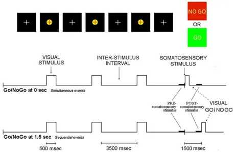
Subjects were seated in a comfortable reclining armchair in front of a computer monitor. A trial of the experimental design is shown inFig. 1. In order to increase expectancy processes (Sutton et al., 1967), 3 visual stimuli (yellow target with black background, 500 ms duration) preceded an electrical somatosensory stimulus. This somatosensory stimulus was obtained by a constant current monophasic pulse of 5 ms, intracutaneously applied by a pin electrode. The stimulus was non-painful and was delivered to the tip of the left index finger over subjective somatosensory threshold. The interval between the onset of two consecu-tive stimuli (3 visualC1 somatosensory) was of 4000 ms. In a first condition named ‘Go/NoGo 0’, the electrical somatosensory stimulus was concomitant with visual stimulus of Go/NoGo task. In this name, ‘0’ indicates the fact that visual Go/NoGo stimulus was delivered at the zerotime, namely the instant at which the somatosensory stimulus was given. The subjects performed either a right hand movement after visual green stimulus (Go stimulus) or no movement after a red stimulus (NoGo stimulus). The occurrence of green and red visual stimuli was randomized (50% of probability to occur for each kind of stimulus). In a
second condition named ‘Go/NoGoC1.5’, the Go/NoGo task was delivered 1.5 s after the somatosensory stimulus given at the zerotime. The two conditions (‘Go/NoGo 0’ and ‘Go/NoGoC1.5’) were performed in two separate recording blocks, whose time order was counterbalanced across subjects. A brief training session served to minimize blinking and eye movements (from 5 s before to 1 s after somatosensory stimulus) and to make stable and reprodu-cible the motor performance during the Go trials. Of note, no appreciable involuntary movement and behavioral error were observed during recordings.
2.3. Electroencephalographic recordings
Bio-signals were recorded (ANT System; bandpass: 0.05–100 Hz, sampling rate: 250 Hz) from 126 electro-encephalographic (EEG) electrodes and two elecro-oculo-graphy electrodes. The EEG electrodes were placed according to an augmented 10–20 system. Linked mastoids served as an electrical reference. To monitor involuntary and voluntary (following the Go stimulus) hand motor responses, electromyographic activity was collected from extensor digitorum muscle. The electrode impedance was kept lower than 5 kU. Acquisition time for all data was set from K10 to C3 s after the onset of the somatosensory stimulation. Indeed, in the other periods of the complete stimulus sequence, the subject could relax the postural and eye control, so that EEG data cannot be used for scientific purposes. Fifty EEG trials were collected in the ‘Go/NoGo 0’ and in the ‘Go/NoGoC1.5’ conditions for each subject.
Fig. 1. Sketch of the experimental design used in the study of expectancy of the Go/NoGo task and consisting in a sequence of three visual stimuli (red cross within a yellow target) and a somatosensory stimulus having a constant inter-stimulus interval of 4 s. This stimulus was followed by a Go/NoGo task represented either by a green visual stimulus triggering a Go response (i.e. right hand movement) or by a red visual stimulus triggering no movement. In the ‘Go/NoGo 0’ condition, the somatosensory stimulus and the Go/NoGo visual stimulus were delivered at the same moment. In the ‘Go/NoGoC1.5’ condition, the Go/NoGo visual stimulus was delivered 1500 ms after the somatosensory stimulus (for interpretation of the reference to colour in this legend, the reader is referred to the web version of this article).
2.4. Preliminary data analysis
The EEG single trials contaminated by blinking, eye movements, and involuntary motor acts were rejected off-line. The spatial resolution of artifact-free EEG data was enhanced by surface Laplacian estimation (regularized 3D spline function), which reduces low spatial frequen-cies of EEG distribution possibly due to head volume conductor effects (Babiloni et al., 1996, 1998, 2001; Nunez, 1996) and eliminates electrode reference influ-ence (Nunez, 1995). In some cases, the Laplacian values at the border electrodes were zeroed because of unreliability of spline Laplacian estimate for these electrodes. Individual data were then interpolated by a spline function (Babiloni et al., 1995), in order to obtain sets at 90 electrode sites of an augmented 10–20 system. This made consistent electrode position across subjects for the subsequent data analyses. The 90 electrodes were disposed over a 3D ‘quasi-realistic’ head model by a ‘spline’ interpolating function.
The single trial analysis was carefully repeated on the Laplacian-transformed EEG data to discard the single trials contaminated by computational artifacts. On an average, the mean of individual artifact-free data was of 28 (G3.2 standard error, SE) single trials for each task. The amount of trial is in line with other research reports on similar topics (Babiloni et al., 2005a,c; Filipovic et al., 2001). Finally, in one out of 14 subjects, trial analysis showed excessive artifact contamination and only on 13 individual data sets are therefore considered.
For the final data analysis, electrodes of interest were C3, C4, P3 and P4. These electrodes overlaid left sensorimotor, right sensorimotor, left posterior parietal, and right posterior parietal cortices, respectively. Of note, the use of much more exploring electrodes was not redundant, since surface Laplacian estimation used the spatial information from all of them to spatially enhance the potentials at the electrodes of interest (Babiloni et al., 1996).
2.5. Estimation of functional coupling: between-electrode coherence analysis
The EEG coherence is a normalized measure of the coupling between two signals at any given frequency (Babiloni et al., 2004c; Halliday et al., 1995; Rappelsberger and Petsche, 1988). From a physiological point of view, EEG coherence reflects functional cooperation among the brain areas under study. The coherence values were calculated for each frequency bin by
CohxyðlÞ Z
jfxyðlÞj 2
fxxðlÞfyyðlÞ
which is the extension of the Pearson’s correlation coefficient to complex number pairs. In this equation, f denotes the spectral estimate of two EEG signals x and y for
a given frequency bin (l). The numerator contains the cross-spectrum for x and y (fxy), while the denominator contains
the respective autospectra for x (fxx) and y (fyy). For each
frequency bin (l), the coherence value (Cohxy) is obtained
by squaring the magnitude of the complex correlation coefficient R. This procedure returns a real number between 0 (no coherence) and 1 (maximal coherence). According to current standards, the EEG coherence values were subjected to hyperbolic tangent transformation to make the coherence values Gaussian.
Here, the EEG coherence was computed between Laplacian-transformed data at C4, C3, P4, and P3 sites. The between-electrode EEG coherence was calculated at ‘baseline or rest’ period (from K5 to K4 s, before the third visual stimulus), ‘PRE-somatosensory stimulus’ period (from K1 s to zerotime), and ‘POST-somatosensory stimulus’ period (from C0.5 to C1.5 s; 1 s period before the visuomotor demands in the ‘Go/NoGo 1.5’ condition and in the corresponding period of the ‘Go/NoGo 0’ condition; in the ‘Go/NoGo 0’ condition, this period anticipated no stimulation). The computation of the EEG coherence was performed from 1 s EEG data segments to obtain coherence values at 1 Hz frequency resolution. The EEG coherence values were calculated within delta–theta (2–7 Hz), alpha (8–14 Hz), beta 1 (15–21 Hz), beta 2 (22– 33 Hz), and gamma (34–45 Hz) frequency bands, according to previous studies (Gerloff et al., 1998; Tecchio et al., 2003; Tiihonen et al., 1989). Furthermore, previous evidence has shown that functional coupling as revealed by EEG coherence can be observed in a wide range of frequencies (von Stein and Sarnthein, 2000). For the statistical analysis of the EEG coherence, individual frequency values within these bands were selected subject-by-subject. This was obtained by selecting the individual frequency showing the maximum coherence value for each subject and for each band.
2.6. Statistical analysis
Statistical comparisons were performed by four-way ANOVA for repeated measures. Dependent variable was event-related EEG coherence (ErCoh). ErCoh is defined as the arithmetical difference between coherence value at the event period (i.e. 1 s expectancy period of interest) and coherence value at the baseline period (i.e. 1 s period before the third visual stimulus). This measure has the advantage to take into account the inter-subject variability of baseline coherence. It should be remarked that the magnitude of ErCoh is usually smaller than the absolute coherence values, since it is obtained computing a difference of coherence values. The ErCoh presents positive values if coherence is higher during the event than baseline period. Vice versa the ErCoh is negative if coherence is lower during the event than baseline period.
In the ANOVA, Mauchley’s test evaluated the sphericity assumption; correction of the degrees of freedom was made by Greenhouse–Geisser procedure. Duncan test was used for post hoc comparisons (P!0.05). The statistical design included the factors Condition (‘Go/NoGo 0’ and ‘Go/NoGoC1.5’), Band (delta–theta, alpha, beta 1, beta 2, gamma), Electrode pair (C4–P4, C3–P3), and Time (PRE-somatosensory stimulus, POST-(PRE-somatosensory stimulus). As aforementioned, the ‘Time’ factor disentangled in time the specific preparatory processes for the somatosensory stimulus and for the visuomotor task. In the ‘sequential’ ‘Go/NoGoC1.5’ condition, 1 s period preceding the somatosensory stimulus could isolate the anticipatory somatosensory processes in the contralateral right hemi-sphere (PRE-somatosensory stimulus), while 1 s period preceding the visual stimulus could isolate the anticipatory visuomotor processes in the contralateral left hemisphere POST-somatosensory stimulus.
The confirmation of the working hypotheses implied that centro-parietal ErCoh increases in the contralateral hemi-sphere during the expectancy of a specific event (i.e. ‘within modality’ comparisons). For example, right centro-parietal ErCoh should specifically increase during the expectancy of contralateral (left) somatosensory hand stimulation, regard-less of the expectancy of hand motor event. Whereas, left centro-parietal ErCoh should specifically increase during the expectancy of contralateral (right) hand motor event, regardless the expectancy of hand somatosensory event. In particular, the confirmation of the working hypotheses implied the following statistical results:
(i) in the ‘Go/NoGo 0’ condition, bilateral centro-parietal ErCoh should be stronger during the anticipation of simultaneous somatosensory and motor events than after that period, in which no corresponding stimulations occurred (i.e. PRE-somatosensory stimulus coherenceOPOST-somato-sensory stimulus coherence);
(ii) in the ‘Go/NoGoC1.5’ condition, centro-parietal ErCoh should be stronger in the right hemisphere during the anticipation of the left (contralateral) somatosensory hand stimulation than after that period in which no further somatosensory stimu-lation occurred—there was just the anticipation of the hand motor event (i.e. PRE-somatosensory stimulus coherence OPOST-somatosensory stimu-lus coherence);
(iii) in the ‘Go/NoGoC1.5’ condition, centro-parietal ErCoh should be stronger in the left hemisphere during the anticipation of the right (contralateral) hand motor event than before that period in which no analogous motor event occurred—there was just the anticipation of the left (ipsilateral) somatosen-sory hand stimulation (i.e. POST-somatosensomatosen-sory stimulus coherence OPRE-somatosensory stimulus coherence).
As a note, the statistical analysis considered only EEG data from subjects showing coherence values above statistical threshold posed at P!0.05, i.e. statistically significant coherence values. Statistical threshold level for coherence was calculated based on the number of single valid EEG trials in accordance with the procedure by Halliday et al. (1995). In particular, the statistical threshold was computed as follows
Threshold Z 1K0:05ð1=NK1Þ where N is the number of trials.
3. Results
3.1. Functional coupling as revealed by spectral coherence between electrodes
Fig. 2illustrates the grand average of coherence spectra at centro-parietal (C3–P3, C4–P4) electrode pairs during REST, PRE-somatosensory stimulus, and POST-somato-sensory stimulus periods of the two conditions, namely the ‘Go/NoGo 0’ condition using ‘simultaneous’ sensorimotor events and the ‘Go/NoGoC1.5’ condition using ‘sequen-tial’ sensorimotor events. In all periods and in both conditions, the absolute coherence values ranged from 0.3 to 0.5 for the left centro-parietal electrode pairs (C3–P3) and from 0.4 to 0.6 for the right centro-parietal electrode pairs (C4–P4). Compared to grand average coherence values at the baseline period, the event-related changes of the coherence values are difficult to be disentangled fre-quency-by-frequency in the figure. This task was easier and more effective when the individual reactive frequency for coherence and the event-related changes of the coherence were taken into account by the ErCoh index. In the evaluation of the present absolute coherence values, it should be remembered that they were calculated from surface Laplacian data, in which the coherence due to head volume conduction is deflated (Nunez et al., 1995, 1996). 3.2. Statistical results
Hemispheric asymmetry of the centro-parietal coherence values was statistically evaluated by three ANOVA analyses, one for each period of interest (REST, PRE-somatosensory stimulus, and POST-PRE-somatosensory stimu-lus). Coherence values served as a dependent variable. Condition (‘Go/NoGo 0’, ‘Go/NoGo 1.5’), Hemisphere (left, right), and Band (delta–theta, alpha, beta 1, beta 2, gamma) were used as factors. A statistically significant main effect Hemisphere was observed for all periods of interest (REST: FZ5.62, P!0.05; PRE-somatosensory stimulus: FZ5.18, P!0.05; POST-somatosensory stimu-lus: FZ9.39, P!0.01), pointing to higher coherence values in the right than left centro-parietal electrode pairs,
regardless of the frequency bands and conditions. These results were in line with previous evidence of a similar hemispheric asymmetry in EEG coherence in humans (Tucker et al., 1986) and motivated the statistical analysis of ErCoh for the evaluation of the working hypotheses of the present study.
The ErCoh was computed in the PRE-somatosensory stimulus and POST-somatosensory stimulus periods, by subtracting coherence values calculated at the rest (baseline) period. As aforementioned, the ANOVA design included the factors Condition (‘Go/NoGo 0’ using ‘simultaneous sensorimotor events’, ‘Go/NoGoC1.5’ using ‘sequential sensorimotor events’), Band (delta–theta, alpha, beta 1, beta 2, gamma), Electrode pair (C4–P4, C3–P3), and Time (PRE-somatosensory stimulus, POST-somatosensory stimulus). All selected subjects (NZ13) showed coherence
values higher than statistical threshold as computed with the procedure suggested byHalliday et al. (1995), namely 0.128 (G0.002, SE) for the ‘Go/NoGo 0’ condition and 0.125 (G 0.002 SE) for the ‘Go/NoGoC1.5’ condition. The only statistically significant ANOVA effect was a three-way statistical ANOVA interaction (F1,12Z10.39; P!0.01) among the factors Condition, Time, and Electrode pair.
Fig. 3 illustrates the mean of the ErCoh (GSE) across subjects for the above ANOVA interaction. The ErCoh values were low in amplitude due to the fact that they are differences between absolute coherence values at expect-ancy and baseline periods. Duncan post hoc testing fully confirmed the working hypotheses. In the ‘Go/NoGo 0’ condition, bilateral centro-parietal ErCoh (C3–P3, C4–P4) was stronger (P!0.01) during the anticipation of the simultaneous somatosensory and motor events (i.e.
Fig. 3. Group mean values (GSE) of the event-related coherence (ErCoh) illustrating three-way statistical ANOVA interaction (F1,12Z10.39; P!0.01) among the factors Condition, Time and Electrode pair. The ANOVA design included the factors Condition (‘Go/NoGo 0’, ‘Go/NoGoC1.5’), Band (delta–theta, alpha, beta 1, beta 2, gamma), Electrode pair (C4–P4, C3–P3), and Time (PRE, POST). Asterisks denote statistically significant differences as obtained by Duncan post hoc test (P!0.05).
Fig. 2. Grand average of the coherence spectra computed from centro-parietal (C3–P3, C4–P4) electrode pairs during REST, PRE and POST periods of the ‘Go/NoGo 0’ and ‘Go/NoGoC1.5’ conditions. See Section 2 for details.
PRE-somatosensory stimulus coherence) than after that period (i.e. POST-somatosensory stimulus coherence). In the ‘Go/NoGoC1.5’ condition, centro-parietal ErCoh was stronger (P!0.01) in the right hemisphere during the anticipation of the left (contralateral) somatosensory hand stimulation (i.e. PRE-somatosensory stimulus coherence) than after that period in which no further somatosensory stimulation occurred (i.e. POST-somatosensory stimulus coherence). In the ‘Go/NoGoC1.5’ condition, centro-parietal ErCoh was stronger (P!0.03) in the left hemi-sphere during the anticipation of the right (contralateral) hand motor event (i.e. POST-somatosensory stimulus coherence) than before that period in which no analogous motor event occurred—there was just the anticipation of the left (ipsilateral) somatosensory hand stimulation (i.e. PRE-somatosensory stimulus coherence).
4. Discussion
Is the anticipation of somatosensory or motor events related to an increase of centro-parietal functional coupling at the contralateral hemisphere (as revealed by EEG spectral coherence)? It was observed that the anticipation of the only somatosensory stimulation to the left hand just increased centro-parietal functional coupling in the contralateral right hemisphere. Conversely, the anticipation of the only right-hand movements just increased centro-parietal functional coupling in the contralateral left hemisphere. Finally, the anticipation of concomitant somatosensory and motor events increased the anticipatory centro-parietal functional coupling in both hemispheres. It should be stressed that opposite sides were used for the somatosensory stimulation (left hand) and the motor response (right hand). In that context, despite some contribution for transcallosal flow of information, the anticipatory centro-parietal coupling in the left hemisphere could be functionally ascribed mainly to the motor channel, whereas the anticipatory centro-parietal coupling in the right hemisphere could be ascribed mainly to the somatosensory channel. Of note, we used somatosensory and sensorimotor events rather than two somatosensory or two sensorimotor events, to support the idea that both somatosensory and sensorimotor events induce centro-parietal functional coupling at the contralateral hemisphere. To address the issue of the hemispheric functional asymmetry, future research should consider a design including somatosensory stimulation at the right hand and right-hand sensorimotor events. This was not done in the present study to avoid that excessively long experiments could induce fatigue and bias in attentional processes such as those characterizing the expectancy periods.
Is parieto-central ErCoh specific for the present passive somatosensory and active visuomotor events? Would the modulation of the parieto-central ErCoh occur in other modalities and events such as auditory modality and counting task? Previous studies using
magnetoencephalographic and intracranial EEG recordings have demonstrated that somatosensory stimuli induce responses of primary somatosensory and posterior parietal cortical areas. In particular, some studies showed the responses only in contralateral posterior parietal cortex (Forss et al., 1994; Gobbele et al., 2003), whereas others also found the responses in ipsilateral posterior parietal cortex (Disbrow et al., 2003; Rektor, 2000). As an extension of these findings, the present study showed that preparatory sensory processes are reflected by a functional coupling of EEG rhythms between central and parietal areas only in the hemisphere contralateral to the stimulation (see results of the ‘sequential’ condition). The present results showed that expectancy of the hand somatosensory stimulation specifi-cally enhanced right (contralateral) centro-parietal ErCoh even whether the somatosensory stimulus was task-irrelevant (i.e. it triggered no counting or motor response). Similar effects were observed for the left (contralateral) centro-parietal ErCoh during the expectancy of right hand visuomotor events in which the task implied visual attentional processes and stimulus uncertainty (i.e. visual Go stimuli were delivered 50% of cases). These results would suggest that centro-parietal ErCoh is sensitive not only to task-relevant stimuli (visuomotor modality) but also to stimuli passively received by subjects (somatosensory modality). However, it should be stressed that a fine evaluation of that ErCoh specificity requires future investigations including systematic modulation of atten-tional load, level of stimulus uncertainty, and task-relevance of the stimuli across stimulus modalities and the two hemispheres. Indeed, the present experimental design allowed just the comparison of the centro-parietal ErCoh within each stimulus modality (somatosensory and visuo-motor), due to the unpaired attentional load between the two modalities.
In the present study, the effects on coherence were not specific for EEG bands. From a statistical point of view, parieto-central EEG coherence had similar trends in all frequency bands. This result is at odds with the idea that the power of EEG at different frequency bands is associated with peculiar cognitive-behavioral functions. In reality, there is no general consensus on that idea. Previous evidence has shown that sensorimotor, attentional, and memory processes are all related to the modulation of gamma power at about 40 Hz (Basar et al., 2001; Engel and Singer, 2001; Singer, 2001; Tecchio et al., 2003), but also to the modulation of beta, alpha, and theta power (Klimesch, 1999; Klimesch et al., 1998; Pfurtscheller and Lopes da Silva, 1999; Sarnthein et al., 1998; Stam et al., 2002). It can be speculated that cognitive processes are associated with the parallel modulation of different EEG rhythms within proper neural networks. The functional specificity of these different EEG rhythms might be affected by the extension and kind of the neural networks engaged (Pfurtscheller and Lopes da Silva, 1999; von Stein and Sarnthein, 2000). At the present stage of research, it should be stressed that
variations of EEG band power across the physiological events do not directly indicate the level of cooperation among nodes of the neural networks. That functional cooperation can be roughly disclosed by functional coupling as revealed by coherence analysis or non-linear techniques (Babiloni et al., 2004a; Stam et al., 2005). On the whole, the present results indicate that expectancy of sensorimotor events is correlated with the functional coupling of parietal and central areas as a function of the specific requested information processing (temporal and spatial specificity) and that such a functional coupling reflects a modulation of the EEG rhythms at all main frequency bands.
Anticipatory processes preceding sensorimotor inter-actions might engage a more complex and distributed neural network than the one limited to centro-parietal areas. The whole network would include multiple frontal, parietal and sub-cortical structures subserving, among others, ‘top down’ anticipatory influences (Giesbrecht et al., 2003; Pessoa et al., 2003). However, some reasons imposed the present data analysis design. A relatively small group size prevented the inclusion of additional cortical regions of interest in the statistical design having 4 factors and 11 levels. Furthermore, the limited spatial resolution of EEG techniques discouraged fragmentation of parietal cortex (i.e. inferior vs. dorsal). Finally, the extreme simplicity and repetitive nature of the experimental stimuli sequence might require longer experimental sessions to enlighten the role of fronto-parietal networks. In previous experiments, the same paradigm induced a small anticipatory activation of frontal areas (Babiloni et al., 2003, 2004b, 2005a). The relation-ships of the centro-parietal neural network with the frontal executive and attentional are crucial scientific issues that merit to be addressed by future investigations colleting data from a larger subjects’ group.
In conclusion, the present EEG study tested the hypothesis that the anticipation of predictable sensorimotor events is related to an increase of functional centro-parietal coupling, as revealed by EEG spectral coherence. Before the ‘simultaneous’ somatosensory and visuomotor events involving both hands, the spectral coherence in the centro-parietal regions was high in both hemispheres and at all rhythms. In the condition of ‘sequential’ somatosensory and visuomotor events, the coherence was high in the right centro-parietal areas before the somatosensory event (left hand) and in the left centro-parietal areas before the Go/NoGo event (right-hand movements). These results suggest that the anticipation of somatosensory and motor events increases the contralateral centro-parietal coupling by the temporal synchronization of slow and fast brain rhythms. This means that predictable somatosensory and visuomotor events are anticipated not only by synchroniza-tion of cortical pyramidal neurons generating EEG power in parietal and primary sensorimotor cortical areas (Babiloni et al., 2005a) but also by functional coordination of these areas. The present protocol may be of interest for the study of the neural correlates of integrative information processes
in human centro-parietal cortical areas during sensorimotor and cognitive interactions.
Acknowledgements
We thank Dr Paolo Capotosto for their helpful technical assist. We thank also Prof. Fabrizio Eusebi for his continuous support. The research was granted by Danish Technical Research Council and Association Fatebenefra-telli for the Research (AFaR).
References
Babiloni F, Babiloni C, Fattorini L, Carducci F, Onorati P, Urbano A. Performances of surface Laplacian estimators: a study of simulated and real scalp potential distributions. Brain Topogr 1995;8(1):35–45. Babiloni F, Babiloni C, Carducci F, Fattorini L, Onorati P, Urbano A.
Spline Laplacian estimate of EEG potentials over a realistic magnetic resonance-constructed scalp surface model. Electroencephalogr Clin Neurophysiol 1996;98(4):363–73.
Babiloni F, Carducci F, Babiloni C, Urbano A. Improved realistic Laplacian estimate of highly-sampled EEG potentials by regularization techniques. Electroencephalogr Clin Neurophysiol 1998;106(4): 336–43.
Babiloni F, Cincotti F, Carducci F, Rossini PM, Babiloni C. Spatial enhancement of EEG data by surface Laplacian estimation: the use of magnetic resonance imaging-based head models. Clin Neurophysiol 2001;112(5):724–7.
Babiloni C, Brancucci A, Babiloni F, Capotosto P, Carducci F, Cincotti F, Arendt-Nielsen L, Chen AC, Rossini PM. Anticipatory cortical responses during the expectancy of a predictable painful stimulation. A high-resolution electroencephalography study. Eur J Neurosci 2003; 18(6):1692–700.
Babiloni C, Babiloni F, Carducci F, Cincotti F, Vecchio F, Cola B, Rossi S, Miniussi C, Rossini PM. Functional frontoparietal connectivity during short-term memory as revealed by high-resolution EEG coherence analysis. Behav Neurosci 2004a;118(4):687–97.
Babiloni C, Brancucci A, Arendt-Nielsen L, Del Percio C, Babiloni F, Pascual-Marqui R, Sabbatini G, Rossini PM, Chen ACN. Cortical sensorimotor interactions during the expectancy of a go/nogo task: effects of painful stimuli. Behav Neurosci 2004b;118(5):925–35. Babiloni C, Brancucci A, Arendt-Nielsen L, Babiloni F, Capotosto P,
Carducci F, Cincotti F, Del Percio C, Petrini L, Rossini PM, Chen AC. Attentional processes and cognitive performance during expectancy of painful galvanic stimulations: a high-resolution EEG study. Behav Brain Res 2004c;152(1):137–47.
Babiloni C, Brancucci A, Capotosto P, Arendt-Nielsen L, Chen ACN, Rossini PM. Expectancy of pain is influenced by motor preparation: a high-resolution EEG study of cortical alpha rhythms. Behav Neurosci 2005a;119(2):503–11.
Babiloni C, Brancucci A, Pizzella V, Romani GL, Tecchio F, Torquati K, Zappasodi F, Arendt-Nielsen L, Chen ACN, Rossini PM. Contingent negative variation in the parasylvian cortex increases during expectancy of painful sensorimotor events: a magnetoencephalographic study. Behav Neurosci 2005b;119(2):491–502.
Babiloni C, Brancucci A, Capotosto P, Romani GL, Arendt-Nielsen L, Chen ACN, Rossini PM. Slow cortical potential shifts preceding sensorimotor interactions. Brain Res Bull 2005c;65(4):309–16. Basar E, Schurmann M, Basar-Eroglu C, Demiralp T. Selectively
distributed gamma band system of the brain (Review). Int J Psychophysiol 2001;39(2–3):129–35.
Disbrow E, Litinas E, Recanzone GH, Padberg J, Krubitzer L. Cortical connections of the second somatosensory area and the parietal ventral area in macaque monkeys. J Comp Neurol 2003;462(4):382–99. Engel AK, Singer W. Temporal binding and the neural correlates of sensory
awareness. Trends Cogn Sci 2001;5(1):16–25.
Filipovic SR, Jahanshahi M, Rothwell JC. Uncoupling of contingent negative variation and alpha band event-related desynchronization in a go/no-go task. Clin Neurophysiol 2001;112:1307–15.
Fogassi L, Luppino G. Motor functions of the parietal lobe. Curr Opin Neurobiol 2005.
Forss N, Hari R, Salmelin R, Ahonen A, Hamalainen M, Kajola M, Knuutila J, Simola J. Activation of the human posterior parietal cortex by median nerve stimulation. Exp Brain Res 1994;99(2):309–15. Gerloff C, Richard J, Hadley J, Schulman AE, Honda M, Hallett M.
Functional coupling and regional activation of human cortical motor areas during simple, internally paced and externally paced finger movements. Brain 1998;121:1513–31.
Gevins A, Smith ME, Leong H, McEvoy L, Whitfield S, Du R, Rush G. Monitoring working memory load during computer-based tasks with EEG pattern recognition methods. Hum Factors 1998;40(1):79–91. Giesbrecht B, Woldorff MG, Song AW, Mangun GR. Neural mechanisms
of top–down control during spatial and feature attention. Neuroimage 2003;19(3):496–512.
Gobbele R, Schurmann M, Forss N, Juottonen K, Buchner H, Hari R. Activation of the human posterior parietal and temporoparietal cortices during audiotactile interaction. Neuroimage 2003;20(1):503–11. Gomez CM, Vaquero E, Lopez-Mendoza D, Gonzalez-Rosa J,
Vazquez-Marrufo M. Reduction of EEG power during expectancy periods in humans. Acta Neurobiol Exp (Wars) 2004;64(2):143–51.
Halliday DM, Rosenberg JR, Amjad AM, Breeze P, Conway BA, Farmer SF. A framework for the analysis of mixed time series/point process data—theory and application to the study of physiological tremor, single motor unit discharges and electromyograms. Prog Biophys Mol Biol 1995;64(2–3):237–78.
Klimesch W. Memory processes, brain oscillations and EEG synchroniza-tion. Int J Psychophysiol 1996;24(1–2):61–100.
Klimesch W. EEG-alpha rhythms and memory processes. Int J Psychophysiol 1997;26(1–3):319–40.
Klimesch W. Event-related band power changes and memory performance. In: Pfurtscheller G, Lopes da Silva FH, editors. Event-related desynchronization. Handbook of electroencephalography and clinical neurophysiology, revised series, vol. 6. Amsterdam: Elsevier; 1999. p. 161–78.
Klimesch W, Doppelmayr M, Schimke H, Pachinger T. Alpha frequency, reaction time, and the speed of processing information. J Clin Neurophysiol 1996;13(6):511–8.
Klimesch W, Doppelmayr M, Russegger H, Pachinger T, Schwaiger J. Induced alpha band power changes in the human EEG and attention. Neurosci Lett 1998;244(2):73–6.
Locatelli T, Cursi M, Liberati D, Franceschi M, Comi G. EEG coherence in Alzheimer’s disease. Electroencephalogr Clin Neurophysiol 1998; 106(3):229–37.
Nunez P. Neocortical dynamics and human EEG rhythms. New York: Oxford University Press; 1995.
Nunez P. Spatial analysis of EEG. Electroencephalogr Clin Neurophysiol Suppl 1996;45:37–8.
Pessoa L, Kastner S, Ungerleider LG. Neuroimaging studies of attention: from modulation of sensory processing to top-down control. J Neurosci 2003;23(10):3990–8.
Pfurtscheller G, Lopes da Silva FH. Event-related EEG/MEG synchroniza-tion and desynchronizasynchroniza-tion: basic principles. Clin Neurophysiol 1999; 110(11):1842–57.
Rappelsberger P, Petsche H. Probability mapping: power and coherence analyses of cognitive processes. Brain Topogr 1988;1(1):46–54. Rektor I. Parallel information processing in motor systems: intracerebral
recordings of readiness potential and CNV in human subjects. Neural Plast 2000;7(1–2):65–72.
Sarnthein J, Petsche H, Rappelsberger P, Shaw GL, von Stein A. Synchronization between prefrontal and posterior association cortex during human working memory. Proc Natl Acad Sci USA 1998;95(12): 7092–6.
Sauseng P, Klimesch W, Doppelmayr M, Pecherstorfer T, Freunberger R, Hanslmayr S. EEG alpha synchronization and functional coupling during top–down processing in a working memory task. Hum Brain Mapp 2005;26(2):148–55.
Serrien DJ, Pogosyan AH, Brown P. Influence of working memory on patterns of motor related cortico-cortical coupling. Exp Brain Res 2004; 155(2):204–10.
Singer W. Consciousness and the binding problem. Ann NY Acad Sci 2001; 929:123–46.
Stam CJ, van Cappellen van Walsum AM, Pijnenburg YA, Berendse HW, de Munck JC, Scheltens P, van Dijk BW. Generalized synchronization of MEG recordings in Alzheimer’s disease: evidence for involvement of the gamma band. J Clin Neurophysiol 2002;19(6):562–74. Stam CJ, Montez T, Jones BF, Rombouts SA, van der Made Y,
Pijnenburg YA, Scheltens P. Disturbed fluctuations of resting state EEG synchronization in Alzheimer’s disease. Clin Neurophysiol 2005; 116(3):708–15.
Sutton S, Tueting P, Zubin J, John ER. Information delivery and the sensory evoked potential. Science 1967;155(768):1436–9.
Tecchio F, Babiloni C, Zappasodi F, Vecchio F, Pizzella V, Romani GL, Rossini PM. Gamma synchronization in human primary somatosensory cortex as revealed by somatosensory evoked neuromagnetic fields. Brain Res 2003;986(1–2):63–70.
Thatcher RW, Krause PJ, Hrybyk M. Cortico-cortical associations and EEG coherence: a two-compartmental model. Electroencephalogr Clin Neurophysiol 1986;64(2):123–43.
Tiihonen J, Hari R, Kaukoranta E, Kajola M. Interaural interaction in the human auditory cortex. Audiology 1989;28(1):37–48.
Tomberg C, Desmedt JE. Failure to recognise objects by active touch (astereognosia) results from lesion of parietal-cortex representation of finger kinaesthesis. Lancet 1999;354(9176):393–4.
Tucker DM, Roth DL, Bair TB. Functional connections among cortical regions: topography of EEG coherence. Electroencephalogr Clin Neurophysiol 1986;63(3):242–50.
Urbano A, Babiloni C, Onorati P, Babiloni F. Dynamic functional coupling of high resolution EEG potentials related to unilateral internally triggered one-digit movements. Electroencephalogr Clin Neurophysiol 1998;106(6):477–87.
von Stein A, Sarnthein J. Different frequencies for different scales of cortical integration: from local gamma to long range alpha/theta synchronization. Int J Psychophysiol 2000;38(3):301–13.
Wheaton LA, Nolte G, Bohlhalter S, Fridman E, Hallett M. Synchronization of parietal and premotor areas during preparation and execution of praxis hand movements. Clin Neurophysiol 2005; 116(6):1382–90.