0394-6320 (2007) Copyright © by BIOLIFE, s.a.s. This publication and/or article is for individual use only and may not be further reproduced without written permission from the copyright holder. Unauthorized reproduction may result in financial and other penalties
(S1)43
MODIFICATION OF CONDYLE ANATOMY FOLLOWING A MONOLATERAL BITE RISE: A HISTOLOGICAL STUDY IN RAT
M. D’ATTILIO, A. SCARANO, A. QUARANTA1, F. FESTA, S. CAPUTI and A. PIATTELLI Department of Stomatology and Oral Sciences, Medical and Dental Schools, University of
Chieti-Pescara; 1University of Rome “La Sapienza”, Rome, Italy
Mailing address: Michele D’Attilio, DDS, MSD,
University “G. D’Annunzio”, Via dei Vestini, 31
66100 Chieti, Italy
Tel.: ++39-0871-3554159 Fax: ++39-0871-3554159 e-mail: [email protected]
Key words: articular compression, articular distraction, bite rise, bone remodelling, temporomandibular joint
The aim of this study is to evaluate the histologic modifications of rat articular capsules, after compression and strain. Fifty adults Sprague Dawley rats were used in this study. The rats were divided into 3 groups: a dysfunction group, treatment group and a Control group. In the rats in the first two groups an occlusion hump, made of composite and less than 0,5 mm high, was applied to the upper right molar. The rats were anesthetized with an intraperitoneal injection of benzodiazepine (0.5-1mg for 100 g. of body weight). The composite used consisted of a hollow, plastic cylinder 0.5 mm high, spread over the whole occlusal surface of the right upper molar. The dysfunction group therapy wore the occlusion hump for a week, at the end of which the animals in this group were killed and their temporomandibular joint removed. In the treatment group a second occlusion hump was applied to the left upper molar, in order to obtain a distraction of the previously compressed temporomandibular joint. The rats of the second group wore the second occlusion hump for one week. The rats were then killed with an intraperitoneal injection of Tanax, and the block sections, containing the temporo-mandibolar joint, were retrieved with a diamond disk, and surrounding tissues were washed in saline solution and immediately fixed in 4% formalin for 4 days. In the control group, no treatment was applied. In the first group, bone resorption was observed in the left temporomandibular joint; no osteoclast were, however, present. No other tissue alterations were present. Newly formed bone undergoing remodelling was intensely stained with acid fuchsin. In the second and third groups, no bone remodelling areas were observed. In conclusion, the compression of the temporomandibular joint determined a remodelling of the bone structure of the condyle.
The problems related to the pathologies of the temporomandibular joint (TMJ) have become increasingly interesting as a result of greater knowledge and the improved diagnostic methods as well as the growing incidence of these pathologies (1-7). The optimal condition in the TMJ is the result of a balance between stomatognathic structure and function (1)
When the mandible is in rest position the intra-articular pressure is low, the intra-intra-articular space is slightly widened and the articular pressure is the same in all mandible normal movement. When the intra-articular pressure is higher as a consequence of a reduction of the intra-articular space, the position of the intra-articular structures become altered. So, when the condyle is pushed against the articular disk with a reduction of the articular space it is possible to have a compression pathology; instead, when the condyle is out of the glenoid fossa, with an increase of the intra-articular space it is possible to have a distraction pathology (1, 9).
On the condyle under compression, the capsule is collapsed with a possible ischaemia or hypoxia/anoxia of the tissues. There is also an increase of the intra-articular pressure that stimulates the III and IV type of capsular receptors, producing a feedback reflex arch with a long
jerk causing a further capsule collapse and pain.
On the condyle under distraction, the articular capsule undergoes a progressive strain but the capsules remain histologically healthy if the capsule fibres do not undergo a strain higher than 70%. On the contrary with a strain higher than 70%, the articular capsule loses its normal working capacity (10-11).
To treat then a TMJ problem due to compression, there is a need to restore the correct length of the capsule fibres. This fact will produce a reduction of the intra-articular compression determining the end of the jerk and the pain (1).
The etiology of TMJ disorders can have multiple causes such as a global orthopedic problem along with morphofunctional alterations or physical components. Most often, the occlusal component is the one altered; however, recent studies have shown that for every alteration of temporomandibular function or pure condyle-disk alteration, there is a muscular fibrotic response (2).In such cases it is always possible to find a muscular contraction starting fibrosis. In the treatment of TMJ disease, it is necessary to induce a lengthening of the muscle fibers (12). Two types of responses are possible in the cases of incoordination (13-14).
2. Muscular> activation of the muscular nociceptors. Both responses cause muscular contraction, and physical therapy is necessary to progressively stretch the muscle fibres and lengthen the muscles. The aim of this research is to evaluate the histologic modifications of rat condyles subjected to compression and strain.
MATERIALS AND METHODS
Fifty adult female Sprague Dawley rats weighing 350 g (average age ten months) were used in the present study. The rats were divided into 3 groups:
a) dysfunction group (20 rats); b) reatment group (20 rats); c) control group (10 rats).
In the rats in the first two groups an occlusion hump, made of composite and less than O.5. mm high, was applied to the upper right molar. The composite was spread over the whole right occlusal surface of the teeth. In the first group, the rats wore the hump for one week, at the and of which the animals in this group were killed and their TMJ removed.
In the second group, a second occlusion raise was applied to the left upper molar at the end of the first week in order to obtain a distraction of the previously compressed joint cap. The rats wore the second occlusion raise for an additional week. At the end of this period the rats were killed and their TMJ were removed.
The block sections, containing the TMJ, were retrieved with a diamond disk, and surrounding tissues were washed in saline solution and immediately fixed in 4% formalin for 4 days. The specimens were processed to obtain thin ground sections with the Precise 1 Automated System (Assing, Rome, Italy) (2). The specimens were dehydrated in an ascending series of alcohol rinses and embedded in a glycolmethacrylate resin (Technovit 7200 VLC, Kulzer, Wehrheim, Germany). After polymerization the specimens were sectioned, along their longitudinal axis, with a high-precision diamond disc at about 150 µm and ground down to about 30µm with a specially designed grinding machine. A total of 3 slides were obtained for each implant. The slides were stained with acid and basic fuchsin and toluidine blue. The slides were observed in normal transmitted light under a Leitz Laborlux microscope (Leitz, Wetzlar, Germany). Histomorphometry of percentage of condyle covered by cartilagenous was carried out using a light microscope (Laborlux S, Leitz, Wetzlar, Germany) connected to a high resolution video camera (3CCD, JVC KY-F55B) (JVC®, Yokohama, Japan) and interfaced to a monitor and PC (Intel Pentium III 1200 MMX) (Intel®, Santa Clara, CA, USA). This optical system was associated with a digitizing pad (Matrix Vision GmbH, Oppenweiler, Germany) and a histometry software package with image capturing capabilities (Image-Pro Plus 4.5, Media Cybernetics Inc., Immagini & Computer Snc Milano, Italy). The birefringent organization of bone collagen fibers, determined under polarized-light microscopy, was used to distinguish lamellar from woven bone.
Data Analysis. The differences in the condyle covered by layers of chondrocytes were evaluated. The layers of chondrocytes were expressed as the means ± standard deviation and standard error. The differences between the 3 groups were analyzed by analysis of variance (ANOVA), and statistical significance of multiple comparisons was evaluated using the Fisher PLSD and Scheffe F tests. Significance was set at P ≤ .05.
RESULTS
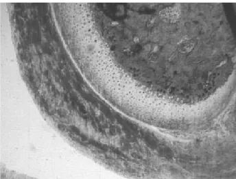
Control group. It was possible to observe the condyle
with a surrounding cartilaginous tissue formed by 4/5 layers of chondrocytes (Fig 1 . Fig 2). Some Haversian systems were visible around the condyle zone. Only in a few areas was it possible to see bone remodelling areas below the cartilaginous layer. Lack of articular cartilaginous tissue or large bone remodelling areas were observed. Some osteoclasts and osteoblasts were present in the central part of the condyle. The percentage of condyle covered by cartilaginous tissue was 91±2.4%.
Dysfunction group. It was possible to show the lack
of the cartilaginous tissue of the condyle and the bone remodelling around the left condyle. This remodelling was particularly evident in the anterior part of the condyle and this led to a variation in the shape and size of the condyle (Fig. 3). Bone resorption with the presence of osteoclasts was observed in the left condyle. An increase in the size of the osteocyte lacunae with the formation of small marrow spaces was also observed. Few osteoclasts, but not osteoblasts were present around the marrow spaces. The remodelling of the condyle was regular especially in the anterior part. This fact was produced by the variation of the occlusal plane, and, in fact, the histologic pattern of the control group was completely different. The percentage of condyle covered by cartilaginous was 39±4.2%.
Treatment group. The histologic pattern was very
similar to the control group excluding the presence of cartilaginous tissue. In this group the cartilaginous tissue was absent (Fig 4). No areas of remodelling of the condyle were present, while few osteoblasts were visible producing new bone. Small osteocyte lacunae and areas of bone necrosis were present. The newly-formed bone was intensely stained with fuchsin acid. In no cases chondroblasts producing articular cartilage were observed. The percentage of condyle covered by cartilaginous is 42±3.4%.
Statistical evaluation. A statistically significant
difference was present in the layers of chondrocytes and in the percentage of condyle covered by cartilaginous tissue between group 3 vs group 2 and 1. (P= 0.0013).
DISCUSSION
Compression of the temporomandibular joint determines a loss of coordination between condyle and disk, with a series of related symptoms also in other regions of the body. The present study was designed to reveal if, after TMJ compression, any anatomical modifications of the condyle were evident. The second goal was to see if it was possible to determine the functional rehabilitation of the condyle after an occlusal plane therapy.
The loss of only one tooth in the posterior segments of the arches has a damaging effect on the TMJ, causing high and posterior displacements and capsular collapse (16-18).
The capsular collagen fibres lose the correct alignment and the characteristic wave shape when intracapsular tension becomes higher (3,4).The collagen fibres become 20-30% elongated, and when stretched this far, normal elasticity is no longer possible and stimulation of type III and IV capsular receptors are used (9). The response is a prolonged muscular contraction which includes a “pain-contraction-pain” cycle. In order to restore the correct relationship between the disk and the condyle, it is necessary to realign the collagen fibres (10,11).
In the past, some experimental studies conducted
in vivo confirmed the presence of structural alterations
due to compression and distraction on the capsular and condylar tissues (5-6). An experiment was carried out on Macaca fascicularis (20). The aim of the study was to observe the effects of a unilateral rise on the condylar
tissues. The decrease of intracapsular vertical dimension was achieved by using a 0.25 mm thick monolateral resin splint for twelve weeks. The capsular complex and muscular insertions were removed from all animals and a microscopic examination showed the existence of a thick, compact connective tissue layer as well as a proliferative cell layer. Deeper inside, a cartilaginous layer with marrow spaces was observed. Higher magnification made it possible to distinguish an external fibrous layer with visible clear areas which may be the result of a disorganization of the fibrils. An ultrastructural examination of the capsules disclosed irregularly-oriented collagen fibres with a disorganization of the fibrils and signs of cell damage.
More recent experimental studies have been carried out to study the articular changes due to compression
Fig. 1. Control group: At low-power magnification it was
pos-sible to observe the condyle with a surrounding cartilaginous tissue. Toluidine blue and acid fuchsin 4X
Fig. 2. At higher magnification it was possible to observe 4/5
layers of chondrocytes. Toluidine blue and acid fuchsin 100X
Fig. 3. Dysfunction group: It was possible to bone remodelling
around the condyle. Toluidine blue and acid fuchsin 50X
Fig. 4. Treatment group: In this area the cartilaginous tissue was
and distraction in the TMJ of rats (21). Compression was induced by fixing a metallic occlusal rise on the upper left second molar for one week.
The study group was made up of 28 Wistar rats approximately 500 grams each; four rats made up the control group, 14 in the therapy group, and ten the dysfunctional group. In both dysfunctional and therapy group, distraction was carried out (21). Then, the following week, the same rise on the opposite side was used on the therapy group only. The microscopic and ultra-structural examination concerned the extracellular matrix. In the articular capsule with compression (right side) the following pattern was observed: an increase of collagen with a decrease of vasal width; collagen fibres irregularly oriented with a disorganization of the fibrils; and hypertrophy of the cells (21). In the articular capsule without compression (on the same side of the rise) the collagen fibres were loose but regularly oriented, and there was a fibroblastic hyperplasia observed in the dysfunctional group. The microscopic examination of the articular capsule subjected to compression and then distraction showed an improvement of the tissue structure. In particular, the collagen fibres were normally oriented, the vasal width was normal, and there were no signs of cell damage. Based on the results of these studies, it can be assumed that the symptomatology is connected to the degree of hypertrophy because of the involvement of the nociceptors (21). During distraction the realignment of the fibres in the characteristic wavelike shape was connected with an almost normal function (21).
Moreover, marked cell proliferation has been associated with increasing mechanical stimuli (7) by other means, such as protrusion of the mandible (7), and posterior relocation of the glenoid fossa (8).
In the present study we have obtained an articular compression on one condyle and an articular distraction on the other condyle by an occlusion hump, made of composite and less than O.5. mm high, applied to the upper right molar. To see the effects of the rebalance a second hump was made (on the first upper left molar) The choice of one week as the period for wearing the unilateral occlusion raise was made by considering the mean life-span of a man (70 years) and of a rat (3 years) and the time necessary in humans for the appearance of the first symptoms of the joint pathology after alteration of occlusion, calculated as 6 months. Expressing the months and years in day and laying out the following proportion:
25550 days: 180 days = 1085: X days, the result of X = 7.64 was obtained.
In conclusion, the present study demonstrates that the condyle, when it is compressed following controlateral bite rise, is anatomically modified both in dimension and shape. It was also possible to show a lack of the cartilaginous
tissue of the condyle. While in the treatment group, after the controlateral bite rise, remodelling zones of the condyle were absent and few osteoblasts were visible.
ACKNOWLEDGEMENTS
This work was partially supported by the National Research Council (CNR), Rome, Italy and by the Ministry of Education, University and Research (MIUR, Rome (Italy). The authors gratefully acknowledge Mr Marcello Piccirilli for the technical support.
REFERENCES
7. Schellhas K.P., M.A. Piper and M.R. Omlie. 1990. Facial skeleton remodelling due to temporomandibular joint degeneration: an imaging study of 100 patients. Am. J. Neuro Radiol. 11:541.
2. Balercia L. and P. Balercia. 1993. Fisiopatologia della deglutizione. Relazioni con occlusione e postura. Il Dentista Moderno 1:55.
3. Cascone P. 1992. Valutazione neuro-muscolare ed esame posturale in pazienti affetti da incoordinazione condilo-meniscale dell’ATM. Minerva Stomatologica 41:79. 4. Colangelo G., F. Festa and S. Colasanto. 1990.
Valutazione clinico-radiografica dei rapporti tra dorso curvo e malocclusione. Mondo Ortodontico XV 4:1. 5. Farrar W.B. 1984. Personal communication Montgomery
Alabama.
6. Festa F., F. D’Emidio and A. Castaldo. 1996. Modificazione dei rapporti occlusali dopo terapia posturale. Ortognatodonzia Italiana V 5:667.
7. Gole D.R. 1993. A clinical observation: a relationship of occlusal contacts to distal muscolature. Cranio 1:55. 8. Rocabado M. and V. Tapia. 1987. Radiographic study of
the cranio-cervical relation in patients under orthodontic treatment and the incidence with related symptoms. Cranio 5:36.
9. Roy S. and F.N. Ghadially. 1967. Synthesis of hyaluronic acid by synovial cells. Path. Bact. 93:555.
10. Rocabado M. 1984. Joint distraction with a functional mandibular orthopedic appliance. J. Craniomandib. Pract. 2:358.
11. Rocabado M. 1983. Arthrokinematics of the temporomandibular joints. Dent. Clin. North Am. 27:573. 12. Festa F. 1985. Joint distraction and condyle advancement
with a modified functional distraction appliance. J. Craniomandib. Pract. 3:343.
13. Luschei E.S. 1981. Neural mechanism of mandibular control: mastication and voluntary biting. In Handbook of physiology. Brooks V.B. ed. American Physiology Society, Bethesda p.1237.
18. Ueno S., K. Kakudo and J. Takasu. 1980. The uptake of the Horseradish Peroxidase in rat temporomandibular joint synovium following alteration in the occlusion. J. Dent. Res. 59:15.
19. Savalle W.P.M. 1988. Some aspect of the morphology of the human temporomandibular joint capsule. Acta Anat. 131:292.
20. Scarpino R.P. 1983. Histopathology associated with malposition of the human temporomandibular joint disk. Oral Surg. 55:382.
21. Colangelo G., P. Turillazzi, F. Festa, A. Modica, L. Brogli and
C. Casini. 1992. Analisi stutturale e ultrastrutturale dell’ATM
nella scimmia (Macaca Fascicularis) dopo l’applicazione di rialzi occlusali. Ortognatodonzia Italiana 1:151.
human masseter muscle to mechanical stimulation of a tooth. Exp. Brain Res. 100:307.
15. Scarano A., M. Quaranta and A. Piattelli. 2003. Bone Sectioning Using the Precise 1 Automated Cutting System. In An YH, KL. Martin Handbook of histology methods for bone and cartilage. Ed. The Humana Press, Totowa, New Jersey, USA, p. 1.
16. Sicher H. 1955. Structural and functional basis for disorders of the temporomandibular articulation. J. Oral Surg. 13:275.
17. Solberg W.K., T.L. Hansson and B. Nordstrom. 1985. The temporomandibular joint in young adults at autopsy: a morphologic classification and evaluation. J. Oral Rehabil. 12:303.