SCUOLA DOTTORALE IN BIOLOGIA
SEZIONE SCIENZE BIOMOLECOLARI E CELLULARI
XXV CICLO
Chemopreventive and Antitumoral Properties of
Resveratrol
Proprietà Chemiopreventive ed Antitumorali del
Resveratrolo
Dottorando: Emiliano Basso
A.A. 2012/2013
Docente Guida: Prof.ssa Renata Cozzi
Coordinatore: Prof. Paolo Mariottini
2
INDEX
Summary ... 4
1 INTRODUCTION ... 10
1.1 The Cancer Chemoprevention ... 10
1.2 Resveratrol in Chemoprevention ... 12
1.3 Resveratrol in Cell Proliferation and Apoptosis ... 15
1.4 Resveratrol and DNA ... 16
1.4.1 RESV and DNA damage in non-cancer cells... 17
1.4.2 RESV and DNA damage in cancer cells... 18
1.5 DNA Topoisomerases ... 20
1.6 REFERENCES ... 23
2 AIM OF THE RESEARCH ... 28
3 PUBLICATIONS ... 29
3.1 Leone, S., Cornetta, T., Basso, E. and Cozzi, R. (2010) Resveratrol induces DNA double-strand breaks through human topoisomerase II. Cancer Lett, 295, 167-72. ... 30
3.2 Leone, S., Basso, E., Polticelli, F. and Cozzi, R. (2012) Resveratrol acts as a topoisomerase II poison in human glioma cells. Int J Cancer, 131, E173-8. ... 36
3.3 Basso, E., Fiore, M., Leone, S., Degrassi, F. and Cozzi, R. (2012) Effects of Resveratrol on Topoisomerase II-α activity: induction of micronuclei and inhibition of chromosome egregation in CHO-K1 cells. Mutagenesis (in press). ... 42
4 DISCUSSION ... 48
4
Summary
A great interest has grown during last decades about the use of natural dietary compounds to inhibit the onset of several human diseases, among them cancer. The study of preventive effects of phytochemicals respect to cancer incidence is called chemoprevention.
A chemopreventive agent must be able to act in one or more steps of carcinogenesis blocking or suppressing the onset and progression of cancer. The stilbene resveratrol (3,5,4’-trans-hydroxystilbene) (RESV) has attracted a great interest as it could exert many potential beneficial effects in human health.
In my host lab it has been previously described the capacity of RESV to re-establish Gap Junction Intercellular Communications (GJIC) in glioma cells, with a concomitant delay in the S-phase of cell cycle. Gliomas are highly malignant tumors owning a strong resistance to conventional therapies and the cells lost GJIC in the promotion/progression steps of carcinogenesis. So we wondered which kind of regulatory factors could be spread across cells after RESV treatment.
During my PhD project we investigated the antiproliferative activity exerted by RESV and we demonstrated that its chemopreventive role could be in part explained through the interaction with Topoisomerase II-α (TOPO2) activity.
In the first part we analysed the effects of RESV on cell cycle progression and DNA damage induction in glioma cells. At first we performed a cytofluorimetric biparametric assay in order to follow the phosphorylation of the histone H2AX (γ-H2AX) during cell cycle. γ-H2AX is a variant form of the histone H2A that is phosphorylated in response to DNA double strand breaks (DSBs) induction. For this reason it is considered a hallmark of DSBs. We found a delay in the S-phase with a concomitant induction of γ-H2AX. These data have let us to suppose a putative interaction between RESV and TOPO2.
TOPO2 is an enzyme mainly expressed in proliferating cells, from the S-phase, where it is involved in DNA supercoiling removal, to the G2/M phase, where it decatenates intertwined chromatids and drives a proper chromosome segregation. Through an in vitro decatenation test we demonstrated that RESV can affect TOPO2 activity.
The inhibition of TOPO2 activity can occurs through two ways: the catalytic inhibition, that inactivates the enzyme without inducing DNA DSBs and the TOPO2-poisoning, that through the stabilization of the complex DNA-TOPO2, namely the “cleavage complex”, causes DNA DSBs induction.
5
In the second part of my PhD project we investigated whether RESV could act as a TOPO2-poison in glioma cells. We performed a Docking Simulation and we observed in silico that RESV was able to act as bridge between DNA and TOPO2 establishing several hydrogen bonds with both molecules. Starting from this preliminary data we verified the stabilization of cleavage complexes by RESV in glioma cells too, through the In Vivo Complex of Enzyme Assay (ICE). Then we analysed the consequences of the TOPO2-poisoning in terms of DNA damage induction and DNA damage response signalling pathways activation. We performed the Cytokinesis-Block Micronucleus Assay (CBMN) in order to detect the DNA DSBs induction and we found that RESV caused a significant increase of micronuclei frequency. Afterwards we analysed the activation of factors involved in the DNA damage response pathway, namely ATM/ATR and their kinase effectors Chk2 and Chk1. We found that RESV activates ATM that in turn causes the activation of Chk2. As H2AX is one of the substrates of ATM, we analysed the phosphorylation of H2AX immediately after RESV treatment and 24 hours of recovery time. Our data showed that RESV causes the induction of γ-H2AX that persists until 24 hours. Surprisingly we didn’t observe any effect in the activation of ATR and Chk1.
In the last part of my PhD project we focused the attention on the consequences of TOPO2-poisoning in CHO cells, that are normal but proliferating cells. In particular we analysed the effects of RESV treatment respect to chromosome stability, namely DNA damage induction and inhibition of chromosome segregation.
We found an increase of polyploidy (POL) and endoreduplicated (ENDO) cells as a consequence of chromosome segregation inhibition exerted by RESV. Endoreduplication is a phenomenon that consists in a new DNA synthesis round without a cell division. Then we performed a CBMN assay and we found a significant increase of micronuclei frequency as previously shown in glioma cells. Interestingly the co-treatment with the TOPO2 inhibitor EtBr caused a significant reduction in micronuclei frequency, indicating that both RESV and EtBr are competitor for the binding to TOPO2. Moreover a CBMN assay with an anti-kinetocore staining allowed us to find out that RESV caused the production of micronuclei positive for kinetocore indicating the presence of a whole chromosome. So RESV not only induces DSBs but also causes chromosome loss. In order to analyse the effects of the inhibition of chromosome segregation respect to mitotic progression, we analysed the frequency of mitotic figures after RESV treatment. We found an accumulation of cells in the prometa/metaphase stage at the expense of ana/telophase one. The manifestation of chromosome segregation inhibition is the presence of anaphase bridges at the
6
ana/telophase stage. We found a significant increase of anaphase bridges after treatment with the highest dose of RESV.
On the whole our data confirm the chemopreventive role of RESV in cancer cells and emphasize a new aspect of this compound not investigated in the past. Indeed we showed here for the first time that RESV is able to poisoning TOPO2, inducing in this manner DNA DSBs and activating pathways involved in DNA damage signalling and cell cycle control in cancer cells. Moreover it affects chromosome stability at various levels in CHO cells as a consequence of TOPO2 poisoning. These evidences create a new insight into RESV landscape, supporting the idea that it could be utilised as chemopreventive and/or as an adjuvant of conventional therapies against cancer.
7
Negli ultimi decenni è emerso un grande interesse circa l'uso di composti naturali presenti nella dieta in grado di inibire l'insorgenza di numerose malattie umane, tra cui il cancro. Con questa prospettiva si è ampiamente sviluppata l’idea della chemioprevenzione, come assunzione di composti naturali bioattivi che agiscano come agenti chemiopreventivi.
Un agente chemiopreventivo deve essere in grado di agire in uno o più stadi della cancerogenesi, bloccando o sopprimendo l'insorgenza e la progressione del cancro. Lo stilbene resveratrolo (3,5,4 '-trans-hydroxystilbene) (RESV) ha suscitato molto interesse in quanto potrebbe esercitare molti potenziali effetti benefici per la salute umana.
Nel laboratorio che mi ha ospitato, era già stata descritta in esperimenti in vitro la capacità del RESV di ristabilire le giunzioni intercellulari (Gap Junction Intercellular Communications) in cellule di glioma umano, in concomitanza con l'induzione di ritardo nella progressione della fase S del ciclo cellulare. I gliomi sono tumori particolarmente aggressivi che possiedono una forte resistenza alle terapie convenzionali e le cui cellule perdono le giunzioni nelle fasi di promozione/progressione dello sviluppo tumorale. Da questi risultati è derivata la domanda su quali fattori di regolazione e/o di danno potessero diffondere da una cellula all'altra dopo il ripristino delle GJICs, favorito dal trattamento con RESV.
Durante il mio progetto di dottorato abbiamo valutato l'attività antiproliferativa esercitata dal RESV e abbiamo dimostrato che il suo ruolo chemiopreventivo potrebbe essere in parte spiegato attraverso l'interazione con la topoisomerasi II-α (TOPO2).
Nella prima parte abbiamo analizzato gli effetti del RESV sulla progressione del ciclo cellulare e l’induzione del danno al DNA in cellule di glioma. Dapprima abbiamo eseguito un'analisi citofluorimetrica biparametrica per seguire la fosforilazione dell’istone H2AX (γ-H2AX) durante il ciclo cellulare. γ-H2AX è una forma variante dell’istone H2A che viene fosforilata in seguito alla formazione di rotture a doppio filamento del DNA (DSBs). Per questo motivo viene considerato un marcatore di DSBs. Abbiamo trovato un ritardo nella progressione della fase S con una induzione di γ-H2AX. Questi dati hanno lasciato supporre una probabile interazione tra RESV e TOPO2.
TOPO2 è un enzima principalmente espresso in cellule proliferanti, durante la tarda fase S, dove è coinvolto nella rimozione dei superavvolgimenti del DNA, fino alla fase G2/M, dove decatena i cromatidi e guida una corretta segregazione cromosomica. Abbiamo dimostrato attraverso un test di decatenazione in vitro che il RESV influenza l’attività della TOPO2. L'inibizione della TOPO2 può avvenire attraverso due vie: l'inibizione
8
catalitica, che inattiva l'enzima senza indurre DSBs e l'avvelenamento, che attraverso la stabilizzazione del complesso DNA-TOPO2, cioè il "complesso di taglio" (cleavage complex), provoca DSBs.
Nella seconda parte del mio progetto di dottorato abbiamo valutato se il RESV potesse fungere da “veleno” della TOPO2 in cellule di glioma. Abbiamo eseguito una simulazione al computer (Docking molecolare) e abbiamo osservato in silico che il RESV era in grado di interagire con entrambe le molecole, fingendo da ponte e stabilendo diversi legami idrogeno. A partire da questi dati preliminari, abbiamo verificato la stabilizzazione dei suddetti complessi nelle cellule di glioma, attraverso l’In Vivo Complex of Enzyme Assay (ICE). Quindi abbiamo analizzato le conseguenze dell'avvelenamento della TOPO2 in termini di induzione di danno al DNA e attivazione delle vie di segnalazione del danno. Per questo scopo, abbiamo eseguito il test dei micronuclei, al fine di rilevare l'induzione di DNA DSBs e abbiamo scoperto che il trattamento con RESV causava un aumento significativo della frequenza di micronuclei. Successivamente abbiamo analizzato l'attivazione di fattori coinvolti nella via di risposta al danno al DNA, cioè ATM/ATR e dei loro effettori, le chinasi Chk2 e Chk1, tramite western blotting. Abbiamo trovato che il RESV attiva ATM che a sua volta fosforila Chk2. Poiché H2AX è uno dei substrati di ATM, abbiamo analizzato la fosforilazione di H2AX immediatamente dopo il trattamento con RESV e dopo 24 ore di recupero. I nostri dati hanno mostrato che il RESV provoca l'induzione di γ-H2AX che persiste fino a 24 ore. Sorprendentemente non abbiamo osservato alcuna attivazione di ATR e Chk1.
Nell'ultima parte del mio progetto di dottorato abbiamo focalizzato l'attenzione sulle conseguenze dell'avvelenamento della TOPO2 utilizzando come sistema di studio le cellule di ovario di Hamster cinese (CHO), che sono non tumorali ma proliferanti. In particolare sono stati analizzati gli effetti del trattamento con RESV rispetto alla stabilità dei cromosomi, in termini di danno al DNA e di induzione dell’inibizione della segregazione dei cromosomi.
Abbiamo trovato un aumento di cellule poliploidi (POL) e endoreduplicate (ENDO) come conseguenza dell’inibizione della segregazione cromosomica esercitata dal RESV. L’endoreduplicazione consiste in una nuova sintesi di DNA cui non segue una divisione cellulare. Poi attraverso il test dei micronuclei abbiamo trovato un significativo aumento della frequenza di danno cromosomico, come precedentemente dimostrato in cellule di glioma. Il co-trattamento con l'inibitore della TOPO2, EtBr, ha causato una riduzione significativa della frequenza di micronuclei, indicando che sia il RESV che l’EtBr competono per il legame alla TOPO2. Inoltre attraverso
9
la colorazione anti-cinetocore, abbiamo dimostrato che il RESV causa la produzione di micronuclei positivi per la presenza del centromero, ad indicare che il micronucleo è costituito da un intero cromosoma. Quindi il RESV non solo induce DSBs, ma causa anche la perdita di interi cromosomi. Per analizzare gli effetti della inibizione della segregazione dei cromosomi rispetto alla progressione mitotica, abbiamo analizzato l'eventuale variazione nella frequenza di figure mitotiche dopo il trattamento con RESV. Abbiamo trovato un accumulo di cellule nello stadio di prometa/metafase a scapito dell’ana/telofase. La manifestazione dell’inibizione della segregazione cromosomica è la presenza alla ana/telofase di ponti anafasici di cui abbiamo trovato un significativo aumento dopo il trattamento con la dose più alta di RESV.
Nel complesso i nostri dati confermano il ruolo chemiopreventivo del RESV in cellule tumorali e dimostrano per la prima volta che il RESV è in grado di “avvelenare” la TOPO2, inducendo in questo modo DSBs, attivando meccanismi coinvolti nella segnalazione del danno al DNA e di controllo del ciclo cellulare nelle cellule tumorali. Inoltre esso influenza la stabilità cromosomica a vari livelli in cellule non tumorali proliferanti come conseguenza dell’avvelenamento della TOPO2. Queste evidenze creano un nuovo punto di vista sulle attività del RESV, dando forza all'idea che possa essere utilizzato nella prevenzione del cancro e/o come coadiuvante nelle terapie convenzionali.
10
1 INTRODUCTION
1.1 The Cancer Chemoprevention
In the early stages of the history of medicine it was already clear a relationship between diet and human diseases and in ancient time several remedies for illnesses were based on the use of natural products.
Over the last century it has been laid the foundations for a scientific explanation of the beneficial effects of natural compounds for human beings, in particular against cancer onset. Many epidemiological investigations showed that a diet rich in fruit and vegetables (400g/day) was associated with a reduced cancer risk [1]. In particular people who eat about five servings of fruit and vegetables a day have half as much risk of developing cancer of the digestive and respiratory tract than those who eat fewer than two servings [2].
The beneficial effects of fruit and vegetables have been attributed to “bioactive” compounds, namely non-nutrient components of food. At present many phytochemicals are known to have key roles in the prevention of chronic diseases, such as cancer, diabetes and hypercholesteremia. In 1976 Sporn defined chemoprevention as “the use of natural or synthetic compounds to inhibit, suppress or reverse the development and progression of cancer” [3].
In general a phytochemical could act both as a chemotherapeutic and as a chemopreventive agent. Indeed several drugs used in chemotherapy are derived by natural compounds. The differences lie in both the dose utilised and in side effects. A chemotherapeutic agent is applied at pharmacological doses and usually shows several side-effects whereas a chemopreventive one is assimilated through diet, that is at low doses and it doesn't show any toxic effect. The overall concept is that in healthy population a chemopreventive agent, taken through diet, acts by reducing the risk of cancer onset, while in cancer patients it should be applied during the chemotherapeutic cycle in order to reduce side effects and to provide additive or synergistic effects [4].
In the last two decades many projects have been launched in order to discover bioactive compounds able to act as chemopreventive agents. Actually a huge amount of phytochemicals owning a chemopreventive activity is known and they are grouped in several classes such as vitamins, polyphenols, flavonoids, stilbenes, carotenoids, flavones and many others (Fig. 1).
11
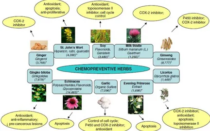
Figure 1. Some of the herbs widely consumed in USA and reported to have chemopreventive efficacy in the literature. The squares indicate the name of the herbs and active phytochemicals present in them. The circles indicate reported mechanism(s) of action for these agents. Numbers in the parentheses (*) represent the estimated US population consuming these herbs. (from Mehta et al. 2010)
Once identified these bioactive compounds, the purpose of the last years was to identify which molecular pathways involved in carcinogenesis they were active. This intense research showed that many phytochemicals can affect one or more of the deregulated pathways of carcinogenesis. Specifically, it has been found they are active in those related to carcinogen metabolism, DNA repair, cell proliferation, apoptosis, cell cycle, angiogenesis and metastasis [5].
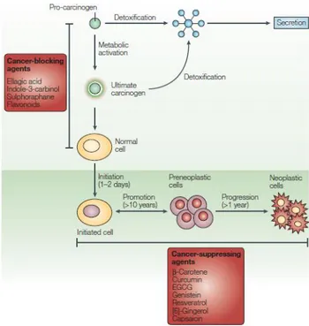
Carcinogenesis is a quite complex process that in a simplistic manner can be divided into three major stages. The initiation involves gene mutation, carcinogen metabolism and aberrant DNA repair. In this initial stage, environmental carcinogens induce one or more simple mutations in genes which control the process of carcinogenesis. The stage of promotion is characterized by deregulation of signalling pathways which normally control cell proliferation and apoptosis. Genes that control cell cycle are often mutated in human cancers. Finally, the stage of progression is characterized by genetic alterations within the karyotype of the cells resulting in chromosomal abnormalities. This stage is characterised by invasion, angiogenesis, and metastatic growth (Fig. 2).
12
Figure 2. The process of carcinogenesis is defined as initiation, promotion and progression. Progression is shown here to include the growth of malignant tumors, invasion and metastasis. In this diagram for each of the stages, various major actions of phytochemicals involving signaling pathways are summarized. (from Surh 2003)
In the last few years different studies have shown that natural constituents of the regular diet influence the process of carcinogenesis. They can act as tumor-blocking agents, tumor-suppressing agents or both. Tumor-blocking agents either prevent DNA damage formation or promote DNA damage removal, and tumor-suppressing agents slow down the process of initiated cells to become invasive cancer cells [2].
Among the plethora of known chemopreventive agents one of the most studied is the stilbene resveratrol, that has excited a great interest in the research community for its potential beneficial effects on human health.
1.2 Resveratrol in Chemoprevention
Resveratrol (3,5,4’-trans-hydroxystilbene) (RESV) is a phytoalexin that protects the plant from injury, ultraviolet (UV) irradiation, and fungal attacks in nature [6]. It was first described in 1940 as a phenolic component
13
of the medicinal herb hellebore [7] and then in 1963 in the Japanese knotweed Polygonum cuspidatum. At the moment it has been detected in at least 100 plants many of which are normal components of human diet. Some examples of main dietary sources of RESV are grape skin (5-10 mg/g), red wine (0.1-1.43 mg/l), cranberry (1.9 mg/g), red currant (1.5 mg/g), raw peanuts (0.15 mg/g). It is also present in the Ayurvedic formula Darakchasawa (0.36 mg/100 ml) and in the Itadori tea (0.97 mg/100 ml) [8]. RESV exists as cis and trans isomeric forms, with trans to cis isomerization facilitated by UV exposure. Its stilbene structure is related to the synthetic estrogen diethylstilbestrol. Two phenolic rings are linked by a styrene double bond to generate 3, 4’,5- trihydroxystilbene (Fig.3).
Figure 3. Structural formulas of trans-resveratrol and cis-resveratrol.
Only in 1992 this molecule polarized the attention of scientific community because it has been identified as the bioactive compound of red wine. This discovery was able to provided an explanation at the so called "French paradox", a term coined in order to explain the correlation between a moderate red wine consumption with a low incidence of cardiovascular diseases despite a high fat diet [9]. Then in 1997 Jang and co-workers demonstrated that RESV acted in vivo as a chemopreventive agent in the three stages of carcinogenesis [10].
Since then the number of publications has been growing exponentially and in vitro, ex vivo and animal model experiments have been providing evidence for bioactivity with clinical potential in cancer chemoprevention and therapy, cardiovascular disease and obesity, hepatic alcoholic or metabolic dysregulation, diabetes, arthritis, osteoporosis and neuroprotection[8].
The chemopreventive property of RESV has been reflected by its ability to block the activation of various carcinogens and/or to stimulate their detoxification, to prevent oxidative damage of target cell DNA, to reduce inflammatory responses and to diminish proliferation of cancer cells. Blockade of angiogenic and metastatic processes of tumor progression, and alleviation of chemotherapy resistance indicate the chemotherapeutic
14
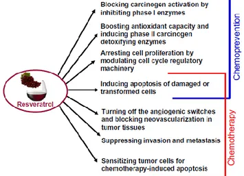
potential of RESV. The induction of apoptosis in various premalignant or cancerous cells by RESV can contribute to both chemopreventive and chemotherapeutic potential of this compound (Fig. 4) [11].
Figure 4. Biochemical mechanisms responsible for chemopreventive and chemotherapeutic potential of resveratrol. (from Kundu & Surh 2008)
The normal cell homeostasis has maintained by a fine tuning of the intracellular signalling network, formed by upstream kinases, that translate extracellular signals into biological responses through the activation of transcription factors. There are in vitro and in vivo data indicating the capacity of RESV to “turn on” or “switch off” several of these players. It inhibits the phopshporylation of the upstream kinases MAPKs, PKC and Akt and suppresses also the activation of transcription factors NF-κK and AP-1 [11].
Despite a huge amount of in vitro and in vivo studies, there are few data indicating a potential therapeutic use of RESV in humans. Indeed a big drawback of this molecule is represented by its poor bioavailability. Pharmacokinetic studies have demonstrated a rapid metabolization exerted by phase II enzymes that leads to the production of sulphate and glucoronide forms. The ingestion up to 5 g a day of RESV didn't show any adverse effect in human volunteers but its maximal concentration in plasma reached values ranging between 0.3 and 2.4 µM [12].
A study group established by the 1st International Conference on Resveratrol and Health in 2010 has worked out a series of guidelines for the use of
15
RESV in clinical trials [13]. A systematic review of literature data showed that there are not sufficient evidences to assert that RESV exerts a preventive effect in human diseases and regard to the toxicity after a nutraceutical intake there are not valid data yet. On the contrary studies in rodent showed that there are sufficient evidences for a chemopreventive role of RESV on the development of cancer skin and there are promising results on the prevention of colon cancer.They concluded that further clinical trials are necessary to elucidate both the chemopreventive role of RESV and the possible side effects in humans.
A big challenge in the cancer fight is to discover molecules able to selectively kill cancer cells with none or few side effects in normal tissues. RESV is a promising chemopreventive agent because it exerts antiproliferative and pro-apoptotic activities at micromolar concentrations in a wide range of cancer cells.
1.3 Resveratrol in Cell Proliferation and Apoptosis
The cell cycle progression is a tightly regulated process based on cyclic events of production and degradation of key enzymes, cyclins (A, B, Ds, E) and cyclin-dependant kinases (Cdk 1, 2, 4, 6). Cyclins are regulator proteins that activate their respective Cdks. The intracellular level of cyclins grows during the interphase reaching a maximum at the onset of mitosis when they undergo degradation [14].
There are many data indicating an antiproliferative effect of RESV in different phases of cell cycle in cancer cells depending on the type considered. The cell cycle block can occur at the G1/S phase through the down regulation of cyclins D1/D2/E, Cdks 2/4/6 and the induction of p21WAF1 and p27KIP1. The arrest in S as well as G2/M phase occurs by inhibition of Cdk7 and p34Cdc2 kinases. RESV is also an upregulator of the oncosuppressor p53 and in addition it causes the cell cycle arrest in cell-lacking p53 [15].
The cell can undergo apoptosis through two ways. The principal is the so called "intrinsic pathway", that involves mitochondria and leads to the release of soluble compounds from the intermembrane space to the cytosol. In most cases, the molecules released activate caspases, leading to the proteolytic cleavage of a series of intracellular proteins, the condensation of nuclear chromatin and the fragmentation of DNA [16]. The intrinsic pathway involves also the tumor suppressor p53, the most commonly mutated gene in human cancer. Normal p53 function leads to cell cycle arrest at the G1 and G2 checkpoints in response to DNA damage. This checkpoint function is executed by the accumulation of p53 followed by the
16
induction of the gadd45, waf1 and mdm2 genes. If DNA repair is not successful, p53 initiates apoptosis, thus preventing the propagation of genetic defects to successive cell generations [17].
The "extrinsic pathway" is triggered by the ligation of death receptor Fas (also known as APO-1 or CD95), to its natural ligand, Fas ligand (FasL, APO-1L, CD95L). Activation of this receptor results in a recruitment of caspase zymogenes into oligomeric complexes and triggers their proteolytic activation [18].
Regarding the first way, RESV affects the respiratory chain producing the release of Reactive Oxygen Species (ROS) and modulates the mitochondria membrane permeability causing a rapid depolarization. This effect is mediated through a downregulation of the expression of anti-apoptotic Bcl-2 proteins [19] and an upregulation of pro-apoptotic Bcl-2 proteins (Bax, Bak, Bad, Bid). RESV is also able to induce apoptosis in a p53 dependent or independent manner, depending on the cell type studied [20].
RESV leads to the activation of the "extrinsic pathway" through a redistribution of receptor molecules Fas in the plasma membrane. This action causes the production of lipid rafts that play an important role in clustering surface receptors, signalling enzymes and adaptor molecules into membrane complexes at specific sites. RESV-induced redistribution of Fas in the rafts could contribute to the formation of the death-inducing signalling complex (DISC) observed in colon cancer cells treated with the polyphenol. It has been also found that in some cell types RESV induces the sphingomyelin/ceramide apoptotic pathway, which in turn plays a crucial role in lipid rafts formation [20,21].
Finally various reports have shown that RESV is able to act on NF-κB pathways, that is an inhibitor of apoptosis, at multiple levels such as down-regulation of NF-κB protein expression, modulation of phosphorylation and transcriptional activity [22].
There are also data indicating that RESV kills cancer cells not only by apoptosis, but also through other mechanisms such as phagocytosis and intracellular killing, senescence and mitotic catastrophe [20].
1.4 Resveratrol and DNA
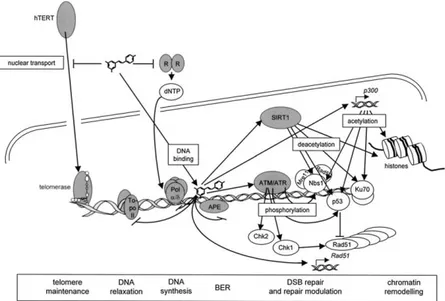
RESV is involved in several aspects of DNA metabolism such as replication, recombination, relaxation, repair and telomere maintenance. It affects directly several enzymes of DNA metabolism, such as DNA polymerases α and δ, ribonucleotide reductase and telomerase (Fig. 5) [23]. RESV is able to maintain DNA integrity because is a scavenger of reactive oxygen species (ROS). At the same time it owns an intrinsic antioxidant
17
capacity and acts as activator of detoxifying enzymes, such as superoxide dismutase, catalase and glutathione peroxidase. These data have been obtained in vitro, whereas it is a matter of debate if RESV possesses a radical scavenger property also in vivo.Moreover in certain conditions, such as other plant polyphenols, it showed a pro-oxidant activity [7].
It has been shown that RESV interacts directly with the DNA molecule. It establishes several hydrogen bonds without any alteration of double helix structure. Indeed it seems to stabilize the molecule and it could represent the molecular basis of the protection against genotoxic injuries [24]. Moreover RESV reverts the intercalation exerted by ethidium bromide without acting itself as an intercalating agent [25]. The Ames test, which measures the reversion of mutations in bacteria, didn’t show any mutagenic effect of RESV [26].
Figure 5. Nuclear activities of resveratrol with relevance for DNA repair. Gray color marks the immediate targets of resveratrol. (from Gatz & Wiesmuller 2008)
1.4.1 RESV and DNA damage in non-cancer cells
With respect to the genotoxic activity in non-cancer cells, interestingly, increasing concentrations of RESV (10–100 μM) in the presence of copper ions were showed to induce DNA damage in human lymphocytes. Moreover it has been demonstrated that the presence of hydroxyl groups is essential
18
for DNA cleavage [27]. Further, the RESV-induced DNA degradation in lymphocytes is inhibited by scavengers of ROS and neocuproine, a Cu(I)-specific sequestering agent [28]. Copper is an important metal ion present in chromatin and is closely associated with DNA bases, particularly guanine [29]. It is also one of the most redox active of all the various metal ions present in the cells. Evidences have shown that polyphenols including RESV do not only bind copper ions but also catalyze their redox cycling [30]. A mechanism was proposed which involves the formation of a ternary complex of DNA–polyphenol–Cu(II) [31]. A redox reaction of this compound and Cu(II) in the ternary complex may occur, leading to the reduction of Cu(II) to Cu(I), whose re-oxidation generates a variety of ROS. These findings demonstrated that the RESV–Cu(II) system for DNA breakage is physiologically feasible and could be of biological significance. In this context Schilder and co-workers have shown that a chronic treatment with RESV induces an increase of ROS in human endothelial cells and this increase is linked to S phase accumulation and finally to cell senescence [32].
As far as the ability of RESV in inducing DNA breaks it is still debated but not much data are present in literature about the induction of chromosomal damage in normal cells. In particular Matsuoka and co-workers showed that RESV is able to induce sister chromatid exchange (SCEs) and micronuclei (MN) in Chinese hamster lung cells at micromolar concentrations [26]. In my host lab a slight increase in chromosome aberrations after 200 μM RESV treatment in Chinese hamster ovary cells has been shown [33].
1.4.2 RESV and DNA damage in cancer cells
DNA damage is a key factor both in the evolution and treatment of cancer. Cellular responses to DNA damage are coordinated primarily by two distinct kinase signaling cascades, the ATM-Chk2 and ATR-Chk1 pathways, which are activated by DNA double and single-strand breaks respectively. In response to DSBs, ATM is required both for ATR-Chk1 activation and to initiate DNA repair via homologous recombination repair (HRR) by promoting formation of single-stranded DNA at damage sites through nucleolytic resection. The ATR-Chk1 pathway is the principal direct effector of the DNA damage and replication checkpoints and, as such, is essential for the survival of many, although not all, cell types [34]. The ATM-mediated posphorylation of H2AX (γ-H2AX) is an hallmark of the cellular response to DNA DSBs. This step is followed by the accumulation and local concentration of a plethora of DNA damage signalling and repair proteins in the vicinity of the lesion and culminating in the generation of
19
distinct nuclear compartments, so-called Ionizing Radiation-Induced Foci [35].
The first demonstration of a DNA cleavage exerted by RESV emerged in 1998. In a plasmid-based DNA cleavage assay, RESV mediated the relaxation of pBR322 at micromolar concentrations in the presence of Cu(II). It was showed that RESV was capable of binding to DNA, and that the Cu(II)-dependent DNA damage is more likely caused by a copper-peroxide complex rather than by freely diffusible oxygen species [36]. On the other hand a chronic administration of non apoptotic doses of RESV induces senescence in colon carcinoma cells through the increase of Radical Oxygen Species (ROS) production by mitochondria [37]. So that even if RESV has been mostly considered an antioxidant, it could act as a radical scavenger in some conditions and during the initial phases of exposure, while its pro-oxidant activity would start according to prolonged exposure time and higher concentrations.
Regarding the genotoxic effect of RESV in cancer cells, there are data that seem to support the hypothesis that DNA strand breaks could be a starting point in the apoptosis induction.
The treatment with a high dose of RESV induces DNA damage, as measured through Comet assay, both in human hepatic cancer cells [38] and in C6 rat glioma cells [39]. In the same cells the effect of RESV on chromosome damage has been measured by micronucleus assay. A dose of 250 μM RESV induced an increase (30%) in micronucleated cells after 24 h of treatment [40].
However, the vast majority of data on RESV ability in inducing DNA damage deal with the phosphorylation of the histone H2AX (γ-H2AX). In peripheral blood and bone marrow mononuclear cells isolated from chronic lymphocytic leukaemia patients, the induction of γ-H2AX and the activation of ATM caused by RESV has been measured. It has been shown that a 40 μM dose of RESV caused an increase in γ-H2AX expression concurrent with increased expression of activated ATM in most but not in all patients. Furthermore in the γ-H2AX-positive patients the increase in the apoptotic response after RESV treatment was significantly higher [41]. Also in human ovarian cancer cells RESV activates ATM/ATR kinases following DNA damage as detected by serine 139 phosphorylation of H2AX. Moreover RESV treatment resulted in an increase in Chk1 phosphorylation (Ser296) as well as in total protein level. Similarly, RESV strongly induced the phosphorylation of Chk2 at Thr68, Thr387 and Ser19 sites together with a moderate increase in total Chk2 protein level [42].
Furthermore, RESV can activate different cell cycle checkpoint mechanisms in response to DNA damage on the basis of the cell line analysed. Human
20
osteosarcoma and lung adenocarcinoma cells showed an upregulation of γ-H2AX at the dose of 50 μM of RESV. Differential expression of BRCA1, cyclin B1, pRb and p21 in U-2OS and A549 cells indicates that RESV can engage various molecular mechanisms to arrest cell cycle progression [43]. More recently Tyagi and co-workers have shown that RESV is able to induce DNA damage as measured through γ-H2AX expression and consequently apoptotic death in head and neck squamous cell carcinoma cells [44]. In human gastric adenocarcinoma cells a treatment with 50-200 µM of RESV caused the phosphorylation of the histone H2AX followed by apoptosis induction [45]. At the same manner in human lung adenocarcinoma cells 50 µM RESV was able to induce the phopsphotylation of ATM on Ser-1981 and a concomitant expression of γ-H2AX [46].
Recently it has been shown that RESV and other polyphenols such as genistein and baicalein, increased the level of ATAD5 protein that is a biomarker of DNA damage. This genotoxic effect was accompanied by apoptosis induction in proliferating cells but unlike conventional genotoxic chemotherapeutic agents, such as cisplatin, that produce mutagenesis at the chromosomal and nucleotide levels, RESV did not increase mutagenesis in the SupF plasmid mutagenesis assay [47].
On the whole these data seem to support the hypothesis that the anti-tumoral activity of RESV is in part due to its capacity to induce DNA damage in cancer cells, so activating the checkpoints responsible for cell cycle arrest and, in some cases, apoptosis. A putative mechanism by which RESV induces DNA DSBs could be the interaction with topoisomerase IIα, that is an enzyme mainly expressed in proliferating cells and involved in several aspects of DNA metabolism.
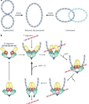
1.5 DNA Topoisomerases
DNA topoisomerases are conserved nuclear enzymes that are involved in the maintaining of DNA topology. In higher eukaryotes there are two classes of topoisomerases: type I enzymes introduce single strand breaks in DNA and type II ones introduce DSBs. Since a single unrepaired DSB has potentially lethal consequences, type II topoisomerases might be viewed as a particularly dangerous way of dealing with the topological problems of DNA. Mammalian cells express two isoforms (α and β) of topoisomerase II. The expression of Topoisomerase IIα (TOPO2) is cell cycle regulated, and this enzyme is essential for the viability of all dividing cells. Many non-dividing cells lack detectable TOPO2 [48].
Reactions catalyzed by eukaryotic TOPO2 include decatenation of linked intact double stranded DNA and relaxation of supercoiled DNA. The
21
enzyme introduces a double strand break in one DNA strand, termed the G or “gate segment”, and will pass a second strand termed the T segment through the break. ATP binding causes the enzyme to form a closed clamp. The closed clamp may also capture another strand (the T strand) that will pass through the break made in the G strand. ATP hydrolysis occurs at two steps in the reaction cycle. The first ATP hydrolyzed may assist in strand passage. The second hydrolysis step (along with release of ADP and Pi) allows the clamp to re-open, and allows release of the G segment (for a distributive reaction). Alternately, the enzyme may initiate another catalytic cycle without dissociating from the G strand (Fig. 6).
Figure 6. a. Reactions catalyzed by eukaryotic TOPO2 include decatenation
of linked intact double stranded DNA and relaxation of supercoiled DNA. The reaction formally requires introduction of a double strand break, strand passage, and break resealing. b. Mechanism of strand passage by TOPO2. (from Nitiss 2009)
One of the central roles of TOPO2 is to solve the topological problems associated with replication. Semi-conservative replication involves the
22
unwinding of duplex DNA and copying of each strand. In the absence of TOPO2 activity the unwinding of the parental duplex leads to the accumulation of positively supercoiled DNA in front of the replication fork. Studies indicated also that TOPO2 plays a key role in chromosome structure and chromosome condensation and it is involved in decatenation of sister chromatids during mitosis. Since TOPO2 carries out a reaction that is essential for chromosome separation at mitosis, a plausible hypothesis is that cells can monitor the successful completion of TOPO2 decatenation, and arrest cell cycle progression if decatenation (or chromosome condensation) is incomplete [48].
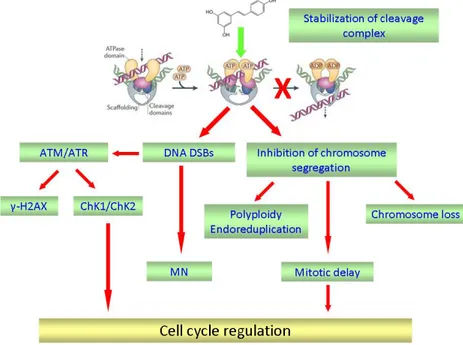
TOPO2 has held the interest of researchers studying cancer owing to the discovery that it is targeted by active anticancer drugs. Drugs targeting TOPO2 are divided into two broad classes. The first class, which includes most of the clinically active agents, leads to increases in the levels of TOPO2–DNA covalent complexes, named “cleavage complex”. Because these agents generate lesions that include DNA strand breaks and protein covalently bound to DNA, they have been termed TOPO2 poisons. A second class of compounds inhibits TOPO2 catalytic activity but does not generate increases in the levels of TOPO2 covalent complexes. Agents in this second class are thought to kill cells through the elimination of the essential enzymatic activity of TOPO2 and are therefore termed TOPO2 catalytic inhibitors.
The cleavage complexes are tightly regulated in the cells; if their level falls too low, cells are unable to undergo chromosome segregation and ultimately die of mitotic failure. On the contrary a higher level of cleavage complexes leads to an accumulation of DNA breaks that can evolve into chromosome translocations and other DNA aberrations. If the accumulation of breaks is overwhelming, they trigger apoptotic pathways and kill the cell [49]. If these DNA strand breaks do not result in cell death, chromosomal translocations may be present in surviving cellular populations [50]. For these reasons, in particular conditions, TOPO2 can change into a genotoxic enzyme.
Polyphenolic compounds, in particular bioflavonoids such as genistein and quercetin, have been shown to enhance DNA cleavage mediated by human TOPO2 isoform α and β [51]. This activity is exerted by increasing the levels of DNA cleavage complexes generated by TOPO2 through a direct interaction of genistein with an ATP binding motif of the enzyme. It has been demonstrated that polyphenolic fractions from grape cell culture are potent inhibitors of human TOPO2 catalytic activity [52].
In two studies RESV was characterized as a catalytic inhibitor of TOPO2 [53,54]. meanwhile another report postulated TOPO I poisoning activity for
23
RESV [55]. However, only a limited number of experiments were performed in this study.
1.6 REFERENCES
1. Gullett, N.P., Ruhul Amin, A.R., Bayraktar, S., Pezzuto, J.M., Shin, D.M., Khuri, F.R., Aggarwal, B.B., Surh, Y.J. and Kucuk, O. (2010) Cancer prevention with natural compounds. Semin Oncol, 37, 258-81.
2. Surh, Y.J. (2003) Cancer chemoprevention with dietary phytochemicals. Nat Rev Cancer, 3, 768-80.
3. Sporn, M.B., Dunlop, N.M., Newton, D.L. and Smith, J.M. (1976) Prevention of chemical carcinogenesis by vitamin A and its synthetic analogs (retinoids). Fed Proc, 35, 1332-8.
4. Mehta, R.G., Murillo, G., Naithani, R. and Peng, X. (2010) Cancer chemoprevention by natural products: how far have we come? Pharm Res, 27, 950-61.
5. Kwon, K.H., Barve, A., Yu, S., Huang, M.T. and Kong, A.N. (2007) Cancer chemoprevention by phytochemicals: potential molecular targets, biomarkers and animal models. Acta Pharmacol Sin, 28, 1409-21.
6. Shakibaei, M., Harikumar, K.B. and Aggarwal, B.B. (2009) Resveratrol addiction: to die or not to die. Mol Nutr Food Res, 53, 115-28.
7. Baur, J.A. and Sinclair, D.A. (2006) Therapeutic potential of resveratrol: the in vivo evidence. Nat Rev Drug Discov, 5, 493-506. 8. Chachay, V.S., Kirkpatrick, C.M., Hickman, I.J., Ferguson, M., Prins, J.B. and Martin, J.H. (2011) Resveratrol--pills to replace a healthy diet? Br J Clin Pharmacol, 72, 27-38.
9. Renaud, S. and de Lorgeril, M. (1992) Wine, alcohol, platelets, and the French paradox for coronary heart disease. Lancet, 339, 1523-6. 10. Jang, M., Cai, L., Udeani, G.O., Slowing, K.V., Thomas, C.F., Beecher, C.W., Fong, H.H., Farnsworth, N.R., Kinghorn, A.D., Mehta, R.G., Moon, R.C. and Pezzuto, J.M. (1997) Cancer chemopreventive activity of resveratrol, a natural product derived from grapes. Science, 275, 218-20.
11. Kundu, J.K. and Surh, Y.J. (2008) Cancer chemopreventive and therapeutic potential of resveratrol: mechanistic. Cancer Lett, 269, 243-61.
24
12. Boocock, D.J., Faust, G.E., Patel, K.R., Schinas, A.M., Brown, V.A., Ducharme, M.P., Booth, T.D., Crowell, J.A., Perloff, M., Gescher, A.J., Steward, W.P. and Brenner, D.E. (2007) Phase I dose escalation pharmacokinetic study in healthy volunteers of resveratrol, a potential cancer chemopreventive agent. Cancer Epidemiol Biomarkers Prev, 16, 1246-52.
13. Vang, O., Ahmad, N., Baile, C.A., Baur, J.A., Brown, K., Csiszar, A., Das, D.K., Delmas, D., Gottfried, C., Lin, H.Y., Ma, Q.Y., Mukhopadhyay, P., Nalini, N., Pezzuto, J.M., Richard, T., Shukla, Y., Surh, Y.J., Szekeres, T., Szkudelski, T., Walle, T. and Wu, J.M. (2011) What is new for an old molecule? Systematic review and recommendations on the use of resveratrol. PLoS One, 6, e19881. 14. Murray, A.W. (2004) Recycling the cell cycle: cyclins revisited.
Cell, 116, 221-34.
15. Athar, M., Back, J.H., Kopelovich, L., Bickers, D.R. and Kim, A.L. (2009) Multiple molecular targets of resveratrol: Anti-carcinogenic mechanisms. Arch Biochem Biophys, 486, 95-102.
16. Green, D.R. and Reed, J.C. (1998) Mitochondria and apoptosis. Science, 281, 1309-12.
17. Greenblatt, M.S., Bennett, W.P., Hollstein, M. and Harris, C.C. (1994) Mutations in the p53 tumor suppressor gene: clues to cancer etiology and molecular pathogenesis. Cancer Res, 54, 4855-78. 18. Ashkenazi, A. and Dixit, V.M. (1998) Death receptors: signaling
and modulation. Science, 281, 1305-8.
19. Shankar, S., Singh, G. and Srivastava, R.K. (2007) Chemoprevention by resveratrol: molecular mechanisms and therapeutic potential. Front Biosci, 12, 4839-54.
20. Delmas, D., Solary, E. and Latruffe, N. (2011) Resveratrol, a phytochemical inducer of multiple cell death pathways: apoptosis, autophagy and mitotic catastrophe. Curr Med Chem, 18, 1100-21. 21. Delmas, D., Rebe, C., Lacour, S., Filomenko, R., Athias, A.,
Gambert, P., Cherkaoui-Malki, M., Jannin, B., Dubrez-Daloz, L., Latruffe, N. and Solary, E. (2003) Resveratrol-induced apoptosis is associated with Fas redistribution in the rafts. J Biol Chem, 278, 41482-90.
22. Sun, C., Hu, Y., Liu, X., Wu, T., Wang, Y., He, W. and Wei, W. (2006) Resveratrol downregulates the constitutional activation of nuclear factor-kappaB. Cancer Genet Cytogenet, 165, 9-19. 23. Gatz, S.A. and Wiesmuller, L. (2008) Take a break--resveratrol in
25
24. Usha, S., Johnson, I.M. and Malathi, R. (2005) Interaction of resveratrol and genistein with nucleic acids. J Biochem Mol Biol, 38, 198-205.
25. Usha, S., Johnson, I.M. and Malathi, R. (2006) Modulation of DNA intercalation by resveratrol and genistein. Mol Cell Biochem, 284, 57-64.
26. Matsuoka, A., Furuta, A., Ozaki, M., Fukuhara, K. and Miyata, N. (2001) Resveratrol, a naturally occurring polyphenol, induces sister chromatid exchanges in a Chinese hamster lung (CHL) cell line. Mutat Res, 494, 107-13.
27. Azmi, A.S., Bhat, S.H. and Hadi, S.M. (2005) Resveratrol-Cu(II) induced DNA breakage in human peripheral lymphocytes: implications for anticancer properties. FEBS Lett, 579, 3131-5. 28. Hadi, S.M., Ullah, M.F., Azmi, A.S., Ahmad, A., Shamim, U.,
Zubair, H. and Khan, H.Y. (2010) Resveratrol mobilizes endogenous copper in human peripheral lymphocytes leading to oxidative DNA breakage: a putative mechanism for chemoprevention of cancer. Pharm Res, 27, 979-88.
29. Kagawa, T.F., Geierstanger, B.H., Wang, A.H. and Ho, P.S. (1991) Covalent modification of guanine bases in double-stranded DNA. The 1.2-A Z-DNA structure of d(CGCGCG) in the presence of CuCl2. J Biol Chem, 266, 20175-84.
30. Hanif, S., Shamim, U., Ullah, M.F., Azmi, A.S., Bhat, S.H. and Hadi, S.M. (2008) The anthocyanidin delphinidin mobilizes endogenous copper ions from human lymphocytes leading to oxidative degradation of cellular DNA. Toxicology, 249, 19-25. 31. Rahman, A., Shahabuddin, Hadi, S.M., Parish, J.H. and Ainley, K.
(1989) Strand scission in DNA induced by quercetin and Cu(II): role of Cu(I) and oxygen free radicals. Carcinogenesis, 10, 1833-9. 32. Schilder, Y.D., Heiss, E.H., Schachner, D., Ziegler, J., Reznicek,
G., Sorescu, D. and Dirsch, V.M. (2009) NADPH oxidases 1 and 4 mediate cellular senescence induced by resveratrol in human endothelial cells. Free Radic Biol Med, 46, 1598-606.
33. De Salvia, R., Festa, F., Ricordy, R., Perticone, P. and Cozzi, R. (2002) Resveratrol affects in a different way primary versus fixed DNA damage induced by H(2)O(2) in mammalian cells in vitro. Toxicol Lett, 135, 1-9.
34. Smith, J., Tho, L.M., Xu, N. and Gillespie, D.A. (2010) The ATM-Chk2 and ATR-Chk1 pathways in DNA damage signaling and cancer. Adv Cancer Res, 108, 73-112.
26
35. Bekker-Jensen, S. and Mailand, N. (2010) Assembly and function of DNA double-strand break repair foci in mammalian cells. DNA Repair (Amst), 9, 1219-28.
36. Fukuhara, K. and Miyata, N. (1998) Resveratrol as a new type of DNA-cleaving agent. Bioorg Med Chem Lett, 8, 3187-92.
37. Heiss, E.H., Schilder, Y.D. and Dirsch, V.M. (2007) Chronic treatment with resveratrol induces redox stress- and ataxia telangiectasia-mutated (ATM)-dependent senescence in p53-positive cancer cells. J Biol Chem, 282, 26759-66.
38. Choi, H.Y., Chong, S.A. and Nam, M.J. (2009) Resveratrol induces apoptosis in human SK-HEP-1 hepatic cancer cells. Cancer Genomics Proteomics, 6, 263-8.
39. Michels, G., Watjen, W., Weber, N., Niering, P., Chovolou, Y., Kampkotter, A., Proksch, P. and Kahl, R. (2006) Resveratrol induces apoptotic cell death in rat H4IIE hepatoma cells but necrosis in C6 glioma cells. Toxicology, 225, 173-82.
40. Quincozes-Santos, A., Andreazza, A.C., Goncalves, C.A. and Gottfried, C. (2010) Actions of redox-active compound resveratrol under hydrogen peroxide insult in C6 astroglial cells. Toxicol In Vitro, 24, 916-20.
41. Podhorecka, M., Halicka, D., Klimek, P., Kowal, M., Chocholska, S. and Dmoszynska, A. (2011) Resveratrol increases rate of apoptosis caused by purine analogues in malignant lymphocytes of chronic lymphocytic leukemia. Ann Hematol, 90, 173-83.
42. Tyagi, A., Singh, R.P., Agarwal, C., Siriwardana, S., Sclafani, R.A. and Agarwal, R. (2005) Resveratrol causes Cdc2-tyr15 phosphorylation via ATM/ATR-Chk1/2-Cdc25C pathway as a central mechanism for S phase arrest in human ovarian carcinoma Ovcar-3 cells. Carcinogenesis, 26, 1978-87.
43. Rusin, M., Zajkowicz, A. and Butkiewicz, D. (2009) Resveratrol induces senescence-like growth inhibition of U-2 OS cells associated with the instability of telomeric DNA and upregulation of BRCA1. Mech Ageing Dev, 130, 528-37.
44. Tyagi, A., Gu, M., Takahata, T., Frederick, B., Agarwal, C., Siriwardana, S., Agarwal, R. and Sclafani, R.A. (2011) Resveratrol selectively induces DNA Damage, independent of Smad4 expression, in its efficacy against human head and neck squamous cell carcinoma. Clin Cancer Res, 17, 5402-11.
45. Wang, Z., Li, W., Meng, X. and Jia, B. (2011) Resveratrol induces gastric cancer cell apoptosis via ROS, but independent of sirtuin1. Clin Exp Pharmacol Physiol.
27
46. Zajkowicz, A. and Rusin, M. (2011) The activation of the p53 pathway by the AMP mimetic AICAR is reduced by inhibitors of the ATM or mTOR kinases. Mech Ageing Dev, 132, 543-51. 47. Fox, J.T., Sakamuru, S., Huang, R., Teneva, N., Simmons, S.O.,
Xia, M., Tice, R.R., Austin, C.P. and Myung, K. (2012) High-throughput genotoxicity assay identifies antioxidants as inducers of DNA. Proc Natl Acad Sci U S A, 109, 5423-8.
48. Nitiss, J.L. (2009) DNA topoisomerase II and its growing repertoire of biological functions. Nat Rev Cancer, 9, 327-37. 49. Baguley, B.C. and Ferguson, L.R. (1998) Mutagenic properties of
topoisomerase-targeted drugs. Biochim Biophys Acta, 1400, 213-22.
50. Felix, C.A. (1998) Secondary leukemias induced by topoisomerase-targeted drugs. Biochim Biophys Acta, 1400, 233-55.
51. Bandele, O.J. and Osheroff, N. (2007) Bioflavonoids as poisons of human topoisomerase II alpha and II beta. Biochemistry, 46, 6097-108.
52. Jo, J.Y., Gonzalez de Mejia, E. and Lila, M.A. (2005) Effects of grape cell culture extracts on human topoisomerase II catalytic. J Agric Food Chem, 53, 2489-98.
53. Cho, K.H., Pezzuto, J.M., Bolton, J.L., Steele, V.E., Kelloff, G.J., Lee, S.K. and Constantinou, A. (2000) Selection of cancer chemopreventive agents based on inhibition of topoisomerase. Eur J Cancer, 36, 2146-56.
54. Jo, J.Y., Gonzalez de Mejia, E. and Lila, M.A. (2005) Effects of grape cell culture extracts on human topoisomerase II catalytic activity and characterization of active fractions. J Agric Food Chem, 53, 2489-98.
55. Webb, M.R. and Ebeler, S.E. (2004) Comparative analysis of topoisomerase IB inhibition and DNA intercalation by. Biochem J, 384, 527-41.
56. Leone, S., Fiore, M., Lauro, M.G., Pino, S., Cornetta, T. and Cozzi, R. (2008) Resveratrol and X rays affect gap junction intercellular communications in human glioblastoma cells. Mol Carcinog, 47, 587-98.
28
2 AIM OF THE RESEARCH
DNA damage is one of the targets of both chemoprevention and chemotherapy and a desirable outcome is the induction of apoptosis and/or the arrest of cell proliferation. Data demonstrating any genotoxic effects of RESV in cancer cells are poor and controversial, probably due to the use of different cell types and to the heterogeneity of the schedules of treatment. In the host lab it has been previously demonstrated that RESV is able to re-establish Gap Junction Intercellular Communication (GJIC) accompanied by a delay in the S-phase of cell cycle in the U87 glioblastoma mulitforme cell line [56]. This aspect is very interesting in relation to the possibility that RESV could enhance the spreading of damage (DNA damage), cell death or cell cycle control signals through GJIC, so increasing the effect of conventional antitumoral therapy.
On the basis of these previous observations, my PhD project concerned the study of chemopreventive/chemotherapeutic activity of RESV in cancer and normal cells, in terms of effects on cell cycle progression and DNA damage induction.
During the first year we analysed the anti-proliferative effects exerted by RESV in glioblastoma cells, associated with the induction of DNA damage, measured through the H2AX phosphorylation.
In the second year we studied the possible molecular interaction between DNA/RESV/TOPO2, through a Docking Simulation. Furthermore we analysed the consequences of this interaction at the level of cellular DNA integrity. The hypothesis was that this interaction could stabilize the cleavage complexes formed by TOPO2 during its activity on DNA.
Finally in the third year we performed a mechanistic study in Chinese Hamster Ovary (CHO) cells, in order to demonstrate whether also in proliferating non-cancer cells, RESV can affect various aspects of DNA metabolism explicable with the interaction with TOPO2 activity. In particular we found that it induces micronuclei but this effect is attenuated by the co-presence of another TOPO2 inhibitor, supporting the idea that DNA damage induction is caused by TOPO2 poisoning. Moreover it affects proper chromosome segregation at mitosis causing chromosome los, inhibiting decatenation and causing a delay in cell proliferation.
29
30
3.1 Leone, S., Cornetta, T., Basso, E. and Cozzi, R. (2010) Resveratrol induces DNA double-strand breaks through human topoisomerase II. Cancer Lett, 295, 167-72.
36
3.2 Leone, S., Basso, E., Polticelli, F. and Cozzi, R. (2012) Resveratrol acts as a topoisomerase II poison in human glioma cells. Int J Cancer, 131, E173-8.
42
3.3 Basso, E., Fiore, M., Leone, S., Degrassi, F. and Cozzi, R. (2012) Effects of Resveratrol on Topoisomerase II-α activity: induction of micronuclei and inhibition of chromosome egregation in CHO-K1 cells. Mutagenesis (in press).
43
48
4 DISCUSSION
A great interest has emerged during last years around the use of natural compounds as a supporting remedy to medicine, in order to prevent the onset of human illnesses or to act as an adjuvant of conventional therapies. Herbs and plants are a natural source of bioactive compounds, namely phytochemicals, that are non-nutrient components of food exerting several beneficial effects for human health, in particular concerning the cure of cancer.
The intense research in the understanding of the way of action of several chemopreventive agents represents a big challenge because could provide the “magic bullet” in the cancer fight. Indeed phytochemicals are normal component of the diet, so they are not expensive, are present at low concentrations and for this reason they are considered safe [1]. Unfortunately the most of in vitro and in vivo studies concerning phytochemicals have been performed at concentrations that unlikely are reached in physiological conditions after a dietary intake. This represents a problem that must be considered for next investigations [2].
Starting from 1997 a growing amount of data, both in vitro and in vivo, have been produced about Resveratrol (RESV) showing its promising capacity in the treatment of cancer and other pathologies. At the moment around 60 clinical trials concerning the assessment of safety of RESV and for a potential use as remedy for various illnesses are in course
(www.clinicaltrials.gov) [3]. Moreover RESV is a compound that acts on
many factors and biochemical pathways and this represents a complication because it becomes difficult to establish a pattern of action [4].
Here, starting from the study of the antiproliferative role of RESV in cancer cells, we have arrived to conclude that its chemopreventive/chemotherapeutic action can be in part explained by the interaction with TOPO2.
Our first observation, through a citofluorimetric bi-parametric assay, concerning a delay in the S-phase of cell cycle accompanied by the maximal induction of DNA DSBs, in terms of phosphorylation of the histone H2AX at this stage in glioma cells, confirmed the extended literature about the anti-proliferative role of the stilbene in cancer cells. On the other hand we showed also that RESV was able to induce DNA DSBs, an aspect poor investigated until now.
Many evidences indicate that RESV is able to arrest cell proliferation at various stages of cell cycle. The stop in the G1-phase can occur through the
49
p53 dependant activation of p21. p21 in turn reduces the activity of the cyclinD/cdk4 complex. The inhibition of the binding of transcription factors (AP1 and NFκB) exerted by RESV causes a decrease in protein expression of cyclin D1, D2, E and cdk4, 6, 2. The G1/S transition is blocked by a reduced hyperphosphorylation of retinoblastoma protein-tumor suppressor (pRb) [5].
The S-phase arrest is mediated by RESV through the disruption of the dephosphorylation process of cdk1 that is a key regulator of the progression in the S-phase. Indeed it increases cyclin A and B expression leading in this manner to an accumulation of inactive cdk1 [6]. Moreover RESV can block cells at this stage acting directly on DNA synthesis, through the inhibition of the ribonucleotide reductase activity and DNA polymerase [7].
The inactivation of cdk1 is also responsible of the accumulation of cells in the G2-phase. The increase of cyclin A and B exerted by RESV causes a block in G2/M [5].
Histone H2AX, a variant of H2A, is phosphorylated at its carboxy-terminal Serine 139 as one of the earliest cellular response to the induction of DNA DSBs [8]. Once phosphorylated, γ-H2AX foci rapidly form over chromatin regions on either side of a DSB facilitating repair through relocalization and focusing factors to the site of breaks. γ-H2AX also functions in the end-joining of DSBs by serving as an anchor for holding broken chromosomal ends in close proximity [9].
So both our results, namely a delay in the S-phase with a concomitant induction of DNA DSBs, have let us to suppose a putative interaction of RESV with TOPO2. We demonstrate here that RESV is able to inhibit the decatenation activity of TOPO2 through an in vitro test, adding a new interesting element for the explanation of its chemopreventive activity. This result is in agreement with what previously showed by Cho et al. who analysed a great number of natural compounds including RESV tested for their TOPO enzymes inhibitory activity [10]. Jo et al. [11] showed that polyphenolic fractions from grape cell culture were potent inhibitor of human TOPO2, utilizing the same assay. Similar results were also achieved by Yamada et al. [12] studying four new RSV oligomers.
TOPO2 is an enzyme mainly expressed in proliferating cells from the S-phase, where it is involved in the resolution of DNA supercoiled, to the G2/M phase, where it is involved in the decatenation of sister chromatids driving in this manner a proper chromosome segregation. These evidences make TOPO2 a preferential target of cancer chemotherapy. In fact,many anticancer drugs have been designed in order to inhibit TOPO2 activity. There are two principal classes of TOPO2 inhibitors: catalytic inhibitors,
50
which act by blocking the catalytic cycle at various stages without inducing any DNA damage and TOPO2 poisons, which stabilize the covalent complex DNA-TOPO2, namely the “cleavage complex”, provoking in this manner DNA DSBs [13].
Utilizing different approaches we demonstrated for the first time that RESV acts as a TOPO2 poison in glioma cells.
An in silico docking simulation provided us a first evidence about a putative stabilization of the covalent complex DNA-TOPO2 exerted by RESV. Then through the In Vivo Complex of Enxyme Assay (ICE) we showed as the stilbene is able to trap both TOPO2 and DNA in a non-covalent manner forming the cleavage complex. The ICE Assay represents a validate test hugely utilised to assess new chemical entities for their ability to inhibit DNA topoisomerases, in particular to examine the topoisomerase covalent complexes in vivo formation [14].
Since one of the consequences of TOPO2 poisoning is the induction of DNA DSBs, we analysed the presence of DNA damage through the Cytokinesis-Block Micronucleus Assay (CBMN) and the activation of DNA damage signalling pathway. Our data showed a significant increase of micronuclei and the activation of the ATM/ATR signalling pathway in glioma cells. The induction of DSBs through TOPO2 poisoning by RESV treatment is also confirmed by γ-H2AX expression immediately after treatment and by its persistence after a recovery period. These data together with the increase in the phosphorylated form of ATM and Chk2 expression lead us to conclude that RESV induced DNA damage is sensed by early signal transducers that activate S phase arrest.
Tyagi and co-workers [15] demonstrated that RESV induces S phase arrest via Tyr15 phosphorylation of Cdc2 in human ovarian carcinoma cells. Further they detected the activation of checkpoint kinases Chk1 and Chk2, which in turn were activated via ATM /ATR kinase in response to DNA damage. In the same study they observed as RESV also increased γ-H2AX, which is known to be phosphorylated by ATM/ATR in response to DNA damage. The involvement of these molecules in RESV-induced S-phase delay was also supported by the data showing that the addition of ATM/ATR inhibitor caffeine reverses RESV-caused activation of ATM/ATR–Chk1/2 as well as the phosphorylation of Cdc25C, Cdc2, γ-H2AX, and S phase arrest. On the contrary RESV showed only marginal S phase arrest in normal human foreskin fibroblasts with undetectable level of γ-H2AX [15].
Studying the effects of RESV on genome stability, Gatz and co-workers showed that it inhibits both homologous recombination (HR) and non